Effect of Chaihu-jia-Longgu-Muli decoction on withdrawal symptoms in rats with methamphetamine-induced conditioned place preference
←
→
Page content transcription
If your browser does not render page correctly, please read the page content below
Bioscience Reports (2021) 41 BSR20211376
https://doi.org/10.1042/BSR20211376

Research Article

Effect of Chaihu-jia-Longgu-Muli decoction on
withdrawal symptoms in rats with
methamphetamine-induced conditioned place
preference

                                                                                                                                                                                           Downloaded from http://portlandpress.com/bioscirep/article-pdf/41/8/BSR20211376/919433/bsr-2021-1376.pdf by guest on 28 August 2021
Zifa Li1,2,* , Yuchen Qi3,* , Kun Liu1,2 , Yiming Cao4 , Hao Zhang1,2 , Chunhong Song1,2 and                                                                 Hualiang Deng4
1 Behavioural Phenotyping Core Facility, Shandong University of Traditional Chinese Medicine, Jinan 250355, China; 2 Chinese Medicine Neuro-Psycho Pharmacology Laboratory
(CMNPPL), Shandong University of Traditional Chinese Medicine, Jinan 250355, China; 3 No. 2 Department of Encephalopathy, Affiliated Hospital of Shandong University of
Traditional Chinese Medicine, Jinan 250011, China ; 4 School of Traditional Chinese Medicine, Shandong University of Traditional Chinese Medicine, Jinan 250355, China
Correspondence: Hualiang Deng (60011803@sdutcm.edu.cn)

                                           Traditional Chinese medicine detoxification prescription Chaihu-jia-Longgu-Muli decoction
                                           (CLMD) relieves depressive symptoms in patients withdrawing from methamphetamine.
                                           In the present study, we assessed the effects of CLMD on methamphetamine with-
                                           drawal in rats. A methamphetamine-intoxicated rat model was established. Rats were ran-
                                           domly divided into the control, model, high-dosage, medium-dosage, and low-dosage
                                           groups, receiving high, medium, and low doses of CLMD, respectively. Weekly body
                                           weight measurements revealed that rats treated with methamphetamine had the low-
                                           est body weight. The conditioned place preference (CPP) experiment revealed that
                                           methamphetamine-intoxicated rats stayed significantly longer in the drug-paired chamber
                                           than the control rats. However, after administering high-dosage CLMD, the amount of time
                                           the rats spent in the drug-paired chamber was significantly less than that of the model rats.
                                           Our open-field test revealed that the model group had lower crossing and rearing scores than
                                           the control group. Additionally, rats that received CLMD treatment exhibited higher cross-
                                           ing and rearing scores than the model rats. Striatal dopamine (DA), 5-hydroxytryptamine
                                           (5-HT), and endorphins (β-EP) and serum interleukin (IL)-1α and IL-2 concentrations were
                                           estimated. Rats in the model group had lower striatal DA, 5-HT, and β-EP and higher serum
                                           IL-1α and IL-2 concentrations than those in the control group. High-dosage CLMD ad-
                                           ministration significantly changed the concentrations of these molecules, such that they
                                           approached normal concentrations. In general, CLMD could prevent the development of
                                           methamphetamine-induced withdrawal symptoms in rats by increasing the DA, 5-HT, and
                                           β-EP and lowering the IL-1α and IL-2 concentrations.

* These authors contributed

equally to this work.                    Introduction
Received: 06 June 2021
                                         Methamphetamine use is associated with an array of symptoms, such as mental excitation, loss of ap-
Revised: 18 July 2021                    petite, insomnia [1], and inclination toward social violence. Neurotoxicity in multiple neurotransmitter
Accepted: 04 August 2021                 systems arises as a result of methamphetamine consumption [2,3]. By altering synaptic plasticity in the
                                         brain, methamphetamine use can result in adverse effects such as dependence, withdrawal syndrome,
Accepted Manuscript Online:
06 August 2021                           and cravings [4,5]. Once a cut-off concentration is reached, withdrawal symptoms manifest [6]. Anxiety
Version of Record published:             and depression are two common symptoms of methamphetamine withdrawal and may be associated with
20 August 2021                           cravings and drug dependency [7].

© 2021 The Author(s). This is an open access article published by Portland Press Limited on behalf of the Biochemical Society and distributed under the Creative Commons Attribution   1
License 4.0 (CC BY).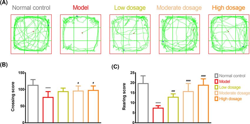
Bioscience Reports (2021) 41 BSR20211376
                                                                                                                                         https://doi.org/10.1042/BSR20211376

              Methamphetamine stimulates the release of several neurotransmitters such as dopamine (DA),
           5-hydroxytryptamine (5-HT), and endorphins (β-EP), which are associated with emotions [8–10]. Metham-
           phetamine directly acts on the dopaminergic neurons and competes with released DA for access to DA transporters
           [11]. It then destroys the DA storage vesicles and facilitates DA antiport using the transporters excreted by DA
           [11]. In addition, methamphetamine can lead to neuronal death in different cerebral areas, such as the striatum.
           Methamphetamine exposure can damage the dopaminergic neurons in the substantia nigra, leading to lower
           DA concentration within the striatum [12,13]. The chemical structure of methamphetamine is similar to that of
           catecholamine-related neurotransmitters; therefore, it can enter into the neuronal ends through 5-HT transporters
           to replace 5-HT in vesicles and cells. During this process, significant levels of 5-HT are consumed, leading to damage
           to the neuronal ends that contain 5-HT [14]. The neurotoxicity associated with the intake of methamphetamine
           causes damage to the dopaminergic and serotoninergic ends of the neurons as well as to the nigrostriatal pathway
           [15]. β-EP produces reward effects by combining with the μ receptor, leading to feelings of satisfaction and euphoria

                                                                                                                                                                                           Downloaded from http://portlandpress.com/bioscirep/article-pdf/41/8/BSR20211376/919433/bsr-2021-1376.pdf by guest on 28 August 2021
           [16]. The μ receptors are widely distributed throughout the central nervous system and are broadly recognized as
           opiate receptors associated with addiction [17].
              The immune system also plays an important role in the pathogenesis of neuropsychiatric disorders, including
           cognitive decline, anxiety, mood changes, and depressive states, as well as increased attention, decreased fatigue, and
           the rush of euphoria [18–20], which are associated with methamphetamine use. Inflammatory biomarkers, especially
           interleukin (IL)-1 (IL-1), are increased by methamphetamine use and are involved in methamphetamine-induced
           neurodegeneration [21–23]. IL-2 (a potent T-cell growth factor) levels have been found to be significantly higher in
           hypothalamic samples taken from methamphetamine-exposed mice [24].
              To date, most studies have focused on exploring the mechanisms of neuropsychiatric disorders and immune dysreg-
           ulation related to methamphetamine use and have not clarified the behavioral changes leading to its abuse or aided
           in the development of rehabilitation medicines with few side effects. Chaihu-jia-Longgu-Muli decoction (CLMD)
           is a detoxifying formulation containing herbal medicine based on the basic theory of traditional Chinese medicine.
           CLMD has been used as a remedy for many years with very few side effects and has been frequently used clinically for
           the treatment of neuropsychiatric disorders [25]. In addition to having few associated toxic effects, CLMD has signifi-
           cant beneficial effects on methamphetamine-induced depressive symptoms, which occur after withdrawal [26]. There
           is a large body of research indicating that this formulation and its derivatives are effective in reducing intimal thicken-
           ing of the carotid artery in animal models. In mice, not only has antidepressant activity and reduction in chronic mild
           stress-induced apoptosis in the hippocampus been observed but also the treatment of insomnia and improvement in
           sleep quality have been noted [25,27–29]. However, little is known about the effect of CLMD on behavioral responses
           to amphetamine withdrawal symptoms. Based on the clinical effects of CLMD seen in methamphetamine-addicted
           people after withdrawal [26,30], the present study was conducted to verify the effects of CLMD on the behavior
           of rats withdrawing from methamphetamine in an attempt to explore the signaling pathways involved. We further
           aimed to estimate the extent of recovery from neuropsychiatric disorders and immune dysregulation resulting from
           CLMD treatment, especially striatal DA, 5-HT, and β-EP and serum IL-1α and IL-2 concentrations, and explored
           the functional mechanisms of CLMD.

           Materials and methods
           Animals
           Sixty SPF male Sprague–Dawley rats weighing 170–190 g were provided by the Beijing Vital River Laboratory Animal
           Technology Co., Ltd., Beijing, China (laboratory animal production license no. SCXK [BJ]2016-0006). All animal
           experiments took place at the Behavioral Phenotyping Core Facility, Shandong University of Traditional Chinese
           Medicine, and the animals were adapted to the following experimental conditions for 1 week: temperature: 21 +     ◦
                                                                                                                          − 1 C;
           humidity: 40 + − 5%; and a 12-h light/dark cycle (light on at 20:00 and off at 8:00). The animals were fed a standard
           diet and filtered water ad libitum. The research plan and experimental procedures followed a protocol approved by
           the Animal Use and Care Committee of Shandong University of Traditional Chinese Medicine, Jinan, China (ethics
           approval reference no. SDUTCM2018-072), and were conducted according to the Guide for the Care and Use of
           Laboratory Animals.

           Preparation of drugs and reagents
           Methamphetamine, which was provided by the Detoxification Surveillance and Treatment Center of China (Shan-
           dong branch), was dissolved in saline immediately before intramuscular injection at a dose of 2 mg/kg. All other
           chemicals used in the present study were purchased from Sinopharm Chemical Reagent Co., Ltd (Shanghai, China).

2   © 2021 The Author(s). This is an open access article published by Portland Press Limited on behalf of the Biochemical Society and distributed under the Creative Commons Attribution
    License 4.0 (CC BY).
Bioscience Reports (2021) 41 BSR20211376
https://doi.org/10.1042/BSR20211376

   CLMD was prepared according to the following steps. First, Radix bupleuri (36 g), ginseng (15 g), fossil fragments
(12 g), oyster (12 g), Radix scutellariae (12 g), ochre (12 g), Cassia twig (12 g), Tuckahoe (12 g), pinellia ternata (9 g),
Rheum officinale (9 g), ginger (12 g), and Chinese dates (41 g) were purchased from the Shandong Pharmaceutical
Company (Jinan, China). The ingredients were boiled twice in a volume of water ten-times that of the ingredients.
Two batches of filtered soup were mixed using filter paper (Nanjing Wanqing Chemical Glassware Instrument Co.,
Ltd.). The filtered soup was then dried by distillation and converted into a freeze-dried powder extract, which was
stored at −20◦ C.

Establishment of the model, drug administration, and animal groups
Fifty-five pre-qualified rats were selected and assigned randomly into five groups: the control, model, high-dosage

                                                                                                                                                                                           Downloaded from http://portlandpress.com/bioscirep/article-pdf/41/8/BSR20211376/919433/bsr-2021-1376.pdf by guest on 28 August 2021
(20 mg/kg), moderate-dosage (10 mg/kg), and low-dosage (5 mg/kg) groups, with 11 rats in each group. The dried
powder extract was weighed for each group and diluted in appropriate volume of water. The absolute volume of
the CLMD liquor administered to each rat was calculated according to their individual body weights so that the
relative administration volume for all the rats was fixed at 0.5 ml/100 g of weight. Dosages administered in the high-,
moderate-, and low-dosage groups were equivalent to 10-, 5-, and 2.5-times the clinical dosages, respectively. All
groups, except the control group, received intramuscular injections of 2 mg/kg methamphetamine daily for 10 days
to establish the methamphetamine-intoxicated rat model for the conditioned place preference (CPP) test (Figure
1). The control group was administered the same dosage of saline by intramuscular injection. After injection, the
corresponding dosages of CLMD were administered by gavage for 18 consecutive days (Figure 1). The rats’ body
weights were measured once every week, and the administrations were adjusted accordingly.

CPP experiment
Before initiating the experiment, the rats were placed inside a CPP apparatus for adaptive feeding for 7 days. The CPP
apparatus consisted of two equal-sized compartments (30 × 30 × 43 cm), one with a white box and the other with
a black box joined by a wall with a sliding door. The ‘non-drug-paired’ chamber was black, while the ‘drug-paired’
chamber was white. The length of time the rats actively stayed within each of the two chambers was recorded, and
those that actively stayed longer in the ‘drug-paired’ chamber than in the ‘non-drug-paired’ chamber were rejected.
   After 8 days of establishing the model (saline injections for the control group and methamphetamine injections for
the model and CLMD treatment groups), the rats were placed inside the ‘drug-paired’ chamber and received intra-
muscular injections of methamphetamine in the morning (a clapboard was placed between the chambers so that the
rats could only stay in the ‘drug-paired’ chamber). The rats were taken out after 30 min. At the same time, the con-
trol experiment was conducted. The rats in the control group were also placed inside the ‘drug-paired’ chamber after
intramuscular injection of the same dosage of saline and then taken out after 30 min. The training phase continued
for 2 days, and the CPP test was conducted on the tenth day (Figure 1).
   No drugs were administered during the CPP test. The rats were placed inside the passage close to the two chambers,
and the clapboard was lifted to allow them to freely move between the two chambers. The test time was 15 min. The
length of time that the rats stayed within each chamber was recorded. The CPP test was re-conducted 24 h after 18
days of CLMD treatment (Figure 1), with a test time of 15 min.

Open-field test
The behavior of the rats was observed by the open-field test. The field test chamber had a dimension of 50 cm × 50
cm × 50 cm, with an open top, black baffles on the sides and bottom, and a Sudoku design at the bottom as a test base.
One day before the test, the rats were placed into the field test chamber for 10 min to adapt them to the experimental
environment. This adaptation was expected to decrease the influence of the strange environment on the activities of
the rats. Upon initiating the experiment, the rats were lightly placed on the central grid of the field test chamber, and
their activity status was recorded for 5 min automatically using a video analysis system. The number of grids crossed,
total length of movement, and number of times they stood erect were recorded. The horizontal and vertical scores of
the rats were calculated. The horizontal score was the total number of grids crossed at the bottom, while the vertical
score was the number of times the rat stood erect, including raising its forepaws into the air or using the wall for
support [31].

© 2021 The Author(s). This is an open access article published by Portland Press Limited on behalf of the Biochemical Society and distributed under the Creative Commons Attribution   3
License 4.0 (CC BY).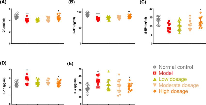
Bioscience Reports (2021) 41 BSR20211376
                                                                                                                                         https://doi.org/10.1042/BSR20211376

                                                                                                                                                                                           Downloaded from http://portlandpress.com/bioscirep/article-pdf/41/8/BSR20211376/919433/bsr-2021-1376.pdf by guest on 28 August 2021
           Figure 1. Experimental design
           Schedule of the experimental design including time course, grouping, and timing of behavioral experiments.

           Estimation of striatal DA, 5-HT, and β-EP concentrations serum and IL-1α
           and IL-2 concentrations
           After the behavioral tests, the rats were anesthetized with pentobarbital sodium and killed by neck breaking. Blood
           samples were collected and centrifuged for 15 min at 3000×g to obtain the serum. The rats were then killed to obtain
           100 mg of striatal tissue, which was put into Eppendorf Safe-Lock tubes after washing the blood stain with phos-
           phate buffered saline (PBS), followed by the addition of 1 ml of PBS. The mixture was then homogenized with a
           tissue grinder and placed at −20◦ C overnight. After two freeze-thaw cycles to damage the cell membranes, the ho-
           mogenate was centrifuged at 5000×g for 5 min at 4◦ C to obtain the supernatant. Striatal DA, 5-HT, and β-EP and
           serum IL-1α and IL-2 concentrations were estimated in strict accordance with the instructions of the following ELISA
           kits: DA ELISA (Wuhan Huamei Biotech Co., Ltd.; batch no. Y06015074), 5-HT ELISA (Wuhan Huamei Biotech Co.,
           Ltd.; batch no. C0150040107), β-EP ELISA (Wuhan Huamei Biotech Co., Ltd.; batch no. C0150050108), IL-2 ELISA
           (Wuhan Huamei Biotech Co., Ltd.; batch no. C23015075), and IL-1α ELISA (Wuhan Huamei Biotech Co., Ltd.; batch
           no. Y02015076).

4   © 2021 The Author(s). This is an open access article published by Portland Press Limited on behalf of the Biochemical Society and distributed under the Creative Commons Attribution
    License 4.0 (CC BY).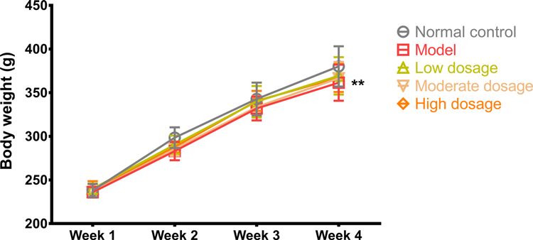
Bioscience Reports (2021) 41 BSR20211376
https://doi.org/10.1042/BSR20211376

                                                                                                                                                                                           Downloaded from http://portlandpress.com/bioscirep/article-pdf/41/8/BSR20211376/919433/bsr-2021-1376.pdf by guest on 28 August 2021
Figure 2. Body weight
Body weight of the rats in the control, model, CLMD low-dosage, CLMD moderate-dosage, and CLMD high-dosage groups. n=12.
**P
Bioscience Reports (2021) 41 BSR20211376
https://doi.org/10.1042/BSR20211376
Downloaded from http://portlandpress.com/bioscirep/article-pdf/41/8/BSR20211376/919433/bsr-2021-1376.pdf by guest on 28 August 2021
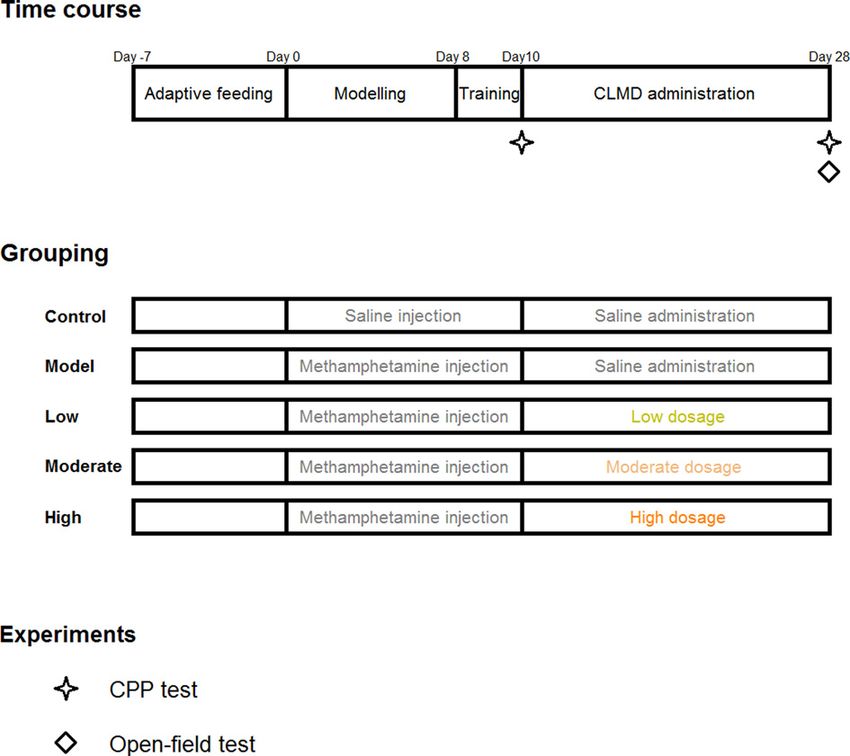
Figure 4. Open-field test
Open-field test results for the control, model, CLMD low-dosage, CLMD moderate-dosage, and CLMD high-dosage groups. (A)
Trajectory for the five groups. (B) Crossing score for the five groups. (C) Rearing score for the five groups. ****PBioscience Reports (2021) 41 BSR20211376
https://doi.org/10.1042/BSR20211376
Downloaded from http://portlandpress.com/bioscirep/article-pdf/41/8/BSR20211376/919433/bsr-2021-1376.pdf by guest on 28 August 2021
Figure 5. Key molecule levels in the striatum and serum
Detection of striatal DA (A), 5-HT (B), and β-EP (C) and serum IL-1 (D) and IL-2 (E) concentrations. *PBioscience Reports (2021) 41 BSR20211376
https://doi.org/10.1042/BSR20211376
Additionally, the neurotoxicity of methamphetamine leads to damage to the dopaminergic and serotoninergic neu-
rons in the nigrostriatal pathway [15]. Therefore, we observed that after 24 h of withdrawal from methamphetamine,
striatal DA and 5-HT concentration was significantly lower in the rats in the model group, suggesting that withdrawal
symptoms experienced by these rats were associated with lower DA and 5-HT concentrations (Figure 5A,B). Fortu-
nately, through CLMD treatment, we also identified that striatal DA and 5-HT concentrations were up-regulated
in the rats in the high-dosage group, exerting certain inhibitory effects on the withdrawal symptoms that might be
associated with the increased striatal DA and 5-HT concentrations (Figure 5A,B).
During the course of addiction, β-EP concentration increases due to the stimulation of methamphetamine [8–10].
This is a similar effect to what occurs with an influx of a large number of exogenous opiates into the body. During
the withdrawal phase, due to the suspension of methamphetamine stimulation, endogenous β-EP concentration is
lower, causing peripheral and central withdrawal symptoms [37], which were also exhibited in our model (Figure 5C).
CLMD treatment up-regulated striatal β-EP concentration, which could also contribute to ameliorating withdrawal
Downloaded from http://portlandpress.com/bioscirep/article-pdf/41/8/BSR20211376/919433/bsr-2021-1376.pdf by guest on 28 August 2021
symptoms.
Changes in serum IL concentrations in the methamphetamine-induced
CPP model
Studies have shown that methamphetamine toxicity-induced neuronal injury is mediated through the activation of
the microglial cell response and tumor necrosis factor system [38,39]. Some drugs that inhibit the immune response
can reduce drug dependence by partly reducing the activation of methamphetamine-dependent microglial cells [40].
Another study reported that microglial activation leads to an inflammatory response in the neurons and that by
inhibiting microglial activation, the expression of inflammatory factors is reduced in a methamphetamine poisoning
rat model [41]. Our finding is consistent with these findings. In our results, higher concentrations of IL-1α and IL-2,
which are two important inflammatory factors, were observed in the methamphetamine-induced CPP model than in
the control rats (Figure 5D,E), suggesting that methamphetamine poisoning was associated with elevated IL-1α and
IL-2 concentrations. CLMD treatment reduced IL-1α and IL-2 concentrations in the methamphetamine-intoxicated
rats (Figure 5D,E); however, the mechanism by which CLMD exerts its effects on IL-1α and IL-2 concentrations was
not clarified.
Conclusions
By relieving or treating anxiety, depressive symptoms, and somnipathy, CLMD inhibited the methamphetamine-
induced formation of CPP, reduced the intensity of the addiction, weakened methamphetamine craving, and resulted
in relief from the effects of previously established CPP.
Data Availability
The datasets analyzed during the study are available from the corresponding author on reasonable request.
Competing Interests
The authors declare that there are no competing interests associated with the manuscript.
Funding
This work was supported by the National Key Research and Development Program of China [grant number
2016YFC0800908-Z05]; and the Natural Science Foundation of Shandong Province [grant number ZR2018BC024].
CRediT Author Contribution
Zifa Li: Data curation, Software, Formal analysis, Investigation, Methodology. Yuchen Qi: Data curation, Software, Formal anal-
ysis, Investigation, Methodology. Kun Liu: Software, Formal analysis, Methodology. Yiming Cao: Software, Formal analysis,
Methodology. Hao Zhang: Software, Formal analysis, Methodology. Chunhong Song: Software, Formal analysis, Methodology.
Hualiang Deng: Supervision, Funding acquisition, Methodology, Writing—original draft, Project administration, Writing—review
and editing. Zifa Li and Yuchen Qi contributed equally to this article.
Acknowledgements
We also thank EdiTar Bio-tech Ltd. (Nanking, China) for language rephrasing and polishing.
8 © 2021 The Author(s). This is an open access article published by Portland Press Limited on behalf of the Biochemical Society and distributed under the Creative Commons
Attribution License 4.0 (CC BY).Bioscience Reports (2021) 41 BSR20211376
https://doi.org/10.1042/BSR20211376
Abbreviations
ANOVA, analysis of variance; CLMD, Chaihu-jia-Longgu-Muli decoction; CPP, conditioned place preference; DA, dopamine; DF
, degree of freedom; IL , interleukin; SPF, specific pathogen free; 5-HT , 5-hydroxytryptamine; β-EP , endorphin.
References
1 Ardani, A.R. et al. (2016) Does abstinence resolve poor sleep quality in former methamphetamine dependents? Sleep Sci. 9, 255–260,
https://doi.org/10.1016/j.slsci.2016.11.004
2 Halpin, L.E. et al. (2014) Ammonia mediates methamphetamine-induced increases in glutamate and excitotoxicity. Neuropsychopharmacology 39,
1031–1038, https://doi.org/10.1038/npp.2013.306
3 Yui, K. et al. (2004) The role of noradrenergic and dopaminergic hyperactivity in the development of spontaneous recurrence of methamphetamine
psychosis and susceptibility to episode recurrence. Ann. N.Y. Acad. Sci. 1025, 296–306, https://doi.org/10.1196/annals.1316.037
Downloaded from http://portlandpress.com/bioscirep/article-pdf/41/8/BSR20211376/919433/bsr-2021-1376.pdf by guest on 28 August 2021
4 Danaceau, J.P. et al. (2007) Persistence of tolerance to methamphetamine-induced monoamine deficits. Eur. J. Pharmacol. 559, 46–54,
https://doi.org/10.1016/j.ejphar.2006.11.045
5 Kitanaka, N. et al. (2010) Withdrawal from fixed-dose injection of methamphetamine decreases cerebral levels of 3-methoxy-4-hydroxyphenylglycol
and induces the expression of anxiety-related behavior in mice. Neurochem. Res. 35, 749–760, https://doi.org/10.1007/s11064-010-0132-4
6 Korcha, R.A. et al. (2014) Intensive motivational interviewing for women with concurrent alcohol problems and methamphetamine dependence. J.
Subst. Abuse Treat. 46, 113–119, https://doi.org/10.1016/j.jsat.2013.08.013
7 McGregor, C. et al. (2005) The nature, time course and severity of methamphetamine withdrawal. Addiction 100, 1320–1329,
https://doi.org/10.1111/j.1360-0443.2005.01160.x
8 Newman, A.H. et al. (2021) New drugs, old targets: tweaking the dopamine system to treat psychostimulant use disorders. Annu. Rev. Pharmacol. 61,
609–628, https://doi.org/10.1146/annurev-pharmtox-030220-124205
9 Sullivan, M.A. and Covey, L.S. (2002) Nicotine dependence: the role for antidepressants and anxiolytics. Curr. Opin. Invest. Drugs 3, 262–271
10 Dfarhud, D. et al. (2014) Happiness & health: the biological factors-systematic review article. Iranian J. Public Health 43, 1468
11 Prakash, M.D. et al. (2017) Methamphetamine: effects on the brain, gut and immune system. Pharmacol. Res. 120, 60–67,
https://doi.org/10.1016/j.phrs.2017.03.009
12 Goodwin, J.S. et al. (2009) Amphetamine and methamphetamine differentially affect dopamine transporters in vitro and in vivo. J. Biol. Chem. 284,
2978–2989, https://doi.org/10.1074/jbc.M805298200
13 Sulzer, D. (2011) How addictive drugs disrupt presynaptic dopamine neurotransmission. Neuron 69, 628–649,
https://doi.org/10.1016/j.neuron.2011.02.010
14 Schenk, J.O. and Chiu, V.M. (2012) Mechanism of action of methamphetamine within the catecholamine and serotonin areas of the central nervous
system. Curr. Drug Abuse Rev. 5, 227–42, https://doi.org/10.2174/1874473711205030227
15 Seiden, L.S. and Sabol, K.E. (1996) Methamphetamine and methylenedioxymethamphetamine neurotoxicity: possible mechanisms of cell destruction.
NIDA Res. Monogr. 163, 251–276, https://doi.org/10.1037/e495672006-014
16 Grossman, A. and Clement-Jones, V. (1983) Opiate receptors: enkephalins and endorphins. Clin. Endocrinol. Metab. 12, 31–56,
https://doi.org/10.1016/S0300-595X(83)80028-0
17 Bond, C. et al. (1998) Single-nucleotide polymorphism in the human mu opioid receptor gene alters beta-endorphin binding and activity: possible
implications for opiate addiction. Proc. Natl. Acad. Sci. U.S.A. 95, 9608–9613, https://doi.org/10.1073/pnas.95.16.9608
18 Fetissov, S.O. and Dechelotte, P. (2011) The new link between gut-brain axis and neuropsychiatric disorders. Curr. Opin. Clin. Nutr. Metab. Care 14,
477–482, https://doi.org/10.1097/MCO.0b013e32834936e7
19 Petra, A.I. et al. (2015) Gut-microbiota-brain axis and its effect on neuropsychiatric disorders with suspected immune dysregulation. Clin. Ther. 37,
984–995, https://doi.org/10.1016/j.clinthera.2015.04.002
20 Kerr, D. et al. (2006) The immune system and neuropsychiatric diseases. Int. Rev. Psychiatry 17, 443–449,
https://doi.org/10.1080/0264830500381435
21 Keshavarzi, S. et al. (2019) Protective role of metformin against methamphetamine induced anxiety, depression, cognition impairment and
neurodegeneration in rat: the role of CREB/BDNF and Akt/GSK3 signaling pathways. Neurotoxicology, https://doi.org/10.1016/j.neuro.2019.02.004
22 Loftis, J.M. and Janowsky, A. (2013) Neuroimmune basis of methamphetamine toxicity. Int. Rev. Neurobiol. 118C, 165–197
23 Moratalla, R., Ares-Santos, S. and Granado, N. (2014) ”Neurotoxicity of Methamphetamine,” in Handbook of Neurotoxicity. (Kostrzewa, R.M., ed.),
pp. 2207–2230, NY: Springer New York, New York, https://doi.org/10.1007/978-1-4614-5836-4˙123
24 Loftis, J.M. et al. (2013) Partial MHC/neuroantigen peptide constructs: a potential neuroimmune-based treatment for methamphetamine addiction. PLoS
ONE 8, e56306, https://doi.org/10.1371/journal.pone.0056306
25 Liu, Y. et al. (2010) SCLM, total saponins extracted from Chaihu-jia-longgu-muli-tang, reduces chronic mild stress-induced apoptosis in the
hippocampus in mice. Pharm. Biol. 48, 840–848, https://doi.org/10.3109/13880200903296154
26 Zhang, J. et al. (2018) Clinical observation on Chaihu Jia Longgu Muli decoction intervening 42 patients with withdrawal depression of
methamphetamine dependent. J. Shandong Univ. TCM 42, 507–510
27 Kim, D.W. et al. (2002) Preventive effects of a traditional Chinese formulation, Chaihu-jia-Longgu-Muli-tang, on intimal thickening of carotid artery
injured by balloon endothelial denudation in rats. J. Pharm. Pharmacol. 54, 571–575, https://doi.org/10.1211/0022357021778691
28 Wang, X. et al. (2019) Immediate and persistent antidepressant-like effects of Chaihu-jia-Longgu-Muli-tang are associated with instantly up-regulated
BDNF in the hippocampus of mice. Biosci. Rep. 39, BSR20181539, https://doi.org/10.1042/BSR20181539
© 2021 The Author(s). This is an open access article published by Portland Press Limited on behalf of the Biochemical Society and distributed under the Creative Commons 9
Attribution License 4.0 (CC BY).Bioscience Reports (2021) 41 BSR20211376
https://doi.org/10.1042/BSR20211376
29 Wang, X. et al. (2020) Chaihu Longgu Muli decoction, a Chinese herbal formula, for the treatment of insomnia: a systematic review and meta-analysis.
Medicine (Baltimore) 99, e22462, https://doi.org/10.1097/MD.0000000000022462
30 Zhou, S. et al. (2018) The clinical effects of Chaihu plus Muli Decoction on the improvement of withdrawal symptoms of methamphetamine addicts.
Chin. J. Drug Depend. 2018, 460–464
31 Wei, S. et al. (2018) Social defeat stress before pregnancy induces depressive-like behaviours and cognitive deficits in adult male offspring: correlation
with neurobiological changes. BMC Neurosci. 19, 61, https://doi.org/10.1186/s12868-018-0463-7
32 Zhou, J.Y. et al. (2010) Effect of rhynchophylline on central neurotransmitter levels in amphetamine-induced conditioned place preference rat brain.
Fitoterapia 81, 844–848, https://doi.org/10.1016/j.fitote.2010.05.007
33 Mori, T. et al. (2016) Differential activation of dopaminergic systems in rat brain basal ganglia by morphine and methamphetamine. Neuroscience 322,
164–170, https://doi.org/10.1016/j.neuroscience.2016.01.043
34 Hyman, S.E. et al. (2006) Neural mechanisms of addiction: the role of reward-related learning and memory. Annu. Rev. Neurosci. 29, 565,
https://doi.org/10.1146/annurev.neuro.29.051605.113009
35 Nordahl, T.E. et al. (2003) Neuropsychological effects of chronic methamphetamine use on neurotransmitters and cognition: a review. J.
Downloaded from http://portlandpress.com/bioscirep/article-pdf/41/8/BSR20211376/919433/bsr-2021-1376.pdf by guest on 28 August 2021
Neuropsychiatry Clin. Neurosci. 15, 317–325, https://doi.org/10.1176/jnp.15.3.317
36 Granado, N. et al. (2013) Methamphetamine and Parkinson’s disease. Parkinsons Dis 2013, 308052, https://doi.org/10.1155/2013/308052
37 Sciorsci, R.L. et al. (2000) High levels of endorphin and related pathologies of veterinary concern. A review. Immunopharmacol. Immunotoxicol. 22,
575–626, https://doi.org/10.3109/08923970009016428
38 McCann, U.D. et al. (2008) Persistent cognitive and dopamine transporter deficits in abstinent methamphetamine users. Synapse 62, 91–100,
https://doi.org/10.1002/syn.20471
39 Goncalves, J. et al. (2010) Methamphetamine-induced neuroinflammation and neuronal dysfunction in the mice hippocampus: preventive effect of
indomethacin. Eur. J. Neurosci. 31, 315–326, https://doi.org/10.1111/j.1460-9568.2009.07059.x
40 Frau, L. et al. (2013) Microglial and astroglial activation by 3,4-methylenedioxymethamphetamine (MDMA) in mice depends on S(+) enantiomer and is
associated with an increase in body temperature and motility. J. Neurochem. 124, 69–78, https://doi.org/10.1111/jnc.12060
41 Yue, X. et al. (2012) CD200 attenuates methamphetamine-induced microglial activation and dopamine depletion. J. Huazhong Univ. Sci. Technol. Med.
Sci. 32, 415–421, https://doi.org/10.1007/s11596-012-0072-0
10 © 2021 The Author(s). This is an open access article published by Portland Press Limited on behalf of the Biochemical Society and distributed under the Creative Commons Attribution
License 4.0 (CC BY).You can also read