The Hippo effector Yorkie activates transcription by interacting with a histone methyltransferase complex through Ncoa6
←
→
Page content transcription
If your browser does not render page correctly, please read the page content below
RESEARCH ARTICLE

             elifesciences.org

                                   The Hippo effector Yorkie activates
                                   transcription by interacting with a histone
                                   methyltransferase complex through
                                   Ncoa6
                                   Yun Qing1,2†, Feng Yin1†, Wei Wang1, Yonggang Zheng1, Pengfei Guo1,
                                   Frederick Schozer3, Hua Deng1, Duojia Pan1*
                                   1
                                    Department of Molecular Biology and Genetics, Howard Hughes Medical Institute,
                                   Johns Hopkins University School of Medicine, Baltimore, United States; 2BCMB
                                   Graduate Program, Johns Hopkins University School of Medicine, Baltimore, United
                                   States; 3Department of Biology, Johns Hopkins University, Baltimore, United States

                                   Abstract The Hippo signaling pathway regulates tissue growth in Drosophila through the
                                   transcriptional coactivator Yorkie (Yki). How Yki activates target gene transcription is poorly
                                   understood. Here, we identify Nuclear receptor coactivator 6 (Ncoa6), a subunit of the Trithorax-
                                   related (Trr) histone H3 lysine 4 (H3K4) methyltransferase complex, as a Yki-binding protein. Like
                                   Yki, Ncoa6 and Trr are functionally required for Hippo-mediated growth control and target gene
                                   expression. Strikingly, artificial tethering of Ncoa6 to Sd is sufficient to promote tissue growth and
                                   Yki target expression even in the absence of Yki, underscoring the importance of Yki-mediated
                                   recruitment of Ncoa6 in transcriptional activation. Consistent with the established role for the Trr
                                   complex in histone methylation, we show that Yki, Ncoa6, and Trr are required for normal H3K4
                                   methylation at Hippo target genes. These findings shed light on Yki-mediated transcriptional
                                   regulation and uncover a potential link between chromatin modification and tissue growth.
                                   DOI: 10.7554/eLife.02564.001

*For correspondence: djpan@
jhmi.edu
†
 These authors contributed         Introduction
equally to this work               The Hippo signaling pathway has recently emerged as a central mechanism in organ size control,
Competing interests: See page 12   tissue regeneration, and stem cell biology (Harvey and Tapon, 2007; Badouel et al., 2009; Pan,
                                   2010; Zhao et al., 2010; Halder and Johnson, 2011; Barry and Camargo, 2013). Initially discovered
Funding: See page 12
                                   in Drosophila for its critical role in restricting imaginal disc growth, the Hippo pathway comprises
Received: 17 February 2014         several tumor suppressor proteins acting through a core kinase cascade that ultimately phosphorylates
Accepted: 11 July 2014             and inactivates the transcriptional coactivator Yorkie (Yki) (Huang et al., 2005). Consistent with its
Published: 15 July 2014            essential role in normal development and tissue homeostasis, YAP, the mammalian counterpart of Yki,
Reviewing editor: Janet Rossant,   encodes a bona fide oncogene and is overexpressed and/or activated in a wide spectrum of human
University of Toronto, Canada      cancers. Elucidating the molecular mechanism by which Yki functions as a transcriptional coactivator
                                   is not only relevant for understanding the fundamental mechanisms of growth control but also has
    Copyright Qing et al. This
                                   important implications for the development of therapeutic strategies targeting the Hippo pathway in
article is distributed under the
                                   cancer and regenerative medicine.
terms of the Creative Commons
Attribution License, which
                                       Posttranslational modifications of histones are important features of transcriptional regulation in all
permits unrestricted use and       eukaryotes. A particularly prevalent modification involved in transcriptional activation is histone H3
redistribution provided that the   methylation. Drosophila contains three COMPASS (complex of proteins associated with Set1)-like
original author and source are     histone H3 lysine 4 (H3K4) methyltransferase complexes, each defined by a distinct methyltransferase
credited.                          subunit, namely, Trithorax (Trx), Trithorax-related (Trr), and dSet1 (Mohan et al., 2011). Previous genetic

Qing et al. eLife 2014;3:e02564. DOI: 10.7554/eLife.02564                                                                              1 of 13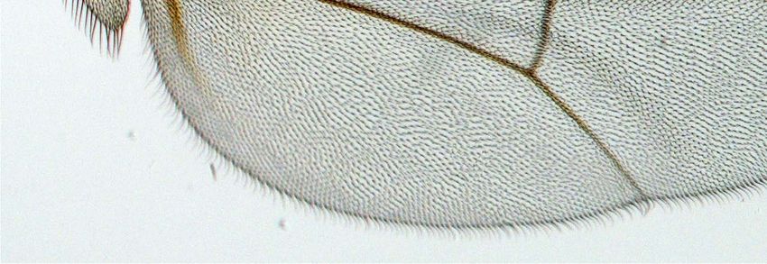
Research article                                                                            Developmental biology and stem cells

                                   eLife digest Cells need to work together for a multi-celled organism, such as a plant or an
                                   animal, to thrive. Many complicated signaling pathways therefore exist that allow cells to
                                   communicate with one another and to control their own activity in response to the signals that they
                                   receive. One such pathway, called the Hippo signaling pathway, regulates when cells grow and
                                   divide, which allows organs and tissues to develop correctly and helps to prevent cancerous tumors
                                   from forming.
                                      Signaling pathways often control the activity of cells by affecting how particular genes are
                                   expressed from DNA. One way of doing this is to activate or inactivate proteins called transcription
                                   factors, which bind to sections of DNA to alter the expression of nearby genes. In fruit flies, the
                                   Hippo signaling pathway stops cells from dividing by inactivating Yorkie, a protein that binds to and
                                   activates certain transcription factors. However, exactly how Yorkie is able to activate these
                                   transcription factors was unclear.
                                      For transcription factors to function correctly, they must be able to reach the stretch of DNA
                                   where they bind. Therefore, another way to alter gene expression is to change how the DNA is
                                   packaged in a cell. This can be done by modifying the proteins that the DNA is wrapped around,
                                   which are called histones, for example by using enzymes called methyltransferases to add methyl
                                   groups to these proteins.
                                      Qing, Yin et al. looked at the Hippo signaling pathway in the fruit fly Drosophila, and found that
                                   the Yorkie protein only activates transcription factors when another protein called Ncoa6—which is
                                   part of a methyltransferase—binds to it. Furthermore, when the Ncoa6 protein was bound directly
                                   to the transcription factor, the tissue grew normally even when the Yorkie protein was not present.
                                   These findings reveal the importance of histone modifications in controlling tissue growth, and
                                   could provide a new direction in the search for cancer treatments.
                                   DOI: 10.7554/eLife.02564.002

                                   analysis has implicated Trx in the maintenance of Hox gene transcription and Trr in ecdysone receptor
                                   (EcR)-mediated gene transcription (Sedkov et al., 2003). Nuclear receptor coactivator 6 (Ncoa6) is a
                                   specific subunit of the Trr complex in both Drosophila and mammals (Mohan et al., 2011). Although its
                                   in vivo function remains undefined in Drosophila, the mammalian Ncoa6 orthologue (also known as
                                   NRC, ASC-2, TRBP, PRIP, and RAP250) is essential for embryonic development (Kuang et al., 2002;
                                   Antonson et al., 2003; Zhu et al., 2003; Mahajan et al., 2004). The mammalian Ncoa6 has been shown
                                   to potentiate the activity of nuclear hormone receptors and other DNA-binding transcription factors,
                                   at least in part, by recruiting the H3K4 methyltransferases (Mahajan and Samuels, 2008). Interestingly,
                                   like YAP, the mammalian Ncoa6 is a pro-survival and anti-apoptotic gene (Mahajan et al., 2004) and is
                                   amplified in multiple cancer types such as breast, colon, and lung cancers (Lee et al., 1999).
                                       In this study, we identify Ncoa6 as a Yki-binding protein that is required for transcriptional regula-
                                   tion by the Hippo signaling pathway. We provide evidence showing that the transcriptional coactivator
                                   function of Yki depends on its ability to interact with Ncoa6 and that the Trr methyltransferase complex
                                   is functionally required for Hippo-mediated growth and gene expression. We further show that Yki,
                                   Ncoa6, and Trr are required for normal H3K4 methylation at Hippo target genes. Thus, Yki functions
                                   as a transcriptional coactivator, at least in part, by recruiting a H3K4 methyltransferase and altering the
                                   chromatin state of target genes.

                                   Results and discussion
                                   We recently reported a genome-wide RNAi screen in Drosophila S2R+ cells using a luciferase reporter
                                   driven by a minimal Hippo Responsive Element (HRE) from the Hippo target gene diap1 (Koontz et al.,
                                   2013). Briefly, Drosophila S2R+ cells were transfected with Yki- and Sd-expressing vectors, together
                                   with HRE-luciferase reporter and Pol III-Renilla expression vector as an internal control. Transfected
                                   cells were then seeded into individual dsRNA-containing 96-well plates. After RNAi depletion, HRE-
                                   luciferase reporter activity was measured and normalized to the Renilla control. This RNAi screen
                                   allowed us to uncover both positive and negative regulators of the HRE-luciferase reporter. We have
                                   previously characterized a negative regulator from the screen, Tgi (Koontz et al., 2013). Here, we
                                   focus on the positive transcriptional regulators (Figure 1—figure supplement1).

Qing et al. eLife 2014;3:e02564. DOI: 10.7554/eLife.02564                                                                              2 of 13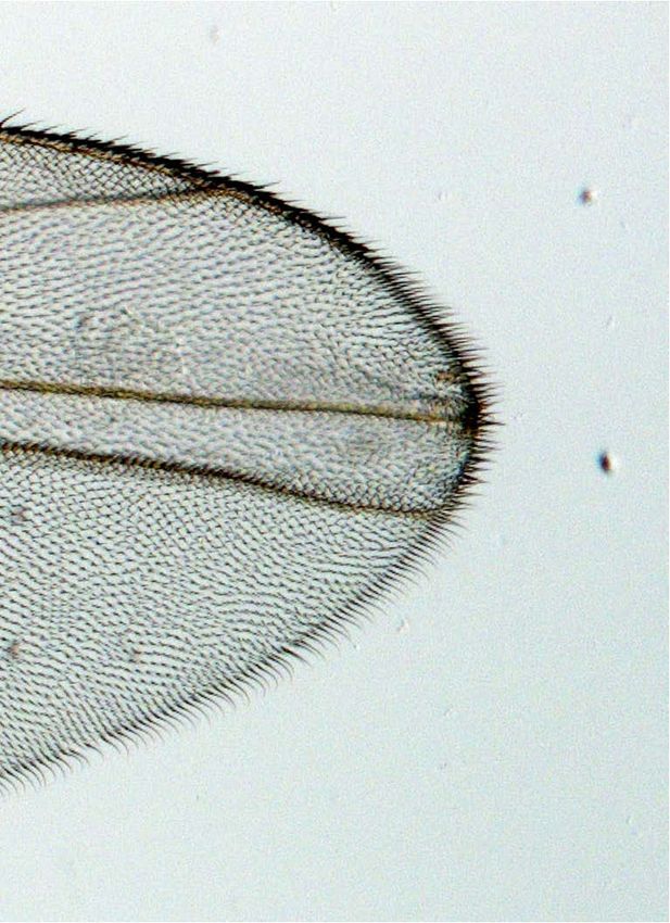
Research article                                                                                    Developmental biology and stem cells

                                        One of the positive regulators identified in our primary screen is Nuclear receptor coactivator
                                     6 (Ncoa6), which was confirmed by repeating the RNAi experiment in triplicate using re-synthesized
                                     dsRNA (Figure 1—figure supplement1). We were particularly interested in Ncoa6 since it contains
                                     three PPxY sequences (Figure 1A), which represent a well-established ligand binding motif for

Figure 1. Ncoa6 physically interacts with Yki and regulates HRE activity. (A) Schematic protein structure of Drosophila Ncoa6 and its human orthologue,
which contain three and two PPxY motifs, respectively. (B) S2R+ cells expressing the indicated constructs were subjected to immunoprecipitation as
indicated. Note the physical interactions between Ncoa6 and Yki, and absence of interactions between Ncoa63m and Yki or between Ncoa6 and YkiWM.
(C) Drosophila S2R+ cells co-transfected with HA-Yki and FLAG-Ncoa6 or FLAG-Ncoa63m constructs were stained for the indicated epitopes. Cells with
or without FLAG expression are marked by arrowheads and arrows, respectively. Both FLAG-Ncoa6 and FLAG-Ncoa63m were localized to the nucleus
(arrowheads), while HA-Yki was more concentrated in the cytoplasm (arrows). FLAG-Ncoa6, but not FLAG-Ncoa63m, induced nuclear accumulation of
HA-Yki (compare arrowheads in the merged channel). (D) Luciferase activity was measured in triplicates in Drosophila S2R+ cells transfected with the
indicated constructs. Ncoa6, but not Ncoa63m, enhanced Yki/Sd-mediated activation of HRE-luciferase reporter. This enhancement was suppressed by
co-expression of Wts. Error bars represent standard deviations. (E) Drosophila S2R+ cells expressing FLAG-tagged Ncoa6 were subjected to ChIP
analysis using control IgG, antibodies against FLAG or antibodies against endogenous Yki. The enrichment of HRE at the endogenous diap1 locus was
measured by real-time PCR. Both Yki and FLAG-Ncoa6 bound to the diap1 HRE.
DOI: 10.7554/eLife.02564.003
The following figure supplement is available for figure 1:
Figure supplement1. Identification of Ncoa6 as a positive regulator of the HRE activity from cell-based RNAi screen.
DOI: 10.7554/eLife.02564.004

Qing et al. eLife 2014;3:e02564. DOI: 10.7554/eLife.02564                                                                                        3 of 13
Research article                                                                            Developmental biology and stem cells

                                   WW domains. Since Yki contains two WW domains, we hypothesized that Ncoa6 may potentiate
                                   Yki-mediated transcriptional activation through physical interactions with Yki. Indeed, epitope-
                                   tagged Ncoa6 and Yki coimmunoprecipitated with each other in Drosophila S2R+ cells (Figure 1B).
                                   This interaction was abolished by mutating the three PPxY motifs in Ncoa6 (Ncoa63m) or the two
                                   WW domains in Yki (YkiWM) (Figure 1B), suggesting that the Ncoa6–Yki interaction was mediated
                                   by Ncoa6's PPxY motifs and Yki's WW domains. In agreement with this conclusion, we found that
                                   wild-type Ncoa6, but not Ncoa63m, promoted nuclear accumulation of Yki in S2R+ cells in co-
                                   transfection assays (Figure 1C). Of note, a recently published Hippo pathway protein–protein
                                   interactome included Ncoa6 as one of 245 proteins that were co-immunopreciated by Yki (Kwon
                                   et al., 2013).
                                       Consistent with our observation that RNAi knockdown of Ncoa6 reduced the HRE reporter
                                   activity, overexpression of Ncoa6, but not the PPxY mutant Ncoa63m, potently enhanced Yki/
                                   Sd-mediated HRE reporter activity in Drosophila S2R+ cells (Figure 1D). In addition, the enhance-
                                   ment of HRE reporter activity by Ncoa6 was significantly suppressed by co-expression of the
                                   kinase Wts (Figure 1D). These results further support the importance of Ncoa6–Yki interactions in
                                   Hippo-responsive transcriptional regulation. Consistent with this notion, chromatin immunopre-
                                   cipitation (ChIP) revealed that Ncoa6, like Yki, binds to the HRE site in the endogenous diap1
                                   gene locus in S2R+ cells (Figure 1E).
                                       Since mutant alleles of Ncoa6 are not available, we used a previously validated transgenic RNAi line
                                   (Herz et al., 2012) to assess the role of Ncoa6 in tissue growth and Hippo target expression in vivo.
                                   Expression of UAS-Ncoa6 RNAi by the dpp-Gal4 driver resulted in a significant decrease in the width
                                   of the dpp-expression domain in adult wings, which corresponds to the region bordered by veins L3
                                   and L4 (Figure 2A). Examination of third instar larval wing imaginal discs revealed a corresponding
                                   decrease in the expression of diap1 and four-jointed (fj), two well-characterized Hippo pathway target
                                   genes (Figure 2B–C,E–F). These results suggest that Ncoa6 is required for normal tissue growth and
                                   expression of Hippo target genes in vivo.
                                       Next, we examined genetic interactions between Ncoa6 and the Hippo pathway. Overexpression
                                   of Yki or RNAi of Wts by the GMR-Gal4 driver leads to increased eye size (Figure 3D,G). Both
                                   phenotypes were suppressed by RNAi knockdown of Ncoa6 (Figure 3E,H). Conversely, knock-
                                   down of Ncoa6 exacerbated the small eye size induced by Sd overexpression (Figure 3J–K). To
                                   further investigate the genetic interactions between Ncoa6 and the Hippo pathway, we used
                                   Mosaic Analysis with a Repressible Cell Marker (MARCM) (Lee and Luo, 1999) to examine the
                                   requirement of Ncoa6 in hpo mutant clones. Ncoa6 knockdown suppressed the overgrowth as
                                   well as the elevated Diap1 expression in hpo mutant clones (Figure 4A–D). In fact, hpo mutant
                                   clones with Ncoa6 knockdown showed a decrease in Diap1 expression, similar to wildtype clones
                                   with Ncoa6 knockdown (Figure 4C–D). These findings further implicate Ncoa6 in Hippo-mediated
                                   growth control and gene expression.
                                       The physical interactions between Yki and Ncoa6, together with the requirement for Ncoa6 in
                                   tissue growth and Hippo target gene expression, suggest that Yki may function as a transcriptional
                                   coactivator by interacting with Ncoa6. Since Sd is the primary DNA-binding transcription partner for
                                   Yki (Koontz et al., 2013), we reasoned that fusing the DNA-binding domain of Sd with Ncoa6 may
                                   directly target Ncoa6 to Hippo target genes and therefore stimulate their transcription in a Yki-
                                   independent manner (i.e., bypassing the genetic requirement for Yki). We tested this hypothesis in
                                   Drosophila wing discs using the MARCM technique. As reported before (Huang et al., 2005), yki
                                   mutant clones grew poorly and the rarely recovered clones always showed decreased Diap1 levels
                                   (Figure 5A–B). MARCM clones expressing a fusion protein between the DNA-binding domain of Sd
                                   and Ncoa6 (SdDB-Ncoa6) resulted in rounded clone morphology and dramatically increased Diap1
                                   levels (Figure 5C). Significantly, the SdDB-Ncoa6 fusion protein, but not wild-type Ncoa6, rescued the
                                   growth defect and the decreased Diap1 levels in yki mutant clone (Figure 5D–E). In fact, yki mutant
                                   clones with SdDB-Ncoa6 overexpression were indistinguishable in clone size and Diap1 expression
                                   compared to wild-type clones with SdDB-Ncoa6 overexpression (Figure 5C–D). Thus, the SdDB–
                                   Ncoa6 fusion protein exhibits gain-of-function activity in a Yki-independent manner. Consistent with
                                   this notion, the SdDB-Ncoa6 fusion protein robustly stimulated the HRE-luciferase reporter in S2R+
                                   cells in a manner that was not suppressed by co-expression of Wts (Figure 5F), in contrast to Wts'
                                   ability to suppress the HRE-luciferase reporter activity stimulated by the co-transfection of Sd, Yki, and
                                   Ncoa6 (Figure 1D).

Qing et al. eLife 2014;3:e02564. DOI: 10.7554/eLife.02564                                                                             4 of 13
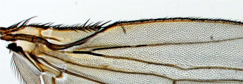
Research article                                                                                   Developmental biology and stem cells

Figure 2. Ncoa6 and Trr are required for normal tissue growth and expression of Hippo target genes in Drosophila imaginal discs. (A) RNAi
knockdown of Ncoa6 and Trr by dpp-Gal4 resulted in decreased area of the dpp expression domain in adult wings. The pictures were taken under the
same magnification. The graph shows quantification of the dpp expression domain (green area in the schematic drawing) relative to the entire wing area
(mean ± SEM, n = 14, ***p
Research article                                                                                Developmental biology and stem cells

                                   Figure 3. Genetic interactions between Ncoa6-Trr and the Hippo pathway. Adult eye images of the indicated
                                   genotypes, all taken under the same magnification. (A) GMR-Gal4/+. Wild-type control. (B) GMR-Gal4/+; UAS-
                                   Ncoa6 RNAi/+. RNAi knockdown of Ncoa6 resulted in a mild decrease in eye size (compare B to A). (C) UAS-
                                   Dicer2/+; GMR-Gal4/+; UAS-trr RNAi/+. RNAi knockdown of Trr resulted in no visible effects on eye size (compare
                                   C to A). (D) GMR-Gal4 UAS-Yki/+. Overexpression of Yki resulted in an increase in eye size (compare D to A). (E)
                                   GMR-Gal4 UAS-Yki/+; UAS-Ncoa6 RNAi/+. RNAi knockdown of Ncoa6 suppressed eye overgrowth induced by Yki
                                   overexpression (compare E to D). (F) UAS-Dicer2/+; GMR-Gal4 UAS-Yki/+; UAS-trr RNAi/+. RNAi knockdown of Trr
                                   suppressed eye overgrowth induced by Yki overexpression (compare F to D). (G) UAS-Wts RNAi/+; GMR-Gal4/+.
                                   RNAi knockdown of Wts resulted in an increase in eye size (compare G to A). (H) UAS-Wts RNAi/+; GMR-Gal4/
                                   UAS-Ncoa6 RNAi. RNAi knockdown of Ncoa6 suppressed eye overgrowth induced by Wts knockdown (compare H
                                   to G). (I) UAS-Dicer2/+; UAS-Wts RNAi/+; GMR-Gal4/ UAS-trr RNAi. RNAi knockdown of Trr did not obviously
                                   suppress eye overgrowth caused by Wts knockdown. (J) GMR-Gal4 UAS-Sd/+. Overexpression of Sd resulted in a
                                   decrease in eye size (compare J to A). (K) GMR-Gal4 UAS-Sd/+; UAS-Ncoa6 RNAi/+. RNAi knockdown of Ncoa6
                                   enhanced the small eye phenotype caused by Sd overexpression (compare K to J). (L) UAS-Dicer2/+; GMR-Gal4
                                   UAS-Sd/+; UAS-trr RNAi/+. RNAi knockdown of Trr enhanced the small eye phenotype caused by Sd overexpression
                                   (compare L to J).
                                   DOI: 10.7554/eLife.02564.006

                                   in adult wings and a corresponding decrease in the Hippo target genes diap1 and fj (Figure 2A,D,G).
                                   Like Ncoa6, Trr knockdown suppressed eye overgrowth induced by Yki overexpression (Figure 3D,F)
                                   and aggravated the small eye phenotype caused by Sd overexpression (Figure 3J,L), although it did not
                                   visibly suppress eye overgrowth induced by Wts RNAi (Figure 3I). We also used MARCM to examine
                                   the requirement of Trr in hpo mutant clones. Similar to Ncoa6, Trr knockdown suppressed the overgrowth
                                   as well as the elevated Diap1 expression in hpo mutant clones (Figure 4E–F). Taken together, these
                                   results implicate the Trr methyltransferase complex in Hippo-mediated growth control and target
                                   gene expression.
                                       The Trr methyltransferase complex in Drosophila mainly affects histone H3K4 monomethylation
                                   with subtle effect on H3K4 di- or trimethylation (Herz et al., 2012; Kanda et al., 2013). To determine
                                   if Yki, Ncoa6, and Trr regulate growth in the Hippo pathway by modulating histone H3K4 methylation,
                                   we first examined the global levels of histone H3K4 methylation in Drosophila wing imaginal discs. It
                                   was reported previously that RNAi knockdown of Trr (using the en-Gal4 driver) led to a strong decrease

Qing et al. eLife 2014;3:e02564. DOI: 10.7554/eLife.02564                                                                                   6 of 13
Research article                                                                               Developmental biology and stem cells

                                   Figure 4. Ncoa6 and Trr are required for Hippo-mediated target gene expression. Wing discs containing
                                   GFP-marked MARCM clones were stained for Diap1 (red). For each genotype, the left most panel shows low
                                   magnification view of the wing disc (Hoechst + GFP), while the remaining three panels show higher magnification
                                   view of the same wing disc (GFP, Diap1 and GFP + Diap1). (A–F) Wing discs containing GFP-marked MARCM
                                   clones (green) of WT control (A), hpo mutant (B), Ncoa6 RNAi (C), hpo mutant with Ncoa6 RNAi (D), Trr RNAi (E),
                                   and hpo mutant with Trr RNAi (F). Note the increased Diap1 levels in hpo mutant clones and the decreased
                                   Diap1 levels in Ncoa6 RNAi or Trr RNAi clones. Also note the decreased Diap1 levels in hpo mutant clones with
                                   Ncoa6 RNAi or Trr RNAi.
                                   DOI: 10.7554/eLife.02564.007

Qing et al. eLife 2014;3:e02564. DOI: 10.7554/eLife.02564                                                                                  7 of 13
Research article                                                                                  Developmental biology and stem cells

                                   Figure 5. Fusion of Ncoa6 with the DNA binding domain of Sd bypasses Yki to stimulate Hippo target gene and
                                   tissue growth. (A–E) Wing discs containing GFP-marked MARCM clones (green) of WT control (A), ykiB5 (B),
                                   SdDB-Ncoa6 overexpression (C), ykiB5 with SdDB-Ncoa6 overexpression (D), and ykiB5 with Ncoa6 overexpression
                                   (E),were stained for Diap1 (red). For each genotype, the left most panel shows low magnification view of the wing
                                   Figure 5. Continued on next page

Qing et al. eLife 2014;3:e02564. DOI: 10.7554/eLife.02564                                                                                      8 of 13
Research article                                                                                  Developmental biology and stem cells

                                   Figure 5. Continued
                                   disc (Hoechst + GFP), while the remaining three panels show higher magnification view of the same wing disc (GFP,
                                   Diap1, and GFP + Diap1). Note the decreased Diap1 expression and undergrowth of ykiB5 clones (B) or ykiB5 clones
                                   with Ncoa6 overexpression (E). SdDB-Ncoa6 overexpression resulted in elevated Diap1 levels in ykiB5 clones (D). (F)
                                   Luciferase activity was measured in triplicates in Drosophila S2R+ cells transfected with the indicated constructs.
                                   Error bars represent standard deviations. Note the Wts-insensitive stimulation of the HRE-luciferase reporter by
                                   SdDB-Ncoa6.
                                   DOI: 10.7554/eLife.02564.008

                                   in H3K4me1 in the posterior compartment of the wing imaginal discs, while it had marginal effects on
                                   H3K4me2 and H3K4me3 levels (Mohan et al., 2011; Herz et al., 2012). It was also showed that RNAi
                                   knockdown of Ncoa6 in the posterior compartment of the wing imaginal disc resulted in a weak reduction
                                   in H3K4me1 levels (Herz et al., 2012), which we confirmed (Figure 6—figure supplement1). We further
                                   examined H3K4me2 and H3K4me3 levels in these imaginal discs, and observed a very subtle decrease
                                   in H3K4me3 levels and no detectable changes in H3K4me2 levels (Figure 6—figure supplement
                                   1B–C). However, when we examined mutant clones of yki in the wing imaginal discs, we could not
                                   detect any changes in the global levels of H3K4me1, H3K4me2, or H3K4me3 (Figure 6—figure
                                   supplement1).
                                       Given the negligible effect of Yki on global levels of H3K4 methylation, we investigated whether
                                   Yki modulates local H3K4 methylation on Hippo target genes. It is well established that H3K4 mono-
                                   methylation marks enhancers and actively transcribed introns, while H3K4 trimethylation is enriched
                                   at active promoters and transcription start site (TSS)-proximal regions (Heintzman et al., 2007;
                                   Kharchenko et al., 2011). Interestingly, a previous genome-wide analysis in Drosophila S2 cells
                                   revealed that diap1 and ex, two well-characterized Hippo target genes, display such differential
                                   enrichment of H3K4me1 and H3K4me3 at the respective region of each gene (Herz et al., 2012)
                                   (Figure 6A). To examine the contribution of Yki, Ncoa6, and Trr to H3K4 methylation at these Hippo
                                   target genes, we knocked down each protein in S2R+ cells and performed ChIP analysis with anti-
                                   bodies against H3K4me1 and H3K4me3. RNAi knockdown of Yki, Ncoa6 or Trr resulted in a decrease
                                   of H3K4me3 in the TSS-proximal region of diap1 and ex (Figure 6B), which normally showed the
                                   strongest enrichment of H3K4me3 marks (Herz et al., 2012) (Figure 6A). RNAi knockdown of Yki,
                                   Ncoa6 or Trr also led to a decrease of H3K4me1 in the intronic HRE of diap1 and an upstream region
                                   of diap1 or ex which normally showed the strongest enrichment of H3K4me1 marks (Herz et al.,
                                   2012) (Figure 6C). Collectively, these data are consistent with the view that Yki activates target gene
                                   transcription by interacting with the Trr methyltransferase complex and modifying the chromatin
                                   state of the target loci.
                                       Despite the ever expanding complexity of upstream inputs into the Hippo pathway, all of them
                                   converge on the transcriptional coactivator Yki. Thus, understanding the molecular mechanisms by
                                   which Yki regulates tissue growth and target gene expression has important implications for devel-
                                   opmental and cancer biology. Previous studies have established that Yki functions primarily as a
                                   coactivator for Sd and that Yki promotes tissue growth by antagonizing Sd's repressor function (Wu
                                   et al., 2008; Koontz et al., 2013). Our current study has extended the previous work by identifying
                                   Ncoa6 as a Yki-binding cofactor that is required for the expression of Yki target genes. The ability of
                                   the SdDB-Ncoa6 fusion protein to rescue the growth and transcriptional defects in yki mutant clones
                                   highlights the importance of Ncoa6 recruitment in the transcriptional output of the Hippo pathway.
                                   Our results further suggest that Ncoa6 recruits the Trr methyltransferase complex to Hippo target
                                   genes and that Yki regulates target gene transcription by modulating local H3K4 methylation.
                                   Consistent with this view, a recent genome-wide chromatin-binding analysis revealed a correlation
                                   between Yki-bound chromatin and peaks of H3K4me3 modification in Drosophila wing discs and
                                   embryos (Oh et al., 2013). We note that, besides the H3K4 methyltransferases, the mammalian
                                   Ncoa6 has been reported to potentiate the activity of transcription factors by interacting with his-
                                   tone acetyltransferase CBP/p300 and several RNA binding proteins (CAPER, CoAA and PIMT)
                                   (Mahajan and Samuels, 2008). Whether these additional mechanisms also contribute to the func-
                                   tion of Ncoa6 in Yki-mediated growth control requires further investigation. Given the biological and
                                   clinical significance of the Hippo pathway, further studies into the molecular mechanism of Ncoa6
                                   will advance our understanding of developmental growth control and facilitate the development of
                                   novel therapeutic strategies.

Qing et al. eLife 2014;3:e02564. DOI: 10.7554/eLife.02564                                                                                      9 of 13
Research article                                                                                  Developmental biology and stem cells

                                   Figure 6. Yki modulates local H3K4 methylation at Hippo target genes. (A) Schematic view of diap1 and ex
                                   genomic loci analyzed by ChIP. Transcriptional start site is labeled as +1, and p1–p5 are a series of primer sets
                                   encompassing following regions of diap1 and ex: diap1: p1: −1951 ∼ −1813, p2: +228 ∼ +377, p3: +3993 ∼ +4104;
                                   ex: p4: −749 ∼ −608, p5: +249 ∼ +393. Note that p3 covers the diap1 HRE. Also shown are the profiles of H3K4me1
                                   (blue line) and H3K4me3 (red line) binding derived from a previously published ChIP-Seq analysis in S2 cells (Herz
                                   et al., 2012). (B and C) RNAi knockdown of Yki, Ncoa6 or Trr resulted in decreased H3K4me3 (B) and H3K4me1
                                   (C) modification on Hippo target genes. ChIP analysis of H3K4me1 or H3K4me3 were performed in Drosophila
                                   S2R+ cells treated with dsRNA of GFP (control), Yki, Ncoa6, or Trr. Chromatins were precipitated by control IgG or
                                   antibodies against H3K4me1 and H3K4me3. The enrichment of ChIP products on diap and ex was measured by
                                   real-time PCR using the indicated primers. ***p
Research article Developmental biology and stem cells
in Ncoa6 using the QuikChange II XL Site-Directed Mutagenesis Kit (Agilent, Santa Clara, CA), replac-
ing tyrosine(Y) with alanine (A). To generate Sd-Ncoa6, Sd DNA binding domain was inserted to the
N-terminal of Ncoa6. FLAG-tag was inserted to the N-terminal of Ncoa6, Ncoa63m, and Sd-Ncoa6 and
cloned into the attB-UAS vector.
Drosophila genetics
Flies with the following genotypes have been described previously: ykiB5, UAS-Yki (Huang et al.,
2005), hpo42−48 (Wu et al., 2003), fj-lacZ reporter fj9-II (Villano and Katz, 1995), UAS-Sd (Halder et al.,
1998), UAS-Wts RNAi (Stock ID VDRC v106174). The UAS-Ncoa6 RNAi and UAS-trr RNAi lines have
been validated previously (Herz et al., 2012) and were obtained from Bloomington Drosophila Stock
Center (Stock ID 34964 and 29,563). attB-UAS-Ncoa6 and attB-UAS-FLAG-SdDB-Ncoa6 transgenes
were inserted into the 86Fa attP acceptor site by phiC31-mediated site-specific transformation
(Bischof et al., 2007).
For the MARCM experiments in Figure 4, the following clones were induced 48–60 hr after egg
deposition and heat shocked at 37°C for 15 min:
UAS-GFP hs-FLP; FRT42D, Tub-Gal80/FRT42D; Tub-Gal4/+
UAS-GFP hs-FLP; FRT42D, Tub-Gal80/FRT42D hpo42−48; Tub-Gal4/+
UAS-GFP hs-FLP; FRT42D, Tub-Gal80/FRT42D; Tub-Gal4/UAS-Ncoa6RNAi
UAS-GFP hs-FLP; FRT42D, Tub-Gal80/FRT42D hpo42−48; Tub-Gal4/UAS-Ncoa6RNAi
UAS-Dicer2/UAS-GFP hs-FLP; FRT42D, Tub-Gal80/FRT42D; Tub-Gal4/UAS-trrRNAi
UAS-Dicer2/UAS-GFP hs-FLP; FRT42D, Tub-Gal80/FRT42D hpo42−48; Tub-Gal4/UAS-trrRNAi
For the MARCM experiments in Figure 5A–E, the following clones were induced 72–84 hr after
egg deposition and heat shocked at 37°C for 10 min:
UAS-GFP hs-FLP; FRT42D, Tub-Gal80/FRT42D; Tub-Gal4/+
UAS-GFP hs-FLP; FRT42D, Tub-Gal80/FRT42D ykiB5; Tub-Gal4/+
UAS-GFP hs-FLP; FRT42D, Tub-Gal80/FRT42D; Tub-Gal4/UAS-SdDB-Ncoa6
UAS-GFP hs-FLP; FRT42D, Tub-Gal80/FRT42D ykiB5; Tub-Gal4/UAS-SdDB-Ncoa6
UAS-GFP hs-FLP; FRT42D, Tub-Gal80/FRT42D ykiB5; Tub-Gal4/UAS-Ncoa6
Drosophila cell culture, transfection, immunoprecipitation,
immunoflurescence, and luciferase reporter assay
Drosophila S2R+ cells were cultured in Schneider's Drosophila Medium (Life Technologies) supple-
mented with 10% fetal bovine serum and antibiotics. HA-Yki and HA-YkiWM have been described pre-
viously (Huang et al., 2005). Luciferase assay was carried out using Dual Luciferase Assay System
(Promega, Madison, WI) and a FLUOstar Luminometer (BMG LabTechnologies, Germany). Tansfection,
immunopreciptation, and immunofluorescence staining of S2R+ cells were performed using standard
protocols as described (Yin et al., 2013).
ChIP assays
ChIP assays were performed according to a previously described protocol (Wang et al., 2009). Briefly,
∼5 × 10⁶ (for ChIP assay with histone methylation antibodies) or 1.5 × 10⁷ (for CHIP assay with Yki or FLAG
antibodies) S2R+ cells were cross-linked with 1% formaldehyde and sonicated to an average fragment
size between 200 bp and 500 bp. Two micrograms of control IgG or specific antibodies, including
rabbit ɑ-H3K4me1 (8895, Abcam, England) and rabbit ɑ-H3K4me3 (8580, Abcam), and 50 µl of protein
G agarose were used in each ChIP assay. The immunoprecipitated DNA was quantified using real-time
PCR. All values were normalized to the input. The primers for analyzing the ChIP NA are provided
as follows:
p1 Forward: TGTTCTTGTTGGTGCTGCTT
p1 Reverse: TTAATGCTGGCATGGTTTCA
p2 Forward: TAAAACTGGGGCTCACCTTG
p2 Reverse: TCGTGTTCACGGAAAATCAA
p3 (HRE) Forward: ACGAACACGAAGACCAAA
p3 (HRE) Reverse: CTCCAAGCCAGTTTGATT
p4 Forward: AAAAGAGGGAAGAGGGAGCA
Qing et al. eLife 2014;3:e02564. DOI: 10.7554/eLife.02564 11 of 13Research article Developmental biology and stem cells
p4 Reverse: GAATCGGAATCGGAACTTGA
p5 Forward: TCGCACTCGCCTCAATTAC
p5 Reverse: CAGCACCAACTTTTCGGAGT
Acknowledgements
We thank Jianzhong Yu, Melissa Jones and Elizabeth Garcia for technical assistance. This study was
supported in part by grants from the National Institutes of Health (EY015708). DP is an investigator of
the Howard Hughes Medical Institute.
Additional information
Competing interests
DP: Reviewing editor, eLife. The other authors declare that no competing interests exist.
Funding
Funder Grant reference number Author
Howard Hughes Medical Yun Qing, Feng Yin, Wei Wang,
Institute Yonggang Zheng, Pengfei Guo,
Frederick Schozer, Hua Deng,
Duojia Pan
National Institutes of NIH EY015708 Yun Qing, Feng Yin, Wei Wang,
Health Yonggang Zheng, Pengfei Guo,
Frederick Schozer, Hua Deng,
Duojia Pan
The funders had no role in study design, data collection and interpretation, or the
decision to submit the work for publication.
Author contributions
YQ, FY, Conception and design, Acquisition of data, Analysis and interpretation of data, Drafting
or revising the article; WW, YZ, Conception and design, Acquisition of data, Analysis and inter-
pretation of data; PG, HD, Acquisition of data; FS, Acquisition of data, Analysis and interpreta-
tion of data; DP, Conception and design, Analysis and interpretation of data, Drafting or revising
the article
References
Antonson P, Schuster GU, Wang L, Rozell B, Holter E, Flodby P, Treuter E, Holmgren L, Gustafsson JA. 2003.
Inactivation of the nuclear receptor coactivator RAP250 in mice results in placental vascular dysfunction.
Molecular and Cellular Biology 23:1260–1268. doi: 10.1128/MCB.23.4.1260-1268.2003.
Badouel C, Garg A, McNeill H. 2009. Herding Hippos: regulating growth in flies and man. Current Opinion in Cell
Biology 21:837–843. doi: 10.1016/j.ceb.2009.09.010.
Barry ER, Camargo FD. 2013. The Hippo superhighway: signaling crossroads converging on the Hippo/Yap
pathway in stem cells and development. Current Opinion in Cell Biology 25:247–253. doi: 10.1016/j.
ceb.2012.12.006.
Bischof J, Maeda RK, Hediger M, Karch F, Basler K. 2007. An optimized transgenesis system for Drosophila using
germ-line-specific phiC31 integrases. Proceedings of the National Academy of Sciences of the United States of
America 104:3312–3317. doi: 10.1073/pnas.0611511104.
Halder G, Johnson RL. 2011. Hippo signaling: growth control and beyond. Development 138:9–22. doi: 10.1242/
dev.045500.
Halder G, Polaczyk P, Kraus ME, Hudson A, Kim J, Laughon A, Carroll S. 1998. The vestigial and scalloped
proteins act together to directly regulate wing-specific gene expression in Drosophila. Genes & Development
12:3900–3909. doi: 10.1101/gad.12.24.3900.
Harvey K, Tapon N. 2007. The Salvador-Warts-Hippo pathway-an emerging tumour-suppressor network. Nature
Reviews Cancer 7:182–191. doi: 10.1038/nrc2070.
Heintzman ND, Stuart RK, Hon G, Fu Y, Ching CW, Hawkins RD, Barrera LO, Van CS, Qu C, Ching KA, Wang W,
Weng Z, Green RD, Crawford GE, Ren B. 2007. Distinct and predictive chromatin signatures of transcriptional
promoters and enhancers in the human genome. Nature Genetics 39:311–318. doi: 10.1038/ng1966.
Herz HM, Mohan M, Garruss AS, Liang K, Takahashi YH, Mickey K, Voets O, Verrijzer CP, Shilatifard A. 2012.
Enhancer-associated H3K4 monomethylation by Trithorax-related, the Drosophila homolog of mammalian Mll3/
Mll4. Genes & Development 26:2604–2620. doi: 10.1101/gad.201327.112.
Qing et al. eLife 2014;3:e02564. DOI: 10.7554/eLife.02564 12 of 13Research article Developmental biology and stem cells
Huang J, Wu S, Barrera J, Matthews K, Pan D. 2005. The Hippo signaling pathway coordinately regulates
cell proliferation and apoptosis by inactivating Yorkie, the Drosophila homolog of YAP. Cell 122:421–434.
doi: 10.1016/j.cell.2005.06.007.
Kanda H, Nguyen A, Chen L, Okano H, Hariharan IK. 2013. The Drosophila ortholog of MLL3 and MLL4, trithorax
related, functions as a negative regulator of tissue growth. Molecular and Cellular Biology 33:1702–1710.
doi: 10.1128/MCB.01585-12.
Kharchenko PV, Alekseyenko AA, Schwartz YB, Minoda A, Riddle NC, Ernst J, Sabo PJ, Larschan E, Gorchakov AA,
Gu T, Linder-Basso D, Plachetka A, Shanower G, Tolstorukov MY, Luquette LJ, Xi R, Jung YL, Park RW, Bishop EP,
Canfield TK, Sandstrom R, Thurman RE, MacAlpine DM, Stamatoyannopoulos JA, Kellis M, Elgin SC, Kuroda MI,
Pirrotta V, Karpen GH, Park PJ. 2011. Comprehensive analysis of the chromatin landscape in Drosophila
melanogaster. Nature 471:480–485. doi: 10.1038/nature09725.
Koontz LM, Liu-Chittenden Y, Yin F, Zheng Y, Yu J, Huang B, Chen Q, Wu S, Pan D. 2013. The hippo effector
yorkie controls normal tissue growth by antagonizing scalloped-mediated default repression. Developmental
Cell 25:388–401. doi: 10.1016/j.devcel.2013.04.021.
Kuang SQ, Liao L, Zhang H, Pereira FA, Yuan Y, DeMayo FJ, Ko L, Xu J. 2002. Deletion of the cancer-amplified
coactivator AIB3 results in defective placentation and embryonic lethality. The Journal of Biological Chemistry
277:45356–45360. doi: 10.1074/jbc.C200509200.
Kwon Y, Vinayagam A, Sun X, Dephoure N, Gygi SP, Hong P, Perrimon N. 2013. The Hippo signaling pathway
interactome. Science 342:737–740. doi: 10.1126/science.1243971.
Lee T, Luo L. 1999. Mosaic analysis with a repressible cell marker for studies of gene function in neuronal
morphogenesis. Neuron 22:451–461. doi: 10.1016/S0896-6273(00)80701-1.
Lee SK, Anzick SL, Choi JE, Bubendorf L, Guan XY, Jung YK, Kallioniemi OP, Kononen J, Trent JM, Azorsa D,
Jhun BH, Cheong JH, Lee YC, Meltzer PS, Lee JW. 1999. A nuclear factor, ASC-2, as a cancer-amplified
transcriptional coactivator essential for ligand-dependent transactivation by nuclear receptors in vivo. The
Journal of Biological Chemistry 274:34283–34293. doi: 10.1074/jbc.274.48.34283.
Mahajan MA, Das S, Zhu H, Tomic-Canic M, Samuels HH. 2004. The nuclear hormone receptor coactivator NRC
is a pleiotropic modulator affecting growth, development, apoptosis, reproduction, and wound repair.
Molecular and Cellular Biology 24:4994–5004. doi: 10.1128/MCB.24.11.4994-5004.2004.
Mahajan MA, Samuels HH. 2008. Nuclear receptor coactivator/coregulator NCoA6(NRC) is a pleiotropic
coregulator involved in transcription, cell survival, growth and development. Nuclear Receptor Signaling
6:e002. doi: 10.1621/nrs.06002.
Mohan M, Herz HM, Smith ER, Zhang Y, Jackson J, Washburn MP, Florens L, Eissenberg JC, Shilatifard A. 2011.
The COMPASS family of H3K4 methylases in Drosophila. Molecular and Cellular Biology 31:4310–4318.
doi: 10.1128/MCB.06092-11.
Oh H, Slattery M, Ma L, Crofts A, White KP, Mann RS, Irvine KD. 2013. Genome-wide association of Yorkie with
chromatin and chromatin-remodeling complexes. Cell Reports 3:309–318. doi: 10.1016/j.celrep.2013.01.008.
Pan D. 2010. The hippo signaling pathway in development and cancer. Developmental Cell 19:491–505.
doi: 10.1016/j.devcel.2010.09.011.
Sedkov Y, Cho E, Petruk S, Cherbas L, Smith ST, Jones RS, Cherbas P, Canaani E, Jaynes JB, Mazo A. 2003.
Methylation at lysine 4 of histone H3 in ecdysone-dependent development of Drosophila. Nature 426:78–83.
doi: 10.1038/nature02080.
Villano JL, Katz FN. 1995. Four-jointed is required for intermediate growth in the proximal-distal axis in
Drosophila. Development 121:2767–2777.
Wang W, Huang L, Huang Y, Yin JW, Berk AJ, Friedman JM, Wang G. 2009. Mediator MED23 links insulin
signaling to the adipogenesis transcription cascade. Developmental Cell 16:764–771. doi: 10.1016/j.
devcel.2009.04.006.
Wu S, Huang J, Dong J, Pan D. 2003. hippo encodes a Ste-20 family protein kinase that restricts cell proliferation
and promotes apoptosis in conjunction with salvador and warts. Cell 114:445–456. doi: 10.1016/
S0092-8674(03)00549-X.
Wu S, Liu Y, Zheng Y, Dong J, Pan D. 2008. The TEAD/TEF family protein Scalloped mediates transcriptional
output of the Hippo growth-regulatory pathway. Developmental Cell 14:388–398. doi: 10.1016/j.
devcel.2008.01.007.
Yin F, Yu J, Zheng Y, Chen Q, Zhang N, Pan D. 2013. Spatial organization of hippo signaling at the plasma
membrane mediated by the tumor suppressor Merlin/NF2. Cell 154:1342–1355. doi: 10.1016/j.
cell.2013.08.025.
Zhao B, Li L, Lei Q, Guan KL. 2010. The Hippo-YAP pathway in organ size control and tumorigenesis: an updated
version. Genes & Development 24:862–874. doi: 10.1101/gad.1909210.
Zhu YJ, Crawford SE, Stellmach V, Dwivedi RS, Rao MS, Gonzalez FJ, Qi C, Reddy JK. 2003. Coactivator PRIP, the
peroxisome proliferator-activated receptor-interacting protein, is a modulator of placental, cardiac, hepatic, and
embryonic development. The Journal of Biological Chemistry 278:1986–1990. doi: 10.1074/jbc.C200634200.
Qing et al. eLife 2014;3:e02564. DOI: 10.7554/eLife.02564 13 of 13You can also read