ELONGATION AT MIDCELL IN PREPARATION OF CELL DIVISION REQUIRES FTSZ, BUT NOT MREB NOR PBP2 IN CAULOBACTER CRESCENTUS
←
→
Page content transcription
If your browser does not render page correctly, please read the page content below
ORIGINAL RESEARCH
                                                                                                                                            published: 27 August 2021
                                                                                                                                      doi: 10.3389/fmicb.2021.732031

                                             Elongation at Midcell in Preparation
                                             of Cell Division Requires FtsZ, but
                                             Not MreB nor PBP2 in Caulobacter
                                             crescentus
                                             Muriel C. F. van Teeseling 1,2*
                                             1
                                              Junior Research Group Prokaryotic Cell Biology, Department Microbial Interactions, Institute of Microbiology,
                                             Friedrich-Schiller-Universität, Jena, Germany, 2 Department of Biology, University of Marburg, Marburg, Germany

                                             Controlled growth of the cell wall is a key prerequisite for bacterial cell division. The
                                             existing view of the canonical rod-shaped bacterial cell dictates that newborn cells
                                             first elongate throughout their side walls using the elongasome protein complex, and
                                             subsequently use the divisome to coordinate constriction of the dividing daughter
                                             cells. Interestingly, another growth phase has been observed in between elongasome-
                                             mediated elongation and constriction, during which the cell elongates from the
                                             midcell outward. This growth phase, that has been observed in Escherichia coli and
                                             Caulobacter crescentus, remains severely understudied and its mechanisms remain
                                             elusive. One pressing open question is which role the elongasome key-component
                            Edited by:
                       Cara C. Boutte,
                                             MreB plays in this respect. This study quantitatively investigates this growth phase in
       University of Texas at Arlington,     C. crescentus and focuses on the role of both divisome and elongasome components.
                         United States
                                             This growth phase is found to initiate well after MreB localizes at midcell, although it does
                      Reviewed by:
                                             not require its presence at this subcellular location nor the action of key elongasome
              Saswat S. Mohapatra,
           Khallikote University, India      components. Instead, the divisome component FtsZ seems to be required for elongation
                   Michael J. Trimble,       at midcell. This study thus shines more light on this growth phase in an important model
     Simon Fraser University, Canada
                                             organism and paves the road to more in-depth studies.
                  *Correspondence:
             Muriel C. F. van Teeseling      Keywords: medial elongation, PBP3-independent peptidoglycan synthesis (PIPS), preseptal peptidoglycan
     muriel.van.teeseling@uni-jena.de        synthesis, cell wall, peptidoglycan, elongasome, divisome

                   Specialty section:
         This article was submitted to      INTRODUCTION
Microbial Physiology and Metabolism,
               a section of the journal     Almost all bacterial cells rely on the cell-spanning macromolecule peptidoglycan (PG) to provide
             Frontiers in Microbiology      integrity and shape their cells (de Pedro and Cava, 2015; van Teeseling et al., 2017). It is therefore of
           Received: 28 June 2021           key importance for bacteria to tightly control growth and remodeling of its PG sacculus throughout
         Accepted: 09 August 2021           their cell cycle (Typas et al., 2012; Egan et al., 2020). The cell cycle of most bacterial cells involves
         Published: 27 August 2021
                                            multiple growth modes, and generally involves that the cells first elongate before they start the
                              Citation:     process of constriction that effectuates cell division (Randich and Brun, 2015; Kysela et al., 2016).
              van Teeseling MCF (2021)      Although some bacteria elongate from one or both poles [e.g., some Alphaproteobacteria (Brown
   Elongation at Midcell in Preparation
                                            et al., 2012) and Actinobacteria (Umeda and Amako, 1983; Flärdh, 2010)], most bacteria grow
of Cell Division Requires FtsZ, but Not
        MreB nor PBP2 in Caulobacter
                                            longer by adding new cell wall dispersed throughout their side walls (Kysela et al., 2016). This
                            crescentus.     dispersed elongation mechanism has been studied in great detail, which unveiled an underlying
          Front. Microbiol. 12:732031.      multi-protein complex called the elongasome. The elongasome harbors the scaffolding protein
     doi: 10.3389/fmicb.2021.732031         MreB, proteins involved in PG synthesis (e.g., RodA and PBP2), as well as proteins that break

Frontiers in Microbiology | www.frontiersin.org                                    1                                          August 2021 | Volume 12 | Article 732031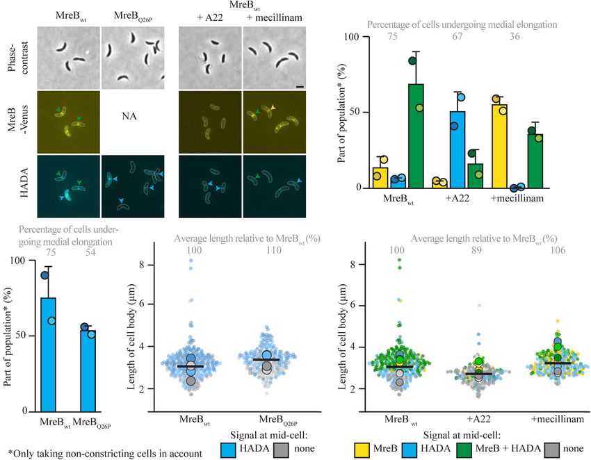
van Teeseling                                                                                                      Medial Elongation in C. crescentus

existing PG bonds to make space for insertion of new material                  Although it remains unclear how widespread this growth
(Egan et al., 2020). It is clear that MreB is a crucial part of             phase is, a similar growth phase has been observed in another
the elongasome, but its exact role remains puzzling (Ducret and             Gram-negative model organism: Caulobacter crescentus (Aaron
Grangeasse, 2021). Instead of a scaffold that actively directs the          et al., 2007). Interestingly, in this species a relatively large part
movement of all other elongasome proteins, MreB seems to have               of the elongation takes place by the medial elongation growth
a subtler coordinating role (Dion et al., 2019; Dersch et al., 2020),       phase (as it is called in C. crescentus). As in E. coli, medial
in which its movement in part seems to depend on PG synthases               elongation in C. crescentus requires FtsZ. It furthermore seems
that take the lead (Dominguez-Escobar et al., 2011; Garner et al.,          that PG synthesis at midcell starts shortly after the arrival of
2011; van Teeffelen et al., 2011; Özbaykal et al., 2020).                   FtsZ at midcell. Also in C. crescentus, MreB was seen to form
    The other important growth phase taking place in bacteria               a band at midcell, dependent on FtsZs localization at this site
is the constricting process, which incorporates cell wall material          (Figge et al., 2004; Gitai et al., 2004). Indirect evidence suggested
at midcell, thereby decreasing the diameter of the cell while               that MreBs arrival at midcell is followed quickly by the onset of
generating the new poles. In parallel to the elongasome, this               medial elongation (Aaron et al., 2007). A more in-depth study is
growth phase is supported by a dedicated multiprotein complex,              needed to further elucidate the role of MreB in medial elongation
called the divisome. In addition to specific PG synthase (e.g.,             in C. crescentus, as it is believed to play a role in this process by
FtsW and PBP3) and hydrolase enzymes, this complex features                 some (Randich and Brun, 2015), but non-quantitative indications
the scaffolding protein FtsZ (Egan et al., 2020). Very recent               by others (Aaron et al., 2007) show it might not be necessary
studies point toward two divisome subcomplexes, one consisting              for medial elongation. Furthermore, it remains unclear which PG
of the treadmilling FtsZ (Bisson Filho et al., 2017; Yang et al.,           synthases are involved. All in all, the mechanisms of this growth
2017) with its anchors (Squyres et al., 2021) that seems to act as a        phase remain puzzling and it is an open question if the growth
resting stage for the second subcomplex. This second subcomplex             phases in E. coli and C. crescentus are mechanistically the same
consists of PG synthases and seems to be active when it moves               process. In order to build a base toward better understanding
around the circumference of the cell as a separate subcomplex               of the importance and mechanisms of this growth phase, these
(Yang et al., 2021). Interestingly, it seems that after constriction        questions should be addressed.
has passed a certain threshold with the help of FtsZ, constriction             This study thus focuses on medial elongation in C. crescentus
is finished by the synthase subcomplex (Monteiro et al., 2018;              and attempts to build a quantitative base of information about
Silber et al., 2021).                                                       the growth phase in this model organism. Specifically, the
    Apart from these two thoroughly studied growth phases, some             timing of events surrounding MreBs arrival at midcell and
model organisms display an additional growth phase that takes               the onset of medial elongation are elucidated. Furthermore,
place after elongasome-guided elongation and before divisome-               the importance of MreB for this growth phase is studied and
guided constriction. During this process, that has been first               quantified using multiple approaches. Additionally, it is shown
described in Escherichia coli (Wientjes and Nanninga, 1989), the            that the elongasome-specific PBP2 is not required for this mode
cells elongate exclusively from the midcell outward. Interestingly,         of growth, whereas the presence of FtsZ is crucial. It thus seems
this process has been scarcely studied. One observation that                that preseptal PG synthesis in E. coli and medial elongation
might start to explain what happens in the transition from                  in C. crescentus are governed by the same or very similar
elongasome to divisome-guided growth, is that, at least in                  molecular mechanisms.
E. coli, FtsZ arrives at midcell and is then flanked by rings of
divisome components, including MreB (Vats and Rothfield, 2007;
Vats et al., 2009). This led to the hypothesis that either MreB             MATERIALS AND METHODS
itself guides growth at midcell or it transfers other elongasome
components to FtsZ that then steers elongation at midcell (Potluri          Bacterial Strains and Growth Conditions
et al., 2012). Interestingly, however, all of the tested divisome           All strains that are analyzed in this study are derivatives of
and elongasome proteins, except for FtsZ and its anchor ZipA                the synchronizable C. crescentus wild-type CB15N (Evinger and
were found dispensable for this growth mode in E. coli (de Pedro            Agabian, 1977; Marks et al., 2010). Strain MT309 carries a
et al., 1997; Varma and Young, 2009; Potluri et al., 2012). Most            chromosomally integrated venus-mreB under the control of the
notably, E. coli was shown to still grow via medial elongation              xylX promoter (Billini et al., 2019). Strain JAT790 contains
when the divisome-specific essential PG synthase PBP3, and                  the mreB variant G165A at the native mreB locus and a
MreB and its cognate synthase PBP2 were absent or non-                      fluorescently labeled copy of the same mreB variant venus-
functional. More in-depth studies in E. coli suggest that a very            G165A mreB under the control of the xylX promoter (Dye et al.,
early divisome consisting of FtsZ anchored by ZipA and FtsA                 2011). Another mreB variant, mreBQ26P , is present at the native
recruits the PG synthases PBP1A and 1B to midcell to drive this             locus instead of the normal MreB in strain CJW1715 (Aaron
preseptal elongation [which has also been described as PBP3-                et al., 2007). In the FtsZ depletion strain YB1585, the sole full
independent peptidoglycan synthesis (PIPS)] (Pazos et al., 2018).           copy of ftsZ is chromosomally integrated under the control of
It remains puzzling, however, if there is any role for MreB, which          the xylX promotor, whereas a non-functional truncated version
after all forms rings next to, and even directly interacts with,            (consisting of the first 163 codons) is present at the native
FtsZ (Fenton and Gerdes, 2013), shortly before preseptal PG                 locus of ftsZ (Wang et al., 2001). Strain information is detailed
synthesis commences.                                                        in Table 1.

Frontiers in Microbiology | www.frontiersin.org                         2                                    August 2021 | Volume 12 | Article 732031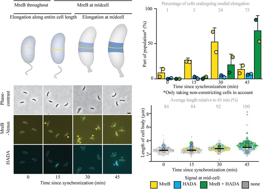
van Teeseling                                                                                                                Medial Elongation in C. crescentus

TABLE 1 | Description of all strains used in this study.                               of the different fluorescent channels per cell were extracted
                                                                                       using BacStalk. In order to compare the percentage of cells
Strain                         Genotype                         Source
                                                                                       undergoing medial elongation in the different conditions, the
CB15N                   C. crescentus wild-type       Evinger and Agabian, 1977;       fraction of non-constricting cells (n = 100 per replicate) that
                                                           Marks et al., 2010          shows HADA at midcell (either in the presence or absence) was
CJW1715                CB15N PmreB :mreBQ26P               Aaron et al., 2007          evaluated, as well as the information if a fluorescently tagged
JAT790                 CB15N PmreB :mreBG165A               Dye et al., 2011           MreB (in the relevant strains) was present at midcell in the
                        Pxyl :venus-mreBG165A
                                                                                       same cell. Measurements were exported to Excel 2016 (Microsoft,
MT309                  CB15N Pxyl :venus-mreB              Billini et al., 2019
                                                                                       United States), where average and standard deviations were
YB1585                   CB15N PftsZ :Pxyl -ftsZ           Wang et al., 2001
                                                                                       calculated. Images were processed using Fiji (Schindelin et al.,
                                                                                       2012) and Adobe Illustrator CS5 (Adobe Systems, United States).
                                                                                       Graphs representing cellular dimensions were created using
   All strains were cultivated in peptone-yeast extract (PYE)
                                                                                       SuperPlotsOfData (Goedhart, 2021) and show constricting, as
medium (Pointdexter, 1964) at 28◦ C, while shaking at 210 rpm.
                                                                                       well as non-constricting cells.
Gene expression from the xylose-inducible xylX promoter
(Meisenzahl et al., 1997) was induced for 75 min with 0.03%
xylose. The following inhibitors were used: A22 (10 µg/ml) and                         RESULTS
mecillinam (150 µg/ml).
   Synchronization of C. crescentus was achieved by density
                                                                                       Onset of Medial Elongation Takes Place
gradient centrifugation, using Percoll (Tsai and Alley, 2001).
After synchronization, cells were released in PYE medium (with                         After MreB Accumulation at Midcell
0.03% xylose and/or inhibitors, when applicable) and allowed to                        One of the biggest outstanding questions concerning medial
grow at 28◦ C, while shaking at 210 rpm.                                               elongation is if it depends on the presence of the elongasome
                                                                                       component MreB at midcell. As a first step in addressing this
                                                                                       question, the relative timing of arrival of MreB at midcell
Peptidoglycan Labeling
                                                                                       versus the onset of medial elongation was analyzed. For this,
To indicate areas of active PG insertion and remodeling,
                                                                                       C. crescentus cells expressing a fluorescent fusion to MreB were
cells were stained with the fluorescent dye hydroxycoumarin-
                                                                                       synchronized and stained with a short pulse of the fluorescent
carbonyl-amino-D-alanine (HADA) (Kuru et al., 2012). For this,
                                                                                       PG precursor HADA to visualize both the intracellular location
cells at multiple time points after synchronization were incubated
                                                                                       of MreB and PG insertion in the same cells. This analysis showed
with 500 µM HADA in PYE (including 0.03% xylose, where
                                                                                       that in almost all cells, MreB arrives at midcell before medial
applicable) while shaking at 300 rpm for 2 min at 28◦ C. Cells
                                                                                       elongation commences (Figure 1 and Supplementary Figure 1).
were then washed once in PYE medium without HADA and
                                                                                       MreB starts to accumulate at midcell already quite early in the
immediately prepared for imaging.
                                                                                       cell cycle, as 9 and 28% of cells respectively 0 and 15 min
                                                                                       after synchronization show MreB at midcell (Figures 1B,C).
Microscopy and Image Analysis                                                          Elongation at midcell starts later, as 3 and 24% of the cells show
After synchronization and PG labeling, cells were immobilized on                       a HADA enrichment at midcell respectively 15 min and 30 min
pads consisting of 1% agar in water (to prevent further growth)                        after synchronization. With time and as cells get longer, the
and immediately imaged with an Axio Observer Z1 microscope                             fraction of (non-constricting) cells showing HADA incorporation
(Zeiss, Germany). Samples were excited with the use of an X-Cite                       at midcell, as well as the maximum intensity of the HADA signal
120PC metal halide light source (EXFO, Canada) and ET-YFP                              increases (Supplementary Figure 1A), indicating that medial
and/or ET-DAPI filter cubes (Chroma, United States) were used                          elongation takes over as a growth phase (Figures 1C,D). The
for fluorescence detection. Images were obtained through a Zeiss                       sequence of events therefore seems to be that cells first recruit
Plan-Apochromat 100×/1.4 oil immersion Ph3 objective and                               MreB to midcell, and as they become longer PG insertion then
recorded with a pco.edge sCMOS camera (PCO, Germany) using                             starts at midcell, where MreB is still present, as most cells
VisiView (Visitron Systems, Germany) software. Dimensions of                           that show HADA signal also have MreB at midcell, at least in
the cells (length and width) and their fluorescent properties                          the early stages. As HADA signal at midcell is very scarcely
were analyzed with the image analysis software tool BacStalk                           seen without MreB being at midcell at early stages, it remains
(Hartmann et al., 2020) using standard settings for stalked cells.                     unclear if the presence of elongasome components at midcell is
All cells were manually inspected to remove wrongly detected                           a prerequisite for medial elongation or if medial elongation takes
cells. The remaining cell outlines generated by BacStalk, as well                      place independently.
as the underlying phase contrast images, were inspected by eye to
determine if cells were constricting. For each replicate, a number                     Medial Elongation Can Start but
of cells was analyzed until 100 non-constricting cells were present
in the analysis, next to a varying number of constricting cells
                                                                                       Proceeds Slower (at the Population
(between 0 and 80, dependent on the conditions). Each cell                             Level) When MreB’s Arrival to Midcell Is
was then inspected to determine if HADA and/or fluorescently                           Delayed
labeled MreB had accumulated at midcell (the midcell region was                        To answer the question if medial elongation can only take
determined by eye) in respect to the background. The maxima                            place when elongasome components, specifically MreB, are

Frontiers in Microbiology | www.frontiersin.org                                    3                                   August 2021 | Volume 12 | Article 732031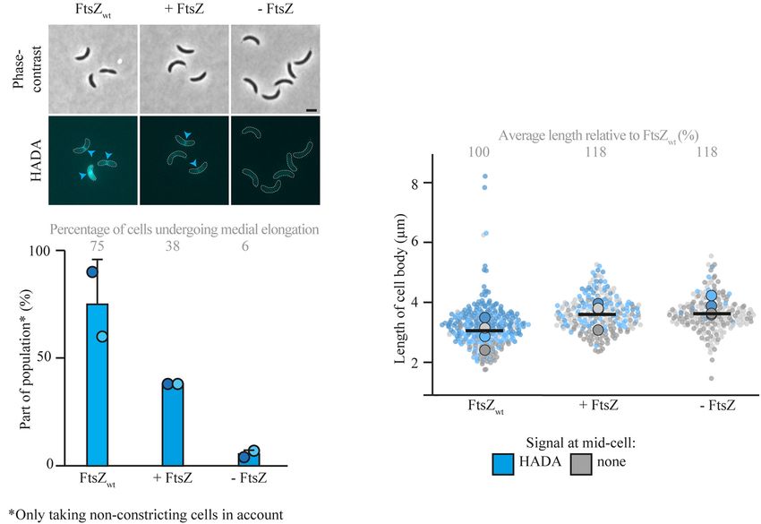
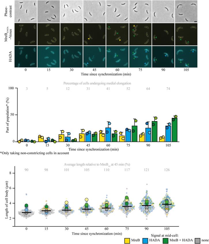
van Teeseling                                                                                                                              Medial Elongation in C. crescentus

  FIGURE 1 | MreB accumulates at midcell clearly before elongation at midcell commences. Intracellular localization of MreB (yellow) and peptidoglycan insertion and
  remodeling (blue) summarized in a cartoon (A), based on the experiments shown in (B–D). Light microscopy images (B) and respective quantification (C) of strain
  MT309 (Pxyl -venus-mreB) grown for the indicated duration after synchronization, stained with a short pulse of HADA show that with time MreB accumulates at
  midcell and cell wall remodeling at midcell commences with a delay. Cells in the representative images (B) are outlined and midcell localization is indicated by
  arrowheads: colocalizing MreB and HADA are indicated in green, only MreB in yellow, and only HADA in blue. Quantification of fluorescence signals (C) at midcell
  (using the color scheme outlined above) was performed on 100 non-constricting cells per replicate for two replicates per timepoint (value for each replicate is
  indicated by circles, replicate 1 is shown in a darker shade and replicate 2 in a light shade). Bars indicate the average of two replicates and error bars indicate
  standard deviation. In addition, the average percentage of non-constricting cells showing HADA (either in presence or absence of MreB) is given for each timepoint.
  Analysis of the length of cell bodies (of both constricting and non-constricting cells, in total at least 100 cells per replicate) shows that cells become longer with time
  and cells that are incorporating HADA are relatively longer (D). Cell lengths are shown in superplots, where each small dot indicates the value for a single cell. The
  color of each dot follows the color scheme outlined above, in order to identify differences in cell length for the different subpopulations, and shows different shades to
  differentiate between the replicates. The large dots show the average cell length for each localization subpopulation per replicate. The horizontal black bar indicates
  the average length over all cells and the numbers above the graph express this average length as a percentage of the average length of the cells after 45 min.

present at midcell, additional experiments were performed. As                             with a higher maximum intensity, at midcell. However, the
a first step, a strain expressing the MreB variant MreBG165A                              fraction of cells that undergoes medial elongation and the
instead of the native MreB (and an inducible MreBG165A                                    maximum HADA intensity increase slower in the strain with
fused to a fluorescent protein) was investigated (Figure 2 and                            the delayed MreB variant than in the wild-type MreB (compare
Supplementary Figure 2), as this strain was previously shown                              Figure 1C with Figure 2B and Supplementary Figure 1A
to have a delayed recruitment of MreB to midcell and a longer                             with Supplementary Figure 2A). This population-wide delay
cell cycle (Dye et al., 2011). Indeed, it could be confirmed                              of medial elongation becomes very clear when comparing the
that MreB arrived later at midcell by approximately 15 min,                               timepoint at which 75% of non-constricting cells shows a HADA
as respectively 11 and 17% of cells showed MreB localization                              signal at midcell: for cells expressing MreBwt this takes place
at 15 and 30 min (Figures 2A,B). Interestingly, the onset                                 45 min after synchronization and for cells with MreBG165A
of medial elongation was comparable to a strain with native                               this percentage is reached only slightly before 105 min after
MreB, with respectively 5 and 12% of cells showing HADA                                   synchronization, at which point the cells had more time to
at midcell 15 and 30 min after synchronization (Figure 2B).                               elongate and were on average longer than their counterparts
Like in the strain with the native MreB variant, with time                                expressing MreBwt . Taken together, the observations that (a)
and increasing cell length (Figure 2C), more and more (non-                               HADA accumulation at midcell can take place before MreB
constricting) elongating cells show a HADA accumulation,                                  reaches the same location and (b) a considerable fraction

Frontiers in Microbiology | www.frontiersin.org                                       4                                            August 2021 | Volume 12 | Article 732031
van Teeseling                                                                                                   Medial Elongation in C. crescentus

of cells shows HADA signal at midcell in the absence of                  percentage of constricting cells as compared to the untreated
MreB (Figure 2B) suggests that medial elongation does not                strain (Supplementary Figure 3D)] as mecillinam inhibits PBP2
require the presence of MreB at midcell, although a timely               that normally aids in dispersed elongation. All in all, it becomes
presence of MreB does speed up medial elongation at the                  clear that medial elongation does not depend on both tested
population level.                                                        elongasome components.

                                                                         Medial Elongation Requires FtsZ
Medial Elongation Takes Place in the                                     After showing that medial elongation can take place in the
Absence of MreB and PBP2 at Midcell                                      absence of selected elongasome components, the focus was
To further look into the role of MreB and other elongasome               moved toward divisome components. Research in E. coli (de
components for the onset of medial elongation, multiple parallel         Pedro et al., 1997; Varma and Young, 2009; Potluri et al., 2012)
routes were taken. First, a strain carrying a MreB variant               and non-quantitative experiments in C. crescentus (Aaron et al.,
(MreBQ26P ) that does not get recruited to midcell at all (Aaron         2007) have implicated FtsZ in medial elongation. To investigate
et al., 2007) was followed. This experiment confirmed that               the requirement of FtsZ for medial elongation in more detail,
medial elongation can take place without MreB being present              cells of the FtsZ depletion strain YB1585 were cultivated and
at midcell, as the majority of cells showed a HADA signal                synchronized. After synchronization, cells were either grown in
at midcell 45 min after synchronization (Figures 3A,B). As               inducing (PYE with xylose) or depleting (PYE without xylose)
compared to cells expressing MreBwt the cell length of this strain       conditions and PG incorporation was investigated via short pulse
was slightly longer, again showing that MreB localization at             labeling with HADA 45 min after synchronization (Figure 4 and
midcell is not needed for cellular elongation (Figure 3C). The           Supplementary Figure 4). As compared to a strain with wild-
fraction of cells that shows medial elongation [and the maximum          type amounts of FtsZ (MT309, grown in PYE without xylose),
intensity of the HADA signal (Supplementary Figure 3A)] is               cells of YB1585 grown in both inducing and depleting conditions
lower than in strain MT309 at the same timepoint, this might             were slightly longer (Figure 4C) and were less likely to constrict
be caused by a delay in the cell cycle, indicated by a lower             (especially in the absence of FtsZ) (Supplementary Figure 4B),
percentage of constricting cells (Supplementary Figure 3C).              but showed less medial elongation (Figure 4B). The drop in
In parallel, the localization of MreBwt and HADA where                   fraction of cells undergoing medial elongation was especially clear
studied upon treatment of strain MT309 (Pxyl -venus-mreB) with           for cells depleted of FtsZ, which contributed to only 6% of the
the MreB inhibitor A22 (Figures 3A,D,E and Supplementary                 population as opposed to 75% in cells with wild-type levels of
Figures 3B,D,E). As expected, only a low fraction of cells showed        FtsZ (MT309), demonstrating that FtsZ is required for medial
MreB localization after treatment with A22 (Figure 3D) and               elongation (Figures 4A,B).
cells became slightly wider (Supplementary Figure 3E) as is
typical for cells treated with A22 (Tropini et al., 2014). In line
with the previous results on MreB variants, also upon A22                DISCUSSION
treatment cells showed medial elongation (as shown by the
accumulation of HADA, either in the absence or presence of               The results obtained in this study suggest that medial elongation
MreB, at midcell), almost up to the same extent as cells untreated       in C. crescentus requires FtsZ but not MreB at midcell, similar to
with A22 (Figure 3D).                                                    PIPS in E. coli (de Pedro et al., 1997; Varma and Young, 2009;
    The elongasome does not only consist of the actin-homolog            Potluri et al., 2012). Multiple experiments showed that medial
MreB, but includes multiple proteins involved in PG synthesis            elongation can take place upon MreB inhibition or its inability
and remodeling, such as the elongasome-specific synthase                 to get recruited to midcell. In this light, the finding that delayed
PBP2. PBP2 was shown to colocalize with FtsZ and MreB at                 recruitment of MreB to midcell results in a slower increase in
midcell in C. crescentus cells that have undergone an osmotic            the fraction of cells undergoing medial elongation is puzzling
challenge (Hocking et al., 2012). This localization was shown            and needs further investigation. Possibly, this specific mutation in
to be dependent on FtsZ, but independent on MreB, which                  MreB has altered interaction properties with its binding partners,
urges the question if PBP2 might be involved in medial                   thereby affecting the process of medial elongation. The previous
elongation. To investigate this question, PG incorporation and           observation that the strain carrying this MreB variant has a longer
MreBwt localization were followed in the presence of the                 cell cycle (Dye et al., 2011), might suggest all cells need to spend
PBP2 inhibitor mecillinam (Figures 3A,D,E and Supplementary              a certain time in the medial elongation phase before they can
Figures 3B,D,E). Upon treatment with mecillinam, the cell                proceed to constriction.
width increased as compared to untreated cells, as has been                 The question which PG synthase(s) are involved in
reported before for mecillinam treatment (Tropini et al., 2014).         medial elongation in C. crescentus remains unanswered.
The results were similar to these for MreB: medial elongation            The experiments presented here suggest PBP2 is not required
still took place upon inhibition of PBP2, suggesting that medial         and the same was suggested for PBP3 (Aaron et al., 2007), which
elongation does not depend on this PBP. The fraction of cells            again fits to what is described for PIPS in E. coli. PIPS in E. coli
that showed medial elongation was lower as compared to samples           was reported to depend on two of its three bifunctional PBPs
without mecillinam, but this might well be caused by a short             (bPBPs): PBP1A and PBP1B, that can take over each other’s
delay in the cell cycle [also exemplified by a drop in the               role (Potluri et al., 2012; Pazos et al., 2018). C. crescentus has

Frontiers in Microbiology | www.frontiersin.org                      5                                    August 2021 | Volume 12 | Article 732031
van Teeseling Medial Elongation in C. crescentus FIGURE 2 | Medial elongation can precede MreB accumulation at midcell in a strain with delayed MreB recruitment. Light microscopy images (A) and respective quantification (B) of strain JAT790 (PmreB -G165A mreB Pxyl -venus-G165A mreB) grown for the indicated duration after synchronization, stained with a short pulse of HADA show that medial elongation commences in the absence of MreB at midcell in part of the population. Cells in the representative images (A) are outlined and midcell localization is indicated by arrowheads: colocalizing MreB and HADA are indicated in green, only MreB in yellow, and only HADA in blue. Quantification of fluorescence signals (B) at midcell (using the color scheme outlined above) was performed on 100 non-constricting cells per replicate for two replicates per timepoint (value for each replicate is indicated by circles, replicate 1 is shown in a darker shade and replicate 2 in a light shade). Bars indicate the average of two replicates and error bars indicate standard deviation. In addition, the average percentage of non-constricting cells showing HADA (either in presence or absence of MreB) is given for each timepoint. Analysis of the length of cell bodies (of both constricting and non-constricting cells, in total at least 100 cells per replicate) shows that cells become longer with time and cells that are incorporating HADA are relatively longer (C). Cell lengths are shown in superplots, where each small dot indicates the value for a single cell. The color of each dot follows the color scheme outlined above, in order to identify differences in cell length for the different subpopulations, and shows different shades to differentiate between the replicates. The large dots show the average cell length for each localization subpopulation per replicate. The horizontal black bar indicates the average length over all cells and the numbers above the graph express this average length as a percentage of the average length of the cells carrying MreBwt after 45 min (strain MT309, indicated in Figure 1). Frontiers in Microbiology | www.frontiersin.org 6 August 2021 | Volume 12 | Article 732031
van Teeseling                                                                                                                                Medial Elongation in C. crescentus

  FIGURE 3 | Medial elongation can take place in the absence of elongasome components at midcell. Light microscopy images (A) and respective quantification of
  strains CJW1715 (PmreB -Q26P mreB) (B) and MT309 (Pxyl -venus-mreB), synchronized and subsequently grown in the absence of additives and (for MT309) in the
  presence of the MreB inhibitor A22 (10 µg/ml) or the PBP2 inhibitor mecillinam (150 µg/ml) (D). Cells were harvested 45 min after synchronization and subjected to a
  short pulse HADA staining. Cells in the representative images (A) are outlined and midcell localization is indicated by arrowheads: colocalizing MreB and HADA are
  indicated in green, only MreB in yellow and only HADA in blue. Quantification of fluorescence signals (B,D) at midcell (using the color scheme outlined above, with
  the exception that in the case of strain CJW1715 MreB was not fluorescently labeled, and therefore only blue can be used) was performed on 100 non-constricting
  cells per replicate for two replicates per timepoint (value for each replicate is indicated by circles, replicate 1 is shown in a darker shade and replicate 2 in a light
  shade). Bars indicate the average of two replicates and error bars indicate standard deviation. In addition, the average percentage of non-constricting cells showing
  HADA (either in presence or absence of MreB) is given for each timepoint. Data shown for MreBwt in both cases are from the same two replicates of strain MT309
  (also shown in Figure 1). Analysis of the length of the cell bodies (of both constricting and non-constricting cells, in total at least 100 cells per replicate) shows that
  cells expressing MreBQ26P are slightly longer than cells expressing MreBwt (C). Analysis of the length of the cell bodies (of both constricting and non-constricting
  cells, in total at least 100 cells per replicate) for cells of strain MT309 incubated without additives, shows that with A22 cells are a bit shorter and with mecillinam cells
  are slightly longer (E). Cell lengths are shown in superplots, where each small dot indicates the value for a single cell. The color of each dot follows the color scheme
  outlined above, in order to identify differences in cell length for the different subpopulations, and shows different shades to differentiate between the replicates. The
  large dots show the average cell length for each localization subpopulation per replicate. The horizontal black bar indicates the average length over all cells and the
  numbers above the graph express this average length as a percentage of the average length of the cells carrying MreBwt after 45 min (strain MT309, indicated in
  Figure 1).

a higher redundancy of bPBPs with five copies in total: in                                  (Strobel et al., 2014), suggests that in C. crescentus PbpX, PbpY,
addition to one PBP1C homolog (PbpZ), it interestingly has                                  PbpC, and potentially also Pbp1A might all be able to take up the
four PBP1A homologs (PBP1A, PbpC, PbpX, and PbpY), but                                      role as bPBP involved in medial elongation.
lacks PBP1B homologs (Yakhnina and Gitai, 2013; Strobel et al.,                                Another open question when comparing medial elongation
2014). The observation that strains missing all bPBPs except for                            in C. crescentus to PIPS in E. coli is if any (and if so which) of
PbpX or PbpY or PbpC or to a lesser extent PPB1A are viable                                 the proteins anchoring FtsZ is involved in medial elongation.

Frontiers in Microbiology | www.frontiersin.org                                         7                                            August 2021 | Volume 12 | Article 732031
van Teeseling                                                                                                                              Medial Elongation in C. crescentus

  FIGURE 4 | Medial elongation is affected by the absence of FtsZ. Light microscopy images (A) and respective quantification (B) of FtsZ depletion strain YB1585
  (grown under depleting and inducing conditions) and MT309 (Pxyl -venus-mreB), synchronized and subsequently grown for 45 min and subsequently subjected to a
  short pulse HADA staining. Cells in the representative images (A) are outlined and HADA midcell localization is indicated by blue arrowheads. Quantification of
  fluorescence signals (B) at midcell (using the color scheme outlined above) was performed on 100 non-constricting cells per replicate for two replicates per timepoint
  (value for each replicate is indicated by circles, replicate 1 is shown in a darker shade and replicate 2 in a light shade). Bars indicate the average of two replicates and
  error bars indicate standard deviation. In addition, the average percentage of non-constricting cells showing HADA is given for each timepoint. Data shown for
  FtsZwt in both cases are from the same replicates of strain MT309 as shown in Figure 1. Analysis of the cell length (C) of cells of strain YB1585 shows that they are
  slightly elongated as compared to cells harboring FtsZwt . Cell lengths are shown in superplots, where each small dot indicates the value for a single cell. The color of
  each dot follows the color scheme outlined above, in order to identify differences in cell length for the different subpopulations, and shows different shades to
  differentiate between the replicates. The large dots show the average cell length for each localization subpopulation per replicate. The horizontal black bar indicates
  the average length over all cells and the numbers above the graph express this average length as a percentage of the average length of the cells carrying MreBwt
  after 45 min (strain MT309, indicated in Figure 1).

In E. coli, the early divisome protein ZipA works together with                            described to regulate the curvature of FtsZ filaments (Lariviere
FtsA to anchor FtsZ to the membrane and stabilize FtsZ polymers                            et al., 2018), which seems to occur at a later stage than the
into protofilaments (Pichoff and Lutkenhaus, 2002; Krupka et al.,                          role ZipA takes in PIPS. All in all, further studies need to be
2018). Both of these proteins are involved in PIPS, for which ZipA                         performed in order to understand which other proteins are
is essential unless a hyperactive FtsA allele is present (Potluri                          involved in medial elongation. Until these proteins and the
et al., 2012; Pazos et al., 2018). C. crescentus, however, lacks a ZipA                    underlying mechanisms they use to make C. crescentus cells
homolog and FtsA is recruited only later to the divisome (Goley                            elongate from midcell are known, it will be impossible to answer
et al., 2011). Instead, the FtsZ-binding proteins ZapA, ZauP,                              if this growth phase is mechanistically the same as PIPS in E. coli.
FzlC, FtsE, and FzlA are recruited early to the divisome (Goley                            For now, however, all results seem consistent. Hopefully research
et al., 2011; Woldemeskel et al., 2017). If any of these proteins is                       in the future will succeed in elucidating mechanisms behind this
required for medial elongation remains unknown at the moment.                              elusive growth phase and answer the question how widespread
It might be that there is more redundancy in FtsZ anchoring                                it is in bacteria.
proteins that are required for medial elongation compared to
E. coli, as strains lacking ZapA, ZauP (and a combination of
these two), FzlC, and FtsE are viable (Goley et al., 2010; Meier                           DATA AVAILABILITY STATEMENT
et al., 2017; Woldemeskel et al., 2017), suggesting that each of
these alone does not take over the essential role of ZipA in                               The raw data supporting the conclusions of this article
medial elongation. Alternatively, the essential FzlA takes over                            will be made available by the authors, without undue
the role of ZipA, although it is implicated in constriction and                            reservation.

Frontiers in Microbiology | www.frontiersin.org                                        8                                            August 2021 | Volume 12 | Article 732031
van Teeseling                                                                                                                                  Medial Elongation in C. crescentus

AUTHOR CONTRIBUTIONS                                                                           scientific discussions and support. Julie Theriot is acknowledged
                                                                                               for sharing strain JAT790 and Martin Thanbichler and Christine
MvT conceived and designed this project, performed the                                         Jacobs-Wagner for CJW1715.
analyses, and wrote the manuscript.

                                                                                               SUPPLEMENTARY MATERIAL
ACKNOWLEDGMENTS
                                                                                               The Supplementary Material for this article can be found
Julia Rosum and Adrian Kietz are acknowledged for excellent                                    online at: https://www.frontiersin.org/articles/10.3389/fmicb.
technical assistance. The author thanks Martin Thanbichler for                                 2021.732031/full#supplementary-material

REFERENCES                                                                                     Flärdh, K. (2010). Cell polarity and the control of apical growth in Streptomyces.
                                                                                                  Curr. Opin. Microbiol. 13, 758–765. doi: 10.1016/j.mib.2010.10.002
Aaron, M., Charbon, G., Lam, H., Schwarz, H., Vollmer, W., and Jacobs-Wagner, C.               Garner, E. C., Bernard, R., Wang, W., Zhuang, X., Rudner, D. Z., and Mitchinson,
    (2007). The tubulin homologue FtsZ contributes to cell elongation by guiding                  T. (2011). Coupled, circumferential motions of the cell wall synthesis machinery
    cell wall precursor synthesis in Caulobacter crescentus. Mol. Microbiol. 64,                  and MreB filaments in B. subtilis. Science 333, 222–225. doi: 10.1126/science.
    938–952. doi: 10.1111/j.1365-2958.2007.05720.x                                                1203285
Billini, M., Biboy, J., Kühn, J., Vollmer, W., and Thanbichler, M. (2019). A                   Gitai, Z., Dye, N. A., and Shapiro, L. (2004). An actin-like gene can determine
    specialized MreB-dependent cell wall biosynthetic complex mediates the                        cell polarity in bacteria. Proc. Natl. Acad. Sci. U. S. A. 101, 8643–8648. doi:
    formation of stalk-specific peptidoglycan in Caulobacter crescentus. PLoS Genet.              10.1073/pnas.0402638101
    15:e1007897. doi: 10.1371/journal.pgen.1007897                                             Goedhart, J. (2021). SuperPlotsOfData- a web app for the transparent display and
Bisson Filho, A. W., Hsu, Y.-P., Squyres, G. R., Kuru, E., Wu, F., Jukes, C.,                     quantitative comparison of continuous data from different conditions. Mol.
    et al. (2017). Treadmilling by FtsZ filaments drives peptidoglycan synthesis and              Biol. Cell 32, 461–505.
    bacterial cell division. Science 355, 739–743. doi: 10.1126/science.aak9973                Goley, E. D., Dye, N. A., Werner, J. N., Gitai, Z., and Shapiro, L. (2010). Imaging-
Brown, P. J. B., de Pedro, M. A., Kysela, D. T., Van der Henst, C., Kim, J., De Bolle,            based identification of a critical regulator of FtsZ protofilament curvature in
    X., et al. (2012). Polar growth in the Alphaproteobacterial order Rhizobiales.                Caulobacter. Mol. Cell. 39, 975–987. doi: 10.1016/j.molcel.2010.08.027
    Proc. Natl. Acad. Sci. U. S. A. 109, 1697–1701. doi: 10.1073/pnas.1114476109               Goley, E. D., Yeh, Y.-C., Hong, S.-H., Fero, M. J., Abeliuk, E., McAdams, H. H.,
de Pedro, M. A., and Cava, F. (2015). Structural constraints and dynamics of                      et al. (2011). Assembly of the Caulobacter cell division machine. Mol. Microbiol.
    bacterial cell wall architecture. Front. Microbiol. 6:449. doi: 10.3389/fmicb.2015.           80, 1680–1698. doi: 10.1111/j.1365-2958.2011.07677.x
    00449                                                                                      Hartmann, R., van Teeseling, M. C. F., Thanbichler, M., and Drescher, K. (2020).
de Pedro, M. A., Quintela, J. C., Höltje, J. V., and Schwarz, H. (1997). Murein                   BacStalk: a comprehensive and interactive image analysis software tool for
    segregation in Escherichia coli. J. Bacteriol. 179, 2823–2834. doi: 10.1128/jb.               bacterial cell biology. Mol. Microbiol. 220, 140–150. doi: 10.1111/mmi.14501
    179.9.2823-2834.1997                                                                       Hocking, J., Priyadarshini, R., Takacs, C. N., Costa, T., Dye, N. A., and Shapiro,
Dersch, S., Mehl, J., Stuckenschneider, L., Mayer, B., Roth, J., Rohrbach, A.,                    L. (2012). Osmolality-dependent relocation of penicillin-binding protein PBP2
    et al. (2020). Super-resolution microscopy and single-molecule tracking reveal                to the division site in Caulobacter crescentus. J. Bacteriol. 194, 3116–3127.
    distinct adaptive dynamics of MreB and of cell wall-synthesis enzymes. Front.                 doi: 10.1128/jb.00260-12
    Microbiol. 11:1946. doi: 10.3389/fmicb.2020.01946                                          Krupka, M., Sobrinos-Sanguino, M., Jiménez, M., Rivas, G., and Margolin, W.
Dion, M. F., Kapoor, M., Sun, Y., Wilson, S., Ryan, J., Vigouroux, A., et al. (2019).             (2018). Escherichia coli ZipA organizes FtsZ polymers into dynamic ring-like
    Bacillus subtilis cell diameter is determined by the opposing actions of two                  protofilament structures. mBio 9:e01008-18.
    distinct cell wall synthetic systems. Nat. Microbiol. 4, 1294–1305. doi: 10.1038/          Kuru, E., Hughes, H. V., Brown, P. J., Hall, E., Tekkam, S., Cava, F., et al.
    s41564-019-0439-0                                                                             (2012). In situ probing of newly synthesized peptidoglycan in live bacteria
Dominguez-Escobar, J., Chastanet, A., Crevenna, A. H., Fromion, V., Wedlich-                      with fluorescent D-amino acids. Angew. Chem. Int. Ed. Engl. 51, 12519–12523.
    Söldner, R., and Carballido-Lopez, R. (2011). MreB-associated cell wall                       doi: 10.1002/anie.201206749
    biosynthetic complexes in bacteria. Science 333, 225–228. doi: 10.1126/science.            Kysela, D. T., Randich, A. M., Caccamo, P. D., and Brun, Y. V. (2016). Diversity
    1203466                                                                                       takes shape: understanding the mechanistic and adaptive basis of bacterial
Ducret, A., and Grangeasse, C. (2021). Recent progress in our understanding of                    morphology. PLoS Biol. 14:e1002565. doi: 10.1371/journal.pbio.1002565
    peptidoglycan assembly in Firmicutes. Curr. Opin. Microbiol. 60, 44–50. doi:               Lariviere, P. J., Szwedziak, P., Mahone, C. R., Löwe, J., and Goley, E. D. (2018).
    10.1016/j.mib.2021.01.011                                                                     FzlA, an essential regulator of FtsZ filament curvature, controls constriction
Dye, N. A., Pincus, Z., Fisher, I. C., Shapiro, L., and Theriot, J. A. (2011). Mutations          rate during Caulobacter division. Mol. Microbiol. 107, 180–197. doi: 10.1111/
    in the nucleotide binding pocket of MreB acan alter cell curvature and polar                  mmi.13876
    morphology in Caulobacter. Mol. Microbiol. 81, 368–394. doi: 10.1111/j.1365-               Marks, M. E., Castro Rojas, C. M., Teiling, C., Du, L., Kapatral, V.,
    2958.2011.07698.x                                                                             Walunas, T. L., et al. (2010). The genetic basis of laboratory adaptation
Egan, A. J. F., Errington, J., and Vollmer, W. (2020). Regulation of peptidoglycan                in Caulobacter crescentus. J. Bacteriol. 192, 3678–3688. doi: 10.1128/jb.
    synthesis and remodelling. Nat. Rev. Microbiol. 18, 446–460. doi: 10.1038/                    00255-10
    s41579-020-0366-3                                                                          Meier, E. L., Daitch, A. K., Yao, Q., Bhargava, A., Jensen, G. J., and Goley, E. D.
Evinger, M., and Agabian, N. (1977). Envelope-associated nucleoid from                            (2017). FtsEX-mediated regulation of the final stages of cell division reveals
    Caulobacter crescentus stalked and swarmer cells. J. Bacteriol. 132, 294–301.                 morphogenetic plasticity in Caulobacter crescentus. PLoS Genet. 13:e1006999.
    doi: 10.1128/jb.132.1.294-301.1977                                                            doi: 10.1371/journal.pgen.1006999
Fenton, A. K., and Gerdes, K. (2013). Direct interaction of FtsZ and MreB is                   Meisenzahl, A. C., Shapiro, L., and Jenal, U. (1997). Isolation and characterization
    required for septum synthesis and cell division in Escherichia coli. EMBO J. 32,              of a xylose-dependent promoter from Caulobacter crescentus. J. Bacteriol. 179,
    1953–1965. doi: 10.1038/emboj.2013.129                                                        592–600. doi: 10.1128/jb.179.3.592-600.1997
Figge, R. M., Divakaruni, A. V., and Gober, J. W. (2004). MreB, the cell shape-                Monteiro, J. M., Pereira, A. R., Reichmann, N. T., Saraiva, B. M., Fernandes,
    determining bacterial actin homologue, co-ordinates cell wall morphogenesis                   P. B., Veiga, H., et al. (2018). Peptidoglycan synthesis drives an FtsZ-
    in Caulobacter crescentus. Mol. Microbiol. 51, 1321–1332. doi: 10.1111/j.1365-                treadmilling-independent step of cytokinesis. Nature 554, 528–532. doi: 10.
    2958.2003.03936.x                                                                             1038/nature25506

Frontiers in Microbiology | www.frontiersin.org                                            9                                            August 2021 | Volume 12 | Article 732031
van Teeseling Medial Elongation in C. crescentus
Özbaykal, G., Wollrab, E., Simon, F., Vigouroux, A., Cordier, B., Aristov, A., et al. van Teeseling, M. C. F., de Pedro, M. A., and Cava, F. (2017). Determinants
(2020). The transpeptidase PBP2 governs initial localization and activity of the of bacterial morphology: from fundamentals to possibilities for antimicrobial
major cell-wall synthesis machinery in E. coli. eLife 9:e50629. targeting. Front. Microbiol. 8:1264. doi: 10.3389/fmicb.2017.01264
Pazos, M., Peters, K., Casanova, M., Palacios, P., VanNieuwenhze, M., Breukink, Varma, A., and Young, K. D. (2009). In Escherichia coli, MreB and FtsZ direct
E., et al. (2018). Z-ring membrane anchors associate with cell wall synthases to the synthesis of lateral cell wall via independent pathways that require PBP2.
initiate bacterial cell division. Nat. Commun. 9:5090. J. Bacteriol. 191, 3526–3533. doi: 10.1128/jb.01812-08
Pichoff, S., and Lutkenhaus, J. (2002). Unique and overlapping roles for ZipA Vats, P., and Rothfield, L. (2007). Duplication and segregation of the actin (MreB)
and FtsA in septal ring assembly in Escherichia coli. EMBO J. 21, 685–693. cytoskeleton during the prokaryotic cell cycle. Proc. Natl. Acad. Sci. U. S. A. 104,
doi: 10.1093/emboj/21.4.685 17795–17800. doi: 10.1073/pnas.0708739104
Pointdexter, J. S. (1964). Biological properties and classification of the Vats, P., Shih, Y.-L., and Rothfield, L. (2009). Assembly of the MreB-associated
Caulobacter group. Bacteriol. Rev. 28, 231–295. doi: 10.1128/br.28.3.231-295. cytoskeletal ring of Escherichia coli. Mol. Microbiol. 72, 170–182. doi: 10.1111/
1964 j.1365-2958.2009.06632.x
Potluri, L.-P., Kannan, S., and Young, K. D. (2012). ZipA is required for FtsZ- Wang, Y., Jones, B. D., and Brun, Y. V. (2001). A set of ftsz mutants blocked at
dependent preseptal peptidoglycan synthesis prior to invagination during cell different stages of cell division in Caulobacter. Mol. Microbiol. 40, 347–360.
division. J. Bacteriol. 194, 5334–5342. doi: 10.1128/jb.00859-12 doi: 10.1046/j.1365-2958.2001.02395.x
Randich, A. M., and Brun, Y. V. (2015). Molecular mechanisms for the evolution Wientjes, F. B., and Nanninga, N. (1989). Rate and topography of peptidoglycan
of bacterial morphologies and growth modes. Front. Microbiol. 6:580. doi: 10. synthesis during cell division in Escherichia coli: concept of a leading edge.
3389/fmicb.2015.00580 J. Bacteriol. 171, 3412–3419. doi: 10.1128/jb.171.6.3412-3419.1989
Schindelin, J., Arganda-Carreras, I., Frise, E., Kaynig, V., Longair, M., Pietzsch, T., Woldemeskel, S. A., McQuillen, R., Hessel, A. M., Xiao, J., and Goley, E. D.
et al. (2012). Fihi: an open-source platform for biological-image analysis. Nat. (2017). A conserved coiled-coil protein pair focuses the cytokinetic Z-ring in
Methods 9, 676–682. Caulobacter crescentus. Mol. Microbiol. 105, 721–740. doi: 10.1111/mmi.13731
Silber, N., Mayer, C., Matos de Opitz, C. L., and Sass, P. (2021). Progression of the Yakhnina, A. A., and Gitai, Z. (2013). Diverse functions for six glaycosyltransferases
late-stage divisome is unaffected by the depletion of the cytoplasmic FtsZ pool. in Caulobacter crescentus cell wall assembly. J. Bacteriol. 195, 4527–4535. doi:
Commun. Biol. 4:270. 10.1128/jb.00600-13
Squyres, G. R., Holmes, M. J., Barger, S. R., Pennycook, B. R., Ryan, J., Yan, Yang, X., Lyu, Z., Miguel, A., McQuillen, R., Huang, K. C., and Xiao, J. (2017).
V. T., et al. (2021). Single-molecule imaging reveals that Z-ring condensation GTPase activity-coupled treadmilling of the bacterial tubulin FtsZ organizes
is essential for cell division in Bacillus subtilis. Nat. Microbiol. 6, 553–562. septal cell wall synthesis. Science 355, 744–747. doi: 10.1126/science.aak9995
doi: 10.1038/s41564-021-00878-z Yang, X., McQuillen, R., Lyu, Z., Phillips-Mason, P., De La Cruz, A., McCausland,
Strobel, W., Möll, A., Kiekebusch, D., Klein, K. E., and Thanbichler, M. (2014). J. W., et al. (2021). A two-track model for the spatiotemporal coordination of
Function and localization dynamics of bifunctional penicillin-binding proteins bacterial septal cell wall synthesis revealed by single-molecule imaging of FtsW.
in Caulobacter crescentus. J. Bacteriol. 196, 1627–1639. doi: 10.1128/jb. Nat. Microbiol. 6, 584–593. doi: 10.1038/s41564-020-00853-0
01194-13
Tropini, C., Lee, T. K., Hsin, J., Desmarais, S. M., Ursell, T., Monds, R. D., Conflict of Interest: The author declares that the research was conducted in the
et al. (2014). Principles of bacterial cell-size determination revealed by cell-wall absence of any commercial or financial relationships that could be construed as a
synthesis perturbations. Cell Rep. 9, 1520–1527. doi: 10.1016/j.celrep.2014.10. potential conflict of interest.
027
Tsai, J. W., and Alley, M. R. (2001). Proteolysis of the Caulobacter McpA Publisher’s Note: All claims expressed in this article are solely those of the authors
chemoreceptor is cell cycle regulated by a ClpX-dependent pathway. J Bacteriol. and do not necessarily represent those of their affiliated organizations, or those of
183, 5001–5007. doi: 10.1128/jb.183.17.5001-5007.2001 the publisher, the editors and the reviewers. Any product that may be evaluated in
Typas, A., Banzhaf, M., Gross, C. A., and Vollmer, W. (2012). From the regulation this article, or claim that may be made by its manufacturer, is not guaranteed or
of peptidoglycan synthesis to bacterial growth and morphology. Nat. Rev. endorsed by the publisher.
Microbiol. 10, 123–136. doi: 10.1038/nrmicro2677
Umeda, A., and Amako, K. (1983). Growth of the surface of Corynebacterium Copyright © 2021 van Teeseling. This is an open-access article distributed under the
diphtheriae. Microbiol. Immunol. 27, 663–671. terms of the Creative Commons Attribution License (CC BY). The use, distribution
van Teeffelen, S., Wang, S., Furchtgott, L., Huang, K. C., Wingreen, N. S., Shaevitz, or reproduction in other forums is permitted, provided the original author(s) and
J. W., et al. (2011). The bacterial actin MreB rotates, and rotation depends the copyright owner(s) are credited and that the original publication in this journal
on cell-wall assembly. Proc. Natl. Acad. Sci. U. S. A. 108, 15822–15827. doi: is cited, in accordance with accepted academic practice. No use, distribution or
10.1073/pnas.1108999108 reproduction is permitted which does not comply with these terms.
Frontiers in Microbiology | www.frontiersin.org 10 August 2021 | Volume 12 | Article 732031You can also read