Low-Intensity Ultrasound as a Novel Strategy to Improve the Cytotoxic Effect of Oncolytic Reovirus on Colorectal Cancer Model Cells
←
→
Page content transcription
If your browser does not render page correctly, please read the page content below
Research Article

                                                             Intervirology                                                                        Received: May 25, 2021
                                                                                                                                                  Accepted: September 6, 2021
                                                             DOI: 10.1159/000519492                                                               Published online: September 10, 2021

Low-Intensity Ultrasound as a Novel Strategy
to Improve the Cytotoxic Effect of Oncolytic
Reovirus on Colorectal Cancer Model Cells
Negar Sharifi a Hoorieh Soleimanjahi a Manijeh Mokhtari-Dizaji b
Razieh Sadat Banijamali a Maliheh Elhamipour a Hesam Karimi a, c
aDepartment
             of Virology, Faculty of Medical Sciences, Tarbiat Modares University, Tehran, Iran; bDepartment of
Medical Physics, Faculty of Medical Sciences, Tarbiat Modares University, Tehran, Iran; cDepartment of Hepatitis and
AIDS, Pasteur Institute of Iran, Tehran, Iran

Keywords                                                                                         sity ultrasound showed an increased virus titer compared
Colorectal cancer model cells · Oncolytic reovirus T3D ·                                         with unexposed infected cells. Moreover, according to the
Low-intensity ultrasound                                                                         results of the flow cytometry test, it was found that the
                                                                                                 amount of apoptosis in infected cells that are exposed to
                                                                                                 low-intensity ultrasound waves is higher than those infected
Abstract                                                                                         cells. Conclusion: Due to the results of CCID50 and flow cy-
Background: Colorectal cancer is the third most common                                           tometry tests, low-intensity ultrasound increases the cyto-
cancer all over the world, so in the battle to fight this hurdle,                                toxicity level of reovirus in CT26 cells of the cellular colorectal
new therapeutic approaches such as oncolytic viruses (OV)                                        cancer model.                             © 2021 The Author(s)
have attracted much attention because of the fact that they                                                                                            Published by S. Karger AG, Basel

can inherently kill cancer cells. Oncolytic reovirus is one of
the candidates for treatment as a nonpathogenic species
specially reovirus type 3 Dearing (T3D), which can induce ap-                                        Introduction
optosis. To speed up the entry and function of the reovirus,
low-intensity ultrasound, which is a safe system for damage                                          Cancer is a lethal problem globally, which was report-
to the cells and tissues, is a promising approach to be used                                     ed by the World Health Organization as the second lead-
in combination with other therapeutic approach. Methods:                                         ing cause of death after cardiovascular diseases [1].
L929 and CT26 cells were infected with reovirus T3D and                                          Colorectal cancer is one of the major types of cancer that
were exposed to ultrasonic irradiation (1 MHz, 1 W/cm2, and                                      is classified as the third malignancy in the world [2]. Some
20% duty factor) for 10 s. The viruses’ titer level of both                                      popular methods of treating cancer include surgery, ra-
groups was calculated in 2 types of cells by using the CCID50                                    diation, chemotherapy, etc.; besides, in some cases, the
method and compared with each other. Apoptosis, after 24                                         combination of stated methods is used in cancer thera-
h, was measured by the flow cytometry method. Result: The                                        peutics. Nevertheless, recurrent and metastatic diseases
results of CCID50 in infected cells were exposed to low-inten-                                   respond poorly to common therapies and have numerous

karger@karger.com      © 2021 The Author(s).                                                     Correspondence to:
www.karger.com/int     Published by S. Karger AG, Basel                                          Hoorieh Soleimanjahi, soleim_h @ modares.ac.ir
                       This is an Open Access article licensed under the Creative Commons
                       Attribution-NonCommercial-4.0 International License (CC BY-NC)
                       (http://www.karger.com/Services/OpenAccessLicense), applicable to
                       the online version of the article only. Usage and distribution for com-
                       mercial purposes requires written permission.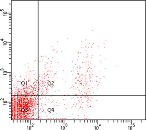
side effects. For example, abnormal DNA methylation                According to the previous published data, antibodies
causes the tumor cells to be developed and processed [3];       can neutralize the replication of viruses by blocking at-
therefore, alternative treatments with high potency and         tachment to the host cell, preventing the penetration of
the least side effects are needed to significantly change the   the host cell membrane, or interfering with uncoating of
overall survival of cancer patients [4–6]. Nowadays, a va-      the virus within the cells, so these viruses need help to
riety of novel adjuvant therapies, particularly oncolytic       conquer and fight limiting factors. It should be supported
viruses (OV), have been developed, which are attractive         by increasing the cell permeability or enhance the migra-
and promising strategies for cancer treatment [6–8].            tion capacity of reovirus to tumor site by application of
    Natural or engineered OV by lysing cells, destruction       mesenchymal stem cells for delivery of oncolytic reovirus
of tumor vessels, and induction of antitumor immunity           in vitro. Using these strategies could open up new win-
can efficiently and precisely affect and eliminate cancer       dows to combined therapy to achieve the excellent out-
cells [6, 9]. OV have a weak pathogenic effect on the nor-      come in fighting to cancers [34].
mal cells, and compared with traditional drugs that re-            Needless to say, by increasing the cell permeability, on-
duce their effectiveness over time, the viruses’ doses in-      colytic effectiveness of reovirus might be enhanced before
crease in the tumor by propagation [10].                        antibody production by promoting its entry, so more reo-
    Reovirus is a double-stranded RNA virus that was de-        viruses can infect target cells; furthermore, based on the
veloped as one of the candidates for cancer treatment           effect of increasing cell permeability by the low-level in-
[11–14]. This virus specifically targets cancer cells with an   tensity of ultrasound, known as a safe way, the goal of this
activated Ras signaling pathway; accordingly, KRAS mu-          study was to investigate the effect of these waves as a way
tations are present in up to 40% of colorectal cancer cases     to raise the number of the oncolytic reoviruses entering
[15–17]. Ras activation enhances viral uncoating and dis-       our target cells.
assembly, raises the production of viral progeny with aug-
mented infection, and animates the release of progeny
through promoted apoptosis [18]. These features are the            Materials and Methods
main therapeutic indices for viruses but are important is-
sues in this type of remedy: if viral particles are unpro-          Cell Culture and Virus Seed Preparation
tected in the bloodstream, they are quickly neutralized by          Murine L929 fibroblasts cells were obtained from Tarbiat
                                                                Modares University, as host cells for reovirus culture and control
the host’s immune system, so they cannot reach the can-         cells group, and murine colon carcinoma (CT26) were gained from
cerous cells [19–21].                                           pastor institute of Iran, as a colorectal cancer model’s cells, cul-
    Since the 1940s, ultrasonic energy, which has a fre-        tured in Dulbecco’s Modified Eagle’s Medium (Gibco, NY, USA),
quency of up to 20 kHz, has been used for imaging and           containing 10% heat-inactivated fetal bovine serum (Gibco, NY,
remedial goals. Thermal effects with high-intensity and         USA), 1% penicillin/streptomycin (Gibco, NY, USA), and grown
                                                                in an incubator at 37°C in a humidified atmosphere with 5% CO2
nonthermal effects with low intensity are 2 kinds of ef-        [35].
fects being composed depending on the energy delivered              The reovirus type 3 Dearing (T3D), obtained from Dr. Shamsi-
by ultrasound [22–25]. In preceding decades, the low-lev-       Shahrabadi (Iran University of Medical Sciences, Iran), was grown
el intensity of ultrasound is being investigated for delivery   in semiconfluent L929 cells’ monolayers in a 75-cm2 flask with
of drugs, genes, viruses, and so on. Moreover, it plays a       MOI 0.1 for virus seed preparation. Infected cells, incubated for 1
                                                                h at 37°C, were washed with phosphate-buffered saline and again
major role in elaborating the influence of the agent treat-     incubated in a new medium until the virus cytopathic effects ap-
ments [6, 26, 27]. Sonoporation is an effect of the low-        peared. Then, they were subjected to 2 cycles of freezing and thaw-
level intensity of ultrasound, which increases the perme-       ing and followed by centrifugation. The supernatants, containing
ability of cell membranes in the presence of ultrasound         produced reovirus T3D, at first, were purified, and then, virus ti-
waves [28]. This nonthermal mechanism of ultrasonic             tration was measured by cell culture infection dose 50 (CCID50).
                                                                Eventually, they were kept at −70°C until use [36].
waves, especially the phenomenon of unstable cavitation,
causes the production of cavities in the radiation environ-        Ultrasound Irradiation
ment, and their destruction in the vicinity of the cell            After we made sure that low-intensity ultrasound has no ad-
membrane causes changes and ultimately increases the            verse effect on the virus seed (by calculating the before and after
permeability of the cell membrane to environmental fac-         virus titers and comparing them with each other) and based on
                                                                the findings of preceding studies, murine L929 fibroblast cells as
tors. This mechanism is reversible; in other words, the         a control and CT26 as target cells were grown in enclosed sterile
membrane returns to baseline by removing the radiation          3 cm polystyrene plates, and when these cells had got 80% conflu-
from the ultrasound [26, 29–33].                                ency infected by viruses at MOI of 1 for considering safety issue

2                    Intervirology                                                     Sharifi et al.
                     DOI: 10.1159/000519492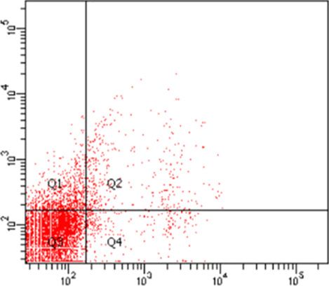
a                                               b                                               c

Fig. 1. Image of L929 cells infected with reovirus at MOI of 1 (titer   (b). c L929 cells infected with reovirus at MOI of 1 exposed to ul-
106) exposed and not exposed to low-intensity ultrasound under          trasound. As shown in the figure of each group, the difference be-
light microscopy after 24 h, magnification ×10. L929 cells control      tween them is completely obvious. To be more specific, c has more
cells (a); L929 cells infected with reovirus at MOI of 1 after 24 h     CPE than b, and in a no CPE is seen.

 a                                               b                                               c

Fig. 2. Image of CT26 cells infected with reovirus at MOI of 1 (titer   (b). c CT26 cells infected with reovirus at MOI of 1 exposed to ul-
106) exposed and not exposed to low-intensity ultrasound under          trasound. There is no CPE in a, and CPE in c is much more in
light microscopy after 24 h, magnification ×10. CT26 cells control      contrast to b.
cells (a); CT26 cells infected with reovirus at MOI of 1 after 24 h

and because of the fact that reproduction of the virus is enough            Determination of Virus Titer by CCID50
for observing the effect of ultrasound waves [37]. After an ad-             When the first development of CPE had been observed (ap-
sorption period of 30 min, unabsorbed viruses were removed by           proximately 18 h after infection with the viruses, CPE can follow
rinsing the cell with phosphate-buffered saline to remove any un-       by a light microscope [37]), L929 and CT26 cells and their super-
bound virus. Besides, they were covered with the Dulbecco’s             natants were collected individually, and then the cells were frozen
Modified Eagle’s Medium. The dishes were held by a permanent            and thawed twice and were centrifuged. Next, both cell lysates and
holder whose bottom was at the distance of 2 cm from the probe          their supernatant were collected and evaluated by CCID50 on L929
and that distance was filled with coupling gel. The plates were         cells. The monolayers of L929 cells in 96-wells plates were inocu-
exposed to ultrasound using an ultrasound machine (Physiomed-           lated with a collected sample of a 10-fold serially dilution of each
Expert, Germany), at room temperature. The ultrasound fre-              group. Inoculated cells were incubated at 37°C in a humidified at-
quency was 1 MHz during the experiments, and it was adjusted            mosphere with 5% CO2 for approximately 72 h. CPE consequenc-
to supply an intensity of 1.0 W cm2 at a duty cycle of 20% for          es examined by comparing with the positive and negative controls
10 s [38, 39].                                                          columns on 96 plates. Virus titers were determined according to

Oncolytic Effect of Reovirus with                                       Intervirology                                                     3
Low-Intensity of Ultrasound                                             DOI: 10.1159/000519492
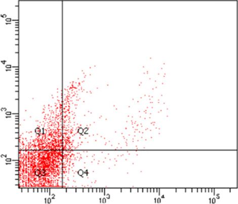
Table 1. Reovirus titers in different groups were calculated after in-
fection, according to the Reed & Munch formula. Data are expressed                                      8           72 h after infection with MOI of 1
as the mean ± SD of 3 independent experiments
                                                                                                        7
Groups         L929          L929 + US     CT26          CT26 + US

                                                                           Logarithm of CCID50/100 μL
                                                                                                        6
Virus titer    105.166       105.749       103.499       104.499
                                                                                                        5

                                                                                                        4

the Reed & Muench formula. log10 50% end point dilution = log10                                         3
of dilution showing mortality next above 50% – (difference of log-
                                                                                                        2
arithms × logarithm of dilution factor) [37].
                                                                                                        1
    Evaluation of Apoptosis by Flow Cytometry Technique
    The cell death was analyzed by the flow cytometry technique.                                        0
First, L929 and CT26 cells were cultured at 3 cm plates and then were                                       Supernant of Supernant of Supernant of Supernant of
infected with reovirus at MOI of 1. After 30 min, infected cells were                                        L929 + US      L929       CT26 + US      CT26
exposed to ultrasounds for 1 min. When CPE was observed, cells
were harvested and analyzed with the Annexin V apoptosis detection
kit FITC (eBioscience, USA, according to the manufacturer’s instruc-     Fig. 3. The rate of reovirus titer on L929 cells and CT26 cells up
tions) and a FACSCanto II flow cytometer (BD Biosciences, NJ,            to72 h of infection with MOI of 1. The outcomes were represented
USA) [40]. We chose exactly after CPE appearance for evaluating          as a logarithm of copies/mL. The results show the cells that are ex-
apoptosis, owing to previous research because the cells die more over    posed to low-intensity ultrasound about 0.5 to 1 log increased in
time due to the increase in the amount of viruses, neither can we see    reovirus titers. In other words, virus entry is much more efficient
the effect of the waves on the increased reovirus entry nor we can       when used with low-intensity ultrasound.
analyze the impact of the ultrasound on viruses, cytotoxicity.

    Statistical Analysis
    All the experiments in this research were performed 3 times in          Remarkable Apoptosis Induction of Reovirus with
3 replicates. Data were expressed as mean ± SD. One-way ANOVA               Low-Intensity Ultrasound
and 2-way ANOVA with Tukey’s test were performed for data                   The cell death percentage through apoptosis in 4 dif-
comparisons. The GraphPad Prism software version 7.04 was used
to plot charts. Asterisks indicated that groups are significantly dif-   ferent groups of L929 (as a control) and 4 different groups
ferent from each other (*p ≤ 0.05; **p ≤ 0.01; ***p ≤ 0.001;             of CT26 was measured, after 24 h, by using the Annexin-
****p ≤ 0.0001).                                                         PI technique (Invitrogen) with flow cytometry (displayed
                                                                         in Fig. 4, 5; Table 2). As shown in Fig. 6, the results dem-
                                                                         onstrated that the rate of apoptosis remarkably increased,
    Results                                                              about 1.5 times more, in the groups exposed to ultra-
                                                                         sound compared to the control groups not exposed to
    Enhancement of Oncolytic Activity of Reovirus by                     ultrasound waves. Furthermore, the rate of apoptosis in
    Low-Intensity Ultrasound                                             different cells is not the same. In other words, the cell
    Approximately 18 h after the test, involving infection               death through apoptosis, by reovirus, in each cell depends
with Reo T3D and then exposure to low-intensity of ul-                   on the type of cell. In addition, ultrasound waves can in-
trasound, CPEs were shown under an inverted micro-                       duce apoptosis alone in CT26 cells, as cancer cells, al-
scope, and then up to 72 h’ postinfection, virus titers of               though it does not have a serious adverse effect on L929,
all groups were calculated based on the Reed & Munch                     which is a normal and control cell.
formula (shown in Fig. 1, 2; Table 1). The increase in the
cytopathic effect of infected cells that are exposed to ul-
trasound is quite evident in comparison with control cells                    Discussion
(shown in Fig. 3), and the virus’s titers were significantly
increased in target cells' groups that were exposed to ul-                  Given the increased prevalence of colorectal cancer,
trasound. The virus titer in L929 cells that were exposed                and its poor response to current treatments, targeted
to ultrasound exceeded 0.5 Log, and the virus titer in                   therapeutic approaches are required [41]. OVs have the
CT26 cells that were exposed to ultrasound’s waves ex-                   ability to selectively replicate in cancer cells and exhibit
ceeded 1.0 Log compared to their control.                                antitumor effects using a variety of mechanisms; further-

4                        Intervirology                                                                                  Sharifi et al.
                         DOI: 10.1159/000519492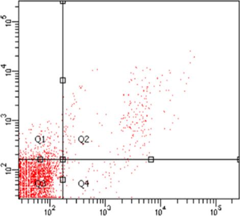
L929                                                        L929 + US
   FITC-A

                               L929 + Reo-OV                                                L929 + Reo-OV + US

                                                           PreCP-Cy5-5-A

                      Fig. 4. Spot chart showing apoptosis induced by reovirus at MOI of 1 in the studied groups of L929 cells after
                      24 h of infection and exposure to low-intensity ultrasound. Annexin V vertical axis and PI horizontal axis: Q1
                      primary apoptosis, Q2 secondary apoptosis, Q3 living cells, and Q4 necrosis. OV, oncolytic viruses.

more, many engineered viruses are undergoing different             of CT26 metastasis [45]; as it came from our flow cytom-
phases of clinical trials; for example, a specific form of         etry results, it is clear that reovirus was capable of killing
herpes simplex virus for the treatment of melanoma has             CRC cells well.
been approved by the US Food and Drug Administration                  The major mechanism of oncolytic reovirus is the abil-
[42]. Oncolytic reovirus is one of the most attractive an-         ity for inducing apoptosis. Thirukkumaran and col-
ticancer agents for clinical trial [43, 44]. As a case in point,   leagues concluded that reovirus induced apoptosis in
Smakman and colleagues concluded that reovirus sero-               breast cancer via the NF-KB pathway [46]. In addition,
type T3D is an effective therapeutic agent in the treatment        Cho and colleagues demonstrated the ability of the reovi-

Oncolytic Effect of Reovirus with                                  Intervirology                                                  5
Low-Intensity of Ultrasound                                        DOI: 10.1159/000519492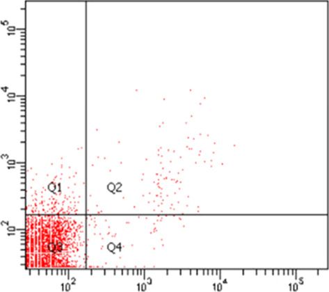
CT26                                                          CT26 + US
    FITC-A

                             CT26 + Reo-OV                                                 CT26 + Reo-OV + US

                                                            PerCP-Cy5-5-A

                     Fig. 5. Spot diagram of the apoptosis induction induced by reovirus at MOI of 1 in the studied groups of CT26
                     cells after 24 h of infection and exposure to low-intensity ultrasound. Annexin V vertical axis and PI horizontal
                     axis: Q1 primary apoptosis, Q2 secondary apoptosis, Q3 living cells, and Q4 necrosis. OV, oncolytic viruses.

rus to induce apoptosis to resistant gastric cancer cells by       virus delivery is the major concern for this method [7].
TRAIL [47]. As a potential outcome of this project, onco-          To overcome this hurdle, combination therapy was pro-
lytic reovirus T3D at MOI of 1 induced apoptosis in CT26           posed [8]. One way is using ultrasound as a new approach
cells.                                                             is proposed, which has previously been used in various
    The most momentous issue during the use of OV is               topics such as drug and DNA molecule release, gene
neutralized by the specific antibody in the body, which            transfer, gene expression, and various other applications
causes that viruses cannot reach their targets alone; thus,        because of its low invasiveness [48, 49]. Baghbani and her

6                    Intervirology                                                        Sharifi et al.
                     DOI: 10.1159/000519492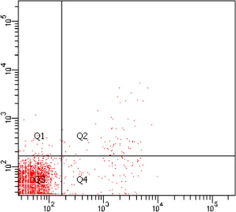
Table 2. The rate of apoptosis in different groups

Group                  L929         L929 + US   L929 + reo   L929 + reo + US                      CT26        CT26 + US        CT26 + reo      CT26 + reo + US

Rate of apoptosis      2.90         3.00        14.10        20.80                                1.50        3.90             7.70            11.80

team came to the conclusion that ultrasound has the po-
tential for being as a drug delivery system, and they had                                    25
seen the perfect anticancer effect of ultrasound with
doxorubicin-loaded PFH nanodroplets in the breast can-                                       20
cer mice model [50]. In the studies of Okunaga and col-

                                                                        Total apoptosis, %
leagues, low-intensity ultrasound increased the entry of
                                                                                             15
HSV-1 into oral squamous cell carcinoma (SCC) and ul-
timately suppressed the growth of rat tumors [39]; be-
sides, Shintani concluded that the exposure of ultrasound                                    10

to Vero cells and oral SCC could accelerate the process of
HSV-1 infection; in addition, ultrasound may be useful                                        5
for increasing the efficiency of HSV-1 infection in viral
therapy for SCC [38]. In recent years, researches have                                        0
been conducted via application of ultrasound in the com-

                                                                                                   29

                                                                                                          S

                                                                                                                 V

                                                                                                                          US

                                                                                                                                 26

                                                                                                                                           S

                                                                                                                                                 V

                                                                                                                                                       US
                                                                                                         /U

                                                                                                                                       /U
                                                                                                               /O

                                                                                                                                               /O
                                                                                              L9

                                                                                                                               CT
bination with OVs in the treatment of various types of

                                                                                                                       V/

                                                                                                                                                     V/
                                                                                                         29

                                                                                                                                      26
                                                                                                              29

                                                                                                                                            26
                                                                                                                     /O

                                                                                                                                                   /O
                                                                                                     L9

                                                                                                                                      CT
                                                                                                              L9

                                                                                                                                            CT
                                                                                                                     29

                                                                                                                                                  26
cancers, but a study investigating the effects of low-inten-

                                                                                                                   L9

                                                                                                                                                CT
sity ultrasound and reovirus had not been performed on
the CT26 cell as a sample of colorectal cancer. Conse-
                                                                     Fig. 6. The amount of total apoptosis in the experimental groups.
quently, the current study has considered the low-inten-             In these groups after 24 h of virus inoculation and exposure to ul-
sity ultrasound advantages, such as the availability of the          trasound waves, apoptosis was measured by the flow cytometry
device, the cheapness of the method, and the improved                technique. The amount of apoptosis in groups infected with reo
treatment efficacy due to the increased probability of cy-           T3D and exposed to ultrasound waves is greater than groups in-
totoxicity of the virus followed by the increase in the cell         fected with reo T3D alone; besides, the rate of apoptosis in groups
                                                                     that are just exposed to ultrasound waves is little. In addition, the
membrane permeability.                                               rate of apoptosis is higher in L929 than in CT26 owing to the fact
    In the present study, low-intensity ultrasound was               that it is the original main host of reo T3D. The figure shows 1 rep-
used to enhance the cytotoxicity of reovirus due to en-              resentative result from 3 independent experiments. OV, oncolytic
hanced virus entry into the target cell. In addition, the            viruses.
behavior of L929 cells as a control and CT26 as a colorec-
tal cancer model cells was investigated. First, we exam-
ined the sensitivity of cells to the ultrasound waves but            of reovirus on various cells is different. Equally impor-
found no apparent destruction after incubation for 24 h.             tant, the L929 cell is the main host of the reovirus. Bani-
Then, the cells were infected with reovirus and were sub-            jamali and colleagues had concluded that the cytopathic
jected to low-intensity ultrasound to investigate differ-            effect of reovirus T3D was seen in both L929 and AD-
ences in virus titers compared to the control group. As the          MSCs cells, but 1 log reduction in virus titer and shedding
results show, the virus titers were significantly higher in          in AD-MSCs was seen compared to L929 cells based on
the cells that are exposed to ultrasound than not exposed.           different characteristics of cells [34, 35].
Therefore, not only can ultrasound increase the rate of                 According to the flow cytometry analysis, reovirus
virus entry into the cells, which improved the virus titer,          T3D is an effective therapeutic agent in the treatment of
but also it can accelerate the process of reovirus infection.        CT26 metastasis and has shown the ability to induce ap-
Moreover, reovirus shedding in exposure cells has in-                optosis in those cells. Likewise, the low-intensity ultra-
creased meaningfully. It can also be noted that the virus            sound effect on the rate of reovirus cytotoxicity could ac-
titers in exposed and unexposed L929 cells to the ultra-             celerate the process of reovirus infection and these waves
sound waves were higher than CT26 cells so that the effect           increased the rate of reovirus cytotoxicity in the cells un-

Oncolytic Effect of Reovirus with                                    Intervirology                                                                               7
Low-Intensity of Ultrasound                                          DOI: 10.1159/000519492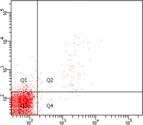
derstudy as we mention above owing to an increase in                               Statement of Ethics
virus entry. Increased apoptotic cell death in both ex-
                                                                                   According to the rules of Tarbiat Modares University, due to
posed cells can result in the efficacy of treatment with OV                    the fact that we used animal cells, so the paper is exempt from
to inhibit the progression of cancer cells and lead to the                     Ethical Committee Approval. The current study was approved that
treatment of cavernous cells.                                                  it does not need the ethical committee code of Tarbiat Modares
                                                                               University. In this study, written informed consent was not re-
                                                                               quired.
    Conclusion

   It can be concluded that using the combination of reo-                          Conflict of Interest Statement
virus with low-intensity ultrasound on murine-derived                              The authors have no conflicts of interest to declare.
cells could increase the oncolytic activity of reovirus and
the rate of cell death. Increased cell death by apoptosis in
L929 and CT26 cells can have positive results in the effi-                         Funding Sources
cacy of treatment with oncolytic reovirus in vivo because
by increasing the speed of virus entry into target cells in                       This project was supported by Tarbiat Modares University’s
this preclinical experiment, it might efficiently prevent                      grant for Master students of medical virology.
the effect of antibodies on the virus neutralization in the
human body. In addition, although the outcome of this
project illustrated a higher rate of cell death, future stud-                      Author Contributions
ies, using a combination of reovirus T3D and low inten-
                                                                                   Negar Sharifi designed and carried out the experience, ana-
sity of ultrasound, should be conducted to measure the                         lyzed data, and wrote the manuscript. Hoorieh Soleimanjahi draft-
factors that induce cell death more accurately by, for ex-                     ed and designed the experience and supervised the research. Mani-
ample, Western blot analysis for cytochrome c release,                         jeh Mokhtari-Dizaji supervised the research. Maliheh Elhamipour
caspase 9, caspase 3 processing, and PARP cleavage;                            performed the experience. Razieh Sadat Banijamali performed the
                                                                               experience. Hesam Karimi performed the experience and cowrote
therefore, more studies are required to evaluate the effi-
                                                                               the manuscript.
cacy of this method.

                                                                                   Data Availability Statement
    Acknowledgments
                                                                                   All data generated or analyzed during this study are included
   The results described in this manuscript were part of the stu-              in this article. Further enquiries can be directed to the correspond-
dent thesis, which was supported by the grant number med-75121                 ing author.
from the Research Deputy of Faculty of Medical Sciences at Tar-
biat Modares University.

    References
 1 Global Burden of Disease Cancer Collabo-            3 Stomper J, Rotondo JC, Greve G, Lübbert M.        using chitosan nanoparticles in breast cancer
   ration; Fitzmaurice C, Allen C, Barber RM,            Hypomethylating agents (HMA) for the              cells. J Nanopart Res. 2015;17(4):168.
   Barregard L, Bhutta ZA, et al. Global, re-            treatment of acute myeloid leukemia and my-     6 Nande R, Howard CM, Claudio PP. Ultra-
   gional, and national cancer incidence, mor-           elodysplastic syndromes: mechanisms of re-        sound-mediated oncolytic virus delivery
   tality, years of life lost, years lived with dis-     sistance and novel HMA-based therapies.           and uptake for increased therapeutic effica-
   ability, and disability-adjusted life-years for       Leukemia. 2021:1–17.                              cy: state of art. Oncolytic Virother. 2015; 4:
   32 cancer groups, 1990 to 2015: a system-           4 Matthay KK, Villablanca JG, Seeger RC,            193.
   atic analysis for the global burden of Disease        Stram DO, Harris RE, Ramsay NK, et al.          7 Marchini A, Scott EM, Rommelaere J. Over-
   Study. JAMA Oncol. 2017; 3(4): 524–48.                Treatment of high-risk neuroblastoma with         coming barriers in oncolytic virotherapy with
 2 Moamer S, Baghestani AR, Pourhoseingholi              intensive chemotherapy, radiotherapy, autol-      HDAC inhibitors and immune checkpoint
   MA, Maboudi AAK, Agha SHS, Zali MR.                   ogous bone marrow transplantation, and            blockade. Viruses. 2016;8(1):9.
   Prognostic factors for survival in patients           13-cis-retinoic acid. Children’s Cancer         8 Babaei A, Soleimanjahi H, Soleimani M, Are-
   with colorectal cancer in Iran between 2004–          Group. N Engl J Med. 1999;341(16):1165–73.        fian E. The synergistic anticancer effects of
   2015: competing risks regression analysis           5 Cengiz BB, Asik MD, Kara G, Turk M, Denk-         ReoT3D, CPT-11, and BBI608 on murine
   with generalized Weibull model. Basic Clin            bas EB. Therapeutic potential of inhibiting       colorectal cancer cells. Daru. 2020;28(2):555–
   Cancer Res. 2017;9(1).                                ABCE1 and eRF3 genes via siRNA strategy           65.

8                          Intervirology                                                                Sharifi et al.
                           DOI: 10.1159/000519492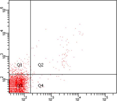
9 Taguchi S, Fukuhara H, Homma Y, Todo T.           23 Barati AH, Mokhtari-Dizaji M. Ultrasound           38 Shintani M, Takahashi G, Hamada M, Oku-
   Current status of clinical trials assessing on-      dose fractionation in sonodynamic therapy.            naga S, Iwai S, Yura Y. Effect of ultrasound on
   colytic virus therapy for urological cancers.        Ultrasound Med Biol. 2010;36(6):880–7.                herpes simplex virus infection in cell culture.
   Int J Urol. 2017;24(5):342–51.                    24 Udroiu I. Ultrasonic drug delivery in Oncol-          Virol J. 2011;8(1):446.
10 Meerani S, Yao Y. Oncolytic viruses in cancer        ogy. J BUON. 2015;20:381–90.                       39 Okunaga S, Takasu A, Meshii N, Imai T,
   therapy. Eur J Sci Res. 2010;40(1):156–71.        25 Yildirim A, Blum NT, Goodwin AP. Colloids,            Hamada M, Iwai S, et al. Ultrasound as a
11 Kemp V, Hoeben RC, van den Wollenberg                nanoparticles, and materials for imaging, de-         method to enhance antitumor ability of onco-
   DJ. Exploring reovirus plasticity for improv-        livery, ablation, and theranostics by focused         lytic herpes simplex virus for head and neck
   ing its use as oncolytic virus. Viruses. 2015;       ultrasound (FUS). Theranostics. 2019; 9(9):           cancer. Cancer Gene Ther. 2015;22(3):163.
   8(1):4.                                              2572.                                              40 Rezazadeh A, Soleimanjahi H, Soudi S,
12 Zhao X, Chester C, Rajasekaran N, He Z,           26 Vilhena NAP. Ultrasound assisted oncolytic            Habibian A. Comparison of the effect of adi-
   Kohrt HE. Strategic combinations: the future         virotherapy: in vitro and in vivo studies; 2015.      pose mesenchymal stem cells-derived secre-
   of oncolytic virotherapy with reovirus. Mol       27 Chowdhury SM, Lee T, Willmann JK. Ultra-              tome with and without reovirus in CT26 cells.
   Cancer Ther. 2016;15(5):767–73.                      sound-guided drug delivery in cancer. Ultra-          Archives of Razi Institute. 2021.
13 Le Boeuf F, Gebremeskel S, McMullen N, He            sonography. 2017;36(3):171.                        41 Li Z, Fan D, Xiong D. Mesenchymal stem cells
   H, Greenshields AL, Hoskin DW, et al. Reo-        28 Zhang L, Liu X, Gao L, Ji Y, Wang L, Zhang            as delivery vectors for anti-tumor therapy.
   virus FAST protein enhances vesicular sto-           C, et al. Activation of piezo1 by ultrasonic          Stem Cell Investig. 2015;2:6.
   matitis virus oncolytic virotherapy in primary       stimulation and its effect on the permeability     42 Ferhat M. Oncolytic viruses: the next major
   and metastatic tumor models. Mol Ther On-            of human umbilical vein endothelial cells.            breakthrough in cancer treatment. Jhvrv.
   colytics. 2017;6:80–9.                               Biomed Pharmacother. 2020;131:110796.                 2017;5(1):00141.
14 Banijamali RS, Soleimanjahi H, Soudi S, Kari-     29 Tang H, Wang CC, Blankschtein D, Langer R.         43 Rahal A, Musher B. Oncolytic viral therapy
   mi H. The effect of oncolytic reovirus infec-        An investigation of the role of cavitation in         for pancreatic cancer. J Surg Oncol. 2017;
   tion on nitric oxide secretion and induction of      low-frequency ultrasound-mediated trans-              116(1):94–103.
   apoptosis in adipose tissue-derived mesen-           dermal drug transport. Pharm Res. 2002;            44 Babaei A, Soleimanjahi H, Soleimani M, Are-
   chymal stem cells. Iran J Med Microbiol.             19(8):1160–9.                                         fian E. Mesenchymal stem cells loaded with
   2018;12(3):218–29.                                30 Hernot S, Klibanov AL. Microbubbles in ul-            oncolytic reovirus enhances antitumor activ-
15 Hirasawa K, Nishikawa SG, Norman KL,                 trasound-triggered drug and gene delivery.            ity in mice models of colorectal cancer. Bio-
   Alain T, Kossakowska A, Lee PW. Oncolytic            Adv Drug Deliv Rev. 2008;60(10):1153–66.              chem Pharmacol. 2021;190:114644.
   reovirus against ovarian and colon cancer.        31 Ebrahiminia A, Mokhtari-Dizaji M, Toliyat          45 Smakman N, van den Wollenberg DJ, Borel
   Cancer Res. 2002;62(6):1696–701.                     T. Dual frequency cavitation event sensor             Rinkes IH, Hoeben RC, Kranenburg O. Sen-
16 Gong J, Mita MM. Activated ras signaling             with iodide dosimeter. Ultrason Sonochem.             sitization to apoptosis underlies KrasD12-de-
   pathways and reovirus oncolysis: an update           2016;28:276–82.                                       pendent oncolysis of murine C26 colorectal
   on the mechanism of preferential reovirus         32 Helfield B, Chen X, Watkins SC, Villanueva            carcinoma cells by reovirus T3D. J Virol.
   replication in cancer cells. Front Oncol. 2014;      FS. Biophysical insight into mechanisms of            2005;79(23):14981–5.
   4:167.                                               sonoporation. Proc Natl Acad Sci U S A. 2016;      46 Thirukkumaran C, Shi ZQ, Thirukkumaran
17 Maitra R, Seetharam R, Tesfa L, Augustine            113(36):9983–8.                                       P, Luider J, Kopciuk K, Spurrell J, et al. PUMA
   TA, Klampfer L, Coffey MC, et al. Oncolytic       33 Man VH, Li MS, Derreumaux P, Wang J,                  and NF-kB are cell signaling predictors of reo-
   reovirus preferentially induces apoptosis in         Nguyen TT, Nangia S, et al. Molecular mech-           virus oncolysis of breast cancer. PloS one.
   KRAS mutant colorectal cancer cells, and syn-        anism of ultrasound interaction with a blood          2017;12(1):e0168233.
   ergizes with irinotecan. Oncotarget. 2014;           brain barrier model. J Chem Phys. 2020;            47 Cho IR, Koh SS, Min HJ, Park EH, Srisuttee
   5(9):2807.                                           153(4):045104.                                        R, Jhun BH, et al. Reovirus infection induces
18 Phillips MB, Stuart JD, Rodríguez Stewart         34 Banijamali RS, Soleimanjahi H, Soudi S, Kari-         apoptosis of TRAIL-resistant gastric cancer
   RM, Berry JT, Mainou BA, Boehme KW. Cur-             mi H. Mesenchymal stem cells support deliv-           cells by down-regulation of Akt activation. Int
   rent understanding of reovirus oncolysis             ery and boost the efficacy of oncolytic reovi-        J Oncol. 2010;36(4):1023–30.
   mechanisms. Oncolytic Virother. 2018;7:53.           ruses in TC-1 tumor cells. J Cell Biochem.         48 Haber T, Baruch L, Machluf M. Ultrasound-
19 Sahin E, Egger ME, McMasters KM, Zhou HS.            2021.                                                 mediated mesenchymal stem cells transfec-
   Development of oncolytic reovirus for cancer      35 Banijamali RS, Soleimanjahi H, Soudi S, Kari-         tion as a targeted cancer therapy platform. Sci
   therapy. Jct. 2013;04(06):1100.                      mi H, Abdoli A, Seyed Khorrami SM, et al.             Rep. 2017;7:42046.
20 Kim J, Hall RR, Lesniak MS, Ahmed AU. Stem           Kinetics of oncolytic reovirus T3D replication     49 Myers R, Grundy M, Rowe C, Coviello CM,
   cell-based cell carrier for targeted oncolytic       and growth pattern in mesenchymal stem                Bau L, Erbs P, et al. Ultrasound-mediated cav-
   virotherapy: translational opportunity and           cells. Cell J. 2020;22(3):283.                        itation does not decrease the activity of small
   open questions. Viruses. 2015;7(12):6200–17.      36 Babaei A, Bannazadeh Baghi H, Nezhadi A,              molecule, antibody or viral-based medicines.
21 Moreno R, Rojas LA, Villellas FV, Soriano            Jamalpoor Z. In vitro anti-cancer activity of         Int J Nanomedicine. 2018;13:337.
   VC, García-Castro J, Fajardo CA, et al. Hu-          adipose-derived mesenchymal stem cells in-         50 Baghbani F, Moztarzadeh F, Mohandesi JA,
   man menstrual blood-derived mesenchymal              creased after infection with oncolytic reovi-         Yazdian F, Mokhtari-Dizaji M. Novel algi-
   stem cells as potential cell carriers for onco-      rus. Adv Pharm Bull. 2021;11(2):361–70.               nate-stabilized doxorubicin-loaded nanodro-
   lytic adenovirus. Stem Cells Int. 2017; 2017:     37 Banijamali RS, Soleimanjahi H, Soudi S, Kari-         plets for ultrasounic theranosis of breast can-
   3615729.                                             mi H. The effect of oncolytic reovirus infec-         cer. Int J Biol Macromol. 2016;93:512–9.
22 Hill CR, Bamber JC, ter Haar GR. Physical            tion on nitric oxide secretion and induction of
   principles of medical ultrasonics: ASA; 2004.        apoptosis in adipose tissue-derived mesen-
                                                        chymal stem cells. Iran J Med Microbiol.
                                                        2018;12(3):218–29.

Oncolytic Effect of Reovirus with                                               Intervirology                                                              9
Low-Intensity of Ultrasound                                                     DOI: 10.1159/000519492
You can also read