Ultrasonography: The Main Diagnostic Tool in Subfertile Women
←
→
Page content transcription
If your browser does not render page correctly, please read the page content below

10.5005/jp-journals-10009-1250
Ekaterini Domali et al
REVIEW ARTICLE

Ultrasonography: The Main Diagnostic Tool in
Subfertile Women
Ekaterini Domali, Konstantinos Kyriakopoulos, Aris Antsaklis

ABSTRACT                                                             diagnosis of the existence of polyp, hyperplasia and
The diagnostic assisted reproductive technology (ART) workup         submucus myoma. Advantage of the method is the possible
includes ultrasonography, hysteroscopy, hysterosalpingography,       direct intervention, treatment of the lesion and biopsy
magnetic resonance imaging (MRI) and laparoscopy where               performance. Disadvantage of the method is the anesthetic
appropriate. Ultrasound represents the mainly used imaging           issuing and the cost. The usage of the minimal diameter
modality for assessing the female genital tract. Recent develop-
ments, i.e. the introduction in the daily praxis of hydrosono-       hysteroscopic instruments, which provoke minimal
graphy, elastography and the use of contrast media, enhanced         discomfort, made the method more easily to apply.4,7 There
by the application of three-dimensional (3D) and four-               is an ongoing debate, if hysteroscopy should be included as
dimensional (4D) software produce images of high resolution.         routine in the standard workup before IVF cycles.4,8,9 Some
All these offer the possibility of multiplanar approach and create
fast techniques that result in specific and detailed reports. The    researchers suggest that the correction of the lesion, if it
comparably short period of training for the medical doctors could    exists, the dilatation of the cervical canal during introduction
transform the ultrasonography in the leading diagnostic tool even    of the instrument and/or a series of immune reactions that
in nonexperienced hands. It is noteworthy, that in suspicion of      are released after endometrium relative damage represent
malignancy, patients should be referred to more experienced
teams.                                                               some of the reasons that hysteroscopy increases the
                                                                     percentage of success of IVF cycles.9-11
Keywords: Three-dimensional ultrasound, Subfertility,
                                                                         Hysterosalpingography is the most widely used method
Diagnosis.
                                                                     in the diagnostic protocol concerning subfertility.12,13 This
How to cite this article: Domali E, Kyriakopoulos K, Antsaklis A.    method provides the possibility to examine the patency of
Ultrasonography: The Main Diagnostic Tool in Subfertile Women.
Donald School J Ultrasound Obstet Gynecol 2012;6(3):270-285.         the tubes, their orientation in the pelvis and to clarify the
                                                                     possible existence of adhesions that could affect externally
Source of support: Nil
                                                                     their mobility. On the other hand, it offers the possibility
Conflict of interest: None declared                                  indirectly to evaluate the anatomical structure of the
                                                                     endometrial cavity. Disadvantage of the method consists
INTRODUCTION                                                         the radiation issuing, increased feeling of pain reported by
                                                                     the women, cost as well as the necessity to program the
The term subfertility includes the failed achievement of
                                                                     method by making an appointment. The relative
gestation after 1 year of unprotected sexual intercourse. In
                                                                     disadvantage of the method is the elevated number of
last decade, the number of pairs that request assisted               positive false results as compared to hysteroscopy because
reproductive technology (ART) methods augmented                      of increased sensitivity but decreased specificity observed.
considerably. It has been calculated that around 2% of the               MRI, characterized as a second level diagnostic study,
born children in the Western world belong to the in vitro            offers the possibility to investigate the anatomical structure
fertilization (IVF) group.1-3 The increase in age of the             of the uterus, to evaluate the ovarian lesions, if they exist
women that desire to become pregnant could represent the             and to examine the patency of the tubes. Increased sensitivity
main reason. The long period of infertility, the increased           and specificity is observed concerning the diagnosis of deep
number of cycles, the male factor, the ovarian pathology,            nodes of endometriosis, especially the smaller one and/or
the anatomical uterine defects and the tubal obstruction             localized in retroperitoneal space. MRI represents a really
interpret also in the subfertile etiology.1,4,5                      specific method, but the elevated cost as well as the absence
    The diagnostic ART workup includes ultrasound scan,              of the direct availability limits significantly its daily use.14,15
hysteroscopy (evaluation of the endometrium and the                      Laparoscopy may participate as a diagnostic tool in the
endomyometrial junction), hysterosalpingography                      workup of IVF protocols. It provides directly the possibility
(evaluation of tubal patency), magnetic resonance imaging            of examination of the whole pelvis and the anatomical-
(MRI) and laparoscopy (estimation of the pelvis and                  included structures. Simultaneously, it is possible one to
adnexa).6                                                            intervene and to correct the possible observed lesions.
    Hysteroscopy offers the possibility to investigate directly      Additionally, it enters considerably in the diagnosis and the
the anatomical defects of uterine cavity and the precise             treatment of the deep nodes of endometriosis. Despite its

270
                                                                                                                              JAYPEE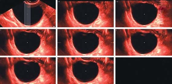
DSJUOG

                                                                 Ultrasonography: The Main Diagnostic Tool in Subfertile Women

advantages, this method remains an interventional, highly          adhesions and subsequently abortions. The diagnostic
costly method, not familiar in women that demands                  ultrasound examination, as it is performed in our days, leads
anesthesia issuing.5,16                                            to clear identification, delimitation and description of the
    Ultrasound approach of female genital tract constitutes        normal and/or abnormal endometrium. Targeted studies
a method simple, repeatable, real-time monitoring without          have been conducted concerning the normal endometrial
radiation, painless, cost-effective and familiar to                cavity, meaning endometrial receptivity (model of
gynecologists as well as to women. In the past few years,          vascularity) and anatomical model of endometrium
occurred a dramatic improvement in the technological               (congenital anomalies, polyps and submucus myomas.17-19
profile of ultrasound scan, regarding the technical clinical
applications as well as the knowledge and experience of            Normal Endometrial Pattern
the medical doctors applying these.
    Ultrasonography enters in the daily clinical practice for      In order to approach the pathology of endometrial cavity, it
the examination of the uterus, adnexa and anatomic                 is required to present the physiological endometrium during
structures of the pelvis (transvaginal). Its usefulness has        3D and 4D ultrasound examination in the different parts of
been extended in the exploration of the gynecological case         the normal menstrual cycle (Fig. 1A). In premenopausal
into the abdominal investigation (transabdominal), into the        woman, the triple layer endometrium is delimitated in early
functional profile of the bladder (translabial), and into the      follicular phase. Progress of the menstrual cycle, provokes
examination of microscopic lesions identified in the pelvis        more hyperechogenic layers and more hypoechogenic
as deep endometriotic lesions and cervical lacerations             uniform interior space. In the luteal phase of the cycle,
(transrectal). Individual developing techniques as                 endometrium is appeared as thick hyperechogenic region
hydrosonography (infusion of saline in the endometrial             in the centrum of the body of the uterus. During 4D
cavity) and use of means, such as SonoVue and EX-Em                ultrasound, we can clearly see the horns of a normal endo-
foam eject the attribution of diagnostic process.                  metrial cavity submerging into the adjacent myometrium.
    During ART protocols, ultrasound scan enters daily into             In the postmenopausal woman, the normal endometrium
the follow-up of the controlled ovarian stimulation, the           appears as a hyperechogenic line that uniformly delineates
estimation of follicular maturity at the time of human             the body of the uterus (Fig. 1B).
chorionic gonadotropin. It is really important to avoid the             Ultrasound examination of endometrial cavity is
appearance of OHSS. In addition, ovum sampling is realized         performed during 5th to 10th day of the menstrual cycle. In
under ultrasound.                                                  suspicion of congenital anomalies, submucus myoma and
    Many variables interfere in the achievement of                 polyp it should be realized during the second part of the
successful clinical pregnancy. We believe and we will try          cycle. 20 The introduction of 3D- and 4D-enhanced
to prove through the presentation of gynecological subfertile      applications of hydrosonography in the daily clinical praxis
cases investigated in our department that the enhanced             remove these restrictions via the smooth enlargement of
ultrasound scan by the application of the three-dimensional        the endometrial lips that it provokes.
(3D) and four-dimensional (4D) software, hydrosonography
and issuing of contrast agents constitutes the main diagnostic     Congenital Anomalies
tool in the daily clinical practice that could guide (permits
or excludes) further surgical treatment, if necessary,             The observation of alterations in the normal described
overlapping henceforth the other diagnostic methods.               endometrial pattern is not a rare condition in daily clinical
                                                                   praxis. Anatomical disturbances, resulting from abnormal
ENDOMETRIUM                                                        development of Mullerian ducts, consist a reason of failed
Successful implantation is strongly correlated to normal           achievement of pregnancy as well as early and/or late
anatomical pattern of endometrium as well as to endometrial        abortion.20 Frequently, in our department we observed
receptivity during a short window phase, where the                 arcuatus morphology of endometrial cavity in different
endometrium erases the implantation of the blastocyst.             degrees. The latter, does not seem to affect seriously the
Initially, endometrial receptivity was defined based on            achievement of pregnancy but shows a remarkable frequency
histological criteria. This procedure demanded D and C,            in subfertile women. In addition, we identified cases of
anesthetic issuing, increased cost and increased worry of          bicornuate uterus, didelphus, septate and uterus that show
the women for consequent provocation of endometrial                disturbed development of the horns (Figs 2A to S).

Donald School Journal of Ultrasound in Obstetrics and Gynecology, July-September 2012;6(3):270-285                         271
Ekaterini Domali et al

      Fig. 1A: 3D and 4D appearance of normal endometrium during follicular phase (a, b and c), periovulatory (d, e and f) and
                                     during luteal phase of the menstrual cycle (g, h and i)

BODY OF THE UTERUS
Structural Disturbances
Recurrently, we detect and describe lesions concerning the
anatomical structure of endometrium and/or myometrium
in subfertile women. Polyps and submucus myomas
originating from endometrium and adenomyosis and
intramural myomas originating from myometrium are the
most frequently observed. In some cases, masses, of not
specified origination (endometrium or myometrium) affect
the body of the uterus and the endometrial cavity causing
significant diagnostic and clinical complications in the
applied IVF protocols. In regard to their size, localization
and extension they can influence the anatomical pattern of
                                                                            Fig. 1B: 3D appearance of endometrial cavity in
normal endometrium and therefore, impact the successful                                postmenopausal women
achievement and/or continuing of the pregnancy.21
    The appearance of lesions that affect the classical            ultrasound examination usually with a centrally located
architectonical structure of endometrium and, therefore, the       feeding vessel (pedicle artery). These lesions are commonly
successful implantation of the fertilized egg is not a rare        combined with clinical symptoms of menometrorrhagia
condition. These lesions originate from endometrium, they          (Figs 3A to I).
remain in the cavity or they may extend in the adjacent                Adenomyosis consists of groups of hyperplastic bundles
myometrium. They may present as hyper- or hypoechogenic            of smooth muscles that surround ectopic implantations of
masses during two-dimensional (2D) as well as 3D and 4D            the endometrium. It can be seen as the diffused pattern of

272
                                                                                                                           JAYPEE
DSJUOG

                                                                    Ultrasonography: The Main Diagnostic Tool in Subfertile Women

Figs 2A to S: (A and B) triple-layer endometrium during the follicular phase of the menstrual cycle; smooth hyperechogenic region was
detected in the middle of the cavity; via 4D application, endometrium was isolated and its arcuatus morphology became obvious,
(C and D) endometrium during the luteal phase of the cycle; 4D software revealed its bicornual morphology, (E and F) bicornual endometrial
morphology; empty pregnancy sac in the left horn, (G and H) abnormal appearance of thick hyperechogenic endometrium during the
luteal phase of the cycle in 3D images; 4D software showed hypoplastic right horn, (I to L) investigation of endometrium in different levels
during 3D examination resulted in failure of appearance of both horns; during 4D application, appearance of unicornuate uterus, (M to P)
hyperechogenic endometrium during the luteal phase of the cycle and abnormally thickened anterior uterine wall in 3D application; via
4D software, didelphys uterus and intramural myoma were revealed, (Q to S) in 3D images, it was observed hyperechogenic endometrium
as well as a hyperechogenic region in the cervix; via 4D software, septate uterus consisting of two endometrial cavities and two discrete
cervices was observed; The one endometrial cavity was hypoplastic, communicating to the greater one

the disease, occupying the whole body of the uterus or as                  Adenomyosis is highly correlated to the coexistence of
the localized pattern creating the adenomyomas. By                     myomas. Identification of well-described masses in the uterine
blocking the normal contractile behavior of the myometrium             wall usually with circular-surrounded circulation indicates the
it interferes in the causes of subfertility.22,23                      diagnosis of myomas (Fig. 4B). The latter, in correlation to their
     The application of the 3D ultrasound examination results          localization (submucus, intramural, subserous) and/or their size
in the clear presentation of disorganized smooth muscle                (>5 cm) could affect negatively the outcome of the IVF program.
bundles of the myometrium (Fig. 4A). The combination of                Application of 3D/4D software permits the clear description of
ultrasound and clinical data increases significantly the               the myomas, the exact identification of their position and
accuracy of the method.                                                predominantly their relationship to the endometrial cavity.
Donald School Journal of Ultrasound in Obstetrics and Gynecology, July-September 2012;6(3):270-285                                    273
Ekaterini Domali et al

Figs 3A to I: (A) 3D presentation of hypogenic lesion that fulfill the endometrial cavity, (B) 4D presentation of possible polyp that destroys
the anterior endometrial lip, (C) 3D appearance of hyperechogenic mass in the endometrium, (D and E) via Doppler, centrally located
vessel was clearly observed in 4D images, (F) hydrosonography enhanced by 3D software revealed undoubtedly the polyp that originated
from the anterior endometrial lip, (G and H) hyperechogenic smooth disturbance of the anterior endometrial lip; (I) hydrosonography
enhanced by 3D software revealed two polypoid lesions; 4D real-time application cancelled the possibility of existence of polyps and the
final report included the diagnosis ‘normal endometrium’

    Recently the elastography gains part of the diagnostic              technology, the measurement is realized automatically by
workup of uterine disturbances.24 This ultrasound method                taking a single volume of each ovary. A variety of colors
calculates the percentage of elastic profile of the                     marks the various stimulated follicles (Fig. 6). The software
myometrium that could be increased in cases of myomas                   calculates simultaneously the 3Ds of each marked follicle
and decreased in cases of adenomyosis. The results are                  as well as its volume.
presented via three basic colors; blue for the hard tissue,
red for the smooth and green for the in-between (Fig. 4C).              Ovarian Pathology
    Despite the significant help that 3D and 4D hydrosono-
                                                                        In the past few years, a revolution occurred in the diagnostic
graphy offers to the clinician, there are some gray zone cases
                                                                        efficacy of transvaginal ultrasound concerning the ovarian
(described in the follow image; Figs 5A to I) that still remain
                                                                        lesions. Logistic regression models, like international
obscure without standardized ultrasonographic criteria. In
                                                                        ovarian tumor analysis (IOTA), orientate diagnosis with
these cases, the clinician should refer to experts, in order to
                                                                        high accuracy, concerning the benign or malignant nature
avoid a possible underlining malignancy.
                                                                        of the lesion.25-27 The extension of ultrasound in the 3D
ADNEXAL MODEL                                                           and the 4D enhances the diagnostic faculty. This extended
                                                                        ultrasonography requires relatively low-grade familiariza-
Ovulation Induction
                                                                        tion of the clinical doctors with technological parameters.
The follow-up of ovulation induction is taken partly through            On the other hand, it offers clear and readable images
ultrasound measurement of the 3Ds of the stimulated follicle            decreasing considerably the rate of necessity of expert
in each ovary separately. Applying the 3D ultrasound                    ultrasonographers.

274
                                                                                                                                    JAYPEE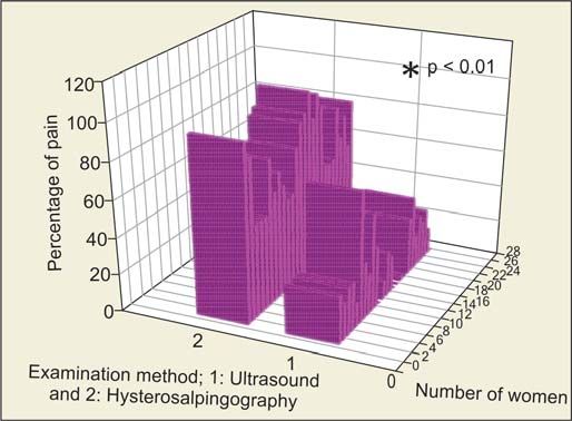
DSJUOG

                                                                    Ultrasonography: The Main Diagnostic Tool in Subfertile Women

Fig. 4A: Presentation of 3D and 4D images; (a and b) disorganization of the ultrasound appearance of smooth myometrial muscles
implies the underline pathophysiology of adenomyosis, (c and d) lesion of mixed echogenicity, observed in the posterior uterine wall,
could be attributed to the existence of malignancy; identification of minimal vascularity pattern cancels the probability of malignancy and
introduces the possibility of underline adenomyosis in the differential diagnosis, (e and f) 4D appearance of thickened endometrial cavity;
the woman underwent hysteroscopic excision of polyp 6 months ago; hydrosonography enhanced by 4D software revealed protrusion of
myometrium in the endometrial that could be attributed to iatrogenic cause of adenomyosis

Fig. 4B: (a) 3D presentation of an intramural myoma that it seems to occupy the whole uterine wall, (b) working on 3D images, endometrial
cavity was revealed, (c) 4D application showed the whole endometrial cavity in close relationship to the intramural myoma, (d and e)
similar case of an intramural posterior myoma; its position was clearly seen via 4D application

                                                                                          Fig. 4C: Predominance of the blue color was
                                                                                          observed in the region occupied by the
                                                                                          intramural myoma; while prevalence of the red
                                                                                          color was saw in the nearby region of uterine
                                                                                          wall, probably affected by adenomyosis

Donald School Journal of Ultrasound in Obstetrics and Gynecology, July-September 2012;6(3):270-285                                   275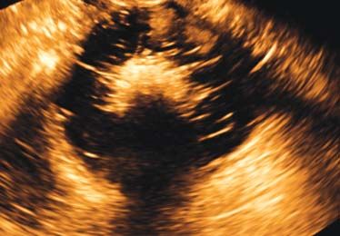
Ekaterini Domali et al

Figs 5A to I: 3D and 4D ultrasound examination. Case I: Woman, 28-year-old, primary infertility; lesion of mixed echogenicity (A), highly
vascularized (B) localized on the anterior uterine wall probably affecting the endometrium (C); enhance hydrosonography and 3D/4D
application failed to distend the endometrial lips (D and E); the histological report by two investigators was completely different, one
suggesting that it is a stromal endometrial sarcoma of low malignancy, while the other one supported that this lesion is a cellular
leiomyoma, Case II: Woman, 39-year-old, secondary infertility; similar images obtained during equal methodology; (F) lesion of mixed
echogenicity; (G) highly vascularized; (H and I) enhance hydrosonography and 3D/4D application failed to distend the endometrial lips
histological report recorded the presence of placenta and trophoblastic tissue without any sign of malignancy

Endometriosis                                                         echogenicity with hyperechogenic areas in their walls show
It represents a pathological procedure that enters                    a 63-fold increased possibility of malignancy and demand
considerably (20-48%) in the subfertile pathophysiology.28            more detailed and careful examination. Identification of
The majority of the endometriotic lesions (88%) are                   papillary projections, transforming the regular wall of
identified in the ovary. The decision of their surgical               endometriotic cysts to irregular, implies the possibility of
excision is crucial. It has been shown that the percentage of         covered malignancy.38,39
ovarian failure after surgical intervention is not negligible.29          Ovarian endometriosis during transvaginal sonography
    Transvaginal ultrasound constitutes an accurate method            may also look like bilocular cysts of double echogenicity,
(sensitivity and specificity around 98%) for the diagnosis            where an invisible septum orientates two discrete foci
of endometriomas.30-33 Hard and soft markers enhance the              (Fig. 7C).
diagnostic capacity as well as the safe follow-up of the                  Unilocular cysts of low echogenicity could imply the
disease.34-36                                                         diagnosis of endometriosis but also multilocular cysts with
    Endometriosis, in 3D images, may appear as unilocular             thick septum with/or without ground glass appearance which
cyst, with thick septum, ground glass echogenicity and                could also be endometriotic masses (Fig. 7D).
sparse wall vascularity (Fig. 7A).                                        The presence of adhesions to the pelvis may indirectly
    Hyperechogenic foci in the cyst wall are not so                   indicate the existence of underlining pathophysiology of
infrequent but also not always innocuous (Fig. 7B). Patel et          endometriosis. These could be identified directly during
al (1999) 37 suggested that unilocular cysts, of low                  transvaginal examination as fine, hyperechogenic strains

276
                                                                                                                               JAYPEE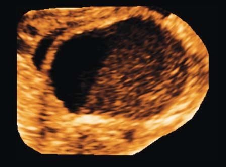
DSJUOG

                                                                 Ultrasonography: The Main Diagnostic Tool in Subfertile Women

              Fig. 6: Presentation and calculation simultaneously of the stimulated follicles during ovulation induction

          Fig. 7A: Typical images of endometrioma during 3D ultrasound procedure; it appears as a unilocular cyst with
                              regular cyst wall, ground glass echogenicity and low-grade vascularity

Donald School Journal of Ultrasound in Obstetrics and Gynecology, July-September 2012;6(3):270-285                            277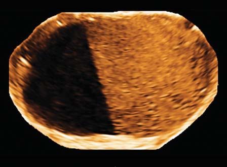
Ekaterini Domali et al

Fig. 7B: Endometrioma showing irregular cyst wall during 2D ultrasound examination. (a) in 3D images, hyperechogenic papillary
projections are obvious, (b) the absence of these features in the e plane during niche mode application, (c to e) proved the nonsolid
nature of the projections, indicating the presence of normal blood clots on the cyst wall

                                Fig. 7C: Two different cases of double echogenicity endometriomas

or indirectly through the observation of a nailed uterus that       These are mainly hypogenic structures with irregular borders
does not follow the movements of the probe and/or ear sign          that remain stable despite the movements of the probe. The
of the uterus. In addition, the observation of kissing ovaries      uterosacral ligament is the driver point to explore the
supports the diagnosis of the existence of adhesions                rectovaginal space, where deep nodes are usually situated.
(Fig. 7E).                                                          The whole examination is based on the reaction of the
    Furthermore, through transvaginal ultrasound, the               patient. The symptom of pain of various degrees indicates
existence of deep nodes of endometriosis is investigated.           the possible existence of deep node.40

278
                                                                                                                           JAYPEE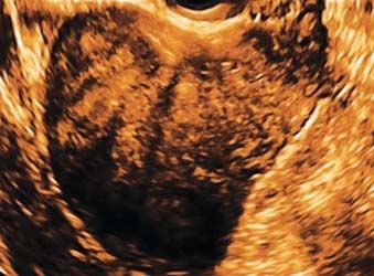
DSJUOG

                                                                   Ultrasonography: The Main Diagnostic Tool in Subfertile Women

Fig. 7D: ‘Atypical' endometriomas during 3D ultrasound; (a) unilocular cyst of low level echogenicity, (b) bilocular of ground glass
echogenicity mass, (c) bilocular lesion of ground glass and low level echogenicity and (d) bilocular lesion, containing an endometrioma
and a hemorrhagic corpus luteum

     Fig. 7E: Indirect signs that imply the underlining presence of adhesions, (a) flattened endometrial cavity flexed to lower level
                 appearing as being under a strong downward pull, (b and c) joined strongly ovaries (kissing ovaries)

   Figs 8A to D: (A and B) Unilocular cyst, originated from the right ovary, which shows mixed echogenicity, regular cyst wall and
      circular pattern of vascularity; (C and D) hemorrhagic ovarian lesion that shows similar pattern in both 3D and 4D images

Donald School Journal of Ultrasound in Obstetrics and Gynecology, July-September 2012;6(3):270-285                                      279
Ekaterini Domali et al
Transrectal ultrasound obtains more clear images of deep cases, they are characterized by mixed echogenicity while
small endometriotic implants including rectum and the observation of a clear image of Rokitansky node is not
parametrium in the examination. It seems that this option a rare condition. In some cases they show a uniformly doted
of ultrasound examination shows increased sensitivity and morphology, provoking problems in the differential
specificity as compared to MRI findings, avoiding the diagnosis from endometriosis. The accurate characterization
rectum peristalsis and its produced bias notably in the of a mass as endometriotic or teratoma is crucial because
sigmoid and ileocecal junction areas.41,42 the two entities demand differentiated therapeutic approach.
Application in the daily praxis of 3D software and The absence of vascularity, the diffuse borders of the mass
especially niche mode and TUI, offers the possibility to without a specific surrounded cyst wall consist ultrasono-
describe in greater detail the extent of the endometriotic graphic remarks that help the investigator to discriminate
implant in the rectovaginal septum and the relationship with the teratoma from an endometriotic cyst. Additionally, the
the rectosigmoid junction or ureter.43 absence of acousting streaming and/or movements of the
internal contents following the movements of the probe in
Hemorrhagic Cysts
combination to the absence of any reaction from the patients
Ovarian lesions characterized by mixed echogenicity are supporting findings of the diagnosis of teratomas
predominantly in both 2D and 3D ultrasound method. Low (Figs 9A to D). The size of the lesion >5 cm recommends
echogenicity may also be observed. High vascularity pattern surgical excision because they are not responsive to
may be appeared surrounding the cyst (ring of the foyer). hormonal therapy and their biologic behavior leads to
Regarding the diagnostic accuracy, the real-time increase of the dimensions of the mass complicating the
ultrasonography (2D and 4D) seems to be superior as achievement of pregnancy that consist the main target of
compared to 3D static image (Figs 8A to D). The main the woman in the ART protocols.30,44
diagnostic point of these masses is the combination of firstly
the appearance of movements of the internal contents of Serous Cystadenomas
the cyst that follow the smooth movements of the probe
and secondary the absence of any feeling of pain of the Unilocular ovarian cysts, presenting ultrasonographic
examined patient. characteristics that support the benign nature of the lesion
are not rare in premenopausal women (Figs 10A to E). An
Teratomas and Dermoid Cysts unresponsive pattern of the cyst, meaning without alteration
They represent the 15 to 20% of germ cell tumors, apparent and/or increase in size, after 6 months hormonal therapy
more frequently in young women. In the majority of the indicates the nonfunctional nature of the mass and leads
Figs 9A to D: 3D images presenting in: (A and B) A teratoma of mixed echogenicity and minimal vascularity, observed in the outer
margin of the cyst; (C) a dermoid cyst of low diffused doted echogenicity easily dispersed to endometriotic lesion; (D) mature cystic
teratoma without specific borders and clear appearance of centrally located Rokitansky node
280
JAYPEEDSJUOG
Ultrasonography: The Main Diagnostic Tool in Subfertile Women
Figs 10A to E: 3D images obtained during ultrasonographic examination of a patient, 31 years old, asymptomatic reporting primary
infertility. (A) Shows a unilocular cyst, anechoic with regular cyst wall, (B) the TUI software confirmed the regularity of the cyst wall and
the absence of any internal content, (C, D and E) the ground plan of the lesion in three different axes is shown
Figs 11A to F: (A) 3D and 4D images presenting a case of hydrosalpinx, where a sausage-like structure with irregular cyst wall is
observed, (B) an incomplete septum appears with high clarity, (C) four cases of abscess; these lesions may appear as a multilocular cyst
of low echogenicity, thick irregular septum (>3 mm), papillary projections and irregular cyst wall, (D) unilocular cyst showing low echogenicity
and extremely thick wall, (E) multilocular solid mass with regular thick septum (>3 mm) and low echogenicity, (F) finally as a solid
structure with extremely high vascularization pattern
obligatory to its surgical excision. Very often, it results to of the tubular (burgeoning-swollen) tube as well as the
the histological diagnosis of ovarian serous cystadenoma. internal septum. The severe inflammatory process (abscess)
may be more ‘impressive’ regarding the clinical situation
Infections
and the ultrasonographic findings. It can be marked by high
It is really often to diagnose hydrosalpinx in young women. fever, abdominal sensitivity and/or pain during
It appears as a sausage-like structure with irregular cyst wall gynecological examination in combination with complex
and obvious incomplete septum in 3D and 4D ultrasound ultrasound pictures of ovarian lesions (mixed echogenicity,
(Figs 11A to F). Especially, the 4D-real-time investigation solid parts, irregular cyst wall and excessive vascularization
of the lesion leads to the achievement of real-time images pattern; Figs 11A to F). All these symptoms and signs that
Donald School Journal of Ultrasound in Obstetrics and Gynecology, July-September 2012;6(3):270-285 281Ekaterini Domali et al
indicate the inflammation subside progressively by the exams were recorded; gold standard was the report of the
issuing of antibiotic therapy. hysterosalpingography. Ultrasound (3D, 4D) was performed
and it was repeated with a contrast agent. At the end of the
FUNCTIONAL PROFILE OF THE TUBES whole procedure (mean duration 15 minutes, ultrasound
Ultrasound method using contrast agents represents a restively with contrast agentDSJUOG
Ultrasonography: The Main Diagnostic Tool in Subfertile Women
Fig. 12C: Woman, 36 years old, primary infertility, (a) incomplete shading of the endometrial cavity during issuing of contrast agent and
ultrasound examination, (b) 3D clear appearance of endometrial polyp, (c) woman 37 years old, secondary infertility; arcuatus appearance
of the uterus, possible intramural myoma nearby the right horn, patent struggle tubes showing beaded-like path
Besides, the application of the 3D and 4D software it surged considerably to 79% during hysterosalpingo-
allowed us through the shading of the structure of the whole graphy. Thus, in the absence of pain as well as the avoidance
tube, the evaluation of their morphology, the regularity or of radiation exposure of the patients, the use of contrast
the irregularity of their sequence, their cool mobility or in agent in subfertile patients in enhancing the diagnostical
the contrary their fixation because of the underline adhesions efficiency of the specialized ultrasound method, seems to
(Fig. 12B). All these details, easily and directly obtained have certain advantages compared to the widely used
during this way of ultrasound examination could not be hypersalpingography.
observed during 2D ultrasound methods.
Finally, the passage of the contrast agent through the CONCLUSION
body of the uterus may reveal possible structural defects Based on our experience, we strongly suggest that the
concerning the body of the uterus, such as polyps or myomas introduction in the daily clinical praxis of the enhanced
(Fig. 12C). ultrasound method, meaning 3D and 4D ultrasonography,
A statistically significant difference was observed transforms the ultrasound in the main diagnostic tool even
concerning the differentiation of the feeling of pain in inexperienced hands. It allows directly to evaluate and
comparing the recorded subjective impression of the patients to diagnose through quite clear images possible anatomical
(Fig. 13). defects of the female genital tract. Simultaneously, it
Applying the graphic patterns of the patients to (%) provides the possibility to investigate the functional profile
percentage, we have established that the feeling of pain was of the tubes and their relationship to the nearby structures
limited to a mere 18% during ultrasound examination, while in the pelvis without pain and without radiation. The latter
was impossible in the conventional ultrasound examination.
This method demands only a short period of training for
the doctors in order to become familiar to the technical
points and availability.
It is noteworthy to underline that damages that could
imply the possibility of malignancy should be referred to
more experienced doctors whose specialty is focused on
ultrasound examination.
REFERENCES
1. Wright VC, Chang J, Jeng G, Macaluso M. Assisted reproductive
technology surveillance—United States, 2005. MMWR Surveill
Summ 2008;57:1-23.
2. Kremer JA, Bots RS, Cohlen B, Crooij M, van Dop PA, Jansen
CA, et al. Ten years of results of in vitro fertilisation in the
Fig. 13: 3D mesh plot presentation of the feeling of pain recorded Netherlands 1996-2005. Ned Tijdschr Geneeskd 2008;152:
by the women during ultrasound and hysterosalpingography 146-52.
examination. Significantly, lower disturbance has been observed 3. van Leeuwen FE, Klip H, Mooij TM, van de Swaluw AM,
during ultrasound examination as compared to hysterosalpingo- Lambalk CB, Kortman M, et al. Risk of borderline and invasive
graphy ovarian tumours after ovarian stimulation for in vitro fertilization
Donald School Journal of Ultrasound in Obstetrics and Gynecology, July-September 2012;6(3):270-285 283Ekaterini Domali et al
in a large Dutch cohort. Hum Reprod 2011 Dec;26(12): A retrospective cohort study. Reprod Biol Endocrinol 2010
3456-65. Mar;24(8):30.
4. Taylor E, Gomel V. The uterus and fertility. Fertil Steril 19. He RH, Gao HJ, Li YQ, Zhu XM. The associated factors to
2008;89:1-16. endometrial cavity fluid and the relevant impact on the IVF-ET
5. Torre A, Poully JL, Wainer B. Anatomic evaluation of the female outcome. Reprod Biol Endocrinol 2010 May;14(8):46.
of the infertile couple. J Gynecol Obstet Biol Reprod (Paris) 20. American College of Obstetricians and Gynecologists: ACOG
2010;39(8 Suppl 2):34-44. technology assessment in obstetrics and gynecology no. 5:
6. Pundir J, El Toukhy T. Uterine cavity assessment prior to IVF. Sonohysterography. Obstet Gynecol 2008;112(6):1467-69.
Source ACU, Guy’s & St Thomas’ NHS Trust, London, UK. 21. Steinkeler JA, Woodfield CA, Lazarus E, Hillstrom MM. Female
Women Health (Long Engl) 2010 Nov;6(6):841-48. infertility: A systematic approach to radiologic imaging and
7. Bosteels J, Weyers S, Puttemans P, Panayotidis C, Van diagnosis. Radiographics 2009 Sep-Oct;29(5):1353-70.
Herendael B, Gomel V, et al. The effectiveness of hysteroscopy 22. Matalliotakis IM, Katsikis IK, Panidis DK. Adenomyosis: What
in improving pregnancy rates in subfertile women without other is the impact on fertility? Curr Opin Obstet Gynecol
gynaecological symptoms: A systematic review. Hum Reprod 2005;17:261-64.
Update 2010;16(1):1-11. 23. Kunz G, Beil D, Huppert P, Noe M, Kissler S, Leyendecker G.
8. Makrakis E, Hassiakos D, Stathis D, Vaxevanoglou T, Adenomyosis in endometriosis-prevalence and impact on
Orfanoudaki E, Pantos K. Hysteroscopy in women with fertility. Evidence from magnetic resonance imaging. Hum
implantation failures after in vitro fertilization: Findings and Reprod 2005;20:2309-16.
effect on subsequent pregnancy rates. J Minim Invasive Gynecol 24. Hobson MA, Kiss MZ, Varghese T, Sommer AM, Kliewer MA,
2009;16(2):181-87. Zagzebski JA, et al. In vitro uterine strain imaging: Preliminary
9. Oliveira FG, Abdelmassih VG, Diamond MP, Dozortsev D, results. J Ultrasound Med 2007 Jul;26(7):899-908.
Nagy ZP, Abdelmassih R. Uterine cavity findings and 25. Timmerman D, Valentin L, Bourne TH, Collins WP, Verrelst
hysteroscopic interventions in patients undergoing in vitro
H, Vergote I. Terms, definitions and measurements to describe
fertilization—embryo transfer who repeatedly cannot conceive.
the sonographic features of adnexal tumors: A consensus opinion
Fertil Steril 2003;80:1371-75.
from the International Ovarian Tumor Analysis (IOTA) Group.
10. Takahashi K, Mukaida T, Tomiyama C, Oka C. High pregnancy
Ultrasound Obstet Gynecol 2000 Oct;16(5):500-05.
rate after hysteroscopy with irrigation in uterine cavity prior to
26. Timmerman D, Ameye L, Fischerova D, Epstein E, Melis GB,
blastocyst transfer in patients who have failed to conceive after
Guerriero S, et al. Simple ultrasound rules to distinguish between
blastocyst transfer. Fertil Steril 2000;4, S206. Future Science
benign and malignant adnexal masses before surgery:
Group Women’s Health 2010;6(6):847.
prospective validation by IOTA group. BMJ 2010 Dec
11. Raziel A, Schachter M, Strassburger D, Bern O, Ron-El R,
14;341:c6839. doi: 10.1136/bmj.c6839.
Friedler S. Favorable influence of local injury to the
27. Van Holsbeke C, Van Calster B, Testa AC, Domali E, Lu C,
endometrium in intracytoplasmic sperm injection patients with
Van Huffel S, et al. Prospective internal validation of
high-order implantation failure. Fertil Steril 2007;87:198-201.
mathematical models to predict malignancy in adnexal masses:
12. Roma Dalfó A, Ubeda B, Ubeda A, Monzón M, Rotger R,
Ramos R, et al. Diagnostic value of hysterosalpingography in Results from the international ovarian tumor analysis study. Clin
the detection of intrauterine abnormalities: A comparison with Cancer Res 2009 Jan 15;15(2):684-91.
hysteroscopy: AJR Am J Roentgenol 2004;183(5):1405-09. 28. Halis G, Meschner S, Ebert AD. The diagnosis and treatment of
13. Brown SE, Coddington CC, Schnorr J, Toner JP, Gibbons W, deep infiltrating endometriosis. Dtsch Arztebl Int 2010
Oehninger S. Evaluation of outpatient hysteroscopy, saline Jun;107(25):446-55.
infusion hysterosonography, and hysterosalpingography in 29. Benaglia L, Somigliana E, Vighi V, Ragni G, Vercellini P,
infertile women: A prospective, randomized study. Fertil Steril Fedele L. Rate of severe ovarian damage following surgery for
2000;74(5):1029-34. endometriomas. Hum Reprod 2010 Mar;25(3):678-82.
14. Kinkel K, Chapron C, Balleyguier C, Fritel X, Dubuisson JB, 30. Sokalska A, Timmerman D, Testa AC, Van Holsbeke C, Lissoni
Moreau JF. Magnetic resonance imaging characteristics of deep AA, Leone FP, et al. Diagnostic accuracy of transvaginal
endometriosis. Hum Reprod 1999;14:1080-86. ultrasound examination for assigning a specific diagnosis to
15. Izumi Imaoka, Akihiko Wada, Michimasa Matsuo, MD Masumi adnexal masses. Ultrasound Obstet Gynecol 2009
Yoshida, Hajime Kitagaki, Kazuro Sugimura, MDMRI maging Oct;34(4):462-70.
of disorders associated with Female Infertility: Use in Diagnosis, 31. Alborzi S, Zarei A, Alborzi S, Alborzi M. Management of
Treatment, and Management Radiographics 2003;23:1401-21. ovarian endometrioma. Clin Obstet Gynecol 2006;49:480-91.
Published online 10.1148/rg.236025115. 32. Moore J, Copley S, Morris J, Lindsell D, Golding S, Kennedy
16. Lim CP, Hasafa Z, Bhattacharya S, Maheshwari A. Should a S. A systematic review of the accuracy of ultrasound in the
hysterosalpingogram be a first-line investigation to diagnose diagnosis of endometriosis. Ultrasound Obstet Gynecol 2002;
female tubal subfertility in the modern subfertility workup? Hum 20:630-34.
Reprod 2011 May;26(5):967-71. Epub 2011 Feb 26. 33. Valentin L. Imaging in gynecology. Best Practice and Research
17. Wang L, Qiao J, Li R, Zhen X, Liu Z. Role of endometrial Clinical Obstetrics and Gynaecology 2006;20:881-906.
blood flow assessment with color Doppler energy in predicting 34. Guerriero S, Ajossa S, Mais V, et al. The diagnosis of
pregnancy outcome of IVF-ET cycles. Reprod Biol Endocrinol endometriomas using colour Doppler energy imaging. Hum
2010 Oct;18:8-122. Reprod 1998;13:1691-95.
18. Chen SL, Wu FR, Luo C, Chen X, Shi XY, Zheng HY, Ni YP. 35. Jermy K, Luise C, Bourne T. The characterization of common
Combined analysis of endometrial thickness and pattern in ovarian cysts in premenopausal women. Ultrasound Obstet
predicting outcome of in vitro fertilization and embryo transfer: Gynecol 2001;17:140-44.
284
JAYPEEDSJUOG
Ultrasonography: The Main Diagnostic Tool in Subfertile Women
36. Muzii L, Bellati F, Plotti F, Manci N, Palaia I, Zullo MA, et al. 45. Luciano DE, Exacoustos C, Johns DA, Luciano AA. Can
Ultrasonographic evaluation of postoperative ovarian cyst hysterosalpingo-contrast sonography replace hysterosalpingo-
formation after laparoscopic excision of endometriomas. J Am graphy in confirming tubal blockage after hysteroscopic
Assoc Gynecol Laparosc 2004;11:457-61. sterilization and in the evaluation of the uterus and tubes in infertile
37. Patel MD, Feldstein VA, Chen DC, Lipson SD, Filly RA. patients? Am J Obstet Gynecol 2011 Jan;204(1):79.e1-5.
Endometriomas: Diagnostic performance of US. Radiology 46. Exacoustos C, Zupi E, Szabolcs B, Amoroso C, Di Giovanni A,
1999;210:739-45. Romanini ME, Arduini D. Contrast-tuned imaging and second-
38. Tanaka YO, Yoshizako T, Nishida M, Yamaguchi M, Sugimura generation contrast agent SonoVue: A new ultrasound approach
K, Itai Y. Ovarian carcinoma in patients with endometriosis: to evaluation of tubal patency. J Minim Invasive Gynecol 2009
MR imaging findings. AJR Am J Roentgenol 2000;175: Jul-Aug;16(4):437-44.
1423-30. 47. Hamed HO, Shahin AY, Elsamman AM. Hysterosalpingo-
39. Wu TT, Coakley FV, Qayyum A, Yeh BM, Joe BN, Chen LM. contrast sonography versus radiographic hysterosalpingography
Magnetic resonance imaging of ovarian cancer arising in in the evaluation of tubal patency. Int J Gynaecol Obstet
endometriomas. J Comput Assist Tomogr 2004;28:836-38. 2009;105(3):215-17.
40. Guerriero S, Alcázar JL, Ajossa S, Pilloni M, Melis GB. Three-
dimensional sonographic characteristics of deep endometriosis. ABOUT THE AUTHORS
J Ultrasound Med 2009;28:1061-66.
41. Abrao MS, Gonçalves MO, Dias JA Jr, Podgaec S, Chamie LP, Ekaterini Domali
Blasbalg R. Comparison between clinical examination, Lecturer, First Department of Obstetrics and Gynecology, University
transvaginal sonography and magnetic resonance imaging for of Athens Medical School, ‘Alexandra’ Hospital, Athens, Greece
the diagnosis of deep endometriosis. Hum Reprod 2007;22:
3092-97. Konstantinos Kyriakopoulos
42. Biscaldi E, Ferrero S, Remorgida V, Rollandi GA. Bowel
endometriosis: CT-enteroclysis. Abdom Imaging 2007;32: First Department of Obstetrics and Gynecology, University of Athens
441-50. Medical School, ‘Alexandra’ Hospital, Athens, Greece
43. Guerriero S, Ajossa S, Gerada M, Virgilio B, Angioni S, Melis
GB. Diagnostic value of transvaginal tenderness-guided Aris Antsaklis (Corresponding Author)
ultrasonography for the prediction of location of deep
endometriosis. Hum Reprod 2008;23:2452-57. Professor and Chairman, First Department of Obstetrics and
44. Caspi B, Weissman A, Zalel Y, Barash A, Tulandi T, Shoham Gynecology, University of Athens Medical School, ‘Alexandra’
Z. Ovarian stimulation and in vitro fertilization in women with Hospital, 80 Vas Sofias Ave, 115 28 Athens, Greece, Phone: +30-210-
mature cystic teratomas. Obstet Gynecol 1998;92:979-81.doi: 7770-461, Fax: +30-210-3381-457, e-mail: adeptobgyn@yahoo.gr
10.1016/S0029-7844(98)00313-5. arisants@otenet.gr, aanstak@med.uoa.gr
Donald School Journal of Ultrasound in Obstetrics and Gynecology, July-September 2012;6(3):270-285 285You can also read