NHS Fetal Anomaly Screening Programme Handbook - Valid from August 2018
←
→
Page content transcription
If your browser does not render page correctly, please read the page content below
NHS Fetal Anomaly Screening
Programme Handbook
Valid from August 2018
NHS FASP Programme Handbook

About Public Health England
Public Health England (PHE) exists to protect and improve the nation’s health and wellbeing,
and reduce health inequalities. We do this through world-leading science, knowledge and
intelligence, advocacy, partnerships and the delivery of specialist public health services.
We are an executive agency of the Department of Health and Social Care, and a distinct
delivery organisation with operational autonomy. We provide government, local government,
the NHS, Parliament, industry and the public with evidence-based professional, scientific
and delivery expertise and support.
Public Health England, Wellington House, 133-155 Waterloo Road, London SE1 8UG
Tel: 020 7654 8000 www.gov.uk/phe
Twitter: @PHE_uk Facebook: www.facebook.com/PublicHealthEngland

About PHE screening
Screening identifies apparently healthy people who may be at increased risk of a disease
or condition, enabling earlier treatment or better informed decisions. National population
screening programmes are implemented in the NHS on the advice of the UK National
Screening Committee (UK NSC), which makes independent, evidence-based
recommendations to ministers in the four UK countries. The Screening Quality Assurance
Service ensures programmes are safe and effective by checking that national standards
are met. PHE hosts the UK NSC secretariat.

www.gov.uk/topic/population-screening-programmes
Twitter: @PHE_Screening Blog: phescreening.blog.gov.uk

For queries relating to this document, please contact: phe.screeninghelpdesk@nhs.net

© Crown copyright 2018
You may re-use this information (excluding logos) free of charge in any format or
medium, under the terms of the Open Government Licence v3.0. To view this licence,
visit OGL. Where we have identified any third party copyright information you will need
to obtain permission from the copyright holders concerned.

Published August 2018
PHE publications                               PHE supports the UN
gateway number: 2018374                        Sustainable Development Goals

                                               2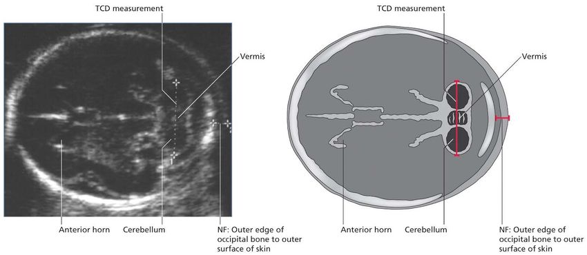
NHS FASP Programme Handbook

Contents
About Public Health England                                2
About PHE screening                                        2
Executive summary                                          5
Introduction                                               6
The NHS Fetal Anomaly Screening Programme                  7
 General principles of screening                           7
 Background                                                7
 Screening policy                                          8
Markers used in screening tests                            9
  Biochemical markers                                      10
  Effect of vaginal bleeding on biochemical markers        10
  Ultrasound markers                                       10
Down’s syndrome, Edwards’ syndrome and Patau’s syndromes   11
  Down’s syndrome                                          11
  Edwards’ syndrome                                        11
  Patau’s syndrome                                         12
Screening Tests                                            12
  The early pregnancy scan                                 12
  First trimester combined test                            13
  Second trimester quadruple test                          14
  Screening in twin pregnancies                            14
  National standards for T21/T18/T13 screening             14
The 18+0 to 20+6 week fetal anomaly ultrasound scan        15
  18+0 to 20+6 NHS FASP ultrasound scan base menu          17
  Fetal cardiac protocol                                   18
  Normal variant                                           19
  Image capture, storage and archiving                     19
  Training and professional competence                     20
  Safety of ultrasound                                     20
Diagnostic testing                                         21
 Results of diagnostic testing                             22
 Audit                                                     22
 Non-invasive prenatal testing (NIPT)                      23
Quality Assurance                                          23
Key performance indicators                                 24
Screening safety incidents                                 25
Glossary                                                   26

                                             3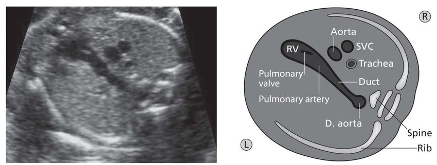
NHS FASP Programme Handbook

Appendix 1.                                       31
18+0 to 20+6 FASP ultrasound scan base menu       31
Appendix 2 – Ultrasound images and schematics     33

                                              4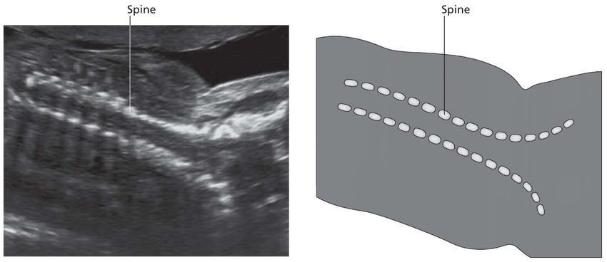
NHS FASP Programme Handbook

Executive summary
The NHS Fetal Anomaly Screening Programme (FASP) has produced this handbook with
support from the members of the programme advisory and task groups and in collaboration
with health care professionals from around England.

This practical guidance supports healthcare professionals and stakeholders in the operational
delivery of the screening pathway. New screening coordinators will find the handbook a source
of information to support their induction and practice.

The handbook provides an update of recent changes to the programme. It refers to supporting
documents and clinical guidance that providers should take into account to deliver a high
quality screening programme.

Commissioners and screening and immunisation teams will find the handbook puts these
documents into the context of the day to day work of the screening coordinators, midwives,
sonographers, screening laboratory teams and obstetricians and fetal medicine specialists who
make up the FASP multi-disciplinary team (MDT).

The handbook provides:
     • structure and governance of the NHS FASP programme
     • a comprehensive outline of each of the conditions
     • clarity on the screening test and terminology
     • hyperlinks to information and supporting documents
     • detail on the delivery of each step of the screening pathway
     • key practice points to consider
     • updates on current quality assurance, data collection and audit processes

The handbook will be updated regularly to ensure it continues to be a valid reference
document.

                                               5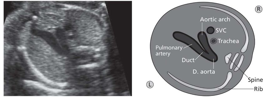
NHS FASP Programme Handbook

Introduction
The purpose of this handbook is to bring together in one publication the NHS Fetal Anomaly
Screening Programme’s (FASP’s) guidelines and recommendations that relate to the
screening pathway and are not covered in detail in the other handbooks.

Throughout the document the following are used interchangeably:
     • Down’s syndrome is referred to as T21
     • Edwards’ syndrome as T18
     • Patau’s syndrome as T13

This handbook is part of a suite of documents that are reviewed and update annually.

   1. Service specifications. Down’s syndrome, Edwards’ syndrome and Patau’s syndrome
      (No. 16) and 18+0 to 20+6 (18 weeks and 0 days of pregancy to 20 weeks and 6 days
      of pregnancy) fetal anomaly scan (No.17). These documents outline the service and
      quality indicators expected by NHS England and the recommendations and
      standards of the UK National Screening Committee (UK NSC).
   2. Programme Standards. These define a set of standards relating to screening for
      Down’s syndrome, Edwards’ syndrome and Patau’s syndrome and the 18+0 to 20+6
      week fetal anomaly scan.
   3. Handbook for laboratories. This sets out the requirements for laboratory staff
      involved in the pathways for first trimester screening for Down’s syndrome, Edwards’
      syndrome and Patau’s syndrome and second trimester biochemical screening for
      Down’s syndrome.
   4. Ultrasound Practitioner’s Handbook. This sets out the requirements for ultrasound
      practitioners involved in the pathway for first trimester screening for Down’s
      syndrome, Edwards’ syndrome and Patau’s syndrome.

                                                6
NHS FASP Programme Handbook

The NHS Fetal Anomaly Screening
Programme
General principles of screening

Screening is a process of identifying apparently healthy people who may be at increased
risk of a disease or condition. They can then be offered information, further tests and
appropriate treatment to reduce their risk and/or any complications arising from the disease
or condition. Further information regarding the general principles of screening can be found
on GOV.UK.

Background

NHS FASP, based in Public Health England, is an expert team that ensures national
consistency and provides expertise. They support and manage on-going roll out,
technical and professional development of the programme and ensure quality and
safety standards are maintained and continuously improved.

The screening programme has evolved from its establishment in 2001 when the
majority of screening was performed using maternal biochemistry in the second
trimester. The recommended method of screening is now first trimester screening,
combining maternal age, biochemistry and ultrasound measurement of fetal nuchal
translucency to provide a pregnant woman with her chance of having a baby with
Down’s syndrome, or Edwards’ syndrome and Patau’s syndrome.

The offer of a fetal anomaly scan is recommended and where accepted should be
undertaken between 18+0 to 20+6 weeks of pregnancy. The fetal anomaly scan base
menu sets out the fetal anatomy to be examined. The fetal anomaly scan screens for 11
conditions.

NHS FASP has established standardisation of the following to enable commissioners
and quality assurance teams to assess the quality of the service provided. These
include:
      • national standards, guidance and chance cut-off for Down’s syndrome,
         Edwards’ syndrome and Patau’s syndrome screening programme in the light of
         emerging evidence
      • a statistical service to ensure that laboratories have access to the best advice in
         maintaining their population medians
      • specifications for chance calculation software to make sure that all laboratories
         calculate chance in a uniform way

                                                7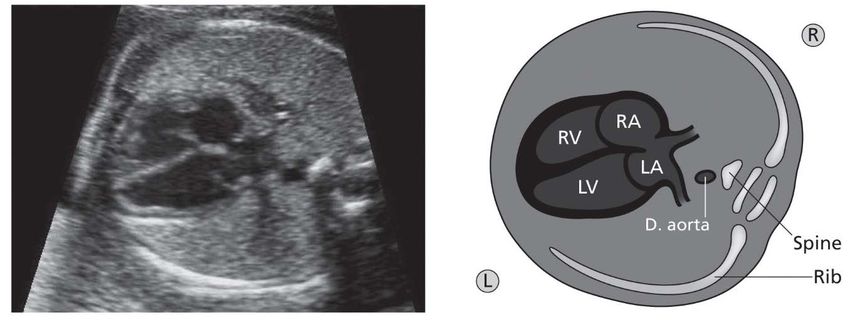
NHS FASP Programme Handbook

     • use of a base menu and fetal cardiac protocol to enable consistency in the
       structures examined as part of the 18+0 to 20+6 week fetal anomaly scan

Screening policy

NHS FASP offers screening to all eligible pregnant women in England to assess the chance
of the baby being born with Down’s syndrome, or Edwards’ syndrome or Patau’s syndrome
or a number of fetal anomalies (structural abnormalities of the developing fetus).

NHS FASP requires that there is equal access to uniform and quality-assured screening
across England and women are provided with high quality information so they can make an
personal informed choice about their screening and pregnancy options. Education and
training resources are available for staff covering all stages of the process, from informing
women of test availability, through to understanding and supporting their decisions.

The screening policy is to offer screening to assess the chance of the baby being born with
Down’s syndrome or Edwards’ syndrome or Patau’s syndrome. The test of choice for both
singleton and twin pregnancies is first trimester combined screening. Women can choose:

     •   not to have screening
     •   to have screening for T21 and T18 / T13
     •   to have screening for T21 only
     •   to have screening for T18 / T13 only

The first scan usually takes place between 10 to 14 weeks and includes a blood sample
taken to test for Down’s syndrome and/or Edwards’ syndrome and Patau’s syndrome, with a
second scan for fetal anomalies between 18+0 to 20+6 weeks. The timing of the scan allows for
further diagnostic tests if required and ensures women have time to consider decisions about
continuing their pregnancy.

The second scan is designed to identify anomalies which indicate:
     • conditions that may benefit from treatment before or after birth
     • that the birth should be in an appropriate hospital/centre and/or to optimise
       treatment after the baby is born
     • that the baby may die shortly after birth

Some women may choose not to be screened at all and it is important that this choice is
respected.

                                                8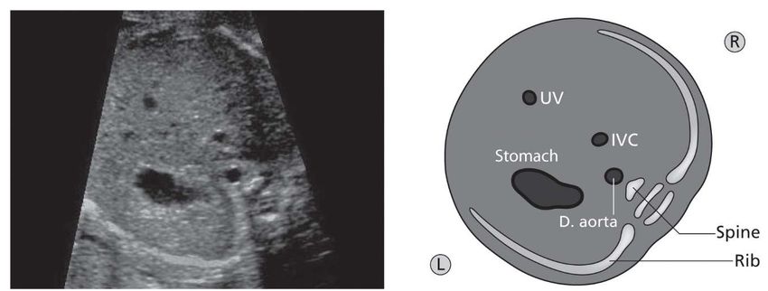
NHS FASP Programme Handbook

Markers used in screening tests
All women have a chance of having a baby with Down’s syndrome, Edwards’ syndrome
or Patau’s syndrome and this chance increases with age. The older a mother, the more
chance she has of having a baby with the one of these conditions.

Table 1: Example for a woman who is 16 weeks pregnant

 Maternal age                 Chances of having a        Probability
                              pregnancy affected by
                              Down’s syndrome
 20 years                     1 in 1500                  0.07%
 30 years                     1 in 900                   0.1%
 40 years                     1 in 100                   1%

Figure 1: Graph to illustrate the likelihood of a pregnancy affected by Down’s syndrome,
Edwards’ syndrome or Patau’s syndrome according to maternal age showing T21 (black
line), T18 (blue line), T13 (green line). The chance is at the time of the 12 weeks scan.

                                             9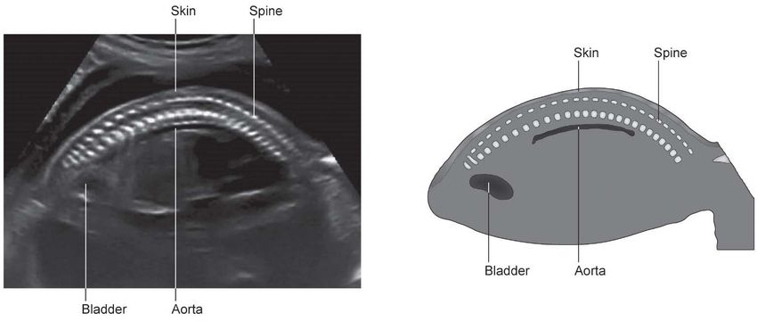
NHS FASP Programme Handbook

Table 2: Reframing chance

          Chance of an affected pregnancy        Chance of an unaffected pregnancy
          1 in 4                25%              3 in 4                    75%
          1 in 5                20%              4 in 5                    80%
          1 in 10               10%              9 in 10                   90%
          1 in 20               5%               19 in 20                  95%
          1 in 30               3%               29 in 30                  97%
          1 in 50               2%               49 in 50                  98%
          1 in 100              1%               99 in 100                 99%
          1 in 200              0.5%             199 in 200                99.5%

Can be applied to any screening test where the result is reported as a probability.

Biochemical markers

There are five analytes (commonly referred to as markers) measured by the laboratory that
are used to calculate the likelihood of a pregnancy being affected by Down’s syndrome,
Edwards’ syndrome or Patau’s syndrome – six if human chorionic gonadotropin (hCG) and
its free beta subunit are considered as two separate analytes. Refer to the laboratory
handbook for more information on biochemical markers.

Effect of vaginal bleeding on biochemical markers

There are concerns that a history of significant maternal vaginal bleeding might change the
levels of biochemical markers used in the combined test. NHS FASP recommends women
are offered the combined test in the normal way (calculating the chance based on maternal
age, Nuchal Translucency (NT), free beta hCG and PAPP-A levels), as current evidence
suggests that the biochemical marker levels are not significantly different in women with this
history.

Ultrasound markers

Please refer to the laboratory and ultrasound practitioner’s handbooks for more detailed
information on ultrasound markers.

                                               10
NHS FASP Programme Handbook
Down’s syndrome, Edwards’ syndrome and
Patau’s syndrome
Inside the cells of our bodies there are tiny structures called chromosomes. These
chromosomes carry the genes that determine how we develop. There are 23 pairs of
chromosomes in each cell. Changes can occur in the sperm or egg cells, which can lead to
a baby having an extra chromosome.
Down’s syndrome
People with Down’s syndrome (T21) have extra chromosome 21 in the cells of their
body.
A person with Down’s syndrome will have some level of learning disability. This means
they will find it harder than most people to understand and to learn new things. They
may have communication challenges and difficulty managing some everyday tasks.
People with Down’s syndrome have distinctive facial features but they do not all look the
same. Most children with Down’s syndrome attend mainstream schools but will require
additional support.
Some health problems are more common in people with Down’s syndrome. These
include heart conditions and problems with hearing and vision. Many health problems
can be treated but, unfortunately, around 5% of babies will not live past their first
birthday.
For babies without serious health problems, survival is similar to that of other children
and most people with Down’s syndrome will live into their 60s or longer.
People with Down’s syndrome can have a good quality of life and most say they enjoy
their lives. With support, many more people with Down’s syndrome are able to get jobs,
have relationships and live semi-independently in adulthood.
Edwards’ syndrome
Babies with Edwards’ syndrome have an extra copy of chromosome 18 in all or some
cells. A condition caused by the presence of an extra copy (three instead of two) of
chromosome 18.
Sadly, the survival rates are low and of those babies born alive only around 10% live
past their first birthday. Some babies may survive to adulthood but this is rare.
11NHS FASP Programme Handbook
All babies born with Edwards’ syndrome will have a learning disability and a wide range
of physical challenges, which can be extremely serious. They may have problems with
their heart, respiratory system, kidneys and/or digestive system. Babies with Edwards’
syndrome may have a low birthweight.
Patau’s syndrome
Babies with Patau’s syndrome have an extra copy of chromosome 13 in all or some
cells.
Sadly, the survival rates are low and of those babies born alive only around 10% live
past their first birthday. Some babies may survive to adulthood but this is rare.
All babies born with Patau’s syndrome will have a learning disability and a wide range of
physical challenges, which can be extremely serious. They may have problems with
their heart, respiratory system, kidneys and/or digestive system. Around half of babies
with Patau’s syndrome will have a cleft lip and palate. Babies with Patau’s syndrome
may have a low birthweight.
Despite their difficulties, children with Edwards` syndrome or Patau`s syndrome can
slowly make progress in their development.
Older children with either Edwards` syndrome or Patau`s syndrome are likely to attend
a specialist school.
Screening tests
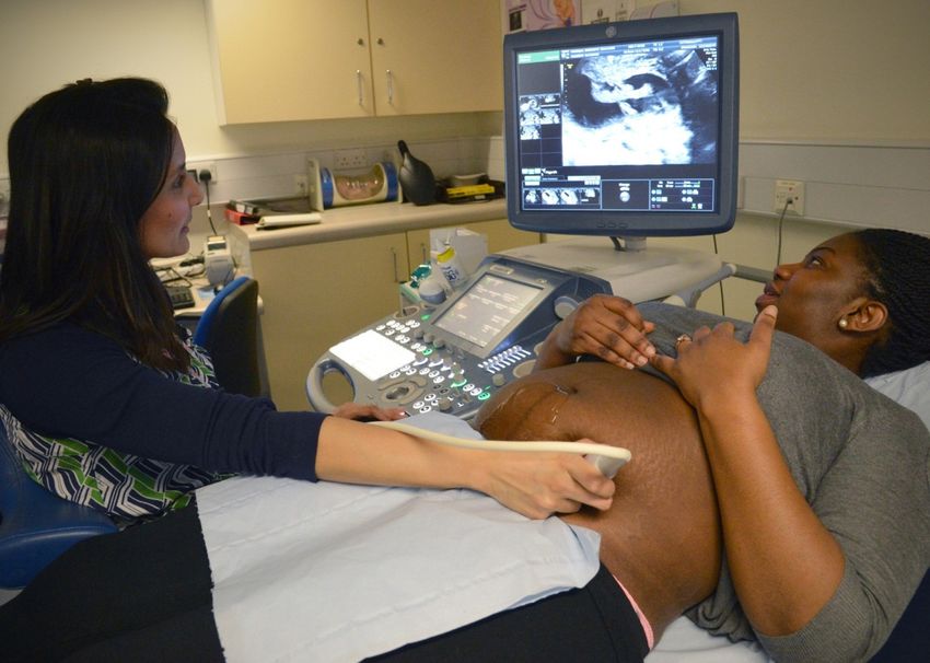
The early pregnancy scan
The scan has several purposes. It is to:
• confirm viability
• ascertain if it is a singleton or multiple pregnancy
• estimate gestational age
• detect major structural anomalies that may be identified in early pregnancy, e.g.
anencephaly
If the woman accepts screening for T21 and/or T18/T13 syndromes, the scan is one
component of the screening test. Ultrasound scanning in pregnancy should, in the first
instance, be performed transabdominally.
12NHS FASP Programme Handbook
Assessment techniques and biometric charts used for fetal measurements must meet
nationally agreed standards. BMUS – Loughna et al 2009
First trimester combined test
The combined test uses maternal age, the nuchal translucency measurement (NT) and
two biochemical markers, free beta hCG and PAPP-A, together with the gestational age
calculated from the crown rump length (CRL) measurement, to calculate the chance
of the pregnancy being affected by T21 or T18/T13. The optimal time to perform the
combined test is between 11+2 weeks to 14+1 weeks of gestation, which corresponds to
a CRL of 45.0 mm to 84.0 mm.
In cases where screening is accepted but it is not possible to obtain the NT
measurement at the first appointment, at least one other attempt should be offered. This
may be on the same day or at a later date. If it is not possible to obtain an accurate NT
measurement despite ‘twice on the couch’, then further attempts do not have to be
offered and the woman should be referred in to the second trimester screening
pathway.
If the ultrasound measurement shows that the CRL is less than 45.0 mm, the woman
should be recalled for a further scan to measure the NT. If the CRL is greater than 84.0
mm, the second trimester quadruple test should be offered.
The first trimester combined test allows earlier decision making for parents. In practice,
two models are available for performing the combined test:
1. a maternal blood specimen may be taken (from 10 weeks onwards) prior to the
ultrasound scan. The biochemistry results can then be made available at the time of the
NT scan and the combined test result can be calculated at the time of the appointment.
Although the result may be calculated by the sonographer, the laboratory remains
responsible for the software that calculates the screening result. Midwifery and/or
ultrasound departments must have a process in place to share ultrasound
measurements and final screening results with the laboratory to enable timely audit of all
results
2. a maternal blood specimen may be taken at the time of the ultrasound scan and
the combined test result made available within a few days of the biochemistry
results being authorised by the laboratory
When calculating a chance for T21 and/or T18/T13, the NT measurement must be used
in combination with a maternal serum screening test. The NT measurement must not be
used in isolation.
Where women have chosen not to accept screening for Down’s syndrome, Edwards’
syndrome and Patau’s syndrome, but choose to accept an early pregnancy scan,
13NHS FASP Programme Handbook
structural anomalies may still be identified, including an NT of ≥ 3.5mm. It is not within
NHS FASP’s remit to provide guidance regarding the clinical care of women who have
declined screening but they should be aware that any such anomaly will be reported
and signposted as per local clinical guidelines for care and management.
NHS FASP recommends that the Down’s syndrome and/or Edwards’ syndrome and
Patau’s syndrome screening chance generated from first trimester combined screening
must not be recalculated up or down following the initial screening test or at the 18+0 to
20+6 fetal anomaly ultrasound scan due to the presence or absence of a single
ultrasound marker of less predictive power than increased nuchal fold (Smith-Bindman
et al, 2001).
For further information regarding the scan element of the combined screening test
please see the Ultrasound practitioner’s handbook.
Second trimester quadruple test
The quadruple test uses maternal age and four biochemical markers measured from 14+2
weeks until 20+0 weeks - AFP, hCG (total, intact or free beta subunit), uE3 and Inhibin-A.
Although this combination of markers has a lower detection and a higher screen positive
rate than the combined test, it is the nationally recommended screening strategy in the
second trimester.
There will always be a need for a screening test in the second trimester for those women
who book too late for first trimester testing or when an NT measurement cannot be obtained
(despite twice on the couch) in the first trimester. Further information regarding the
practicalities of a solution to combining dating and screening requirements at the early
pregnancy scan are explored in more detail in the following article: Chudleigh et al (2011)
Screening in twin pregnancies
Women with a twin pregnancy are eligible for combined screening or quadruple screening
dependent on gestational age. For detailed information regarding screening in twin
pregnancies please see section 6 of the Laboratory Handbook
National standards for T21/T18/ T13 screening
The national standards seen in Table 3 state the threshold for the national programme and
will be reported on each year by the Down’s syndrome screening Quality Assurance
Support Service (DQASS).
14NHS FASP Programme Handbook
Table 3. National standards – thresholds for performance
Screening Thresholds
strategy Acceptable Achievable
T21 Standardised DR 85%
Standardised SPR 1.8-2.5% Standardised SPR 1.9-2.4%
T18/T13 Standardised DR 80%
Standardised SPR 0.1-0.2% Standardised SPR 0.13-0.17%
T21/T18/T13 Standardised DR 80%
Standardised SPR 1.8-2.5% Standardised SPR 1.9-2.4%
Quadruple Standardised DR 80%
(T21) Standardised SPR 2.5-3.5% Standardised SPR 2.7-3.3%
*The DR and SPR for the quadruple test relate to singleton pregnancies only
The 18+0 to 20+6 week fetal anomaly
ultrasound scan
NHS FASP recommends the offer of a mid-pregnancy scan which is undertaken between
18+0 to 20+6 weeks of pregnancy to screen for major fetal anomalies. The examination
should be undertaken in accordance with the 18+0 to 20+6 NHS FASP ultrasound scan base
menu and fetal cardiac protocol.
Some providers are able to arrange the fetal anomaly scan later within the
recommended window, that is closer to 20 weeks as opposed to 18 weeks. Where this
occurs, services must be able to facilitate referrals for further investigations and options
for pregnancy choices in a timely manner and within the required national timeframes.
Ongoing audit of practice should be in place to monitor conformity. The screening
pathway must be completed by 23+0 weeks of pregnancy.
Women who wish to have a fetal anomaly ultrasound scan, but do not wish to be informed if
abnormalities are found, should be advised that all significant findings seen on the scan will
be reported and therefore they should consider not having fetal anomaly ultrasound
screening.
The main structures to be assessed at the 18+0 to 20+6 week scan are defined as shown in
Table 4. Abnormalities of these structures can indicate a number of specific conditions.
15NHS FASP Programme Handbook
Other conditions may be detected using this ultrasound screening test, but there are
insufficient data to confidently predict the standard which should be achieved.
11 conditions are specified that indicate:
• conditions that may benefit from treatment before or after birth
• that the birth should be in an appropriate hospital/centre and/or to optimise
treatment after the baby is born
• that the baby may die shortly after birth
(Fetal Anomaly Ultrasound Screening Programme Study: Literature Survey June
2007)
Table 4: The conditions screened for as a minimum in England
Conditions Detection rate (%)
Anencephaly 98
Open spina bifida 90
Cleft lip 75
Diaphragmatic hernia 60
Gastroschisis 98
Exomphalos 80
Serious cardiac anomalies includes the following: 50
• Transposition of the Great Arteries (TGA)
• Atrioventricular Septal Defect (AVSD)
• Tetralogy of Fallot (TOF)
• Hypoplastic Left Heart Syndrome (HLHS)
Bilateral renal agenesis 84
Lethal skeletal dysplasia 60
Edwards’ syndrome (Trisomy 18) 95**
Patau’s syndrome (Trisomy 13) 95**
**Detections rates will be reviewed once sufficient data is received following
implementation of screening as part of the combined screening strategy
It is accepted that an ultrasound scan at this time can also constitute part of general
clinical practice and management as well as screening. The two are closely linked.
Although it is not the remit of the screening programme to set out standards or guidance
on the management of these areas, it is acknowledged that by not incorporating a
16NHS FASP Programme Handbook
reference to them in the 18+0 to 20+6 NHS FASP ultrasound scan base menu, it may
give the impression that they should not be noted during the ultrasound scan. The
examination of placental position and amniotic fluid, whilst not part of the screening
protocol, is good clinical practice.
There is no requirement to determine fetal gender within the NHS FASP in England; it is
not part of the 18+0 to 20+6 NHS FASP ultrasound scan base menu. There is no
programme requirement to recall the woman if the fetal sex is not identified due to poor
visualisation or difficult fetal position.
18+0 to 20+6 NHS FASP ultrasound scan base menu
The 18+0 to 20+6 NHS FASP ultrasound scan base menu (Appendix 1) specifies
measuring techniques and defines the anatomical structures to be assessed. This
promotes consistency in the examination.
The fetal anatomy to be examined and measured is:
1. head circumference demonstrating HC measurement and measurement of the
atrium of the lateral ventricle
2. suboccipitobregmatic view demonstrating measurement of the transcerebellar
diameter
3. coronal view of lips with nasal tip
4. abdominal circumference demonstrating AC measurement
5. femur length demonstrating FL measurement
6. sagittal view of spine including sacrum and skin covering
These 6 specific fetal anatomical sections should be captured and archived at
examination. A hard copy image of each section (see appendix 1) and report should be
recorded and appropriately stored in any combination of the following formats:
• ultrasound clinical information storage system
• auditable electronic hospital information system
• ultrasound request/report form
• in the woman’s hand-held notes
The head circumference (HC), abdominal circumference (AC) and femur length (FL)
measurements should be taken to assess growth velocity in a pregnancy where the
expected date of delivery (EDD) was previously assigned in line with nationally
approved charts and tables.
If the EDD was not previously assigned, the pregnancy should be dated by HC or FL.
Loughna P, et al (2008)
17NHS FASP Programme Handbook
Fetal cardiac protocol
The views required are:
1. Situs/Laterality
2. Four-Chamber: Transverse section of the thorax including a complete rib and
crux of the heart
3. Aorta/Left Ventricular Outflow Tract: This view shows the outflow tract of the left
ventricle
4. Pulmonary/Right Ventricular Outflow Tract: This view shows the outflow tract of
the right ventricle only; or the Three-Vessel View (3VV): This view shows the
outflow tract of the right ventricle including the pulmonary artery
5. The 3 vessel and trachea view (3VT): a transverse view of the fetal upper
mediastinum; it depicts the main pulmonary artery in direct communication with
the ductus arteriosus, the transverse aortic arch and the superior vena cava
A single repeat scan must be offered and completed by 23+0 weeks gestation in cases
where the image quality of the first examination is compromised by one of the following:
• increased maternal body mass index (BMI)
• uterine fibroids
• abdominal scarring
• sub-optimal fetal position
The woman should be rescanned on the same day or offered a new appointment
according to local clinical assessment.
If the first examination is sub-optimal and the sonographer is suspicious of a possible
fetal anomaly, a second opinion should be sought and a referral made for further
investigation of the anomaly suspected. This should be documented. There is no
requirement to rescan. Refer in these circumstances.
Where an adequate assessment of the fetal anatomy remains compromised after the
repeat scan, there is no requirement to offer further ultrasound examination for
completion of screening. The woman should be informed that the screening is
incomplete and this should be recorded.
18NHS FASP Programme Handbook
Normal variant
The introduction of a national Down’s syndrome, Edwards’ syndrome and Patau’s
syndrome screening programme in early pregnancy has changed the way in which the
18+0 to 20+6 fetal anomaly scan findings are interpreted. NHS FASP recommends that
an established screening test result should not be recalculated at this time.
The screening programme is increasingly delivering higher detection rates for lower screen
positive rates. Therefore, women who are found to be ‘lower chance’ through testing in either
first or second trimesters, or who have declined screening for Down’s syndrome, Edwards’
syndrome and Patau’s syndrome should not be referred for further assessment of
chromosomal anomaly even if normal variants such as the examples below (whether one or
more are identified) are seen at the 18+0 to 20+6 week fetal anomaly screening scan. The
term ultrasound “soft marker” should no longer be used.
1. Choroid plexus cyst(s)
2. Dilated cisterna magna
3. Echogenic foci in the heart
4. Two vessel cord
However, the appearances listed below (previously classified as “markers”) are examples of
findings which should be reported and the woman referred for further assessment and treated
as for any other suspected fetal anomaly.
1. Nuchal fold (equal to or greater than 6mm)
2. Ventriculomegaly (atrium equal to or greater than 10mm)
3. Echogenic bowel (with density equivalent to bone)
4. Renal pelvic dilatation (AP measurement greater than 7 mm)
5. Small measurements compared to dating scan (significantly less than 5th centile on
national charts)
Image capture, storage and archiving
The required images are detailed on the 18+0 to 20+6 NHS FASP ultrasound scan base menu
(see appendix 1). Ultrasound images should be captured, stored and archived on an electronic
reporting system. There should be a permanent electronic record of all imaging studies. All
imaging studies should be accompanied by an electronic report available with the images.
Every provider should be able to upload ultrasound scan reports and images on an auditable
electronic reporting system in order to provide minimum audit data. All required images should
be captured, stored and archived for the purposes of a complete maternal record and to fulfil
medico-legal requirements.
19NHS FASP Programme Handbook
Training and professional competence
All ultrasound practitioners must hold minimum certification as specified by NHS FASP in
Service Specification No 17.
All providers should have multidisciplinary education and training programmes for health
professionals involved in obstetric ultrasound and antenatal screening. All diagnostic
ultrasound procedures must be undertaken by health professionals who are fully trained
in the use of the specialised equipment and in the safe use of ultrasound.
All practitioners undertaking ultrasound screening should be funded by the provider to
attend relevant continuous professional development (CPD) training.
Safety of ultrasound
All health professionals working with ultrasound equipment should be aware of the
Royal College of Radiologists (RCR) and Society and College of Radiographer’s
(SCoR) standards for the provision of an ultrasound service.
All health professionals should adhere to the British Medical Ultrasound Society (BMUS)
recommended scanning time limits for obstetric scanning. British Medical Ultrasound
Society Guidelines for the safe use of diagnostic ultrasound equipment 2009.
Ultrasound machinery used for the 18+0 to 20+6 weeks fetal anomaly scan should be
capable of producing images of diagnostic quality and include the following features (as
a minimum):
• adequate display/screen size for sufficient clear visualisation
• magnification facility
• cineloop function
• callipers that have a precision to one decimal point (ie 0.1 mm)
• adjustable signal processing facilities
• tissue-specific pre-sets for individual clinical applications
• appropriate probe relevant to gestational age
• doppler and harmonic function
20NHS FASP Programme Handbook
Diagnostic testing
Pregnant women should not be offered a diagnostic test for Down’s syndrome,
Edwards’ syndrome or Patau’s syndrome based on their age-related chance alone.
Diagnostic testing or invasive prenatal diagnosis (IPD) can include Chorionic Villus
Sampling (CVS) or amniocentesis. The procedure should be performed by specially
trained health professionals and women may be required to attend a tertiary centre for
the procedure.
CVS is an abdominal or sometimes cervical invasive procedure performed under
continuous ultrasound guidance. The CVS can be performed from 10 weeks, but is
usually only performed from 11 weeks of pregnancy, to obtain a sample of placental
tissue for chromosomal or genetic analysis. Up to 1 out of every 100 women who have
a CVS will miscarry.
Amniocentesis is an invasive procedure undertaken from about 15 completed weeks
(15+0) onwards to obtain a sample of amniotic fluid surrounding the fetus. Using an
aseptic technique whilst under continuous ultrasound guidance, a sterile needle is
passed through the mother’s abdomen, uterus and amniotic sac. A sample of amniotic
fluid is aspirated and sent for chromosomal or genetic analysis. Up to 1 out of every 100
women who have an amniocentesis will miscarry.
The reason for offering the woman the test should be explained, for example:
• a history of an inherited disorder
• a previous pregnancy or a child with a chromosome disorder
• a raised chance of Down’s syndrome or Edwards’ syndrome or Patau’s
syndrome following screening
• suspected anomaly following an ultrasound scan
In twin pregnancies invasive prenatal diagnosis should be conducted at a tertiary fetal
medicine unit due to the specialised nature of the procedures and the increased risk of
miscarriage, and in line with Royal College of Obstetrics and Gynaecology and National
Institute for Health and Care Excellence (NICE) guidelines.
Where the indication for undertaking prenatal diagnosis (PND) is a higher chance screening
result, the sample is sent to the genomic laboratory for quantitative fluorescence polymerase
chain reaction (QF-PCR) testing.
In some cases a confined placental mosaicicm (CPM) may be present. Confined placental
mosaicism is the presence of a chromosome anomaly in the placenta of a fetus with a normal
karyotype. Therefore, to minimise the risk of making decisions regarding the ongoing
21NHS FASP Programme Handbook
pregnancy on the result of a CVS that reflects the cells of the placenta with an unaffected fetus,
the national screening programme recommends that:
• where the QF-PCR result from a CVS sample indicates that the baby may be
affected by Down’s syndrome, Edwards’ syndrome or Patau’s syndrome and in
the absence of any suspected or identified structural anomalies on ultrasound
scan, a culture result should be used to confirm the QF-PCR result prior to any
decisions being made regarding ongoing care or termination of the pregnancy
• where structural anomalies are present on ultrasound scan and the QF-PCR
result indicates that the baby may be affected by Down’s syndrome, Edwards’
syndrome or Patau’s syndrome, the clinician should discuss the options for
ongoing pregnancy care with the woman
If karyotyping is offered following an anomaly suspected on ultrasound scan, the woman
should be informed that subtle chromosomal changes and single gene defects will not
normally be detected. The implications of this should be explained, ie not all inherited
conditions will be identified. The woman should be informed of the usual reporting times
for karyotyping and/or QF-PCR before the procedure.
Results of diagnostic testing
All providers should have a written pathway for communication of results. The process
for communicating results should be discussed and agreed with the woman before the
procedure. All women must be informed of the CVS or amniocentesis result by an
appropriately trained person. When a CVS or amniocentesis is performed at a tertiary
centre, that centre should provide written results to the referring clinician. The woman
should be informed of the results of diagnostic testing as per local policy.
Audit
Each department performing CVS and amniocentesis procedures should maintain a
register of procedures performed and outcome of pregnancy. To facilitate audit,
pregnancy outcome forms should be completed and returned to the screening
laboratory, or other locally agreed collating centre, at the end of the pregnancy. The
provider should develop a written pathway for the completion and return of pregnancy
outcome forms to the centre collecting the data.
Patient evaluation of service provision is an integral aspect of overall service audit and
should be included as part of the audit and performance management framework.
Information should be shared with the National Congenital Anomaly and Rare Disease
Registration Service (NCARDRS) for quality and monitoring of screening programme
outcomes.
22NHS FASP Programme Handbook
Non-invasive prenatal testing (NIPT)
During pregnancy, the placenta sheds DNA into the mother`s bloodstream which results
in the mother`s blood containing both her own and placental DNA. This is known as
total cell free DNA (cfDNA) and in most cases, the placental DNA will be the same as
fetal DNA. The total cfDNA is extracted from a maternal blood sample, and sequenced
and counted. This sequence is then compared to a reference range to see if any DNA is
over represented for chromosomes 21, 18 or 13. An overrepresentation of these
chromosomes means that there is a higher chance of the fetus being affected by
Down`s syndrome, Edwards` syndrome or Patau`s syndrome.
Current availability of NIPT
The UK NSC has recommended introducing NIPT into the NHS FASP screening
pathway for Down’s syndrome, Edwards’ syndrome and Patau’s syndrome as an
evaluative roll out. This means that any necessary changes can be made in a timely
fashion. NIPT will be an additional option for those women who have a higher chance (1
in 2 to 1 in 150) of having a baby with Down’s syndrome, Edwards’ syndrome or Patau’s
syndrome following first trimester combined or second trimester quadruple screening
(singleton pregnancies only). Planning is under way with a view to implement the offer
of NIPT as an additional option in the current screening pathway during 2018 to 2019.
Quality assurance
Each NHS screening programme has a defined set of standards that providers have to meet to
ensure that local programmes are safe and effective. Quality assurance (QA) is the process of
checking that these standards are met and encouraging continuous improvement and
includes:
• advising on the development of national quality standards
• monitoring of how services meet (or fail to meet) standards
• providing expert screening advice for incident management
• facilitating quality review of services, including peer advice
• supporting on a day-to-day basis, those involved in commissioning or providing
screening services
QA covers the entire screening pathway; from identifying who is eligible to be invited to
screening, through to referral and treatment where required/appropriate.
23NHS FASP Programme Handbook
The aim of QA is to maintain minimum standards and drive continuous improvement in the
performance of all aspects of screening to ensure that all women and their babies have
access to high quality screening wherever they live. QA is essential in order to minimise
harm and maximise benefits of screening.
Formal QA visits to local screening programmes provide the forum for a peer review of
the whole multidisciplinary screening pathway, and an assessment of the effectiveness
of team working within the local screening programme and associated referral sites.
• regional teams advise providers and commissioners about reducing risks in
local screening programmes
• they assess the robustness of local arrangements through audit, as part of peer
review and in the investigation of any incidents as they occur
• they act as a conduit for information and dialogue at national, regional and local
levels, additionally sharing good practice
• participation in a formal process of QA is the responsibility of each local
screening programme
• the performance of the local programmes is monitored in a variety of ways such
as review of statistics, regional meetings or informal visits, all of which offer a
valuable insight into the activity of a local programme
Key performance indicators
Key performance indicators (KPIs) for the NHS screening programmes were introduced
to provide a way of measuring how well the screening programmes are doing in
important areas. They contribute to the quality assurance of screening programmes but
are not, in themselves, sufficient to quality assure or performance manage screening
services. They help local screening services to identify potential problems so they can
be put right and have led to changes in practice and implementation of measures to
prevent errors occurring in the screening pathway.
There are currently three KPIs for the fetal anomaly screening programme.
KPI FA1 relates to the completion of the request form to ensure all required information
is available at the point of calculation of the screening risk to prevent delays in
screening and reduce inaccuracies in screening results.
KPI FA2 is a measure of coverage of the 18+0 to 20+6 week scan. This is a measure to
make sure that all women who have been offered and accepted screening have
completed screening by 23+0 weeks of pregnancy.
24NHS FASP Programme Handbook
KPI FA3 is a measure of coverage of Down’s syndrome, Edwards’ syndrome and
Patau’s syndrome screening. This is a measure to make sure screening is offered to
everyone who is eligible and that each individual who chooses to accept screening has
a conclusive screening result.
Screening safety incidents
A screening safety incident is any unintended or unexpected incident(s) that could have
or did lead to harm to one or more persons who are eligible for NHS screening; or to
staff working in the screening programme.
A screening safety incident can affect populations as well as individuals. It is an actual
or possible failure in the screening pathway and/or at the interface between screening
and the next stage of care. Although the level of risk to an individual in an incident may
be low, because of the large numbers of people offered screening, this may equate to a
high corporate risk. It is important to ensure that there is a proportionate response
based on an accurate investigation and assessment of the risk of harm. Due to the
public interest in screening, the likelihood of adverse media coverage with resulting
public concern is high even if no harm occurs.
Find more information and guidance about managing screening safety incidents here.
25NHS FASP Programme Handbook
Glossary
Amniocentesis
An invasive procedure undertaken from about 15 completed weeks (15+0) onwards to
obtain a sample of amniotic fluid (liquor) surrounding the fetus. Using an aseptic
technique whilst under continuous ultrasound guidance, a sterile needle is passed
through the mother’s abdomen, uterus and amniotic sac. A sample of amniotic fluid is
aspirated with a syringe and sent for analysis to test for a range of chromosomal and
inherited disorders. Up to 1 out of every 100 women who have an amniocentesis will
miscarry.
Amniotic fluid
Also known as ‘liquor’, this is the fluid surrounding the fetus during pregnancy. It
contains substances and cells from the fetus, which can be removed by amniocentesis
and examined.
Biochemical markers
Analytes (commonly referred to as markers) measured by the laboratory that are used
to calculate the likelihood of a pregnancy being affected by a condition or syndrome.
Chance
The likelihood that an event will occur.
Chance cut-off
Determines those women who are in the ‘higher chance’ group and considered ‘screen
positive’.
Chorionic villus sampling (CVS)
An abdominal or cervical procedure performed under continuous ultrasound guidance
after 10 completed weeks in pregnancy to obtain a sample of placental tissue for
chromosomal or genetic analysis. The range of chromosomal and genetic conditions
that can be detected is similar to those for amniocentesis. Up to 1 out of every 100
women who have a CVS will miscarry.
Combined test
Between 11+2 weeks and 14+1 weeks of pregnancy, a combination of the nuchal scan
measurement and a blood sample from the mother which measures the concentration
of pregnancy associated plasmprotein-A (PAPP-A), and free beta human chorionic
gonadotrophin (free beta hCG). Together with the mother’s age and the gestation of the
pregnancy, these are used to estimate the chances that the fetus is affected with Down’s
syndrome, Edwards’ syndrome or Patau’s syndrome.
26NHS FASP Programme Handbook
Crown rump length (CRL)
Ultrasound measurement between the top of the head (crown) to the bottom of the
buttocks (rump). To be eligible for first trimester combined screening as part of the NHS
screening programme the CRL should measure between 45.0mm and 84.0mm.
Detection rate
The proportion of affected individuals with a positive screening result.
Diagnostic test
Refers to the process involved in obtaining a definite diagnosis. For example the
diagnostic test on an amniocentesis sample (invasive procedure) is the full karyotype or
QF-PCR.
Down’s Syndrome (trisomy 21)
Down’s syndrome is caused by an extra copy of chromosome 21 in all or some cells of
the body.
A person with Down’s syndrome will have some level of learning disability. This means
they will find it harder than most people to understand and to learn new things. They
may have communication challenges and difficulty managing some everyday tasks.
People with Down’s syndrome have distinctive facial features but they do not all look the
same.
Some health problems are more common in people with Down’s syndrome. These
include heart conditions and problems with hearing and vision. Many health problems
can be treated but unfortunately around 5% of babies will not live past their first
birthday.
Edwards’ Syndrome (trisomy 18)
Babies with Edwards’ syndrome have an extra copy of chromosome 18 in all or some
cells. A condition caused by the presence of an extra copy (three instead of two) of
chromosome 18.
Sadly the survival rates are low and of those babies born alive only around 10% live
past their first birthday. Some babies may survive to adulthood but this is rare.
All babies born with Edwards’ syndrome will have a learning disability and a wide range
of physical challenges, which can be extremely serious. They may have problems with
their heart, respiratory system, kidneys and/or digestive system.
Babies with Edwards’ syndrome may have a low birthweight.
Fetal anomaly
Structural abnormalities with how the fetus has developed.
Fetal anomaly ultrasound scan
A detailed ultrasound scan, sometimes called the mid-pregnancy or 20-week scan. It is
a screening test offered to all pregnant women and is usually carried out between 18
27NHS FASP Programme Handbook
and 21 weeks of pregnancy. It produces a 2-dimensional black and white image that
gives only a side view of the baby and it checks for major physical anomalies in the
baby; although it can't pick up every anomaly.
Gestational age
The duration of an ongoing or completed pregnancy, measured from the first day of the
last menstrual period (usually about two weeks longer than that measured from
conception). Gestational age is usually measured in weeks and days.
Invasive diagnostic procedure
A method used to obtain a sample used to aid diagnosis , for example, amniocentesis
or chorionic villus sampling.
Marker
An identifiable physical location on a chromosome whose inheritance can be monitored.
Markers can be expressed regions of DNA (genes) or some segment of DNA with no
known coding function but whose pattern of inheritance can be determined.
Nuchal scan (Nuchal transluscency scan NT)
Between 11+2 weeks and 14+1 weeks of pregnancy the thickness of fluid in the tissue
space within the nape of the fetal neck, the nuchal translucency can be measured. An
increased amount of fluid may indicate that the fetus has Down’s syndrome, structural
or genetic anomaly. By combining the mother’s age and the gestation of the pregnancy
with information from the scan an individual statistical chance of an anomaly can be
given for that particular pregnancy. If the chance is between 1 in 2 and 1 in 150 a
diagnostic test, such as CVS, will be offered.
Patau’s Syndrome (trisomy 13)
Babies with Patau’s syndrome have an extra copy of chromosome 13 in all or some
cells.
Sadly the survival rates are low and of those babies born alive only around 10% live
past their first birthday. Some babies may survive to adulthood but this is rare.
All babies born with Patau’s syndrome will have a learning disability and a wide range of
physical challenges, which can be extremely serious. They may have problems with
their heart, respiratory system, kidneys and/or digestive system. Around half of babies
with Patau’s syndrome will have a cleft lip and palate. Babies with Patau’s syndrome
may have a low birthweight.
Prenatal
Relating to the period before birth
28NHS FASP Programme Handbook
Quadruple test
Second trimester test to calculate the chance of the pregnancy being affected by
Down’s syndrome, usually based on the measurement of AFP, uE3, free beta hCG (or
total hCG), and inhibin-A together with the woman’s age.
Quality assurance (QA)
A system for monitoring and maintaining high standards in every aspect of a screening
programme.
Screening
Testing people who do not have or have not recognised the signs or symptoms of the
condition being tested for, either with the aim of reducing risk of an adverse outcome, or
with the aim of giving information about risk.
Screening pathway
The whole system of activities needed to deliver high quality screening. It ranges from
identifying and informing those to be offered screening through to the treatment and
follow up of those found to have abnormality, and support for those who develop
disease despite screening
Screen positive rate (SPR)
The number of women who receive a higher chance result.
Screening programme
The whole system of activities needed to deliver high quality screening. It ranges from
identifying and informing those to be offered screening through to the treatment and
follow up of those found to have abnormality, and support for those who develop
disease despite screening
Screening safety incident
An unintended or unexpected incident(s) that could have or did lead to harm to one or
more persons who are eligible for NHS screening; or to staff working in the screening
programme.
Screening test
A test or inquiry used on people who do not have or have not recognised the signs or
symptoms of the condition being tested for. It divides people into lower and higher
chance groups.
Syndrome
Combination of symptoms and signs grouped together to form a disorder.
29NHS FASP Programme Handbook
Throughput
Number of samples undertaken per cycle
Trisomy
Three copies of a particular chromosome rather than two.
Ultrasound scan
A ultrasound scan is a safe and painless test that uses sound waves to make images. It
is like radar.
30NHS FASP Programme Handbook
Appendix 1.
18+0 to 20+6 FASP ultrasound scan base
menu
Structure/Area Detail Fetal Measurements* Images/measurements
to capture/archive
Head and neck Head shape *Head circumference Yes, to include HC
(HC) measurement, CSP,
• Skull
posterior horn and
• Brain measurement of the
Cavum septum Measurement not ventricular atrium at the
• Neck pellucidum (CSP) required level of the glomus of
Ventricular Atrium (VA) *Atrium of the lateral the choroid plexus
Ventricle
Cerebellum *Transcerebellar Yes, to include
diameter (TCD) measurement of the
TCD in the
suboccipitobregmatic
view
Nuchal Fold ( NF) Distance between the Yes, if measurement ≥
outer border of the 6mm
Measure if appears large
occipital bone and the
outer skin edge
• Facial Features Coronal view of lips & Measurement not Yes
nasal tip required
• Lungs Visceral situs/laterality of Measurement not
No
heart required
• Heart
a) Four chamber view
(FCV)
b) Aorta (Ao) arising No
from left ventricle
c) Pulmonary artery (PA) No
arising from right
ventricle, or the 3 vessel
view (3VV)
d) 3 vessel and trachea No
view (3VT)
31NHS FASP Programme Handbook
Structure/Area Detail Fetal Measurements* Images/measurements
to capture/archive
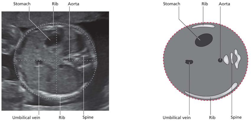
Abdominal content Stomach & position Measurement not Yes
required
*Abdominal
circumference (AC)
Short intra-hepatic
section of the umbilical
vein (UV)
Abdominal wall and
cord insertion
Diaphragm Measurement not
required
Kidneys Measurement not Yes, if AP renal pelvis
required unless renal diameter measures
Measure AP renal pelvis
pelvis AP diameter >7mm
diameter if it appears
>7mm
large
Bladder Measurement not
required
Spine Vertebrae Measurement not Yes, image either
required sagittal or coronal plane
• Cervical Skin covering
• Thoracic
• Lumbar
• Sacral
Limbs Femur, tibia & fibula *Femur length Yes, image and measure
(both legs) a single femur only
• Upper & lower
Metatarsals (both feet) Digit count not required
Radius, ulna, humerus Measurement not
(both arms) required
Metacarpals (both Digit count not required
hands)
Uterine cavity Placenta According to local
policy/protocol
• Uterine content
Amniotic fluid According to local
policy/protocol
32NHS FASP Programme Handbook
Appendix 2 – Ultrasound images and
schematics
Head circumference (HC) and ventricular atrium (VA)
Transcerebella diameter (TCD) and nuchal fold (NF)
33NHS FASP Programme Handbook
Lip and nasal tip
Abdominal circumference (AC)
34NHS FASP Programme Handbook
Femur length (FL)
Sagittal spine
35NHS FASP Programme Handbook
Coronal upper spine
Coronal lower spine
36NHS FASP Programme Handbook
Visceral situs/laterality
4 chamber view (4CH)
37NHS FASP Programme Handbook
Aorta (AO)/left ventricular outflow tract
Pulmonary artery (PA)/right ventricular outflow tract or 3 vessel view (3VV)
3 vessel and trachea view (3VT)
38You can also read