Synthesis, Characterization and DFT investigation of New Metal Complexes of Ni(II), Mn(II) and VO(IV) Containing N,O-donor Schiff Base Ligand
←
→
Page content transcription
If your browser does not render page correctly, please read the page content below
This is a previous version of the article published in Journal of Molecular Structure. 2021, 1231: 129923. https://doi.org/10.1016/j.molstruc.2021.129923

      Synthesis, Characterization and DFT investigation of New Metal Complexes of
             Ni(II), Mn(II) and VO(IV) Containing N,O-donor Schiff Base Ligand

 Djouhra AGGOUN a,b,*, Zakia MESSASMAa,b, Brahim BOUZERAFA c, Raúl BERENGUER d,
                      Emilia MORALLON d, Yasmina OUENNOUGHI a and Ali OURARI a

  a
     Laboratoire d'Electrochimie, d'Ingénierie Moléculaire et de Catalyse Redox (LEIMCR), Faculté de
                 Technologie, Université Ferhat ABBAS de Sétif-1, Sétif 19000, Algeria.
     b
       Département de chimie, Faculté des sciences, Université Ferhat ABBAS de Sétif-1, Sétif 19000,
                                                Algeria.
   c
     Laboratoire de Préparation, Modification des Matériaux Polymériques Multiphasiques (LMPMP),
             Faculté de Technologie, Université Ferhat ABBAS Sétif-1, 19000 Sétif, Algeria.
d
  Departamento de Química Física, Instituto Universitario de Materiales, Universidad de Alicante (UA),
                                      Ap.99, 03080 Alicante, Spain.

Abstract
In this work, a bidentate Schiff base ligand (HL) based on N and O donor heteroatoms has been used to
synthesize a series of new stable metal complexes of general composition of M(L)2 with M = Mn, Ni and
VO. The ligand was obtained from the condensation of 2-methoxybenzylamine with 2,3-
dihydroxybenzaldehyde in methanolic medium. The complexes were formed by mixing the metal acetate
salts and the ligand in 1:2 molar ratio. The resulting three complexes have been characterized by different
analytical techniques, including FT-IR, UV-Vis, mass spectroscopies, thermogravimetry and cyclic
voltammetry, to study their molecular structure and redox/electrochemical properties. Moreover, the effect
of the metal on the complexes electronic properties, and their predicted reactivity, has been studied by
Density Functional Theory (DFT). The spectroscopic results suggest that the metal is bonded to the ligand
through the phenolic-like oxygen and the imine-type nitrogen atoms. Electronic and vibrational absorption
spectra of the nickel complex suggest a square-planar geometry around the central metal ion Ni(II), while
square-pyramidal and octahedral geometry have been proposed for VO(IV) and Mn(II) complexes. The
thermogravimetric analyses of the complexes confirmed the presence of water molecules in their
structures and thermal decomposition led to the formation of metal oxides as residues. The voltammogram
of the Ni(II) complex suggests the existence of quasi-reversible redox system in DMSO solution.

Keywords: N,O donors; Bis-bidentate Schiff base complexes; Spectral studies; Cyclic voltammetry;
Thermogravimetry. DFT Calculations.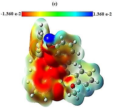
•   Corresponding author. Tel: +213.697.78.81.45
          E-mail address: aggoun81@yahoo.fr (Dj. AGGOUN).

1. Introduction

In recent decades, Schiff bases ligands [1] have attracted lot of attention not only for their structural variety
(fundamental research), but also for their various physicochemical properties opening a wide domain of
applications (practical point of view) [2]. This interest is also due to the structural versatility of Schiff
bases to conform asymmetric compounds (Katsuki and Jacobsen), stabilizing many different complexes
in various oxidation states and controlling the performance of metals in a large variety of useful
transformations [3,4]. Moreover, the coordination geometry of these complexes [5,6] depends upon
various factors, including the size of coordination sphere, electronic configuration of the central metal
ions, non-bonding interactions between atoms in different ligands accompanied with inherent rigidity due
to the presence of aromatic rings.
Since they are getting increasing significance as analytical and electroanalytical reagents [7,8], transition
metal complexes employing Schiff base as ligands are among the most interesting coordination
compounds in the past few years. Their importance was also clearly proved from their good catalytical
activities in numerous photochemical, chemical or electrochemical reactions [9-13]. Especially, transition
metal complexes with N,O-chelating Schiff bases derived from salicylaldehyde are of particular
interest[14-16]. They are essential in organic chemistry as starting compounds, synthetic intermediates
and designer molecules and, therefore, in the field of materials science and industry of chemicals and
bioactive compounds. In addition, they have played a seminal role in the development of modern
coordination chemistry, but they could play also a crucial role in the development of inorganic chemistry.
The design of new bidentate Schiff base ligands containing N and O atoms or functional groups with
electron donor capability is then very interesting for metal complexation application. These ligands can
be prepared by condensation of aldehydes with primary amines. Among different possibilities, we have
previously reported the synthesis of novel N,O-donor Schiff base ligands from the combination of
benzaldehydes with phenylamines [5,14-16]. The donor capability of these ligands can be further tailored
by modifying the nature and relative position of different substituents in these precursors. Hence, new
metal complexes exhibiting different properties can be derived from these new ligands. In a previous
work, we demonstrated that a Schiff base ligand obtained from 2-methoxybenzylamine and 2,3-
dihydroxybenzaldehyde can effectively complex and detect Cu2+, Co2+, Fe2+ and Fe3+ ions by color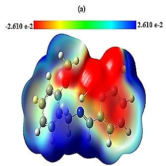
changes [5]. Despite not studied yet, this type of Schiff base ligands could also form interesting complexes
with other redox active species.
Continuing our efforts in the synthesis and study of new transitions metal complexes, this work reports
the synthesis and physicochemical and electrochemical characterization of three new complexes of Ni(II),
Mn(II)     and    VO(IV)     (oxovanadium)      derived     from    2-methoxybenzylamine        and    2,3-
dihydroxybenzaldehyde. Specifically, the study is essentially focused on elucidating the molecular
structure and geometry as well as redox properties of these complexes.

2. Experimental

2.1.     Materials
All the chemicals were purchased from commercial sources and used without any further purification. The
purity of the synthesized compounds was checked by thin-layer chromatography (TLC) using glass plates
precoated with silica gel (60F; Merck).

2.2.     Synthesis of the Schiff base ligand and metal complexes
The Schiff-base N (imine), O-donor ligand (HL) was prepared according the literature [5]. Briefly, 1
mmol of 2-methoxyphenylmethylamine was mixed with 1 mmol of the 2,3-dihydroxybenzaldehyde in
methanol (10 mL). The resulting solution was refluxed for ca. 2 h and allowed to cool. The yellow powder
ligand was used to form new metal complexes.
For the synthesis of the complexes, the required weights of the acetate salts of Ni(II)
(Ni(CH3COO)2·4H2O), Mn(II) (Mn(CH3COO)2·4H2O) and VO(IV) (VO(CH3COO)2), dissolved in
absolute MeOH, were added to a boiling solution of the imine ligand (HL) in absolute MeOH in 1:2 molar
ratio of metal and ligand, respectively. This molar ratio was chosen to form ML2 complexes. The resulting
mixture was stirred under reflux for 4 h, whereupon the complexes precipitated. The obtained powders
were collected by filtration, washed several times with ethanol and then with diethylether to remove any
traces of unreacted starting materials, and finally dried in a vacuum desiccator, which leads to the target
metal complexes. The synthesis of the different complexes and their molecular structure, as proposed from
the data obtained in this work (see section 3), are shown in Scheme 1.
OCH3
                                                                                           HO
       OCH3
                                                                    H
                                              MeOH, Reflux                C    N       O
                      N          OH
                                                                                   M                         , xH2O
                      C                  OH
                  H                           Metal acetate                    O       N    C
                                                                                                 H
                                                                          OH
                                                                        H3CO

                  M= Ni, Mn and VO                                        M(L)2· x H2O

                      OCH3                                                                            OCH3
                                                          OCH3
                       HO                                                                               HO
                                                                 HO
       H                                                  H2O                      H
                                                H
           C N        O                                                                    C N        O
                                                    C N         O
              Ni                      , H2O                                                       V                   , 2H2 O
              O       N C                                 Mn
                                                      O         N C                          O        N C
                             H                                                                    O          H
                                                          H2O         H
           OH                                       OH                                     OH
           H3CO                                                                            H3CO
                                                      H3CO

Scheme 1. Scheme of the synthesis of new complexes of Ni(II), Mn(II) and VO(IV) with the bidentate
Schiff-base ligand.

2.3.       Physico-chemical and electrochemical characterization
The obtained complexes were characterized by elemental analysis by using a (C, N, H, S) LECO analyzer
(Micro TruSpec model). This analysis was repeated three times to confirm the reproducibility of the
results.
FT-IR spectra of the complexes were recorded by preparing KBr pellets on a Perkin-Elmer 1000 FTIR
Spectrophotometer. UV-visible spectroscopy was carried out in DMF solutions on a UNICAM UV-300
spectrophotometer (path length of the cell, 1 cm). For mass spectrometry (LC-MS), a HPLC Agilent 1100
series coupled to UV-Vis and MS with ionic tramp detector (Agilent model 1100 Series LC/MSD Trap
SL) was used.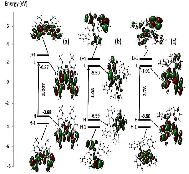
Thermal analysis (TG and DTG) of the complexes was obtained under nitrogen atmosphere using a TGA
Perkin Elmer thermal analyzer. The heating rate was set at 10 °C min-1 and the weight loss was measured
from 25 ºC up to 950 °C.
Cyclic voltammetry was utilized to study the redox and electrochemical properties of the complexes. The
experiments were carried out in an undivided Metrohm cell of 5 cm3 using a Voltalab 40
(potentiostat/galvanostat) PGZ 301 A 0.1 M tetra-n-butylammoniumtetrafluoroborate (TBABF4) solution
in dimethyl sulfoxide (DMSO) was used as supporting electrolyte in all the electrochemical experiments.
The planar tip of a glassy carbon (GC) bar (Ø, 3 mm) was used as the working electrode, a platinum wire
(Pt) as the auxiliary electrode and all the electrode potentials were referred versus as saturated calomel
electrode (SCE) reference electrode. Furthermore, the influence of different scan rates: 25, 50, 75, 100,
200, 300, 400 and 500 mV/s, has been studied.

2.4.   Computational study
Theoretical calculations for HL ligand and its Ni(II), Mn(II) and VO(IV) complexes were performed
using the Gaussian 09 program, based on Density Functional Theory (DFT) [19], using Beck’s three
parameter hybrid exchange functional [20], with Lee-Yang-Parr correlation functional (B3LYP) and 6-
31G (d, p) basis set [21,22]. In order to make comparative study between the ligand and their complexes
with the optimized configurations, HOMO, LUMO, gap properties and Energy calculations for the
compounds were useful to determine binding sites and predicting the reactivity of the compounds. The
Molecular electrostatic potential (MEP) and the global reactivity descriptors of the molecules were also
performed by the DFT.

3. Results and discussion

3.1.   Physico-chemical properties and molecular formula of the complexes
The obtained complexes were intensively colored, stable in air and moisture free powder solids. The
Ni(II), Mn(II), and VO(IV) complexes were not soluble in most common solvents, such as water,
methanol, ethanol, ethyl acetate and acetonitrile. However, they were fairly soluble in N,N-dimethyl
formamide (DMF) and dimethyl sulfoxide (DMSO). Table 1 summarizes some of the physico-chemical
properties and molecular formulae of the complexes. As it can be deduced from this table, the results of
elemental analysis (measured) were found in good agreement with those calculated from the proposed
molecular formulae for the different complexes.
NiII(L)2·H2O: Anal. Calc. for C30H30N2O7Ni: C 61.15; H 5.13; N 4.75 Found: C 60.84; H 4.88; N 4.68
%. UV-Vis (DMF):λmax(n) (nm), εmax(n) [M-1.cm-1]: λmax(1) (276), εmax(1) [16250]; λmax(2) (380), εmax(2) [3820].
(MS-LC): (m/z) 571.1 ([NiL2]+).
MnII(L)2·2H2O: Anal. Calc. for C30H32N2O8Mn: C 59.70; H 5.34; N 4.64 Found: C 60.27; H 4.43; N
4.23 %. UV-Vis (DMF): λmax(n) (nm), εmax(n) [M-1.cm-1]: λmax(1) (329), εmax(1) [10600]; λmax(2) (585), εmax(2)
[890]. (MS-LC): (m/z) 569.4 ([MnL2 + 2H+]).
VOIV(L)2·2H2O: Anal. Calc. for C30H32N2O9V: C 58.54; H 5.24; N 4.55 Found: C 58.08; H 4.62; N 3.47
%. UV-Vis (DMF): λmax(n) (nm), εmax(n) [M-1.cm-1]: λmax(1) (269), εmax(1) [18600]; λmax(2) (310), εmax(2)
[15830]; λmax(3) (430), εmax(3) [4560]. (MS-LC): (m/z) 580.7 ([VOL2 + H+]).
Table 1. Physico-chemical properties and proposed molecular formulae of the synthesized nickel(II),
manganese(II) and oxovanadium(IV) complexes.
                                                                   Elemental analysisd
                     λmax(n)       εmax(n)                                                       Molecular
                                                             c
       Complex                                  LC-MS (m/z)      C (%)    H(%)      N(%)          formula
                     (nm)a       (M-1.cm-1)b
                     (1)   276   (1)   16250                     60.84   4.88        4.68
     NiII(L)2·H2O                               571.1   [NiL2]+ (61.15) (5.13)                C30H30N2O7Ni
                     (2)   380    (2)    3820                                       (4.75)

                     (1)   329   (1)   10600                     60.27     4.43      4.23
    MnII(L)2·2H2O                                       [MnL2
                                                569.4            (59.7)   (5.34)    (4.64)    C30H32N2O8Mn
                     (2)   585     (2)   890            +2H+]
                     (1)   269   (1)   18600
    VOIV(L)2·2H2O          310         15830            [VOL2 + 58.08    4.62        3.47
                     (2)         (2)            580.7      +                                   C30H32N2O9V
                                                          H]    (58.54) (5.24)      (4.55)
                     (3)   430    (3)   4560
a
 Wavelength of maximum absorbance; b Molar attenuation coefficient; c Some of the major fragmentation
peaks observed on the mass spectra of nickel(II), manganese(II) and oxovanadium(IV) complexes; d
Measured C, H, N contents (atomic %) in the complexes and the calculated ones (in brackets) from the
proposed molecular formulae.

3.2.     Spectroscopic characterizations of the complexes
3.2.1. FT-IR Spectroscopy
Figure 1 shows the FT-IR spectra of the synthesized complexes, whereas Table 2 collects the wavelength
of some of the bands assigned to stretching and bending vibrations. The FT-IR spectra of all complexes
(Fig. 1) display bands between 3650 and 3200 cm-1 due to ν(OH) of crystalline or coordinated water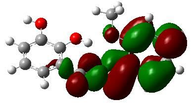
molecules associated with the complex. The absorption bands in the region 3000–2900 cm-1 may be due
to aliphatic and aromatic ν(C-H) [24], whereas the bands between 1050 and 1020 cm-1 may correspond to
ν(C-O-C) stretching vibration of the methoxy group [23]. In addition, the complexes possessed a potential
donor site, like the azomethine group (-C=N), which has tendency to coordinate with metal ions. The
strong bands associated to this group are generally situated between 1660 and 1630 cm-1[25]. Specifically,
for the studied ligand this band was observed at 1644 cm-1[5], but it is found shifted to lower frequencies
by 6 cm-1 in the spectrum of the nickel complex, and shifted to higher frequencies by about 14 cm-1 in the
spectra of the manganese and oxovanadium complexes. This result suggests that the azomethine nitrogen
may be coordinated to the metallic centers. Additionally, the bands at 1247–1253 cm-1 are assigned to C-
O stretch of the phenolate group in the complexes. These bands undergo a displacement to the higher
frequencies comparing with the value of this band in the corresponding ligand (at about 1240 cm-1 [5]) in
compliance with deprotonation and coordination of the phenolate oxygen to the metal [26].
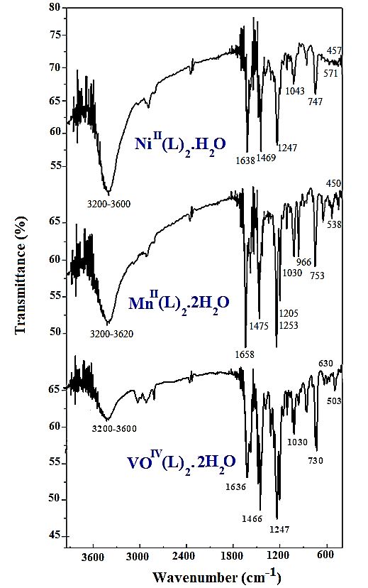
Fig.1. FT-IR Spectra of the obtained nickel(II), manganese(II) and oxovanadium(IV) complexes.
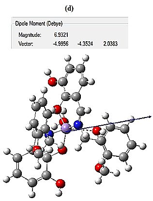
Table 2. Characteristic FT-IR bands (4000–400 cm−1) of the synthesized complexes.
   Complex         ν(O-H)    ν(C-H)    ν(C-H)     ν(C=N)   ν(C-O)   ν(C-O-C)   ν(V=O)   ν(M-O)   ν(M-N)
                             (arom.)   (aliph.)
     Nickel      3600-3200   3077      2951        1638     1247      1043        -      552      445
  Manganese      3620-3200   3079      2945        1658     1253      1030        -      538      450
 Oxovanadium     3650-3200   3080      2950        1658     1253      1023       970     531      443

In oxovanadium complexes, a strong band at 970 cm-1 was observed and can be assigned to ν(V=O)
vibrations [27]. The coordination of the Schiff base ligands to the metal ions is also confirmed by the
appearance of new bands in spectra of metal complexes in the range of 460-440 cm-1, which may be
assigned to the ν(M-N) bond [28]. Another absorption band was also seen, not far from the previous ν(M-
N) frequency, in the region of 560-530 cm-1 corresponding to ν(M-O) bond [28]. Considering these results,
it can be inferred that the two Schiff base molecules coordinating the metallic centers in the three
complexes may act as dibasic bis-bidentate ligands.

3.2.2. UV-Vis Spectroscopy
Electronic (UV–Vis) spectra of all complexes have been recorded in DMF solvent [10 -4M] (Fig. 2). For
all the complexes, an absorption band relatively intense was observed between 260 and 280 nm. This band
could be ascribed to the π–π* transitions of aromatic rings and azomethine groups. In the case of the
electronic spectrum of nickel complex, another band at 380 nm was seen. This band is assigned to the
ligand-metal charge-transfer (LMCT) transition Ophenolate → Ni(II).
As for the spectrum of manganese complex, two bands are observed. The first one located at 329 nm is
characteristic of the charge-transfer (LMCT) transition Mn(III)→Ophenolate, thus appearing shifted to
higher energy when compared to the same band of nickel complex [29,30]. The second appears as a broad
band centered at 585 nm. This electronic transition is attributed to a charge-transfer (LMCT) between pπ-
orbital of the ligand and dπ-orbital of Mn(II) [31,32].
Regarding the spectrum of oxovanadium complex, it shows two types of transitions. The first absorption
band at 310 nm corresponds to n–π* transitions, while the second one at 430nm may be also attributable
to the charge-transfer (LMCT) transition from the phenolic oxygen (Ophenolate) to the empty d orbital’s on
the vanadium [33,34].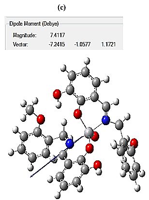
Accordingly, upon coordination, these observed bands appear between 320-600 nm correspond to
square planar geometry for Ni(II) complex [35], octahedral geometry for Mn(II) complex [36] and square-
pyramidal geometry for VO(IV) complex [37].

                                                  NiII(L)2

                                        2         MnII(L)2
                                                                                 0.30
                                                  VOIV(L)2
                                                                                               MnII(L)2
                     Absorbance /a.u.

                                                                                 0.25

                                                             Absorbance / a.u.
                                                                                 0.20

                                                                                 0.15            Weak d-d Transition
                                        1                                        0.10

                                                                                 0.05

                                                                                 0.00
                                                                                         500        600      700         800
                                                                                                Wavelength /nm

                                        0

                                            300      400                                500         600            700
                                                             Wavelength /nm

     Fig.2. UV-Vis Spectra of the obtained nickel(II), manganese(II) and oxovanadium(IV) complexes.

3.2.3. Mass spectrometry
Mass spectrometry is potentially considered as a powerful structural characterization technique in
molecular chemistry. This technique has also been successfully applied in coordination chemistry to
elucidate the major molecular ion peaks of our synthesized Schiff base complexes. This analysis was
carried out by HPLC chromatography coupled with mass spectrometry technique (LC-MS).
The mass spectrum of nickel complex (Fig. 3) displays a fragmentation ion peak with m/z = 571.1, which
may correspond to the parent molecular ion peak [Ni(II)L2]+ (calc. 571.2). On the other hand, a base peak
with m/z equal to 258.1 was observed. This could be assigned to the mass of ligand diprotonated [L+
2H]+. The molecular structure deduced from these mass fragmentation patterns is included in the Scheme
2.
Fig. 3. Mass spectra of nickel(II), manganese(II) and oxovanadium(IV) complexes.
OCH3                            OCH3                                     OCH3
                        HO                              HO                                       HO
  H                                    H                                         H
       C N O                                C N O                                    C N O
               Ni                                  Mn                 + 2H
                                                                                      O V
                                                                                                              +H
               O N C                               O N C                               O N C
                           H                                 H                                           H
     OH                                      OH                                    OH
   H3CO                                    H3CO                                  H3CO

       (m/z=571.1, 22%)                        (m/z=569.4, 22%)                       (m/z=580.7, 100%)

                                                    OCH3                                         OCH3
OCH3                                                                                             HO
                                                    HO
           N        O              H                                             H
           C              OH           C    N O                                      C N O
       H                    + 2H           Mn                                                V                +H
                                                                 -H
                                            O N C                                        O N C
                                                        H                                                H
      (m/z=258.1, 100%)
                                       OH                                          OH
                                                                                 H3CO

NiII(L)2· H2O
                                            (m/z=445.1, 33%)                         (m/z=564.7, 91%)

                                   OCH3
                                                                                             OCH3
                                               N    O
                                                                                             HO
                                               C            OH
                                                                             H
                                           H
                                                                                 C N O
                                                                                         V                   + 3H
                                           (m/z=256.1, 100%)                          O N C
                                                                                                     H
                                                                                 OH

                                       MnII(L)2·2H2O                                 (m/z=445.2, 25%)

                                                                             OCH3

                                                                                         N       O
                                                                                         C               OH
                                                                                     H                     + 2H

                                                                                     (m/z=258.0, 41%)

                                                                                     VOIV(L)2·2H2O
Scheme 2. Proposed major fragment ions deduced from the mass spectra of nickel(II), manganese(II) and oxovanadium(IV) complexes. As for the mass spectrum of the manganese complex, it shows a molecular ion peak with m/z ratio equal to 569.4. This fragment ion may fit with the molecular weight of the expected compound. Moreover, another important fragment with m/z ratio = 445.1 (33.4 %) is observed, which may correspond to the [C21H19O5Mn – H]+ ion (Scheme 2). The fragment with m/z = 256.1, with a relative abundance of 100 %, is assigned to the ligand [L]+, what confirms its high stability and therefore corresponds to the base peak. For the last complex of oxovanadium, it exhibits a strong peak at m/z = 580.7 (100 %), which may be indicative of the molecular peak and also the base peak. This behavior seems to be the main particularity for this compound when compared to the previous complexes of Ni(II) and Mn(II). In addition, the mass spectrum shows again multiple peaks representing successive degradation of this complex leading to the formation of different fragments (Scheme 2), highlighting among them the Schiff base ligand ion peak[L]+ of m/z = 258.0 (41.0 %). All these results confirmed that the synthesized complexes are mononuclear, with two molecular moieties of Schiff base ligands, and they are, therefore, in good agreement with their molecular structure proposed in the Scheme 1. 3.3. Thermogravimetric analysis (TG/DTG) Figure 4 shows the TG and DTG curves recorded for the three metal complexes. As it is observed, upon increasing the temperature, the complexes showed a gradual weight loss. Generally, this weight loss can be initially assigned to the release of water molecules, followed by other successive decompositions leading to the formation of metallic oxides. Particularly, the different decomposition stages, temperature ranges, DTG peak positions, as well as the experimental and calculated mass loss percentages for these complexes are summarized in Table 3. First, the thermogram of nickel complex undergoes decomposition in four steps (Fig. 4A). The first one is accompanied by a weight loss of 3.8 %, which may correspond to the release of one molecule of water (theoretically calculated = 3.05% %). The DTG curve of the second step, for which the temperatures ranged from 220 to 300 °C, clearly indicates a weight loss centered at 242 °C of 17.7 %. This decomposition process is attributed to the removal of the methoxyphenyl moiety (theoretically calculated = 18.15 %). With a weight loss of 13.0 %, the third decomposition step seems to represent the loss of
phenyl moiety (theoretically calculated = 12.89 %). Regarding the last process evidenced at 557.4 °C on
the DTG curve (24.2 % loss), it seems to be due to the removal of the organic entity (C 3H17N2O4)
(theoretically calculated = 24.6 %). Finally, the remaining final residue is identified as NiO with 16 carbon
[38].
For the manganese complex (Fig. 4B), the first decomposition ranges between 138.6-222.5 °C and it
involves a weight loss of 3.0 %, so it is attributed to the removal of one molecule of water (theoretically
calculated = 2.98 %). The second step between 144.7 and 350 °C shows a weight loss of 12.6 %, thus,
being explained by the removal of one molecule of water and two methoxy (-OCH3) moieties from the
ligand (theoretically calculated = 13.25%). The next step observed in the temperature range from 326 to
442 °C implies a weight loss of 20 %, probably representing the decomposition of the C8H7N part
(theoretically calculated = 19.38 %). As for the last step occurring in the range of 451-947 °C (22.6 %
weight loss), it may account for the loss of the remaining organic part (C5H15NO3). These four stages were
denoted by the DTG peaks at about 181.6, 343.4, 375.1 and 578.9, respectively. The final residue may be
manganese oxide (MnO), with carbon atoms (15C) [39].
Finally, the thermal decomposition of the vanadium complex (Fig. 4C) displays also four decomposition
steps in the temperature ranges of 50.5-91.8, 190-250, 269.5-438.9 and 452.4-528.9 ºC and DTG peaks at
70.7, 222.3, 382.6 and 491.0 °C, respectively. With a weight loss of 5.5 %, the first one corresponds to
the loss of two water molecules, as previously mentioned for the manganese complex. The second step
with a weight loss of 12.7 % indicates also the loss of the phenyl group (theoretically calculated = 12.34
%). Because these processes are also observed for nickel and manganese complexes, the obtained results
suggest the existence of a similarity in the thermal decomposition pathway of the complexes. However,
the last decomposition step of the vanadium complex leaving 41.5% residue as VO2 and 14C [40].
0.01
                                   1.00                                 (A)
                                                                                                       0.00

                                                                                                                     d(1-a)/dt/ s-1
                                   0.75              TG
                                                                                                       -0.01
                                                                                    DTG
                                                                                                       -0.02

                            1-a
                                   0.50

                                                                                                       -0.03

                                   0.25
                                                                                                       -0.04

                                   0.00                                                                -0.05
                                               150    300         450         600   750          900

                                                          Temperature/ °C

                                                                                                       0.01
                                   1.0                      (B)
                                                                                                       0.00

                                                                                                                   d(1-a)/dt/ s-1
                                   0.8
                                                                                                       -0.01
                                                      TG
                            1-a

                                   0.6
                                                                                                       -0.02

                                   0.4
                                                                                                       -0.03
                                                                                    DTG
                                   0.2                                                                 -0.04

                                   0.0                                                                 -0.05
                                               150    300         450         600    750         900

                                                          Temperature/ °C

                                                                                                       0.01
                                  1.00                       (C)
                                          TG                                                           0.00
                                                                                                               d(1-a)/dt/ s-1

                                  0.75                                                                 -0.01
                            1-a

                                                                                           DTG         -0.02
                                  0.50

                                                                                                       -0.03

                                  0.25
                                                                                                       -0.04

                                  0.00                                                                 -0.05
                                               150    300         450         600   750      900

                                                          Temperature/ °C

Fig. 4. The TG and DTG of (A)- Ni(II), (B)- Mn(II) and (C)- VO(IV) Schiff base complexes (where α, is
the conversion = 1-w/w0, w = the residual weight in mg and w0 is the initial weight of the sample in mg).
Table 3. Thermal decomposition data for the prepared complexes.
Samples Number Temp. range Tmax/ Mass loss /% Assignments Residue/%
of steps /°C °C Found Found
(Calcd.) (Calcd.)
NiII(L)2·H2O 4 125.9-173.4 144.6 3.8(3.05) Loss of H2O
220.6-300.0 242.0 17.7(18.15) Loss of C7H7O
337.9-450.3 396.8 13.0(12.89) Loss of C6H4 41.3 (41.41)
522.6-613.6 557.4 24.2(24.6) Loss of C3H17N2O4 NiO+ 16C
MnII(L)2·2H2O 4 138.6-222.5 181.6 3.0(2.98) Loss of H2O
144.7-350.0 343.4 12.6 (13.25) Loss of H2O+2(CH3O)
326.7-442.0 375.1 20 (19.38) Loss of C8H7N
451.0-947.0 578.0 22.6(22.7) Loss of C5H15NO3 41.8(41.69)
MnO + 15C
VIVO(L)2·2H2O 4 50.5-91.8 70.7 5.5(5.84) Loss of 2H2O
190.0-250.0 222.3 12.7(12.34) Loss of C6H4
269.5-438.9 382.6 21.6(21.93) Loss of C8H9NO
452.4-528.9 491.0 18.7(19.01) Loss of C2H15NO4 41.5(40.79)
VO2 + 14C
3.4. Electrochemical behavior
Cyclic voltammetry measurements were carried out to investigate the redox properties of the complexes
in solution. Figure 5 displays the voltammetric curves of the nickel complex in the potential range from
+1.6 to -2.2 V and the manganese complex between +1.4 to -1.4 V.120
Schiff base
II
Ni (L)2 . H2O group
90
I
i(µA/cm2)
II
i
N
60
i II
N
I
Azomethine
i
N
30
group
0
-30
-2.4 -1.8 -1.2 -0.6 0.0 0.6 1.2
E(V/SCE)
40
Schiff base
MnIII(L)2 . 2H2O group
30
20
i(µA/cm2)
MnII MnIII
10
0
-10
-20
-1.2 -0.6 0.0 0.6 1.2
E(V/SCE)800
Schiff base
600
VOIV(L)2 . 2H2O
group
400
VIV VV
i(µA/cm2)
200 III IV
V V
0 80
Epa - Epc = 0.28 V (A)
-200 40 E1/2 = +0.33 V
i(µA/cm2)
Ipa/Ipc = 0.92
-400 0
-40 VIV/VV at 100 mV/s
-600
-200 0 200 400 600 800
E(mV/SCE)
-800
-1.0 -0.5 0.0 0.5 1.0 1.5
E(V/SCE)
Fig. 5. Cyclic voltammograms of a GC electrode in a solution containing 10-3 M nickel (A), manganese
(B) or oxovanadium (C) complexes and 0.1M of TBABF4 in DMSO, under argon atmosphere, at a scan
rate of 100 mV/s.
In the case of the nickel complex (see Fig. 5A), the voltammogram displays four redox processes. When
the electrode potential is scanned towards negative values, a reduction peak is observed at -1.53 V, and
its corresponding oxidation pair appears in the reverse scan at -1.38 V (ΔE= Epa - Epc = 150 mV). The
anodic to cathodic peak current density ratio (Ipa/Ipc) of this process is close to 1, which is assigned to
the metal centered NiII/NiI redox couple [41,42]. Next, the reduction at -1.91 V, with a small oxidation
counterpart -1.71 V can be attributed to the irreversible reduction of azomethine group. When the electrode
potential is scanned from 0.0 to +1.60 V, the complex exhibits a large oxidation current from ca. +0.5 V.
A reduction couple is discerned in the reverse scan, so the process is characterized by an E 1/2 = +0.65 V
and peak separation about ΔE =180 mV, and can be assigned to the NiIII/NiII redox couple [43]. Finally,
an oxidation process is clearly observed at +1.34 V, which could probably be assigned to an oxidation of
Schiff-base ligand [44].
The cyclic voltammogram of manganese complex (Fig. 5B) exhibits only one quasi-reversible redox
couple with an anodic–cathodic peak separation (ΔE) equal to 200 mV. Its half wave potential value wasthen calculated as average of the anodic and cathodic peak potentials, which is found to be equal to E1/2 =
-0.49 V. These results suggest that this redox process may be described to MnIII/MnII [45]. On the other
hand, the wave at Epa = +1.12V may probably correspond to the Schiff base ligand oxidation [44].
Cyclic voltammetry of the VIVO(L)2·2H2O complex (see Fig. 5C) was performed in the potential range
of −1.0 to 1.5 V vs. SCE. This voltammogram displays two redox processes. The reduction peak for the
first redox couple is observed at -0.74 V, whereas its corresponding oxidation pair appears in the reverse
scan at -0.53 V, being the peak to peak separation ΔE = 210 mV. According to literature, this redox process
may be ascribed to VIII/VIV [46]. For the second couple (see a magnified voltammogram in the inset of
Fig. 5C) the cathodic peak is centered at Epc= +0.2 V vs. SCE, and the anodic one at Epa = +0.48 V vs.
SCE. This process has been attributed to the VIV/VV redox couple [47] and it is characterized by ΔE =
0.28 V, Ipa/Ipc close to 1 (0.92) and E1/2 = +0.33 V. These two redox systems of the vanadium complex
are proposed to be monoelectronic. The last oxidation wave observed at Epa = +0.82 V may probably
correspond to the Schiff base ligand oxidation [44].
3.4.1. Effect of scan rate on the response of the Ni(II) complex
The cyclic voltammogram of the nickel complex in DMSO solution under the potential range from-0.8 to
-1.7 V shows a well-defined redox system (Fig. 6A) associated to Ni(II)/Ni(I). Specifically, the Epa is
equal -1.33 V and the cathodic peak is centered at -1.47 V, so this redox couple shows a peak separation
(ΔE) equal to 130 mV, and the Ipc/Ipa (1.01) is practically equal to unity, suggesting an electrochemical
process with one electron.
4
Epa - Epc = 0.13 V
5 NiII/NiI (B)
(A)
2
E1/2 = -1.39 V 0
i(µA/cm2)
0
i(µA/cm2)
Ipa/Ipc = 1.01 -5
-2 25 mV/s
-10 .
-4
.
-15 .
-6
500 mV/s
NiII/NiI at 100 mV/s -20
-8
-1.8 -1.6 -1.4 -1.2 -1.0 -0.8 -1.8 -1.6 -1.4 -1.2 -1.0 -0.8
E(V/SCE) E(V/SCE)9
(C) -1,30 (D)
6
-1,35
ipa= 0.413v
1/2
- 0.402 Epa = 0.032 Log v - 1.398
3 2
ip (µA/cm )
R = 0.993
2
2
R = 0.999 -1,40
E (V/SCE)
0 Epc = -0.062 Log v - 1.357
1/2 -1,45 2
ipc= -0.381v + 0.277 R = 0.930
-3 2
R = 0.994
-1,50
-6
-1,55
-9
3 6 9 12 15 18 21 24 1,2 1,4 1,6 1,8 2,0 2,2 2,4 2,6 2,8
1/2 -1 1/2 -1
v (mV.s ) Log v (mV.s )
Fig. 6. (A) Cyclic voltammogram of the synthesized Ni(II) complex (10-3 M) in 0.1 M TBABF4/DMSO
at a scan rate of 100 mV s-1; (B) Cyclic voltammograms of the same complex at different scan rates; (C)
Plot of the cathodic and anodic peak currents versus the square root of sweep rate (v 1/2) and (D) Anodic
and cathodic potentials versus Log ν.
The reversibility of the Ni(II)/Ni(I) redox process in the synthesized nickel complex was studied by cyclic
voltammetry at different scan rates. Fig. 6B depicts the voltammograms of the complex varying the scan
rate between 25 to 500 mV s-1. As it can be observed in the figure, the reversibility of the redox couple is
well maintained even up to 500 mV s-1. The effect of the scan rate on the electrochemical parameters
associated to this redox couple is summarized in the Table 4, whereas Figures 6C and 6D plot the cathodic
and anodic peak current densities versus the square root of the scan rate (v1/2) and the corresponding peak
potentials versus the logarithm of the scan rate(Log v), respectively. The obtained results reveal a linear
relationship between the peak current densities and v1/2 (Fig. 6C). In addition, the anodic and cathodic
peak potentials (Epa, Epc) are found proportional to Log ν (Fig. 6D). These behaviors are indicative of a
diffusion-controlled electron transfer process [48].
Table 4. Electrochemical parameters obtained from cyclic voltammograms of Figure 6.
│IPa/IPc│ 0.95 0.97 1.02 1.01 1.02 1.03 1.01 1.01
ΔE (mV) 100 130 137 140 160 180 210 220
E1/2 (mV) -1400 -1405 -1407 -1410 -1415 -1418 -1422 -14254. DFT calculations for the ligand and complex structures 4.1. Geometry optimization The geometry of the HL ligand structure was optimized to investigate the geometric and electronic characteristics that may control its electron-donation character and, therefore, its reactivity. Calculations showed that the molecule is planar and two donating groups (one hydroxyl and one imino groups) are facing each other around the central point of the molecule Figure 7. The structure was found to be stabilized through hydrogen bonding formed between the hydroxyl and imino groups. This specific orientation suggested that the ligand could be bound to the metal ions in one plane, giving rise to the formation of a square planar geometry around the central metal ion Ni(II), while square-pyramidal and octahedral geometry around the central metal ion VO(IV) and Mn(II) complexes, respectively. The planar geometry was associated with symmetrical charge distribution among atoms. As can be observed from Fig. 7, Table 5, the calculated atomic charges indicate that the most negative atoms are the oxygen and nitrogen, whereas the attached protons are the most positive ones. Again, this charge distribution supports the idea that the HL molecule donates the electrons through these two groups. The orbital occupation of the highest occupied molecular orbital (HOMO) and the lowest unoccupied molecular orbital (LUMO) showed that electrons are delocalized through the whole molecule with calculated energy gap of 4.75 eV.
LUMO
                                                                   E= -0.34 eV

                                                                    ∆E = 4.75 eV

                                                                   HOMO
                                                                   E= -5.09 eV

Figure 7. Proposed structures for the HL ligand, showing (A) the atomic charges distributions in color
range and (B) the HOMO-LUMO orbitals occupation with the calculated energy gap, as optimized by DFT
calculations.
Table 5. Mulliken atomic charges calculated by B3LYP/6-31G(d,p) of the Schiff base ligand HL shown
in Figure 7.
Atom Charge
C1 0.102252
C2 -0.164758
C3 -0.118200
C4 -0.135242
C5 -0.121028
C6 0.269684
H7 0.124027
H8 0.126045
H9 0.130537
H10 0.137220
C11 -0.173253
H12 0.213199
H13 0.141580
H14 0.153624
O15 -0.582906
C16 -0.176495
N17 -0.602434
C18 0.107245
C19 0.231670
C20 -0.131431
C21 -0.157441
C22 -0.139031
C23 0.029718
C24 0.273646
H25 0.110588
H26 0.116375
H27 0.114311
H28 0.157677
O29 -0.558180
O30 -0.601743
H31 0.430699
H32 0.361764
H33 0.144206
H34 0.186075With respect to the calculations for the different complexes, the obtained results show that:
➢ When optimizing Ni2+ ion complex with HL Schiff base ligand, the lowest energy structure has a
square planar geometry with molecular formula [NiII(L)2·H2O]. This geometry is accomplished with
the protonation of the hydroxyl groups to neutralize the Ni2+ ion. The stability of this complex came
from the formation of two four membered rings between Ni2+ and the ligand (Fig. 8a).
➢ VO2+ ion may form a square-pyramidal [VIVO(L)2·2H2O] complex with the HL Schiff base ligand
(Fig. 8b), where the ligand’s donor groups coordinate in one plane with the metal ion. According to
the lower energy of this conformation, the protons may rotate in opposite directions to prevent the
steric interaction between the VO2+ and hydroxyl groups of HL (Fig. 8b).
➢ the optimized structure of the Mn2+ complex may be octahedral. To attain this conformation the results
suggest that, upon coordination, the ligand is deprotonated and that the octahedral geometry is fulfilled
by two water molecules coordinated in a plane perpendicular to the Mn-HL plane (Fig. 8c). As a result,
the donor groups of the ligand may be coordinated with the Mn(II) center in one plane.
All these (theoretical) results well agree with the molecular structures deduced from the analytical
(experimental) techniques.Figure 8. Proposed structures for (a) nickel(II), (b) oxovanadium(IV) and (c) manganese(II) complexes, as optimized by DFT calculations.
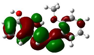
4.2. Frontiers Molecular Orbitals (FMOs)
            The HOMO-LUMO are the main orbitals that take part in the chemical stability of a molecule, so
the calculation of their energy helps to characterize its chemical reactivity. The HOMO highlight is the
orbital that primarily acts as electron donor and the LUMO is the orbital that largely acts as electron
acceptor, and the gap between HOMO and LUMO orbitals is the parameter related with the molecular
chemical stability [49]. The energies of the HOMO and the LUMO orbitals were investigated by using DFT
with the standard B3LYP/6-31G(d,p) model. Figure 9 shows the complexes molecular structures deduced
from calculations, where the positive phases are presented in red and the negative ones in green.
In addition, Table 6 summarizes the calculated HOMO and LUMO energies and some related characteristic
physicochemical parameters (such as, energy gap (∆E), electronic chemical potential (μ), hardness (η) and
electrophilicity (ω)) of the Schiff base ligand HL and its complexes. The chemical hardness is associated
with the stability and reactivity of a chemical system. According to frontier molecular orbitals, chemical
hardness corresponds to the energy gap between HOMO and LUMO. As the energy gap increases, the
molecule becomes harder and more stable (less reactive). On the other hand, the higher the electronic
chemical potential (absolute values), the higher the reactivity. As observed in Table 6, the energy gap,
chemical hardness and electronic chemical potential values calculated for the complexes are generally lower
than that for the ligand. This infers that the ligand is stabilized upon complex formation. In this sense, the
results suggest that the VO(II) complex may be the most stable (according to larger ∆E and η), followed by
the Mn(II) and Ni(II) ones.

Table 6. HOMO, LUMO, energy gap (∆E), chemical hardness (η), electronic chemical potential (μ) and
electrophilicity (ω) calculated for the ligand and its corresponding Ni(II), VO(II) and Mn(II) complexes.
All values were calculated in eV unit.

       Compound                  HOMO         LUMO            ∆Ea          ηb         μc          ωd
       HL                          -5.09       -0.34         4.75         2.37       -2.71       1.54
       Ni(II) complex              -3.88       -0.87         3.06         1.53       -2.37       1.83
       VO(IV) complex              -6.59       -5.50         1.08         0.54       -6.04      33.77
       Mn(II) complex              -3.80       -1.01         2.78         1.39       -2.40       2.11
        a
            ∆E = LUMO-HOMO; b η = 1/2(ELUMO-EHOMO), c μ = 1/2(EHOMO + ELUMO), d ω = μ 2/2η.
Fig. 9. Energy diagram and representation of the HOMO (H) and LUMO (L) energy levels, energy gaps
and frontier molecular orbitals for (a) nickel(II), (b) oxovanadium(IV) and (c) manganese(II) complexes.
The electronic dipole moment {µi (i = x, y, z) and total dipole moment µtot} for title compounds
are collected in Table 7. The results indicate that the value of dipole moment of VO(IV) complex (7.7547
debye) Mn(II) complex (6.9321 debye) Ni(II) complex (6.2395 debye) HL (2.8732 debye). It is well
known that a higher value of dipole moment is important for more active Non-Linear Optical (NLO)properties. NLO effects have been extensively studied over the last decades. Thus, molecules exhibiting
large hyperpolarizabilities have a strong NLO potential and could be used, under certain conditions, for
optoelectronics and a variety of optical devices [50]. Some quantum chemical descriptors which are the
mean electric polarizability (α), the anisotropy of the polarizability (Δα) and first order
hyperpolarizability (β) have been used for explaining the NLO properties in many computational studies
[51]. In the present study, the α, Δα and β values were theoretically predicted (Table 7). According to
these calculations (at the same level of theory as well as same basis set 6-31G(d,p)), the values of α and
Δα found for the VO(IV) complex are higher than for the other compounds. This is in line with the
found lower frontier orbital energy gap (∆E) of the VO(IV) complex (Table 6). On the other hand, in
agreement with its higher dipole moment, the lower β value obtained for the VO(IV) complex indicates
that the this compound possesses better NLO properties than the others, so it becomes a potential candidate
for NLO applications.
Table .7 The electric dipole moments (μ, in debyes), polarizabilities (α, in a.u) and hyperpolarizabilities
(ß, in a.u) components and total values, as well as the anisotropy of the polarizability (Δα) of the HL
ligand and the Ni(II), VO(IV) and Mn(II) complexes calculated with B3LYP/6-31G(d,p).
Parameters HL Ni(II) VO(IV) Mn(II)
complex complex complex
μx -2.4198 2.1894 -5.0781 -5.6321
μy -2.6969 2.0836 -5.8607 -3.1826
μz 0.4168 2.0253 -0.0150 2.4910
μtot 2.8732 6.2395 7.7547 6.9321
αxx -99.9058 -205.1184 -247.6975 -214.1616
αyy -96.0414 -210.9704 -232.5401 -220.8387
αzz -109.8336 -225.7951 -231.2249 -226.6761
αxy 2.7832 0.8714 11.4563 9.8801
αxz 1.4939 8.0543 -16.0336 17.3521
Αyz -0.2142 -10.1195 17.7394 15.3022
α -101.9278 -213.9613 -237.1541 -220.558
∆α° 13.5248 29.1536 48.7295 45.0485ßxxx 64.8155 64.8155 -274.9187 -0.8224 ßxxy -13.9245 -13.9245 -132.9412 -24.9312 ßxyy -8.9802 -8.9882 16.3759 -43.6176 ßyyy 15.4898 15.4898 100.6738 166.7716 ßxxz -39.0351 -39.0351 31.0844 -10.8502 ßxyz -6.7001 -6.7001 67.2330 -18.6250 ßyyz 4.7512 4.7512 3.6709 -4.9014 ßxzz -13.9245 -13.9245 27.0438 -6.0415 ßyzz 8.4708 8.4708 -11.9078 -48.6632 ßzzz 6.8008 6.8008 34.4736 -44.3838 ßtot 51.1132 51.1066 245.1870 123.8642
Figure 10. Direction of dipole moment of Ground state optimized structures for the (a) HL ligand and
the (b) Ni(II), (c) VO(IV) and (d) Mn(II) complexes.
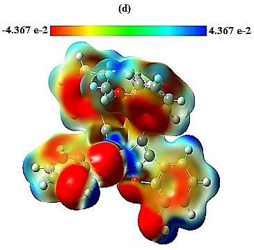
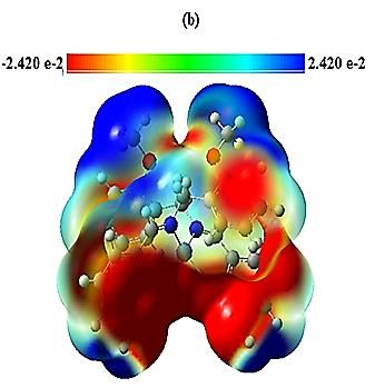
4.3. Molecular Electrostatic Potential (MEP)
The MEP is a property useful to study the attacking sites for electrophilic and nucleophilic substitution
reactions [52,53]. Figure 11 displays the total electron density surfaces mapped with electrostatic potential
of the ligand HL and their complexes. The different values of the electrostatic potential at the surface are
represented by different colors. Thus, the negative regions represented by the red color are the preferable
sites for electrophilic attack. The results show that these negative potentials are generated over the more
electronegative oxygen and nitrogen atoms, whereas the H-atoms exhibiting a positive potential region in
the structures (represented by the blue color) are favorable sites for nucleophilic attack. These last regions
are located around hydrogen and carbon atoms. Finally, the yellow color indicates the slightly rich electron
regions and the green one reflects more neutral zones.
The obtained MEP maps suggest that while the HL ligand and Ni(II) complex may participate in both
electrophilic and nucleophilic reactions, the VO(IV) and Mn(II) complexes seem to better take part in
electrophilic processes.
Figure 11. Molecular electrostatic potential maps for the (a) HL ligand and the (b) Ni(II), (c) VO(IV)
and (d) Mn(II) at the B3LYP/6-31G(d,p) level of theory.

5. Conclusion

In summary, we have successfully synthesized and characterized three new Schiff-base complexes of
nickel(II), manganese(II) and oxovanadium(IV). From the results of FT-IR, UV-Vis, LC-MS and
elemental analysis, the molecular structures of the complexes have been proposed. The bis-bidentate
Schiff-base ligands coordinate to the VO(IV), Mn(II) and Ni(II) ions in a tetradentate mode (ML2) using
the azomethine N and enol O atoms. The assignment of a square-planar geometry for Ni(II), a square-
pyramidal geometry for the VO(IV) and an octahedral geometry for the Mn(II), is supported by electronic
absorption and infrared spectral measurements, as well as quantum chemical calculations. The
electrochemical characterization shows that the complexes exhibit redox activity associated to their metal
centers. Moreover, the calculated molecular properties suggest that the VO(II) complex may be the most
stable compound and that exhibiting best NLO properties. Hence, the present study shows a simple method
for the synthesis of some bis-bidentates Schiff base complexes with interesting properties to be used in
electrochemical and other applications.

Knowledgements

The authors would like to thank the MESRS and DG-RSDT (Ministère de l’Enseignement Supérieur et
de la Recherche Scientifique et la Direction Générale de la Recherche et du Développement
Technologique-Algérie) for their financial support. Financial support from the Ministerio de Economía y
Competitividad (Spain) and FEDER funds (MAT2016-76595-R and RYC-2017-23618) is also gratefully
acknowledged.

References
[1] H. Schiff, Untersuchungenüber Salicinderivate. Ann. Chem., 150 (1869) 193-200.
[2] V.M. Leovac, D.M. Joksovic, V. Divjakovic, L.S. Jovanovic, Z. Saranovic, A. Pevec, Synthesis,
spectroscopic and X-ray characterization of a copper(II) complex with the Schiff base derived from
pyridoxal and aminoguanidine: NMR spectral studies of the ligand, J. Inorg. Biochem., 101 (2007) 1094-
1097.
[3](A) E.N. Jacobsen, & M.H. Wu, in Comprehensive Asymmetric Catalysis: Asymmetric Synthesis and
Induction Catalysts, ed. Yamamoto, H. (Springer, Berlin), 2 (1999) 649-677; (B) T. Katsuki, in Catalytic
Asymmetric Synthesis, ed. Ojima, I. (Wiley-VCH, New York), (2000) 287-325; (C) E. N. Jacobsen,
in Catalytic Asymmetric Synthesis, ed. Ojima, I. (VCH, New York), (1993) 159-202.
[4](A) J. Tisato, F. Refosco, F. Bandoli, Structural survey of technetium complexes, Coord. Chem. Rev.,
135 (1994) 325-397; (B) I.N. Booysen, S. Maikoo, M.P. Akerman, B. Xulu, Novel ruthenium (II) and (III)
compounds with multidentate Schiff base chelates bearing biologically significant moieties, Polyhedron,
79 (2014) 250-257.
[5] D. Aggoun, A. Ourari, R. Ruiz-Rosas, E. Morallon, A selective naked-eye chemosensor derived from
2-methoxybenzylamine and 2,3-dihydroxybenzaldehyde: synthesis, spectral characterization and
electrochemistry of its bis-bidentates Schiff bases metal complexes, Spectrochim. Acta A, 184 (2017)
299-307.
[6] A.A. Alshaheri, M.I.M. Tahir, M.B. Abdul Rahman, T. Begum, T.A. Saleh, Synthesis,
characterization and catalytic activity of dithiocarbazate Schiff base complexes in oxidation of
cyclohexane, J. Mol. Liq., 240 (2017) 486-496
[7] A. Ourari, M. Khelafi, D. Aggoun, A. Jutand, C. Amatore, Electrocatalytic oxidation of organic
substrates with molecular oxygen using tetradentate ruthenium(III)-Schiff base complexes as catalysts,
Electrochim. Acta, 75 (2012) 366-370.
[8] F. Heshmatpour, S. Rayati, M.A. Hajiabbas, P. Abdolalian, B. Neumüller, Copper(II) Schiff base
complexes derived from 2,20-dimethyl-propandiamine: Synthesis, characterization and catalytic
performance in the oxidation of styrene and cyclooctene, Polyhedron, 31 (2012) 443-450.
[9] A. Ghaffari, M. Behzad, M. Pooyan, H.A. Rudbari, G. Bruno, Crystal structures and catalytic
performance of three new methoxy substituted salen type nickel(II) Schiff base complexes derived from
meso-1,2-diphenyl-1,2-ethylenediamine, J. Mol. Struct., 1063 (2014) 1-7.
[10] S. Ilhan, H. Baykara, A. Oztomsuk, V. Okumus, A. Levent, M.S. Seyitoglu, S. Ozdemir, Synthesis
and characterization of 1,2-bis(2-(5-bromo-2 hydroxybenzilidenamino)-4-chlorophenoxy) ethane and its
metal complexes: An experimental, theoretical, electrochemical, antioxidant and antibacterial study,
Spectrochim. Acta A, 118 (2014) 632-642.
[11] S. Mohebbi, S. Eslami, Electrocatalytic oxidation of 2-mercaptoethanol using modified glassy carbon
electrode by MWCNT in combination with unsymmetrical manganese (II) Schiff base complexes, Mate.
Res. Bull., 66 (2015) 219-225.
[12] C.R. Bhattacharjee, P. Goswami, M. Sengupta, Synthesis, electrochemical and antimicrobial studies
of mono and binuclear iron(III) and oxovanadium(IV) complexes of [ONO] donor tridentate Schiff-base
ligands, J. Coord. Chem., 63 (2010) 3969-3980.
[13] A. Kamath, N.V. Kulnarni, P. P Netalkar, V.K. Revankar, Phenoxide bridged tetranuclear Co(II),
Ni(II), Cu(II) and Zn(II) complexes: Electrochemical, magnetic and antimicrobial studies, Spectrochim.
Acta A, 79 (2011) 1418-1424.
[14] K.C. Gupta and A.K. Sutar, Catalytic activities of Schiff base transition metal complexes, Coord.
Chem. Rev., 252 (2008) 1420-1450.
[15] D.A. Atwood and M.J. Harvey, Group 13 compounds incorporating Salenligands, Chem. Rev., 101
(2001) 37-52.
[16] N. Yoshida, K. Ichikawa and M. Shiro, Supramolecular motifs in metal complexes of Schiff bases. Part
5. Zinc(II)-assisted self-assembly of some bis-N,N- and N,O-bidentate Schiff bases and chiral packing modes
in solid state, J. Chem. Soc., Perkin Trans., 2 (2000) 17-26.
[17] H. Hosseini-Monfared, R. Bikas, P. Mahboubi-Anarjan, A.J. Blake, V. Lippolis, N.B. Arslan, C.
Kazak, Oxidovanadium(V) complexes containing hydrazone based O,N,O-donor ligands: Synthesis,
structure, catalytic properties and theoretical calculations, Polyhedron, 69 (2014) 90-102.
[18] A. Ourari, I. Bougossa, S. Bouacida, D. Aggoun, R. Ruiz‑Rosas, E. Morallon, H. Merazig, Synthesis,
characterization and X‑ray crystal structure of novel nickel Schiff base complexes and investigation of
their catalytic activity in the electrocatalytic reduction of alkyl and aryl halides, J. Iran. Chem. Soc.,14
(2017) 703-715.
[19] W. Kohn, L.J. Sham, Self-Consistent Equations Including Exchange and Correlation Effects,
Phys.Rev,140 (1965) 1133-1138.
[20] A.D. Becke, Density‐functional thermochemistry. III. The role of exact exchange, J. Chem. Phys, 98
(1993) 648-5652.
[21] C. Lee, W. Yang, R.G. Parr, Development of the Colle-Salvetti correlation-energy formula into a
functional of the electron density, Phys. Rev, 37 (1998) 785-789.
[22] B. Miehlich, A. Savin, A. Stoll, H. Preuss, Results obtained with the correlation energy density
functionals of becke and Lee, Yang and Parr, Chem. Phys. Lett, 157 (1989) 200-206.
[23] B. Bouzerafa, A. Ourari, D. Aggoun, R. Ruiz-Rosas, Y. Ouennoughi, E. Morallon, Novel nickel(II)
and manganese(III) complexes with bidentate Schiff-base ligand: synthesis, spectral, thermogravimetry,
electrochemical and electrocatalytical properties, Res. Chem. Intermed., 42 (2016) 4839-4858.
[24] A. Ourari, D. Aggoun, Synthesis and spectral analysis of N‑substituted pyrrole salicylaldehyde
derivatives‑electropolymerization     of   a   new    nickel(II)‑Schiff   base   complex      derived   from
6‑[3′‑N‑pyrrolpropoxy]‑2 ‑hydroxyacetophenone and 1,2‑diaminoethane, J. Iran. Chem. Soc., 12
(2015) 1893-1904.
[25] R. Aurkie, M.G. Rosair, R. Kadam, S. Mitra, Three new mono- di-trinuclear cobalt complexes of
selectively and non-selectively condensed Schiff bases with N2O and N2O2 donor sets: syntheses,
structural variations. EPR DNA Bind Stud, Polyhedron, 28 (2009) 796-806.
[26] R.N. Egekenze, Y. Gultneh, R. Butcher, Mn(III) and Mn(II) complexes of tridentate Schiff base
ligands; synthesis, characterization, structure, electrochemistry and catalytic activity, Inorg. Chim. Acta,
478 (2018) 232–242.
[27] A.M. Fayed, S.A. Elsayed, A.M. El-Hendawy, M.R. Mostafa, Complexes of cis-
dioxomolybdenum(VI) and oxovanadium(IV) with a tridentate ONS donor ligand: Synthesis,
spectroscopic properties, X-ray crystal structure and catalytic activity, Spectrochim. Acta A, 129 (2014)
293-302.
[28] B. Bouzerafa, D. Aggoun, Y. Ouennoughi, A. Ourari, R. Ruiz-Rosas, E. Morallon, M.S. Mubarak,
Synthesis, spectral characterization and study of thermal behavior kinetics by thermogravimetric analysis
of metal complexes derived from salicylaldehyde and alkylamine, J. Mol. Struct., 1142 (2017) 48-57.
[29] M. Salehi, F. Faghani, M. Kubicki, M. Bayat, New complexes of Ni(II) and Cu(II) with tridentate
ONO Schiff base ligand: synthesis, crystal structures, electrochemical and theoretical investigation, J.
Iran. Chem. Soc., 15 (2018) 2229–2240.
[30] M.M. Tulu and A.M. Yimer, Catalytic Studies on Schiff Base Complexes of Co(II) and Ni(II) Using
Benzoylation of Phenol, Mod. Chem. Appl., 6 (2018) 1-6.
[31] M.R. Bermejo, M.I. Fernández, E. Gómez-Fórneas, A. González-Noya, M. Maneiro, R. Pedrido, and
M.J. Rodríguez, Self-Assembly of Dimeric MnIII–Schiff-Base Complexes Tuned by Perchlorate Anions,
Eur. J. Inorg. Chem., (2007) 3789–3797.
[32] G. Gonzalez-Riopedre, M.I. Fernandez-Garcıa, A.M. Gonzalez-Noya, M.A. Vazquez-Fernandez,
M.R. Bermejo and M. Maneiro, Manganese-Schiff base complexes as catalysts for water photolysis, Phys.
Chem. Chem. Phys., 13 (2011) 18069–18077.
[33] M. Tűmer, Synthesis and Spectral Characterization of Metal Complexes Containing Tetra- and
Pentadentate Schiff Base Ligands, Synth. React. Inorg. Met. Org. Chem. 30 (2000) 1139-1158.
[34] P.S. Prasad, P. Kumar, K. Bharathi, V. Narayanan, Synthesis of Vanadium (III) Schiff Base Complex
and its Electrocatalytic Sensing Application, Mechanics, Mater. Sci. Eng., (2017) 130-134.
[35] A.R. Cowley, J.R. Dilworth, P.S. Donnelly, A.D. Gee and J.M. Heslop, Acetylacetonate
bis(thiosemicarbazone) complexes of copper and nickel: towards new copper radiopharmaceuticals,
Dalton Trans., (2004) 2404-2412.
[36] B. Debajani, L.R. Ashray, K. Arvind, Synthesis, and characterization of low- and high-spin
manganese(II) complexes of polyfunctional adipoyldihydrazone: Effect of coordination of N-donor
ligands on stereo-redox chemistry, J. Mol. Struct., 1092 (2015) 122-129.
[37] A.A. Hassoon, N.S. Al-Radadi, N. Nawar, M.M. Mostafa, New Square-Pyramidal Oxovanadium (IV)
Complexes Derived from Polydentate Ligand (L1), J. Inorg. Chem., 6 (2016) 23-65.
[38] M. Ikram, S. Rehman, M. Ali, Faridoon, C. Schulzke, R.J. Baker, A.J. Blake, K. Malook, H. Wong,
S.-Ur-Rehman, Urease and α-chymotrypsin inhibitory activities of transition metal complexes of new
Schiff base ligand: Kinetic and thermodynamic studies of the synthesized complexes using TG–DTA
pyrolysis, Thermochim. Acta, 562 (2013) 22-28.
[39] E.M. Zayed, G.G. Mohamed, A.M.M. Hindy, Transition metal complexes of novel Schiff base
Synthesis, spectroscopic characterization, and in vitro antimicrobial activity of complexes, J. Therm. Anal.
Calorim., 120 (2014) 893-903.
[40] A.H. Kianfar, V. Sobhani, M. Dostani, M. Shamsipur, M. Roushani, Synthesis, spectroscopy,
electrochemistry and thermal study of vanadyl unsymmetrical Schiff base complexes, Inorg.Chim. Acta,
365 (2011) 108-112.
[41] M. Pooyan, A. Ghaffari, M. Behzad, H. Amiri Rudbari, G. Bruno, Tetradentate N2O2 type Nickel(II)
Schiff    base    complexes      derived     from meso-1,2-diphenyle-1,2-ethylenediamine:        synthesis,
characterization, crystal structures, electrochemistry, and catalytic studies, J. Coord. Chem. 66 (2013)
4255-4267.
[42] D. Chatterjee, A. Mitra, Olefin epoxidation catalysed by Schiff-base complexes of Mn and Ni in
heterogenised-homogeneous systems, J. Mol. Catal. A: Chem. 144 (1999) 363-367.
[43] N. Bréfuel, C. Lepetit, S. Shova, F. Dahan, J.-P. Tuchagues, Complexation to Fe(II), Ni(II), and
Zn(II) of multidentate ligands resulting from condensation of 2-pyridinecarboxaldehyde with
alpha,omega-triamines: selective imidazolidine/hexahydropyrimidine ring opening revisited, Inorg.
Chem. 44 (2005) 8916-8928.
[44] J.L.N. Xavier, E. Ortega, J.Z. Ferreira1, A.M. Bernardes, V. Pérez-Herranz, An electrochemical
Study of Phenol Oxidation in Acidic Medium, Int. J. Electrochem. Sci. 6 (2011) 622-636.
[45] M.H. Habibi, E. Askari, M. Amirnasr, A. Amiri, Y. Yamane, T. Suzuki, Syntheses, spectral,
electrochemical and thermal studies of mononuclear manganese(III) complexes with ligands derived from
1,2-propanediamine and 2-hydroxy-3 or 5-methoxybenzaldehyde: Self-assembled monolayer formation
on nanostructure zinc oxide thin film, Spectrochim. Acta A, 79 (2011) 666-671.
[46] K. Ramesh, T. K. Lal and R. N. Mukherjee, Synthesis, Spectra and Electrochemistry of Non-
Oxovanadium(IV) Bischelates of Tridentate Schiff Base Ligands. Magnetism of Bis[N-(2-
Hydroxyphenyl)-5- Methylsalicylideniminato]Vanadium(IV), Polyhedron, 11 (1992) 3083-3089.
[47] Pierluca Galloni, Alessia Coletti, Barbara Floris, Valeria Conte, Electrochemical properties of VO
salen complexes, Inorg. Chim. Acta, 240 (2014) 144-148.
[48] J.T. Lundquist, JR. and Richard S. Nicholson, Theory of the potential step-linear scan electrolysis
method with a comparison of rate constants determined electrochemically and by classical methods, J.
Electroanal. Chem.,16 (1968) 445-456.
[49] Y. Sun, X. Chen, L. Sun, X. Guo, W. Lu, Nanoring structure and optical properties of Ga8As8, J.
Chem. Phys. Lett. 381 (2003) 397-403.
[50] M. Albota, D. Beljonne, J.L. Brédas, J.E. Ehrlich, J.Y. Fu, A.A. Heikal, S.E. Hess, T. Kogej, M.D.
Levin, S.R. Marder, D. Mc Cordmaughon, J.W. Perry, H. Rockel, M. Rumi, C. Subramaniam, W.W.
Webb, I.L. Wu and C. Xu, Design of Organic Molecules with Large Two-Photon Absorption Cross
Sections, Science, 281 (1998) 1653-1656.
[51] K. Gokula Krishnan, R. Sivakumar, V. Thanikachalam, H. Saleem, M. Arockia doss, Synthesis,
spectroscopic investigation and computational study of 3-(1-(((methoxycarbonyl)oxy)imino)ethyl)-2H-
chromen-2-one, Spectrochim. Acta Part A, 144 (2015) 29-42.
[52] J.S. Murray, K. Sen, Molecular Electrostatic Potentials, Concepts and Applications, Elsevier,
Amsterdam, 1996.
[53] P. Politzer, D.G. Truhlar, Chemical Application of Atomic and Molecular Electrostatic Potentials,
Plenum, New York, 1981
You can also read