Green synthesis of silver nanoparticles using Tropaeolum majus: Phytochemical screening and antibacterial studies
←
→
Page content transcription
If your browser does not render page correctly, please read the page content below
Green Processing and Synthesis 2021; 10: 85–94
Research Article
Saud Bawazeer*, Abdur Rauf*, Syed Uzair Ali Shah, Ahmed M. Shawky, Yahya S. Al-Awthan,
Omar Salem Bahattab, Ghias Uddin, Javeria Sabir, and Mohamed A. El-Esawi
Green synthesis of silver nanoparticles using
Tropaeolum majus: Phytochemical screening and
antibacterial studies
https://doi.org/10.1515/gps-2021-0003 430–450 nm in the UV-Visible spectra. The FTIR spectral
received September 06, 2020; accepted December 16, 2020 analysis of extract and AgNPs exhibited that peaks at
Abstract: The green biosynthesis of metal nanoparticles 2947.23, 2831.50, 2592.33, 2522.89, and 1,411 cm−1 disap-
of already explored phytomedicines has many advan- peared in the spectra of FTIR spectra of the AgNPs,
tages such as enhanced biological action, increased bio- indicating carboxyl and hydroxyl groups are mainly
availability, etc. In this direction, keeping in view the accountable for reduction and stabilization of AgNPs.
peculiar medicinal value of Tropaeolum majus L., we Atomic force microscopic scan of the synthesized AgNPs
synthesized its silver nanoparticles (AgNPs) by adopting confirmed its cylindrical shape with size of 25 µm. The
eco-friendly and cost-effective protocol by using metha- extracts and AgNPs were investigated for antioxidant poten-
nolic and aqueous extract of T. majus. The synthesized tial by DPPH-free radical essay, which showed that aqueous
AgNPs were characterized by using several techniques extract has significant and dose-independent antioxidant
including UV spectroscopic analysis, FTIR analysis, and activity; however, the synthesized AgNPs showed decline
atomic force microscopy. The methanolic/aqueous extracts in antioxidant activity. The extracts and synthesized AgNPs
of T. majus and synthesized AgNPs were assessed for anti- were also evaluated for antibacterial activity against
oxidant potential and antimicrobial effect. The preliminary Klebsiella pneumonia, Staphylococcus aureus, and Bacillus
screening showed that the T. majus extracts have variety of subtilis. Neither extract nor AgNPs were active against
reducing phytochemicals including tannins, terpenoids, Klebsiella pneumonia. The aqueous and methanolic extract
flavonoids, and cardiac glycosides. The green synthesis of exhibited inhibition against Bacillus subtilis and their
AgNPs was confirmed by the appearance of sharp peak at synthesized AgNPs were active against Staphylococcus aureus.
Our data concluded that the extracts of T. majus have neces-
sary capping and reducing agents which make it capable to
develop stable AgNPs. The aqueous extract of T. majus has
* Corresponding author: Saud Bawazeer, Department of potential antioxidant effect; however, the AgNPs did not
Pharmaceutical Chemistry, Faculty of Pharmacy, Umm Al-Qura enhance its free radical scavenging effect. The bacterial
University, Makkah, P.O. Box 42, Saudi Arabia,
strains’ susceptibility of the extract and AgNPs was changed
e-mail: ssbawazeer@uqu.edu.sa
* Corresponding author: Abdur Rauf, Department of Chemistry,
from Bacillus subtilis to Staphylococcus aureus, respectively.
University of Swabi, Swabi, Anbar, Khyber Pakhtunkhwa, Pakistan, The biological action of AgNPs is changed in case of
e-mail: mashaljcs@yahoo.com antibacterial activity which means that AgNPs might change
Syed Uzair Ali Shah: Department of Pharmacy, University of Swabi, the specificity of T. majus and likewise other drugs.
Swabi, Anbar, Khyber Pakhtunkhwa, Pakistan
Ahmed M. Shawky: Science and Technology Unit (STU),
Keywords: Tropaeolum majus, phytochemicals, silver nano-
Umm Al-Qura University, Makkah, 21955, Saudi Arabia
Yahya S. Al-Awthan: Department of Biology, Faculty of Sciences, particles, antioxidant, antimicrobial activity
University of Tabuk, Tabuk, Saudi Arabia; Department of Biology,
Faculty of Science, Ibb University, Ibb, Yemen
Omar Salem Bahattab: Department of Biology, Faculty of Sciences,
University of Tabuk, Tabuk, Saudi Arabia
Ghias Uddin, Javeria Sabir: Institute of Chemical Sciences,
1 Introduction
University of Peshawar, Peshawar, Khyber Pakhtunkhwa, Pakistan
Mohamed A. El-Esawi: Botany Department, Faculty of Science, Tanta The medicinal plants provided the ailments and cure
University, Tanta, 31527, Egypt throughout human history. Plants are capable of
Open Access. © 2021 Saud Bawazeer et al., published by De Gruyter. This work is licensed under the Creative Commons Attribution 4.0
International License.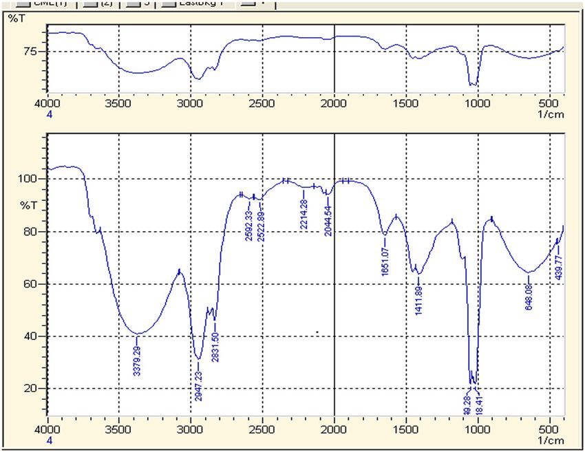
86 Saud Bawazeer et al.
producing diverse range of chemical compounds that are (delphinidin, cyaniding, delphinidin, and pelargonidin deri-
responsible for various biological actions [1]. Even at the vatives), flavonoids (quercetin and kaempferol glycosides),
dawn of twenty-first century, about 90% of potential drug sulfur compounds (glucotropaeolin), phenolic acids (chloro-
molecules have been isolated directly or indirectly [2]. genic acid), cucur-bitacines, and vitamin C [19–23]. Nastur-
WHO estimated that 80% population of Africo-Asian tium plant has been reported for various ailments including
countries utilize herbal medicine in their primary health upper respiratory tract (bronchitis, tonsillitis) as well as
care system [3]. The clinical application of herbal drugs urinary tract diseases [19,22,24], cardiovascular disorders,
faces the same challenges as allopathic medicines like and constipation [25–27]. It is reported as disinfectant,
selectivity, drug delivery, solubility, safety, toxicity, effi- wound-healing, antibiotic [28,29], antiascorbutic [29], and
cacy, and frequent dosing [4,5]. The modern pharma- anticancer activity [30]. Moreover, in the fields of cosme-
ceutical research could overcome the above-mentioned tology and dermatology, its external use for treatment of
challenges by developing novel drug distribution systems diseases of the hair, nails, and skin (itchy dandruff), super-
of herbal medicines, including, micro emulsion, nano- ficial/moderate burns, and rashes has been reported [19].
particles’ solid dispersion, liposomes, matrix system as The leaves and flowers contain glucosinolates resulting in
well as solid lipid nanoparticles [6,7]. its peppery flavour and are commonly used in salads [31].
Nanotechnology is emerging as interdisciplinary In the present study, we aimed to synthesize the
science for resolving various problems in the fields of AgNPs of T. majus, keeping in view its extensive medical
biomedical sciences, pharmacology, and food processing applications. The preliminary investigation of T. majus
[8–10]. It deals with matter at the scale of one billionth of has led us to the presence of high number of flavonoids,
a metre, so the nanoparticle is the most fundamental phenolics, and tannins components which might play
component in the fabrication of a nanostructure. The role in antioxidant effect. Thus, we aimed to synthesize
nanometre-sized particles exhibit interesting and sur- the stable AgNPs of T. majus’ methanolic and aqueous
prising capabilities [11,12]. There are various methods extract by adopting easy, reliable, and simple method and
reported for fabrication of nanoparticles, but metal nano- evaluate their antioxidant and antibacterial activities.
particles like gold and silver are extensively studied due
to their unique electrical and optical properties.
The study of techniques for the synthesis of AgNPs of
various morphologies and sizes is widely accepted 2 Materials and methods
recently [13,14]. Currently, various chemical and physical
methods are applied for the green synthesis of AgNPs,
2.1 Chemicals
but most of them have limitation for being expensive
and inclusion of hazardous solvents [15]. The technique
Silver nitrate (BDH laboratory, BH.15-1TD) was purchased
of green biosynthesis of eco-friendly metal NPs processes
from local market of Peshawar. De-ionized water, potas-
is studied widely, where the bio-extracts are fabricated as
sium bromide, 1,1-diphenyl-2-picrylhydrazyl (DPPH),
NPs [16,17]. It involves the reduction of metal ions by
methanol, sulphuric acid, sodium hydroxide, ammonia,
phytochemicals of extracts to fabricate them as NPs
ammonium hydroxide, hydrochloric acid, and trichloro
[18]. Various reducing phytochemicals are involved in
methane were purchased from Merck.
this process including phenol, tannins, terpenoids, flavo-
noids, alkaloids, quinines, etc. The above-mentioned
technique leads to formation of crystalline NPs ranging
in size between 1 and 100 nm and has several advantages 2.2 Plant collection and extraction
including low cost and easy availability. The extracts’
capability to be fabricated as stable NPs depends on the The plant is obtained from the ground of Botany
phytochemical profile of the plant extract. Department, University of Peshawar, Peshawar. The plant
Tropaeolum majus L. (T. elatum Salisb., Tropaeolaceae) specimen was recognized by taxonomist Ghulam Jillani from
is a fast-growing annual plant native to Peru and Bolivia, Department of Botany, University of Peshawar. The plant
found in South and Central America and subcontinent. The material was collected and dried under shade for about
plant is commonly identified with the name of nasturtium 15 days. The dried plant material (leaves) of Tropaeolum
or nasturtiums. The genus roughly includes 80 species. majus was crushed to make well powder. The powder mate-
The explored constituents of T. majus included caroten- rials were soaked in methanol for 3 days and assessed for
oids (zeaxanthin, lutein, and carotene), anthocyanins normal cold extraction until exhaustion of plant materials.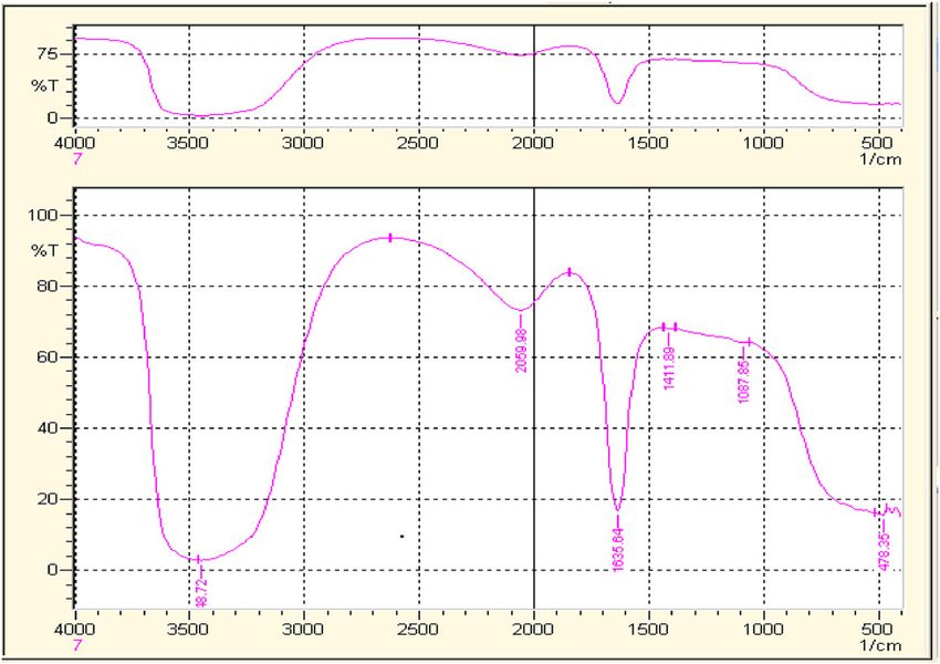
Biosynthesis of silver nanoparticles of Tropaeolum majus 87
The obtained extract was then concentrated under reduced ratios were then heated same as for 1:1. The UV spectra
pressure at low temperature using rotary evaporator. The were taken for each and every step to check and confirm
same procedure was done for aqueous extract by soaking the synthesis of metallic nanoparticles [35].
the powder material with water [32].
2.4.3 Time and temperature effect on synthesis of
AgNPs
2.3 Phytochemical screening
The time and temperature effect of synthesized nano-
Chemical tests for phytochemicals like anthraquinones, tan- particles was performed for confirming the stability of
nins, alkaloids, glycosides, saponins flavonoids, steroids, nanoparticles. The nanoparticles showed maximum absor-
reducing sugars, terpenoids, phlobatanins, anthocyanins, bance was first heated at 70°C for 30 min, and then time
soluble starch, and free reducing sugars were carried out was extended to 60, 90, and 120 min. The temperature
on the methanol extract of the leaves of Tropaeolum majus effect was assessed by heating the same ratio of stock
using standard procedure to identify the constituents as and salt solution at 30°C, 50°C, 70°C, 80°C, and 100°C.
described by Sofowora, Trease, and Evans [32–34]. The stability of nanoparticles was then confirmed by
taking UV spectra [36].
2.4 Synthesis of nanoparticles
2.5 Characterization
The fresh plant materials were cleaned by washing with
water to remove dust and other impurities. The derided 2.5.1 UV-Vis spectroscopy
plant sample was cut into small pieces. Small pieces of
fresh plants materials were put in a conical flask sepa- Ultraviolet-Visible (UV-Vis) spectrophotometry assay is
rately followed by the introduction of methanol. The considered easy and less time-consuming assay for con-
flasks were kept for three days, after that the solutions firmation of the synthesis of nanoparticle [37,38]. The
were filtered and filtrates were concentrated and stored in UV-Vis analysis was done by using a double beam spectro-
the refrigerator at 4°C [35]. photometer (Shimadzu UV spectrophotometer). Simply,
4 mL of the diluted newly synthesized AgNPs solution
was placed in a cuvette and inserted in the UV-Vis spectro-
2.4.1 Preparation of stock and salt solutions photometer. The UV-Vis spectrum was obtained for the
sample using wavelength range of 300–800 nm.
Crude extract (0.1 g) of plant Tropaeolum majus was dis-
solved in 50 mL of methanol, to prepare stock solution of
extract. For the synthesis of metallic nanoparticles, 17 mg 2.5.2 FT-IR analysis
of silver nitrate (AgNO3) was dissolved in 100 mL of dis-
tilled water to prepare 1 mM salt stock solution. The salt Fourier transform infrared (FTIR) is one of the preferen-
solutions were stored in refrigerator in reagents bottle tially implemented methods of infrared spectroscopy.
which was coated by aluminium foil for further use [35]. The IR method involves the principle of allowing the IR
radiation to pass through a sample. During the process,
the sample absorbs some of the radiations, while some of
2.4.2 Processing for synthesis of AgNPs it will be transmitted through sample. The resultant IR
spectrum represented as the molecular absorption and
The prepared stock solutions of extract and salt were com- transmission spectrum, producing a clear idea regarding
bined in different ratios, i.e. 1:1, 1:2, 1:3, and 1:4, by making the molecular composition or fingerprints of the sample.
the extract solution diluted in each successive reaction mix- They are referred fingerprints because of uniqueness of
ture. The reaction mixture 1:1 was first heated at 70°C for 1 h; the IR spectrum, i.e. each molecular structure has its unique
the colour was observed before heating and after some time. infrared spectrum. The FTIR spectrum of Tropaeolum majus,
The modification in colour exhibited the reduction process AgNPs were recorded using a FTIR prestige-21 Shimadzu
and the synthesis of nanoparticles. The above-mentioned FTIR spectrophotometer [39]. FT-IR measurement was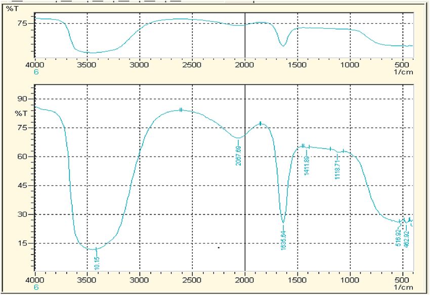
88 Saud Bawazeer et al.
performed and analysed for identification of the chemical where control OD is the absorbance of the blank sample
ratios of the plant crude extracts and synthesized AgNPs. and OD sample is the absorbance of samples or standard
Both (plants crude extracts and synthesized AgNPs) were sample.
analysed in the range from 400 to 4,000. The analysis was
done by following potassium bromide (KBr) pellets method.
Both the spectra of crude and AgNPs were compared to each 2.6.2 Antibacterial activity
other for the confirmation of nanoparticles synthesis.
Three strains of bacteria (Staphylococcus aureus, Klebsiella
pneumonia, and Bacillus subtilis) were used to analyse
2.5.3 Atomic force microscopy (AFM) antibacterial activity of T. majus extract and AgNPs. The
above-mentioned bacterial strains were kept at 4°C in
The atomic force microscopy (AFM) for characterization Muller-Hinton agar. Modified agar well diffusion method
of nanoparticles is the most suited technique [40]. It has was implemented for analysis of antibacterial activity of
the capability of 3D visualization and is effective in gath- crude extract and AgNPs. The cultures were cultivated in
ering both qualitative and quantitative information on triplicates at temperature of 37°C for 1–3 days. Then the
many physical parameters of nanoparticles. The physical broth cultures were transferred to sterilized Petri-dish
parameters that could be analysed with AFM included following addition of 20 mL of the sterilized molten MHA.
size, surface texture (roughness or smoothness), and The amount of 0.2 mL of the extracts and derived AgNPs
morphology. It could also provide statistical information solution were added in corresponding wells through a bore
regarding size, surface area, and volume distributions. into the medium. The streptomycin (2 mg/mL) was used as
AFM is capable of analysing wide range of particle sizes reference antimicrobial agent. To ensure complete diffusion
in the same scan, ranging from 1 nm to 8 µm. Further- of the antimicrobial agent into the medium, inoculation
more, the characterization of nanoparticle with AFM is was extended for 1 h. The inoculated plates were incubated
possible in multiple mediums, for example, controlled at 37°C for 24 h and the diameter of the zone of inhibition
environments, ambient air, and even liquid dispersions. of bacterial growth was measured in the plate in milli-
metres [42,43].
2.6 Biological activities
3 Results and discussion
2.6.1 Antioxidant activity (DPPH radical scavenging
assay) 3.1 Phytochemical screening of plant
T. majus
The antioxidant potential was measured by DPPH radical
scavenging assay [41]. The principle of the assay is Selected plant (Tropaeolum majus) was subjected to
bleaching of the purple-coloured methanol solution of extraction to get methanolic and aqueous extract. Keep-
2,2-diphenyl-1-pierylhydrazyl (DPPH) correspondent to the ing in view the objectives of this research project, the
measurement of the hydrogen atom or electron donation resultant extracts were analysed for presence or absence
anilities in the sample. All the samples were performed of important reducing phytochemicals. The data from
in triplicate. Simply, 3 mL of samples (extracts/AgNPs) preliminary screening of extracts have been shown in
solutions in methanol (containing 10–100 μg) and control Table 1. Both the methanolic and aqueous extracts indi-
(without sample) were mixed with 1 mL solution of DPPH cated the presence of tannins, alkaloids terpenoids, fla-
radical solution (1 mM) in methanol. The solution was vonoids, and cardiac glycosides. These compounds may
allowed to stand for 30 min, then the absorbance was mea- be actively involved in the reduction of silver and gold
sured in dark at wavelength ʎ = 517 nm. The decline of the ions to nanoparticles.
absorbance of DPPH solution is represented as an increase
of the DPPH radical scavenging activity. The following for-
mula was used for calculation of percent radical scavenging
activities (%RSA): 3.2 Characterization of AgNPs
%DPPH = (OD control − OD sample) The newly synthesized AgNPs were characterized by
(1)
× 100/ OD control using UV-Vis spectroscopy, FTIR, and AFM.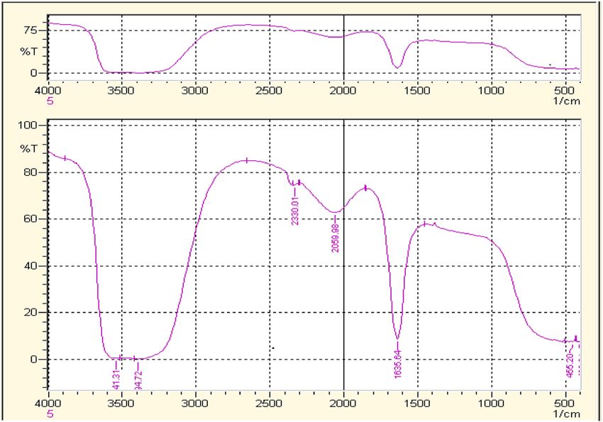
Biosynthesis of silver nanoparticles of Tropaeolum majus 89
Table 1: Phytochemical analysis of aqueous and methanolic 3.2.1.1 UV spectrum of AgNPs synthesized with
extracts of Tropaeolum majus methanolic extract of T. majus
Chemical components Methanolic Aqueous The absorption spectra of synthesized AgNPs were re-
extract extract
corded against methanol. The given spectrum shows
Alkaloids + + the UV-Vis spectra of AgNPs formation using constant
Tannins + + silver nitrate ratio with successive increasing ratios of
Anthraquinone − −
extract at room temperature. The colour of the solution
Glycosides − −
Reducing sugar − −
changed from dark green to orange brown. The different
Saponins − − ratios of the nanoparticles (AgNPs) are shown in the
Flavonoids + + Figure 1. The efficient synthesis was observed in reaction
Phlobatanins − − mixture (1:1) at ʎ = 430 nm.
Steroids − −
Terpenoids + +
Cardiac glycoside + +
Coumarin − − 3.2.1.2 UV spectrum of AgNPs synthesized with aqueous
Emodins − − extract of T. majus
Betacyanin − −
Carbohydrates − −
Monosaccharide’s − − The absorption spectra of synthesized AgNPs were re-
Free reducing sugar − − corded against water. The given spectrum shows the
Combined reducing − − UV-Vis spectra of AgNPs’ formation using constant silver
sugars nitrate concentration with different aqueous extract con-
Soluble starch − −
centrations at room temperature. The different ratios of
the nanoparticles are shown in the Figure 2. The effi-
cient synthesis was observed by 1:1 AgNPs at 450 nm
absorbance.
3.2.1 UV-Vis spectroscopic analysis
The UV-Vis spectroscopy is mostly used for confirmation
of newly synthesized metal NPs. It measures surface 3.2.1.3 Temperature effect on AgNPs synthesis with
plasmon resonance peaks of the metals. The property of methanolic and aqueous extract of T. majus
surface plasmon resonance of the metals makes them
capable of exhibiting distinctive optical properties. The In the present set of experiment, the concentration of silver
addition of T. majus extract to salt solution at room tem- salt and extract solution was kept constant, i.e. 1:1 under
perature caused the solutions to change from dark green variable temperature range to observe any possible effect
to orange brown, the apparent indication of the forma- of temperature on AgNPs synthesis. Temperature effects of
tion of AgNPs. This colour change is reported due to the
reduction of Ag+ to Ag0 by using the active phytochem-
icals present in the extract of T. majus [38]. The variety of 1
1:01
phytochemicals that act as reducing and capping agents 1:03
0.8
1:04
for the synthesis of NPs are already reported. The diverse
range of molecules present in the plant extracts is respon- 0.6
sible for the synthesis of symmetrical NPs [27]. Experi-
0.4
ment was carried out with varying stock solutions of
T. majus extract and salt solution ratio. For monitoring 0.2
of the formation and stability of silver nanoparticles,
0
the absorption spectra of the synthesized silver AgNPs 430 480 530 580 630 680
were recorded against methanol. The peak absorbance -0.2
of AgNPs was observed in wavelength (ʎ) range of 400–
600 nm in methanol solutions which exhibited the suc- Figure 1: UV spectra of the AgNPs solution with methanolic extract of
cessful formation of AgNPs. Tropaeolum majus.90 Saud Bawazeer et al.
1 and aqueous extracts. The maximum absorbance was
0.9 1:01 observed under temperature of 70°C and 90°C.
0.8 1:02
1:03
0.7
0.6
0.5 3.2.2 FTIR spectra analysis of extracts and synthesized
0.4 AgNPs of T. majus
0.3
0.2
The FTIR spectra acquired from the methanolic extract of
0.1
0 T. majus and synthesized AgNPs are demonstrated in
450 500 550 600 650 700 Figures 5 and 6. In Figure 5, a broader peak observed at
3,379 cm−1 is attributed to the O–H bonds stretching, indi-
Figure 2: UV spectra of AgNPs solution with the aqueous extract of cating presence of aromatic alcoholic and phenolic com-
Tropaeolum majus.
pounds. The peaks such as 2947.23, 2831.50, 2592.33, and
2522.89 cm−1 reflect carboxylic acid bond stretching. The
peak of 1635.78 cm−1 shows carbonyl group C]O and
3.5
peak at 1411.89 cm−1 shows C–C stretching. The peak at
3 2,947 indicates the presence of ant symmetric CH2 stretching.
2.5
30c
2
50 €
1.5
70c
1 90c
0.5
0
300
320
340
360
380
400
420
440
460
480
500
520
540
560
580
600
620
640
660
680
700
Figure 3: Temperature effect of methanolic nanoparticles of
Tropaeolum majus.
the methanolic and aqueous extract are shown in Figures 3
and 4, respectively. Data exhibited that increasing tempera-
ture increased synthesis of nanoparticles, both of methanolic
Figure 5: FTIR spectrum of crude water extract of Tropaoelum majus.
3.5
30 °C
3 50 °C
70 °C
2.5
90 °C
2
1.5
1
0.5
0
300
320
340
360
380
400
420
440
460
480
500
520
540
560
580
600
620
640
660
680
700
Figure 4: Temperature effect of aqueous nanoparticles of Figure 6: FTIR spectrum of methanolic nanoparticles of Tropaeolum
Tropaeolum majus. majus.Biosynthesis of silver nanoparticles of Tropaeolum majus 91
The peak at 2,214 is due to the C–H unsaturated stretching.
The peak at 1,661 is due to C]O stretching. The peak at 1,411
is due to OH diff and the peak at 643 indicates the presence
of C–H diff.
When FTIR spectra were compared with those of
AgNPs, the spectra exhibited different IR absorption.
FTIR analysis of the AgNPs obtained after 60 min of reac-
tion was performed to detect involvement of different
functional groups present in T. majus extract (Figure 6).
The peaks at 2947.23, 2831.50, 2592.33, 2522.89, and
1,411 cm−1 disappear in the spectra of FTIR spectra of the
AgNPs. Thus, it means that carboxyl and hydroxyl groups
are considered mainly accountable for reduction and stabi-
lization of AgNPs [44]. Broad bands observed in spectra of
Figure 8: FTIR spectrum water nanoparticles of Tropaeolum majus.
AgNPs, 544.92 and 541.31 cm−1, validated the formation of
AgNPs which were not observed in the crude FTIR spectrum
[45]. The formation of AgNPs is in agreement with already
existing studies on AgNPs synthesis by using various plant
extracts [46].
The FTIR spectra acquired from the aqueous extract
of T. majus and synthesized AgNPs are demonstrated in
Figures 7 and 8, respectively. In Figure 7, the peak of
1635.78 cm−1 shows carbonyl group C]O and peak at
1411.89 cm−1 shows C–C stretching. The peak 1,118 cm−1
exhibits C–N stretching. While comparing the FTIR spectra
of synthesized AgNPs (Figure 8), only peak 1,118 cm−1 is
missing which attributes to C–N stretching and seems to
be mainly responsible for aqueous AgNPs synthesis.
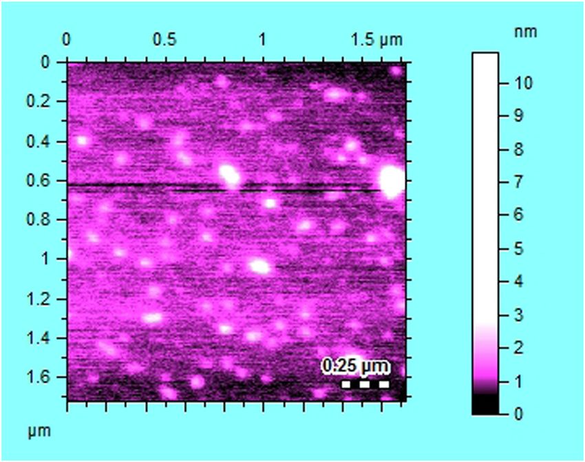
3.2.3 Atomic force microscopic analysis
Figure 9: AFM scan of AgNPs synthesized from methanolic extract of
The AFM for characterization of nanoparticles is the most Tropaeolum majus.
suited technique [40]. It has the capability of 3D visuali-
zation and is effective in gathering both qualita- tive and quantitative information on many physical para-
meters of nanoparticles. The physical parameters that
Table 2: Antioxidant activity of T. majus extract and its synthesized
AgNPs
Conc. Percent DPPH (%)
(µg/
Aqueous Aqueous Methanolic Methanolic
mL)
extract AgNPs (%) extract (%) AgNPs (%)
(%)
20 96.5 23.4 7.7 42
40 94.9 23.9 10 12.2
60 93.7 26.6 24 11.7
80 93.6 34.7 31.6 9.5
100 92.2 40.6 45.1 5.4
150 90 45.7 49 4
Figure 7: FTIR spectrum crude water extract of Tropaeolum majus.92 Saud Bawazeer et al.
Table 3: Antibacterial activity of T. majus extract and its synthesized AgNPs
Microorganism strain Gram stanning Aqueous extract Aqueous AgNPs Methanolic extract Methanolic AgNPs Streptomycin
Klebsiella pneumonia − 0 0 0 0 26
Staphylococcus aureus + 0 12 0 10 28
Bacillus subtilis + 10 0 12 0 28
could be analysed with AFM included size, surface aqueous extracts and their AgNPs were screened
texture (roughness or smoothness), and morphology. It against three strains of bacteria shown in Table 3.
could also provide statistical information regarding size, The antibacterial effect was compared to standard
surface area, and volume distributions. AFM is capable of drug of Streptomycin. The aqueous and methanolic
analysing wide range of particle sizes in the same scan, extract exhibited inhibition against Bacillus subtilis
ranging from 1 nm to 8 µm. Furthermore, the character- and their synthesized AgNPs were active against Sta-
ization of nanoparticle with AFM is possible in multiple phylococcus aureus. Both the above-mentioned bac-
mediums, for example, controlled environments, ambient terial strains are gram-positive; however, the effect is
air, and even liquid dispersions. The AFM scan of the far less than standard drug.
AgNPs synthesized from methanolic extract of T. majus
is shown in Figure 9. The shape of synthesized AgNPs was
observed to be cylindrical and its size was 25 μm.
4 Conclusion
3.3 Biological activities Our data concluded that the extracts of T. majus have
necessary capping and reducing agents which make it
3.3.1 Antioxidant activity of AgNPs vs respective extract capable to develop stable AgNPs. The aqueous extract
of T. majus of T. majus has potential antioxidant effect; however, the
AgNPs did not enhance its free radical scavenging effect.
The free radical scavenging potential of the methanolic The bacterial strains’ susceptibility of the extract and
and aqueous extracts of T. majus versus their synthesized AgNPs was changed from Bacillus subtilis to Staphy-
AgNPs is presented in Table 2. The maximum percent lococcus aureus, respectively. The biological action of
antioxidant potential was exhibited by aqueous extract AgNPs is changed in case of antibacterial activity which
of T. majus at all applied doses. This was found to be means that AgNPs might change the specificity of
dose-independent, i.e. the scavenging ability was almost T. majus and likewise other drugs.
same at 20 µg/mL (minimum dose) to 150 µg/mL (maxi-
mum dose). The scavenging ability of AgNPs synthesized Research funding: The work is funded by grand number
with aqueous extract was markedly reduced, but it has 14-MED333-10 from the National Science, Technology
shown dose-dependent effect, i.e. 23.4% at 20 µg/mL and Innovation Plan (MAARIFAH), the King Abdul-Aziz
(minimum dose) to 45% at 150 µg/mL (maximum dose). City for Science and Technology (KACST), Kingdom of
Similarly, methanolic extract has shown dose-dependent Saudi Arabia. We thank the Science and Technology
effect and is reduced in case of AgNPs. Our data exhibited Unit at Umm Al-Qura University for their continued
that antioxidant potential of T. majus reduced when fab- logistic support.
ricated as AgNPs, probably the reducing phytochemicals
would be consumed during metal reduction. Author contributions: Saud Bawazeer: writing-original
draft; Abdur Rauf: writing-review editing, methodology;
Syed Uzair Ali Shah: formal analysis; Ahmed M. Shawky:
3.3.2 Antibacterial activity visualization; Yahya S. Al-Awthan and Omar Salem
Bahattab: English corrections of the manuscript; Ghias
The synthesized AgNPs of various plants, such as Uddin and Javeria Sabir: project administration;
Acalypha wilkesiana and Tithonia diversifolia, has been Mohamed A. El-Esawi: resources and proofreading of
documented for antimicrobial potential [47–49]. To ana- the final manuscript. All authors read and approved the
lyse the antibacterial effect of T. majus, crude and manuscript for submission.Biosynthesis of silver nanoparticles of Tropaeolum majus 93
Conflict of interest: One of the authors (Abdur Rauf) is a [19] Bruneton J. Pharmacognosy, phytochemistry, medicinal
member of the Editorial Board of Green Processing and plants. 2nd edn. Paris, Hampshire: Inter-cept Ltd., TecDoc,
Synthesis. Lavoisier Publishing Inc.; 1999.
[20] Garzón GA, Wrolstad RE. Major anthocyanins antioxidant
activity of Nas-turtium flowers, Tropaeolum majus. Food
Chem. 2009;114:44–49.
[21] Niizu PY, Rodriguez-Amaya DB. Flowers leaves of Tropaeolum
References majus L. richsources of lutein. J Food Sci. 2005;70:S605–9.
[22] Strzelecka H, Kowalski J, (Eds.). Encyklopedia zielarstwa i
ziołolecznictwa. 1st edn. Warsaw: PWN; 2000. p. 365–6 (in
[1] Daniel AD, Sylvia U, Ute R. A historical overview of natural Polish).
products in drug discovery. Metabolites. 2012;2(2):303–36. [23] Wojciechowska B, Wizner L. Cucurbitacines in Tropaeolum
[2] Veeresham C. Natural products derived from plants as a majus L. fruits. Herba Pol. 1983;29:97–101.
source of drugs. J Adv Pharm Technol Res. 2012;3(4):200–1. [24] Fournie P, (Ed.). Le livre des plantes médicinales et
[3] Fabricant DS, Farnsworth NR. The value of plants used in vénéneuses de France. 1st edn. Paris: Paul Lechevalier; 1947
traditional medicine for drug discovery. Env Health Persp. (in French).
2001;109(1):69. [25] Corrêa MP. Dicionário das plantas úteis do Brasil e das
[4] Yadev D, Suri S, Choudhary AA, Sikender M, Hemant BMN. exóticas cultivadas, vol. 2. Rio de Janeiro: Imprensa Nacional;
Novel approach, herbal remedies and natural products in 1978. p. 67.
pharmaceutical science as nano drug delivery systems. Int J [26] Ferreira RBG, Vieira MC, Zárete NAH. Análise de crescimento
Pharm Tech Res. 2011;3(3):3092–116. de Tropaeolum majus ‘jewel’ em func¸ ão de espac¸ amentos
[5] Ratnam DV, Ankola DD, Bhardwaj V, Sahana DK, Kumar MN. entre plantas. Rev Brasileira de Plantas Medicinais.
Role of antioxidants in prophylaxis and therapy: a pharma- 2004;7:57–66.
ceutical prospective. J Control Rel. 2006;113:189–207. [27] Ferro D. Fitoterapia: conceitos clínicos. São Paulo: Atheneu;
[6] Kumar K, Rai AK. Miraculous therapeutic effects of herbal 2006. p. 410.
drugs using novel drug delivery systems. Int Res J Pharm. [28] Chevallier A. The encyclopedia of medicinal plants. London:
2012;3:27–33. Dorling Kindersley; 1996.
[7] Sharma AT, Mitkare SS, Moon RS. Multicomponent herbal [29] Torres-Jimenez IB, Quintana-Cardenes IJ. Comparative ana-
therapy: a review. Int J Pharm Sci Rev Res. 2011;6:185–7. lysis on the use of medicinal plants in traditional medicine in
[8] Saini R, Saini S, Sharma S. Nanotechnology: the future Cuba and The Canary Islands. Revista Cuba de Plantas
medicine. J Cutan Aesthet Surg. 2010;3(12):32–33. Medicinales. 2004;9(1):lil-394333.
[9] Bionanotechnology. In: Ramsden JJ, Ed. Nanotechnology: an [30] Duke JA, Ayensu ES. Medicinal plants of China. Michigan:
introduction. 2nd edn. United states of America: Elsevier; Reference Publications Algonac; 1985.
2016. p. 358. [31] Jens L, Birger LM. Synthesis of benzylglucosinolate in
[10] Rajiv S, Santosh S. Nanotechnology: the future medicine. Tropaeolum Majus L. Plant Physiol. 1993;102:609–13.
J Cutan Aesthet Surg. 2010;3(1):32–3. [32] Taylor RA, Otanicar TP, Herukerrupu Y, Bremond F,
[11] Raffaele C, Luca ID, Luise AD, Petillo O, Calarco A, Peluso G. Rosengarten G, Hawkes ER, et al. Feasibility of nanofluid-
New therapeutic potential of nanosized phytomedicine. based optical filters. Appl Opt. 2013;52(7):1413–22.
J Nanosci Nanotechnol. 2016;16(8):8176–87. [33] Reiss Gunter, Hutten Andreas. Magnetic nanoparticles.
[12] Chakraborty K, Shivakumar A, Ramachandran S. Nano-tech- In: Sattler KD, (Ed.). Handbook of nanophysics: nanoparticles
nology in herbal medicines: a review. Int J Herb Med. and quantum dots. Boca Raton, FL: CRC Press; 2010. p. 1–2.
2016;4(3):21–7. [34] Khan FirdosAlam. Biotechnology fundamentals. Boca Raton,
[13] Mohan YM, Premkumar T, Geckeler KE. Fabrication of silver FL: CRC Press; 2012. p. 328.
nanoparticles in hydrogel networks. Macromol Rapid [35] Belloni J, Mostafavi M, Remita H, Marignier JL, Delcourt AMO.
Commun. 2006;2(27):1346–54. Radiation-induced synthesis of mono- and multi-metallic
[14] Mazzocut A, Coutino-Gonzalez E, Baekelant W, Sels BF, clusters and nanocolloids. N J Chem. 1998;22(11):1239.
Hofkens J, Vosch TJ. Fabrication of silver nanoparticles with [36] Pang YX, Xujin B. Influence of temperature, ripening time and
limited size distribution on TiO2 containing zeolites. Phys calcination on the morphology and crystallinity of hydroxy-
Chem Chem Phys. 2014;16:18690–3. apatite nanoparticles. J Eur Ceram Soc. 2003;23(10):1697–704.
[15] Beer C, Foldbjerg R, Hayashi Y, Sutherland DS, Autrup H. [37] NanoComposix. UV/Vis/IR spectroscopy analysis of nanopar-
Toxicity of silver nanoparticlesnanoparticle or silver ion? ticles. NanoComposix. 2012;1(1):1–6.
Toxicol Lett. 2012;208:286–92. [38] Khattak A, Ahmad B, Rauf A, Bawazeer S, Farooq U, Patel S,
[16] Iravani S. Green synthesis of metal nanoparticles using plants. et al. Green synthesis, characterisation and biological eva-
Green Chem. 2011;13:2638–42. luation of plant-based silver nanoparticles using Quercus
[17] Makarov VV, Love AJ, Sinitsyna OV, Makarova SS, Yaminsky IV, semecarpifolia Smith aqueous leaf extract. IET Nanobiotechnol.
Kalinina NO. Green nanotechnologies: synthesis of metal 2018;13(1):36–41. doi: 10.1049/iet-nbt.2018.5063.
nanoparticles using plants. Acta Naturae. 2014;6:35–44. [39] Islam NU, Jalil K, Shahid M, Rauf A, Muhammad N, Khan A,
[18] Seddik K, Smain A, Arrar L, Baghiani A. Effect of some phenolic et al. Green synthesis and biological activities of gold nano-
compounds and quercus tannin on lipid peroxidation. World particles functionalized with Willow Salix Alba. Arab J Chem.
Appl Sci J. 2010;21(8):1144–9. 2019;12(8):2914–25.94 Saud Bawazeer et al.
[40] Islam NU, Jalil K, Shahid M, Muhammad N, Rauf A. Pistacia and Ag nanoparticles using Camellia sinensis extract. Mater
integerrima gall extract mediated green synthesis of gold Lett. 2008;62:3103–5.
nanoparticles and their biological activities. Arab J Chem. [46] Abbasi Z, Feizi S, Taghipour E, Ghadam P. Green synthesis of
2015;12(8):2310–19. silver nanoparticles using aqueous extract of dried Juglans
[41] Ahmad B, Hafeez N, Bashir S, Azam S, Khan I, Nigar S. regia green husk and examination of its biological properties.
Comparative analysis of the biological activities of bio- Green Process Synth. 2017;26:477–85.
inspired gold nano-particles of Phyllantus emblica fruit and [47] Dada AO, Adekola FA, Dada FE, Adelani-Akande AT, Bello MO,
Beta vulgaris bagasse with their crude extracts. Pak J Bot. Okonkwo CR, et al. Silver nanoparticle synthesis by Acalypha
2015;2(47):139–46. wilkesiana extract: phytochemical screening, characteriza-
[42] Singleton VL, Rossi JA. Colorimetry of total phenolics with tion, influence of operational parameters, and preliminary
phospomolybdic-phosphotungstic acid reagents. Am J Enol antibacterial testing. Heliyon. 2019;5:10.
Vitic. 1965;16:144–58. [48] Dada AO, Inyinbor AA, Idu EI, Bello OB, Oluyori AP, Adelani-
[43] Barandozi NF. Antibacterial activities and antioxidant capacity Akande TA, et al. Effect of operational parameters, character-
of Aloe vera. Org Med Chem Lett. 2013;3:5. doi: 10.1186/2191- ization and antibacterial studies of green synthesis of silver
2858-3-5. nanoparticles, using Tithonia diversifolia. PeerJ.
[44] Mistry B. A handbook of spectroscopic data chemistry: UV, IR, 2018;6:e5865. doi: 10.7717/peerj.5865.
PMR, CNMR and mass spectroscopy. Gujrat: Oxford Book [49] Zaman K, Bakht J, Malikovna BK, Elsharkawy ER, Khalil AH,
Company; 2009. Bawazeer S, et al. Trillium govanianum wall. ex. Royle rhi-
[45] Alfredo VNR, Victor SM, Marco CLA, Rosa GEM, Miguel CLA, zomes extract medicated silver nanoparticles and their anti-
Jesus AAA. Solventless synthesis and optical properties of Au microbial activity. Green Process Synth. 2020;9:503–14.You can also read