Green synthesis, characterization, and antimicrobial activity of silver nanoparticles prepared using Trigonella foenum-graecum L. leaves grown in ...
←
→
Page content transcription
If your browser does not render page correctly, please read the page content below
Green Processing and Synthesis 2021; 10: 421–429
Research Article
Humaira Rizwana, Mona S. Alwhibi, Hadeel A. Aldarsone, Manal Ahmed Awad,
Dina A. Soliman, and Ramesa Shafi Bhat*
Green synthesis, characterization, and
antimicrobial activity of silver nanoparticles
prepared using Trigonella foenum-graecum L.
leaves grown in Saudi Arabia
https://doi.org/10.1515/gps-2021-0043
received March 09, 2021; accepted June 17, 2021
1 Introduction
Abstract: Silver nanoparticles (AgNPs) are widely used for The emergence of multidrug-resistant strains of various
medical applications particularly as antimicrobial agents pathogens like fungi, bacteria, and viruses has raised
against multidrug-resistant microbial strains. Some plants serious concern globally [1]. According to WHO, intensive
stimulate the reduction of Ag ions to AgNPs. In this study, and indiscriminate use of antimicrobials has led to the
we prepared AgNPs via the green synthesis approach using exponential increase in multidrug resistance [2], limiting
fenugreek leaves grown in Saudi Arabia. Furthermore, we the line of treatment [3]. Hence, alternative approaches to
characterized these AgNPs and evaluated their antimicro- develop new antimicrobial or modify the existing ones
bial activities against pathogenic yeast, bacteria, and fungi. are being researched with high priority.
The ultraviolet-visible peak at 380 nm confirmed the bio- Nanoparticles (NPs) have been widely explored and
synthesis of NPs. Transmission electron microscopy ana- studied, thus opening new perspectives in the field of med-
lyses revealed particle size in the range of 9–57 nm with a
icine, especially against multidrug resistance. Various metal
spherical shape. Dynamic light scattering results confirm
NPs and nanocomposites like iron oxide [4], copper oxide
slight aggregation as the average particle size was shown
[5], zinc oxide [6], graphene oxide [7], gold [8], and silver
as 68.71 nm and a polydispersity index of 0.083. The energy-
oxide have been used in different fields like medicine, agri-
dispersive X-ray spectroscopy results showed an intense
culture, paint, and packaging industries [9–11]. Among
peak at 3 keV, indicating the presence of elemental AgNPs.
them, silver nanoparticles (AgNPs) have been applied and
The synthesized AgNPs efficiently inhibit the growth of both
exploited the most giving promising results against drug-
Gram-positive and Gram-negative bacteria; however, varying
resistant microbes. Recent studies have shown excellent
degree of inhibition was shown toward fungi. The potent
antimicrobial activity of AgNPs against bacteria in synergism
antimicrobial ability of the synthesized NPs can be attributed
with antibiotics at concentrations that were not cytotoxic to
to their small size and round shape. Among all test organ-
human cells [12]. Many scientific communities started using
isms, the growth of Candida albicans and Helminthosporium
AgNPs in therapeutic applications especially in many com-
sativum was remarkably affected by AgNPs treatment.
mercial items like soap, catheters, and bandages to curb
Keywords: green synthesis, Trigonella foenum-graecum L., infection against pathogenic agents [13–17]. Also, some sur-
silver nanoparticles, antimicrobial activity gical instruments, face masks, and bone cementing materials
are incorporated with AgNPs for their excellent antibacterial
properties [13,14]. Nowadays, silver sulfadiazine is being
* Corresponding author: Ramesa Shafi Bhat, Department of replaced by silver NPs for wound healing treatment [16].
Biochemistry, College of Science, King Saud University, P.O. Box Nanobiotechnology deals with the synthesis of metal
11495, Riyadh, Saudi Arabia, e-mail: rbhat@ksu.edu.sa NPs via nontoxic and ecofriendly methods using the sol-
Humaira Rizwana, Mona S. Alwhibi, Hadeel A. Aldarsone, Dina A.
vent medium, reducing agent, and nontoxic NP stabilizer
Soliman: Department of Botany and Microbiology, College of
Science, King Saud University, P.O. Box 11495, Riyadh, Saudi Arabia
[18]. In the past few years, different methods have been
Manal Ahmed Awad: King Abdullah Institiute for Nanotechnology, employed to synthesize NPs, of which the biogenic method
King Saud University, Riyadh, 11451, Saudi Arabia using plant extracts as reducing agents has drawn a lot of
Open Access. © 2021 Humaira Rizwana et al., published by De Gruyter. This work is licensed under the Creative Commons Attribution 4.0
International License.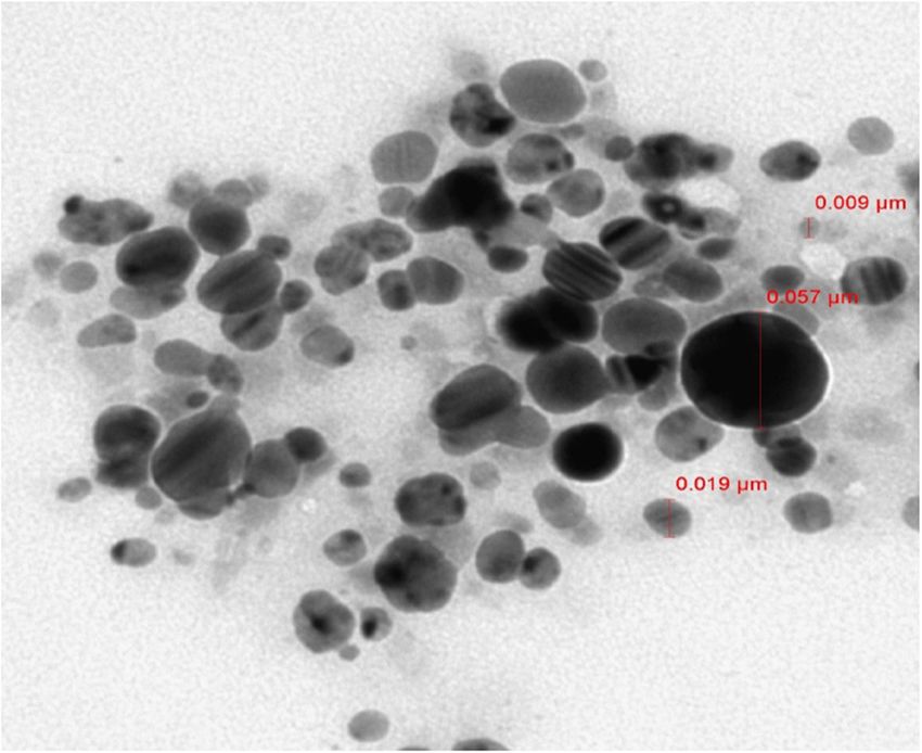
422 Humaira Rizwana et al.
attention. Although many chemical and physical methods of healthy leaves were washed, chopped, and stirred at
are employed to synthesize AgNPs, these methods either 70°C in 0.1 L of double-distilled water (1:10 volume/weight
require high energy or may produce toxic by-products ratio) for half an hour. After cooling to room temperature,
[19,20], which are not encouraging for biomedical uses. the broth was filtered and stored at 4°C. The plant was
This has prompted new investigations in the field of green identified by Professor Najat Bukhari and Dr Mona
synthesis [21]. Plants are a good source of active com- Al Wahibi – Taxonomist, Department of Botany and
pounds, which can stimulate the reduction of Ag ions to Microbiology, King Saud University.
AgNPs [22]. Although the exact mechanism for bioreduction
still needs further elucidation, various studies have shown
the safe, effective, and ecofriendly application of green
synthesized AgNPs in the field of biomedicine [23–25]. 2.2 Biosynthesis of AgNPs
Almost all the parts of plants like leaves, roots, and seeds
can act as potential reducing agents during the synthesis of The freshly prepared leaf extract was added to the silver
AgNPs without using any catalyst or a stabilizer [26]. Plants nitrate solution with a final concentration of 5 mM. The
contain various active biomolecules such as amino acids, reduction of Ag ions to AgNPs in the plant extract was
alkaloids, proteins, polysaccharides, phenolics, terpenoids, monitored through the color change from yellow to brown
and flavones, which act as reducing and capping agents [27]. and was confirmed by the UV-visible spectroscopy (UV-Vis)
Among these biomolecules, phenols and flavonoids exhibit analysis (Figure 1).
unique chemical properties and can reduce and wrap NPs.
This is because these biomolecules contain hydroxyl and
carboxyl groups, which can bind to the metal [28].
Fenugreek (Trigonella Foenum-gracium L.) also called as 2.3 Characterization of the prepared NPs
Helba in Arab countries is an herb that belongs to the
Leguminosae family. This plant is used in Chinese and The synthesis of AgNPs was further ascertained by using the
Ayurvedic medicines to treat many diseases in humans due to UV-Vis spectrophotometer (Thermo Scientific 1500, USA) at
its antibacterial properties. Fenugreek (T. foenum-graecum L.) different intervals for 3 h. The absorbance of the reaction
leaves have been used by humans since ancient times because mixture was measured over the range of 200–700 nm. The
of their medicinal properties. These leaves are rich in flavonoids, FTIR (functional group) analysis of the extract and the
alkaloids, vitamins, and amino acids and are known to nor- synthesized NPs was carried out by FTIR spectrometer at
malize blood glucose, blood lipids, and plasma cholesterol a scan range of 400–4,000 cm−1 (Thermo Scientific-Nicolet-
levels in patients. Different kinds of fungus are sensitive toward 6700, USA). The average size of NPs was measured by Zeta
defensin proteins present in fenugreek leaves. In Saudi Arabia, sizer (Nano–ZS-90 Malvern) after diluting the samples with
fenugreek leaves are commonly used to ease childbirth and to pure water. Transmission electron microscopy (TEM) was
increase breast milk production in lactating mothers. also used for determining the average size of the synthe-
It is well known that a plant grown under different sized AgNPs. The samples were prepared by placing a drop
climatic conditions shows different active components of the synthesized AgNPs on a grid coated with copper and
[29,30]. A study by Al-Jasass and Al-Jasser [31] demon- imaged under the transmission electron microscope (JEOL
strated that fenugreek grown in Saudi Arabia is rich in JEM-1400 Plus). The elemental composition of NPs was
nutrients and contains appreciable amounts of bioactive determined by the EDX analysis. A thin film of AgNPs
compounds. Hence, in this study, we used fenugreek grown was prepared on a glass slide by adding the sample drop-
in Saudi Arabia for the synthesis of AgNPs. We also evalu- wise and allowing the solvent to evaporate after which it
ated the antimicrobial activity of the synthesized AgNPs. was coated with platinum and observed under the FESEM
(FESEM-JSM-7610F, JAPAN).
2 Material and methods
2.4 Antimicrobial activity
2.1 Leaf extract
The bacterial strains and Candida used in this study were
Fresh green leaves of fenugreek cultivated in the Qaseem provided by King Khalid Hospital, Riyadh Saudi Arabia.
region of Saudi Arabia were used in this study. Ten grams Staphylococcus aureus (Gram positive), Escherichia coli,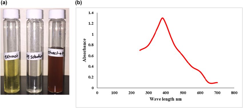
Green Synthesis of silver nanoparticles using T. Foenum-gracium L. extracts 423
Figure 1: (a) Color change of the reaction mixture (from yellow to brown) and (b) UV-Vis spectra showing the absorbance of the AgNPs
synthesized using Trigonella foenum-graecum leaves.
Pseudomonas aeruginosa (Gram negative), Candida albi- 2.4.1 Statistical analysis
cans (yeast), Helminthosporium sativum, Fusarium solani,
Fusarium oxysporum, and Alternaria alternata were pro- All experiments were carried out in triplicate, and the
vided by the Department of Botany and Microbiology, results are reported as means ± SD Values of percentage
King Saud University. The antibacterial activity of the mycelial growth inhibition were subjected to analysis of
synthesized AgNPs against bacteria and C. albicans was variance and Turkey HSD test, and the difference between
evaluated using the well diffusion method. Conversely, the values was considered significant at P < 0.05.
the antifungal activity of the synthesized AgNPs against
fungi was evaluated using the poison food method. The
overnight prepared suspensions of bacteria and Candida
(0.5 McFarland; 106 CFU mL−1) were aseptically spread on 3 Results and discussion
Muller Hinton agar plates with the help of sterile cotton
swabs. After drying, 6 mm wells were punched in the NPs interact with specific wavelengths of light depending
lawned agar with a sterile cork borer. Each well was on their size, shape, and cluster state. Hence, UV-Vis
loaded with 50 µL of the synthesized NPs. The treated spectroscopy is a primary technique for confirming the
culture plates were incubated at 37°C for 24 h, after which synthesis of NPs. In this study, the reduction of Ag ions to
the diameter of the clear area around each well was mea- AgNPs with the help of plant extract was indicated by the
sured (in millimeters). This area represented the zone of color change of the reaction mixture (from yellow to
inhibition. The in vitro antifungal assay of the synthe- brown), as shown in Figure 1a. The mixture showed a
sized NPs was carried out using potato dextrose agar surface plasmon resonance (SPR) peak at 380 nm, con-
(PDA). The synthesized NPs (1 mL) were placed on a firming the presence of AgNPs. AgNPs can show SPR peak
9 cm Petri plate followed by the addition of 19 mL of between 380 and 470 nm subjected to their size and
molten PDA. After the solidification of the media, a fungal shape as small-sized AgNPs absorbed at smaller wave-
plug (6 mm) was placed at the center. The plate was then length [32]. SPR is due to free electrons vibrating in metal
incubated at 28 ± 2°C until it showed full growth. The NPs resonating with the light waves. The absorption
radial growth of the fungal mycelium was measured for of NPs is related to their particle size. A decrease in the
all the fungi used in this study, and the percentage of size of NPs shifts their SPR peak toward shorter wave-
growth inhibition was calculated as follows: lengths [33].
Fenugreek leaves are rich in active compounds [34],
PI = CR–RI/C, (1)
which can facilitate the green synthesis of NPs. Plant
where CR is the radial growth of fungal mycelia on the sources that are rich in active secondary metabolites act
control plate and RI is the radial growth of fungal mycelia as capping agents due to which aggregation of synthesized
on the plate treated with AgNPs. nanoparticles is reduced [35]. These active compounds can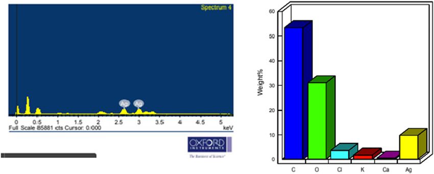
424 Humaira Rizwana et al.
be analyzed by IR spectroscopy. The green method of to the OH stretching due to phenols, and a strong peak at
synthesizing metal NPs is a three-step process. It begins 1,603 and 1,410 cm−1 is due to N–H and C–H bending cor-
with the activation phase where metal ions in salt are responding to amide and aromatic groups. A peak at
reduced with the help of reducing agents present in the 1,075 cm−1 is due to the C]C stretch. However, the IR
plant extract. In the growth phase, reduced metal ions spectrum of the synthesized NP (Figure 2b) shows a shift
combine to form NPs. NPs are finally capped by plant in the peaks and also the appearance of a new peak at
metabolites for stability [36]. The IR spectrum (Figure 2a) 2,927 cm−1. The peaks at 3,396 and 1,603 cm−1 in leaf
of the extract shows peaks at 3,396 cm−1, which corresponds extract were replaced by peaks at 3,430 and 1,622 cm−1
(a)
39.2
38
36
34
32
30 893.95
28 841.63
26
24
%T 22
20 619.64
543.20
18
16
14 3396.46
12
1410.38 1075.79
10
8
1603.57
6
5.0
4400. 0 4000 300 0 2000 1500 100 0 500 400. 0
cm-1
(b)
56.6
54
573.77
52
50 1076.52
48
46 1622.80
1382.30
44
%T 2927.94
42
40
38
36
3430.25
34
32
30.0
4 4 00 . 0 40 0 0 3 0 00 20 0 0 1 5 00 1 00 0 50 0 40 0 . 0
cm-1
Figure 2: IR spectra of (a) Trigonella foenum-graecum aqueous leaf extract and (b) AgNPs.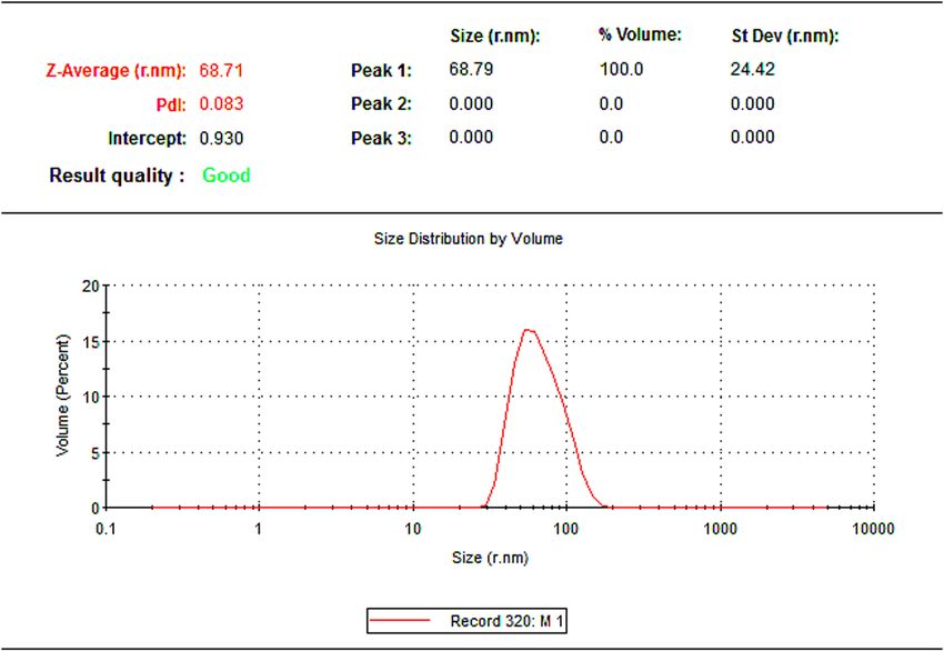
Green Synthesis of silver nanoparticles using T. Foenum-gracium L. extracts 425
Figure 3: DLS results of the AgNPs.
Similar to our findings, the IR spectrum of AgNPs synthe-
sized from seeds of fenugreek had shown the presence of
OH, COOH, and N]O groups [39].
In this study, we used dynamic light scattering (DLS)
to determine the particle size distribution of the synthe-
sized AgNPs (Figure 3a and b). The Z-average mean size
of the synthesized AgNPs was 68.71 nm, and the polydis-
persity index (PDI) was 0.083. DLS is an efficient tech-
nique to determine the PDI of NPs. The size of the NPs
prepared in this study was satisfactory because PDI
values of more than 0.7 indicate a broad size distribution
[31]. The particle size of AgNPs ranged from 9 to 57 nm
(Figure 4), which is in agreement with the zeta size read-
ings to some extent. Our results showed the low PDI
value, which indicates low aggregation, but DLS shows
the hydrodynamic diameter that includes core plus any
Figure 4: TEM images of the AgNPs.
molecule attached or adsorbed on the surface unlike
TEM, which gives the size of NPs in a dried form [40].
respectively in synthesized NP. The resulting peaks in NP The energy-dispersive spectrum (EDX) of the synthe-
were much narrower and less intense, thus indicating sized NPs exhibited strong signals in the silver region,
encapsulation. The reduction and capping of the synthe- and an absorption peak was observed at 3 keV (Figure 5).
sized AgNPs might be due to the presence of phenols, The peak in this region is due to SPR, indicating the for-
flavonoids, carboxylic, or amide groups, which were seen mation of AgNPs [41]. The weight percent of silver was 10.
in the IR spectrum. Previous reports on fenugreek have The presence of other elements including carbon, oxygen,
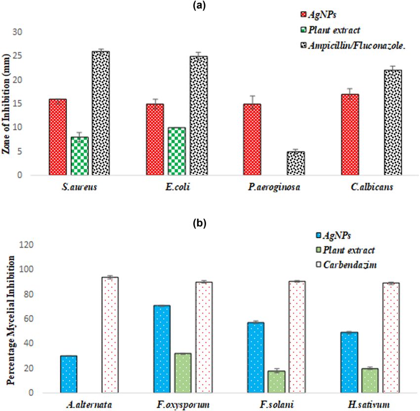
shown polyphenols as a major phytoconstituents [37,38]. chlorine, potassium, and calcium was also observed in the426 Humaira Rizwana et al. Figure 5: EDX elemental analysis of the AgNPs. Figure 6: (a) Antimicrobial activity of the Trigonella foenum-graecum aqueous leaf extract and synthesized AgNPs and (b) antifungal activity of the T. foenum-graecum aqueous leaf extract and synthesized AgNPs. Ampicillin was used as a standard antibiotic for bacteria; fluco- nazole was used as a standard antifungal against Candida albicans; and carbendazim as a standard antifungal against fungi. The value shown represents the mean of the three replicates (±SD). Significant difference in means (P < 0.05) was determined by analysis of variance.
Green Synthesis of silver nanoparticles using T. Foenum-gracium L. extracts 427
spectrum with the weight percent of 53, 31, 3, 2, and 1 wt%, biogenic AgNPs [56]. However, the mechanism under-
respectively. lying the inhibition of the fungal growth by AgNPs is
AgNPs are used as antimicrobial agents in detergents unclear. Some studies have reported that AgNPs reduce
and disinfectants because many pathogenic microbes the ergosterol level of fungal cells. Since ergosterol plays
including bacteria, fungi, and yeast are resistant to che- a vital role in cell functioning, any disturbance in its
mical fungicides and disinfectants [42]. In this study, we concentration leads to the instability of the cell structure,
evaluated the antimicrobial efficiency of AgNPs against which may result in cell damage or cell death [57].
pathogenic bacteria, fungi, and yeast. AgNPs signifi-
cantly inhibited the growth of both the Gram-positive
and Gram-negative bacteria (Figure 6). However, varying
degree of inhibition was shown toward fungi. This anti- 4 Conclusion
bacterial efficacy of the synthesized NPs can be attributed
to their small size and round shape. Our results are con- Fenugreek leaves extract act as a reducing and stabilizing
sistent with the previous reports that suggest the potent agent in the process of synthesizing AgNPs. Prepared NPs
antimicrobial activity of synthesized NP, which was attri- were of small size and round shape and validating were a
buted to their small size and round shape [43–46]. Hence, potential excellent antifungal and antibacterial agent.
the significant antibacterial efficacy of the AgNPs observed
in the present study indicates that NPs could penetrate the Acknowledgment: The author extends their appreciation
thick and rigid cell walls of Gram-positive bacteria and to the researchers supporting project number (RSP-2021/
also the tough lipopolysaccharide membrane of Gram- 173) of King Saud University Riyadh Saudi Arabia.
negative bacteria, unlike many other plant-synthesized
NPs that are potent only against Gram-negative bacteria Funding information: The author extends their apprecia-
[47,48]. The antibacterial properties of the plant-derived tion to the researchers supporting project number (RSP-
AgNPs can be also be attributed to their ability to permeate 2021/173) of King Saud University Riyadh Saudi Arabia.
the cell and interact with the genetic material (DNA) and
other important constituents, thereby disturbing its integ- Author contributions: Humaira Rizwana: project admin-
rity, which ultimately leads to cell death. In addition, the istration; Mona S. Alwhibi: project administration and
adherence of NPs to negatively charged cell surfaces alters resources; Hadeel A. Aldarsone: methodology; Manal
the chemical and physical properties of the cell wall and Ahmed Awad: methodology; Dina A. Soliman: metho-
the membrane. These changes affect the stability of the cell dology; Ramesa Shafi Bhat: writing – original draft.
by disturbing its osmoregulation, permeability, respira-
tion, and electron transport [49–51]. A recent study shows Conflict of interest: The authors state no conflict of
that the AgNPs synthesized from fenugreek caused bac- interest.
terial cell death due to maximum leakage of proteins in the
4 h of treatment due to an increase in the cell membrane
permeability.
AgNPs synthesized in this study significantly inhib- References
ited the growth of C. albicans. Similar findings have been
reported earlier with Candida sp. [52]. Panacek et al. [53] [1] Tenover FC. Mechanisms of antimicrobial resistance in bac-
reported that even low concentrations of AgNPs can sig- teria. Am J Med. 2006;119(6 Suppl 1):S3–10. Discussion
nificantly inhibit the growth of fungi. They also reported S62–70. doi: 10.1016/j.amjmed.2006.03.011. PMID: 16735149.
[2] World Health Organization. 10 Facts on antimicrobial resis-
that AgNPs do not show toxicity toward human anti-
tance; 2018. Available online: http://www.who.int/features/
fungal drugs used for controlling several pathogenic
factfiles/antimicrobial_resistance/en/ [accessed on
fungi. The plant-derived AgNPs have also been reported 10 June 2018].
to be effective for controlling plant pathogenic fungi such [3] Roca I, Akova M, Baquero F, Carlet J, Cavaleri M, Coenen S,
as Magnaporthe grisea and Bipolaris sorokiniana [54]. et al. The global threat of antimicrobial resistance: science for
Botrytis cinerea and Alternaria alternata treated with intervention. N Microbes N Infect. 2015 16;6:22–9.
doi: 10.1016/j.nmni.2015.02.007.
AgNPs show severe damaging effects on conidial and
[4] Dinali R, Ebrahiminezhad A, Manley-Harris M, Ghasemi Y,
hyphal structures [55]. Similarly, wood-degrading fungi Berenjian A. Iron oxide nanoparticles in modern microbiology
such as Chaetomium globosum, Gloeophyllum abietinum, and biotechnology. Crit Rev Microbiol. 2017
and Phanerochaete sordida are significantly inhibited by Aug;43(4):493–507. doi: 10.1080/1040841X.2016.12677085.428 Humaira Rizwana et al.
[5] Ingle AP, Duran N, Rai M. Bioactivity, mechanism of action, and [20] Cele T. Preparation of nanoparticles. IntechOpen.
cytotoxicity of copper-based nanoparticles: a review. Appl 2020;15:10.5772.
Microbiol Biotechnol. 2014;98:1001–9. [21] Barkat MA, Harshita, Beg S, Naim MJ, Pottoo FH, Singh SP,
[6] Gao Y, Arokia Vijaya Anand M, Ramachandran V, et al. Current progress in synthesis, characterization and
Karthikkumar V, Shalini V, Vijayalakshmi S, et al. applications of silver nanoparticles: precepts and prospects.
Biofabrication of zinc oxide nanoparticles from aspergillus Recent Pat Antiinfect Drug Discov. 2018;13(1):53–69.
niger, their antioxidant, antimicrobial and anticancer activity. [22] Zhang XF, Liu ZG, Shen W, Gurunathan S. Silver nanoparticles:
J Clust Sci. 2019;30:937–46. synthesis, characterization, properties, applications, and
[7] Mariadoss AVA, Saravanakumar K, Sathiyaseelan A, Wang MH. therapeutic approaches. Int J Mol Sci. 2016 Sep 13;17(9):1534.
Preparation, characterization and anti-cancer activity of gra- doi: 10.3390/ijms17091534.
phene oxide-silver nanocomposite. J Photochem Photobiol B. [23] Mittal AK, Kaler A, Mulay AV, Banerjee UCJ. Synthesis of gold
2020;210:111984. nanoparticles using whole cells of geotrichum candidum.
[8] Peng J, Liang X. Progress in research on gold nanoparticles in Nanopart. 2013;2013:1–6. doi: 10.1155/2013/150414.
cancer management. Med (Baltim). 2019;98(18):e15311. [24] Makarov VV, Love AJ, Sinitsyna OV, Makarova SS, Yaminsky IV,
doi: 10.1097/MD.0000000000015311. Taliansky ME, et al. “Green” nanotechnologies: synthesis
[9] Ertem E, Gutt B, Zuber F, Allegri S, Le Ouay B, Mefti S, et al. of metal nanoparticles using plants. Acta Naturae.
Core-shell silver nanoparticles in endodontic disinfection 2014 Jan;6(1):35–44.
solutions enable long-term antimicrobial effect on oral bio- [25] Rheder DT, Guilger M, Bilesky-José N, Germano-Costa T,
films. ACS Appl Mater Interfaces. 2017 11;9(40):34762–72. Pasquoto-Stigliani T, Gallep TBB, et al. Synthesis of biogenic
10.1021/acsami.7b13929. silver nanoparticles using Althaea officinalis as reducing
[10] Nakazato G, Kobayashi , R, Seabra AB, Duran N. Use of agent: evaluation of toxicity and ecotoxicity. Sci Rep.
nanoparticles as a potential antimicrobial for food packaging. 2018;8:12397. doi: 10.1038/s41598-018-30317-9.
In: Grumezescu A, editor. Food preservation. 1st ed. [26] Singh A, Gautam PK, Verma A, Singh V, Shivapriya PM,
Cambridge, MA, USA: Academic Press; 2016. Shivalkar S, et al. Green synthesis of metallic nanoparticles as
[11] Holtz RD, Lima BA, Filho AGS, Brocchi M, Alves OL. effective alternatives to treat antibiotics resistant bacterial
Nanostructured silver vanadate as a promising antibacterial infections: a review. Biotechnol Rep (Amst). 2020 Jan
additive to water-based paints. Nanomed Nanomed NBM. 31;25:e00427. doi: 10.1016/j.btre.2020.e00427.
2012;8:935–40. [27] Iqbal J, Abbasi BA, Munir A, Uddin S, Kanwal S, Mahmood T.
[12] Abo-Shama UH, El-Gendy H, Mousa WS, Hamouda RA, Facile green synthesis approach for the production of chro-
Yousuf WE, Hetta HF, et al. Synergistic and antagonistic mium oxide nanoparticles and their different in vitro biological
effects of metal nanoparticles in combination with antibiotics activities. Microsc Res Tech. 2020 Jun;83(6):706–19.
against some reference strains of pathogenic microorganisms. doi: 10.1002/jemt.23460.
Infect Drug Resist. 2020 7;13:351–62. doi: 10.2147/ [28] Castillo-Henríquez L, Alfaro-Aguilar K, Ugalde-Álvarez J, Vega-
IDR.S234425. Fernández L, Montes de Oca-Vásquez G, Vega-Baudrit JR.
[13] Lee SH, Jun BH. Silver nanoparticles: synthesis and application Green synthesis of gold and silver nanoparticles from plant
for nanomedicine. Int J Mol Sci. 2019 17;20(4):865. extracts and their possible applications as antimicrobial
doi: 10.3390/ijms20040865. agents in the agricultural area. Nanomaterials (Basel). 2020
[14] Pascu B, Negrea A, Ciopec M, Davidescu CM, Negrea P, Sep 7;10(9):1763. doi: 10.3390/nano10091763.
Gherman V, et al. New generation of antibacterial products [29] Guo H, Chamberlain SA, Elhaik E, Jalli I, Lynes A-R, Marczak L,
based on colloidal silver. Mater [Basel]. 2020 29;13(7):1578. et al. Geographic variation in plant community structure of salt
[15] Nakamura S, Sato M, Sato Y, Ando N, Takayama T, Fujita M, marshes: species, functional and phylogenetic perspectives.
et al. Synthesis and application of silver nanoparticles (Ag PLoS ONE. 2015;10(5):e0127781. doi: 10.1371/
NPs) for the prevention of infection in healthcare workers. Int J journal.pone.0127781.
Mol Sci. 2019 24;20(15):3620. doi: 10.3390/ijms20153620. [30] Liu W, Liu J, Yin D, Zhao X. Influence of ecological factors on the
[16] Paladini F, Pollini, M. Antimicrobial silver nanoparticles for production of active substances in the anti-cancer plant
wound healing application: progress and future trends. Mater Sinopodophyllum hexandrum (Royle) T.S. Ying. PLoS One.
(Basel). 2019;12(16):2540. doi: 10.3390/ma12162540. 2015 Apr 15;10(4):e0122981. doi: 10.1371/
[17] Butrón Téllez Girón C, Hernández Sierra JF, DeAlba-Montero I, journal.pone.0122981.
Urbano Peña MLA, Ruiz F. Therapeutic use of silver nanopar- [31] Al-Jasass FM, Al-Jasser MS. Chemical composition and fatty acid
ticles in the prevention and arrest of dental caries. Bioinorg content of some spices and herbs under Saudi Arabia condi-
Chem Appl. 2020;12:8882930. tions. Sci World J. 2012;2012:1–5. doi: 10.1100/2012/859892.
[18] Mariadoss AVA, Ramachandran V, Shalini V, Agilan B, [32] Rahman A, Kumar S, Bafana A, Lin J, Dahoumane SA, Jeffryes C.
Franklin JH, Sanjay K, et al. Green synthesis, characterization A mechanistic view of the light-induced synthesis of silver
and antibacterial activity of silver nanoparticles by Malus nanoparticles using extracellular polymeric substances of
domestica and its cytotoxic effect on (MCF-7) cell line. Microb chlamydomonas reinhardtii. Molecules. 2019 Sep
Pathog. 2019;135:103609. 27;24(19):3506. doi: 10.3390/molecules24193506.
[19] El Shafey AM. Green synthesis of metal and metal oxide [33] Peng S, McMahon JM, Schatz GC, Gray SK, Sun Y. Reversing
nanoparticles from plant leaf extracts and their applications: the size-dependence of surface plasmon resonances. Proc Natl
a review. Green Process Synth. 2020;9:304–39. doi: 10.1515/ Acad Sci USA. 2010 Aug 17;107(33):14530–4. doi: 10.1073/
gps-2020-0031. pnas.1007524107. Epub 2010 Jul 29.Green Synthesis of silver nanoparticles using T. Foenum-gracium L. extracts 429
[34] Rahmani M, Hamel L, Toumi-Benali F, Dif MM, Moumen F, J Braz Chem Soc. 2010;21:949–59. doi: 10.1590/S0103-
Rahmani H. Determination of antioxidant activity, phenolic 50532010000600002.
quantification of four varieties of fenugreek Trigonella foenum [46] Pal S, Tak YK, Song JM. Does the antibacterial activity of silver
graecum L. seed extract cultured in west Algeria. J Mater nanoparticles depend on the shape of the nanoparticle? A
Environ Sci. 2018;9(6):1656–61. study of the Gram-negative bacterium Escherichia coli. Appl
[35] Marslin G, Siram K, Maqbool Q, Selvakesavan RK, Kruszka D, Env Microbiol. 2007;73(6):1712–20. doi: 10.1128/
Kachlicki P, et al. Secondary metabolites in the green synth- AEM.02218-06.
esis of metallic nanoparticles. Materials (Basel). 2018 Jun [47] Morones JR, Elechiguerra JL, Camacho A, Holt K, Kouri JB,
3;11(6):940. doi: 10.3390/ma11060940. Erratum in: Materials Ramírez JT, et al. The bactericidal effect of silver nanoparticles.
(Basel). 2019 Mar 08;12(5): PMID: 29865278; PMCID: Nanotechnology. 2005;16(10):2346–53. doi: 10.1088/0957-
PMC6024997. 4484/16/10/059.
[36] Malik P, Shankar R, Malik V, Sharma N, Mukherjee TKJ. Green [48] Haytham M, Ibrahim M. Green synthesis and characterization
chemistry based benign routes for nanoparticle synthesis. of silver nanoparticles using banana peel extract and their
Nanopart. 2014;2014:302429. doi: 10.1155/2014/302429. antimicrobial activity against representative microorganisms.
[37] Wani SA, Kumar P. Fenugreek: a review on its nutraceutical J Radiat Res Appl Sci. 2015;8:265–75. doi: 10.1016/
properties and utilization in various food products. J Saudi Soc j.jrras.2015.01.007.
Agric Sci. 2018;17(2):97–106. doi: 10.1016/ [49] Shrivastava S, Bera T, Roy A, Singh G, Ramachandrarao P,
j.jssas.2016.01.007. Dash D. Characterization of enhanced antibacterial effects of
[38] Devasena T, Nathiya S, Durga M. Therapeutic role of Trigonella novel silver nanoparticles. Nanotechnology. 2007;18:103–12.
foenum-Graecum [fenugreek] – a review. Int J Pharm Sci Rev [50] Marambio-Jones C, Hoek EMV. A review of the antibacterial
Res. 2014;27:74–80. effects of silver nanomaterials and potential implications for
[39] Varghese R, Almalki MA, Ilavenil S, Rebecca J, Choi KC. Silver human health and the environment. J Nanopart Res.
nanopaticles synthesized using the seed extract of Trigonella 2010;12:1531–51. doi: 10.1007/s11051-010-9900-y.
foenum-graecum L. and their antimicrobial mechanism and [51] AshaRani PV, Low Kah Mun G, Hande MP, Valiyaveettil S.
anticancer properties. Saudi J Biol Sci. 2019;26:148–54. Cytotoxicity and genotoxicity of silver nanoparticles in human
[40] Clayton KN, Salameh JW, Wereley ST, Kinzer-Ursem TL. cells. ACS Nano. 2009 Feb 24;3(2):279–90. doi: 10.1021/
Physical characterization of nanoparticle size and surface nn800596w. PMID: 19236062.
modification using particle scattering diffusometry. [52] Senthil B, Devasena T, Prakash B, Rajasekar A. Non-cytotoxic
Biomicrofluidics. 2016 Sep 21;10(5):054107. doi: 10.1063/ effect of green synthesized silver nanoparticles and its anti-
1.4962992. bacterial activity. J Photochem Photobiol B. 2017 Dec;177:1–7.
[41] Eaton P, Quaresma P, Soares C, Neves C, de Almeida MP, doi: 10.1016/j.jphotobiol.2017.10.010Epub 2017 Oct 7.
Pereira E, et al. A direct comparison of experimental methods PMID: 29028495
to measure dimensions of synthetic nanoparticles. [53] Panacek A, Kolar M, Vecerova R, Prucek R, Soukupova J,
Ultramicroscopy. 2017 Nov;182:179–90. doi: 10.1016/ Rystof V, et al. Antifungal activity of silver nanoparticles
j.ultramic.2017.07.001. against Candida spp. J Biomater. 2009;30:6333–40.
[42] Das J, Das MP, Velusamy P. Sesbania grandiflora leaf extract [54] Jo Y-K, Kim BH, Jung G. Antifungal activity of silver ions and
mediated green synthesis of antibacterial silver nanoparticles nanoparticles on phytopathogenic fungi. Plant Dis.
against selected human pathogens. Spectrochimica Acta Part 2009;93:1037–43. doi: 10.1094/PDIS-93-10-1037.
A: Mol Biomolecular Spectrosc. 2013;104:265–70. [55] Ouda SM. Antifungal activity of silver and copper nanoparti-
[43] Hedberg J, Skoglund S, Karlsson ME, Wold S, Odnevall I, cles on two plant pathogens, Alternaria alternata and Botrytis
Hedberg Y. Sequential studies of silver released from silver cinerea. Res J Microbiol. 2014;9:34–42.
nanoparticles in aqueous media simulating sweat, laundry [56] Narayanan KB, Park HH. Antifungal activity of silver nano-
detergent solutions and surface water. Env Sci Technol. particles synthesized using turnip leaf extract (Brassica
2014;48:7314–22. rapa L.) against wood rotting pathogens. Eur J Plant Pathol.
[44] Rai MK, Deshmukh SD, Ingle AP, Gade AK. Silver nanoparti- 2014;140(2):185–92. doi: 10.1007/s10658-014-0399-4.
cles: the powerful nanoweapon against multidrug-resistant [57] Radhakrishnan VS, Reddy Mudiam MK, Kumar M, Dwivedi SP,
bacteria. J Appl Microbiol. 2012 May;112(5):841–52. Singh SP, Prasad T. Silver nanoparticles induced alterations in
doi: 10.1111/j.1365-2672.2012.05253.x. multiple cellular targets, which are critical for drug suscepti-
[45] Durán N, Marcato PD, Conti RD, Alves OL, Costa FTM, bilities and pathogenicity in fungal pathogen (Candida albi-
Brocchi M. Potential use of silver nanoparticles on pathogenic cans). Int J Nanomed. 2018 May 3;13:2647–63. doi: 10.2147/
bacteria, their toxicity and possible mechanisms of action. IJN.S150648.You can also read