Assessing SABU (Serum Anti Bisa Ular), the sole Indonesian antivenom: A proteomic analysis and neutralization efficacy study - CyberLeninka
←
→
Page content transcription
If your browser does not render page correctly, please read the page content below
www.nature.com/scientificreports

              OPEN            Assessing SABU (Serum Anti
                              Bisa Ular), the sole Indonesian
                              antivenom: A proteomic analysis
      received: 09 May 2016
  accepted: 27 October 2016   and neutralization efficacy study
Published: 21 November 2016
                              Choo Hock Tan1, Jia Lee Liew1,*, Kae Yi Tan2,* & Nget Hong Tan2

                              Serum Anti Ular Bisa (SABU) is the only snake antivenom produced locally in Indonesia; however, its
                              effectiveness has not been rigorously evaluated. This study aimed to assess the protein composition
                              and neutralization efficacy of SABU. SDS polyacrylamide gel electrophoresis, size-exclusion liquid
                              chromatography and shotgun proteomics revealed that SABU consists of F(ab’)2 but a significant
                              amount of dimers, protein aggregates and contaminant albumins. SABU moderately neutralized
                              Calloselasma rhodostoma venom (potency of 12.7 mg venom neutralized per ml antivenom, or 121.8 mg
                              venom per g antivenom protein) and Bungarus fasciatus venom (0.9 mg/ml; 8.5 mg/g) but it was weak
                              against the venoms of Naja sputatrix (0.3 mg/ml; 2.9 mg/g), Naja sumatrana (0.2 mg/ml; 1.8 mg/g) and
                              Bungarus candidus (0.1 mg/ml; 1.0 mg/g). In comparison, NPAV, the Thai Neuro Polyvalent Antivenom,
                              outperformed SABU with greater potencies against the venoms of N. sputatrix (0.6 mg/ml; 8.3 mg/g),
                              N. sumatrana (0.5 mg/ml; 7.1 mg/g) and B. candidus (1.7 mg/ml; 23.2 mg/g). The inferior efficacy of
                              SABU implies that a large antivenom dose is required clinically for effective treatment. Besides,
                              the antivenom contains numerous impurities e.g., albumins that greatly increase the risk of
                              hypersensitivity. Together, the findings indicate that the production of SABU warrants further
                              improvement.

                              Indonesia is a vast archipelago extending more than 5000 km from east to west in the equatorial region. Its rich
                              herpetofauna includes more than 10 venomous snake species that distribute in two major ecozones divided by the
                              Wallace’s line. On the eastern side of the Wallace’s line on the Sahul Shelf, there are the Australian elapid fauna,
                              while snakes inhabiting islands west of the Wallace’s line on the Sunda Shelf are mostly common or similar species
                              found in the Malay Archipelago. Java and Sumatra are two huge, densely populated islands on the Sunda Shelf,
                              and they are also natural habitat to many Indonesian snakes. In these islands, the spitting cobras (Naja sputatrix
                              in Java and Lesser Sunda; Naja sumatrana in Sumatra and Kalimantan), the Malayan krait (Bungarus candidus)
                              (Sumatra and Java) and the Malayan pit viper (Calloselasma rhodostoma in Java) are listed under WHO Category
                              1 of medical importance1. Other species of medical importance include the Russell’s viper (Daboia siamensis)
                              and green pit vipers of Trimeresurus complex, the geographical distributions of which are relatively limited in the
                              country. Although snakebite is likely affecting the Indonesian population at a large scale1, unfortunately, compre-
                              hensive epidemiological study of snakebite in this country remains extremely scarce2.
                                  Snakebite envenomation has been aptly described as a disease of poverty that affects heavily the poor or rural
                              population in the developing tropical countries3,4. Prior to the year 2015, it was obscurely listed under “Other
                              Categories” of the Neglected Tropical Diseases by the WHO, lacking systematic attention and official global sup-
                              port program. In 2015, the world saw the de-listing of this critical health problem from the mentioned list of
                              WHO Neglected Tropical Diseases. In fact, the persistent underestimation of snakebite morbidity and mortality
                              has made it the most neglected condition among many other diseases in the tropics5, and toxinology experts have
                              called on WHO and governments to re-establish snakebite as a neglected tropical disease6. Regional toxinologists
                              are also taking up proactive approaches to tackle the various challenges associated with snakebite envenomation.

                              1
                                Department of Pharmacology, Faculty of Medicine, University of Malaya, 50603 Kuala Lumpur, Malaysia.
                              2
                               Department of Molecular Medicine, Faculty of Medicine, University of Malaya, 50603 Kuala Lumpur, Malaysia.
                              *
                               These authors contributed equally to this work. Correspondence and requests for materials should be addressed to
                              C.H.T. (email: tanchoohock@gmail.com)

Scientific Reports | 6:37299 | DOI: 10.1038/srep37299                                                                                           1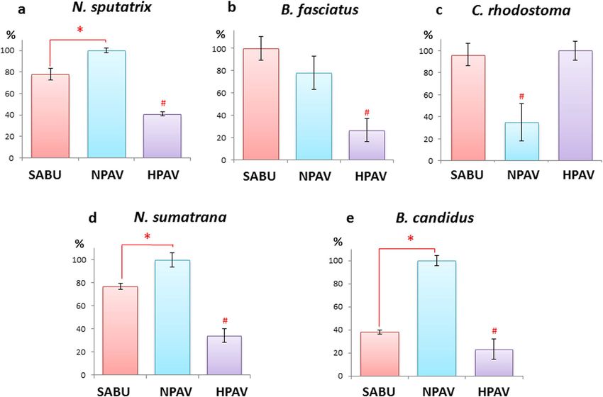
www.nature.com/scientificreports/
One of the basic steps to overcome the problem is to have a rigorous assessment of antivenom in order to ensure
the supply of an affordable and efficacious antivenom product7. Various techniques have been adopted for
antivenom assessment, including the use of high performance liquid chromatography to profile antivenom pro-
teins8, and enzyme-linked immunosorbent assay as well as affinity chromatography (antivenomic approach) to
characterize the immunological binding between antivenom and toxins7,9. Nevertheless, in vivo study remains
indispensable to determine the efficacy of an antivenom in neutralizing the overall toxic effect of snake venom.
The measurable dose-response data obtained from in vivo study will provide an objective reference for the com-
parison of efficacy between different antivenom products5,10,11.
®
In Indonesia, the only local antivenom available is marketed as Biosave , which is more commonly known
as SABU (Serum Anti Bisa Ular), manufactured by the state-owned enterprise BioFarma. SABU is formulated as
a trispecific or trivalent antivenom for clinical use in Indonesia (except the region east of the Wallace’s line and
West Papua). It is derived from the sera of horses which have been hyperimmunized against the venoms from
three snake species of Indonesian origin: the Javan spitting cobra (Naja sputatrix, ular sendok Jawa), the Malayan
pit viper (Calloselasma rhodostoma, ular tanah) and the banded krait (Bungarus fasciatus, ular welang). This is an
antivenom packaged in liquid form, demanding cold-chain transport and stringent storage condition maintained
between 2–8 °C. Anecdotally, SABU is not widely available in many regions of the country, and reports of the use
and efficacy of SABU have been lacking. The effectiveness and limitation of this antivenom have not been rigor-
ously evaluated, leaving the manufacturer and healthcare community clueless about its usefulness and weakness
in snakebite envenomation treatment. In this study, we investigated the quality of SABU including analysis of its
protein composition and neutralization capacity against the toxic effects induced by the venoms of important
snakes in Indonesia. Parallel to this, the performance of SABU was compared to two other antivenom prod-
ucts available commercially in Southeast Asia, i.e. Neuro Polyvalent Antivenom (NPAV) and Hemato Polyvalent
Antivenom (HPAV) which are produced by The Thai Red Cross Society, Queen Saovabha Memorial Institute,
Bangkok. It is hoped that the findings will provide insights into the strength and weakness of the Indonesian
antivenom, and shed light on how the production and the use of the antivenom can be optimized.
Results
Protein determination. SABU has a protein concentration of 104.3 ± 0.5 mg/ml (of undiluted liquid
antivenom, 5 ml), equivalent to approximately 520 mg protein per vial of antivenom. NPAV, in comparison, has
a protein concentration of 75.3 ± 0.6 mg/ml (of reconstituted antivenom, 10 ml), equivalent to approximately
750 mg protein per vial of antivenom. HPAV has a protein concentration of 43.0 ± 0.5 mg/ml (of reconstituted
antivenom, 10 ml), equivalent to approximately 430 mg protein per vial of antivenom.
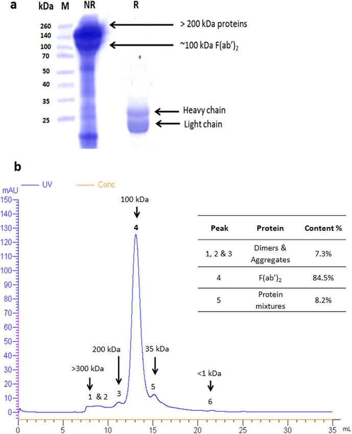
Electrophoretic and chromatographic profiling of antivenom. The electrophoretic profile of SABU
was shown in Fig. 1a. Non-reducing SDS-PAGE of SABU revealed the presence of major proteins with molecular
mass above 100 kDa. On reducing SDS-PAGE, these proteins were observed mainly as two major bands at 21
and 25 kDa. Size-exclusion (or gel filtration) fast protein liquid chromatography (FPLC) resolved the antivenom
into 5 peaks corresponding to the elution of proteins of different molecular masses (Fig. 1b). The major proteins
were eluted in Peak 4, estimated to constitute 84.5% of total antivenom proteins based on the peak area under
the curve. The calibrated estimated molecular mass for peak 4 proteins is approximately 100–110 kDa. SABU
was also found to contain a significant amount of proteins above 200 kDa (Peaks 1–3) (7.3%) and in the range of
30–60 kDa, detected in Peak 5 (8.2%) (Fig. 1b).
Proteomic analysis with liquid chromatography-tandem mass spectrometry (LC-MS/MS). The
shotgun LC-MS/MS analysis revealed the protein composition of fractions collected from the size-exclusion
FPLC (Table 1; Fig. 1b). The protein scores, mass spectral data (intensities, masses and charges of ions) and
amino acid sequences were provided in supplementary file Table S1. Collectively, immunoglobulin chains were
detected as the main composition throughout Peak 1 to Peak 3, constituting 5.73% of total antivenom proteins.
These peaks represented high molecular mass proteins (>200 kDa). Peak 4 (corresponding to the molecular mass
range of 100–120 kDa) also contained immunoglobulin chains as its main component, and these immunoglob-
ulin chains accounted for 84.04% of total antivenom proteins. The immunoglobulin chains were composed of
heavy chains and light chains in a ratio of approximately 1:1. Peak 5, with molecular mass of 30–60 kDa, consisted
mainly of equine serum albumins (two isoforms were identified) as well as some fragments of serum proteins
such as immunoglobulin heavy/light chains, serum fibronectin and serotransferrin. Peak 5 constituted 8.2% of
total antivenom proteins, serum albumins, contributed to approximately 5.5% of the total antivenom proteins in
SABU. The overall protein composition of SABU is shown in Fig. 2.
Immunological binding of venom antigens. The immunological binding activities of SABU toward the
antigens of the five venoms tested were shown in Fig. 3. Compared with NPAV (binding activity = 100%), SABU
was found to be significantly weaker in binding the venom antigens of N. sputatrix, N. sumatrana and B. candi-
dus (p 0.05). Against C. rhodsotoma venom antigens, SABU was shown to be as effective as
HPAV in immunological binding (95–100%). In assays that tested antivenom binding of elapid venoms of cobras
and kraits, HPAV served as the negative control. On the other hand, NPAV served as the negative control in assay
that tested antivenom binding of C. rhodostoma venom.
Neutralization of procoagulant (thrombin-like) activity of C. rhodostoma venom. The minimal
coagulant dose (MCD) of C. rhodostoma venom on bovine fibrinogen was determined to be 0.53 ± 0.06 μg. Both
SABU and HPAV were equally effective to neutralize the procoagulant effect of the venom at 2 MCD. The effective
Scientific Reports | 6:37299 | DOI: 10.1038/srep37299 2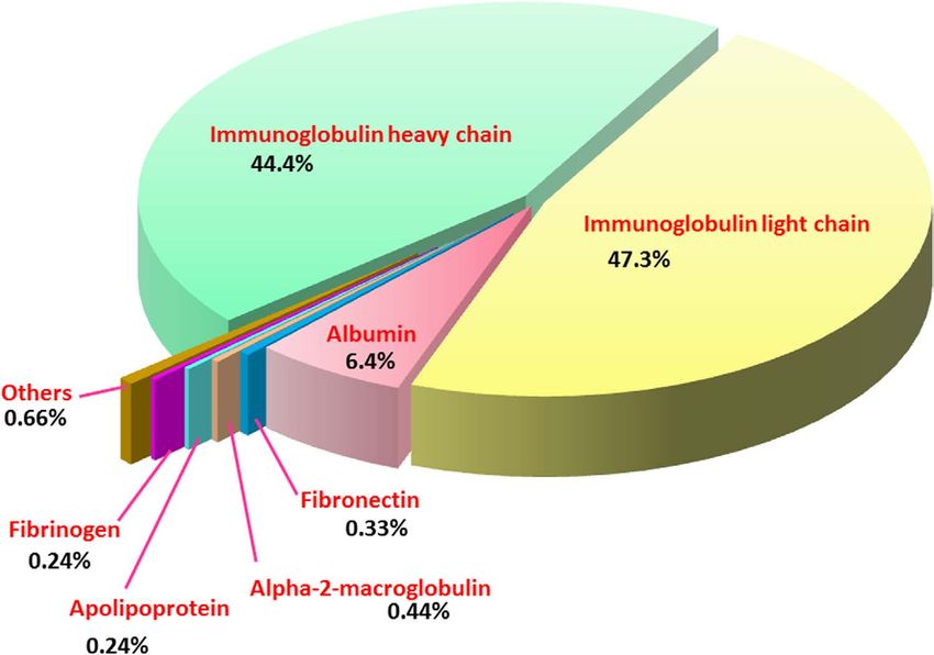
www.nature.com/scientificreports/
Figure 1. Profiling of Serum Anti Bisa Ular (SABU), the Indonesian tri-specific antivenom. (a) SDS-PAGE
of SABU under non-reducing (NR) and reducing (R) conditions. M: Molecular mass standard. (b) Size-exclusion
FPLC of SABU (flow rate = 0.5 ml/min). Inlet shows the protein abundance estimated by peak areas under the
curve.
High Molecular mass proteins Molecular Major peak proteins Molecular weight Moderate/Low Molecular mass proteins
weight > 150 kDa ~100–120 kDa Molecular weight < 60 kDa
Protein Percentage Protein Percentage Protein Percentage
Immunoglobulin chains 5.73 Immunoglobulin chains 84.04 Serum albumin 5.47
Serum albumin 0.71 Serum albumin 0.21 Immunoglobulin chains 1.96
Alpha-2-macroglobulin 0.28 Fibrinogen chain 0.21 Alpha-2-macroglobulin 0.17
Apolipoprotein-A 0.21 Apolipoprotein-A 0.03 Fibronectin 0.32
Alpha-1-antiproteinase 0.16 Serotransferrin 0.11
Haptoglobin 0.1 Plasminogen 0.10
Serotransferrin 0.07 Complement factor B 0.04
Fibrinogen chain 0.02 Inter-alpha-trypsin inhibitor 0.01
Inter-alpha-trypsin inhibitor 0.004 Kininogen 0.01
Antithrombin-III 0.003 Fibrinogen chain 0.004
Attractin 0.003 Carboxypeptidase 0.003
Total 7.27 Total 84.50 Total 8.2
Table 1. Composition of Indonesian antivenom SABU (Serum Anti Bisa Ular) according to molecular
mass as separated by size-exclusion fast protein liquid chromatography.
dose that prolonged the onset of clotting to 3 times of that induced by 2 MCD was determined to be 0.4 ± 0.0 μl
antivenom, or 2.65 mg venom per ml of the antivenom (SABU and HPAV). For consistency, the effective doses
Scientific Reports | 6:37299 | DOI: 10.1038/srep37299 3www.nature.com/scientificreports/
Figure 2. Proteome of the Indonesian tri-specific antivenom, Serum Anti Bisa Ular (SABU).
Figure 3. Immunological binding activities of antivenoms toward (a) Naja sputatrix; (b) Bungarus fasciatus;
(c) Calloselasma rhodostoma; (d) Naja sumatrana; (e) Bungarus candidus. Values were percentages of
normalized mean absorbance ± S.E.M. *Significant difference, pwww.nature.com/scientificreports/
i.v. LD50 ED50 ER50 P Normalized P ED50 ER50 P Normalized P
Venom (μg/g) Challenge (μl) (mg/ml) (mg/ml) (mg/g) Challenge (μl) (mg/ml) (mg/ml) (mg/g)
Homologous to SABU SABU (Indonesian tri-specific antivenom) NPAV (Thai Neuro Polyvalent Antivenom)
0% survival of mice (n = 4) at maximum antivenom dose 25% survival of mice (n = 4) at maximum antivenom
5 LD50 5 LD50
0.90a of 200 μl dose of 200 μl
Naja sputatrix (Java)
(0.59–1.36) 0.51 1.04
2.5 LD50 111.25 0.30 2.9 2.5 LD50 50.00 0.62 8.3
(0.33–0.76) (0.68–1.56)
Bungarus fasciatus 0.45 1.10 1.08
5 LD50 44.94 0.88 8.5 5 LD50 43.75 0.86 11.5
(Java) (0.30–0.68) (0.73–1.66) (0.72–1.63)
HPAV (Thai Hemato Polyvalent Antivenom)
Calloselasma 1.35 15.83
5 LD50 9.38 12.67 121.8 7.92
rhodostoma (Java) (0.78–2.06) (9.15–24.16) 5 LD50 18.75 6.34 147.5
(4.58–12.09)
Heterologous to SABU SABU (Indonesian tri-specific antivenom) NPAV (Thai Neuro Polyvalent Antivenom)
Naja sumatrana 0.39 0.24 0.67
5 LD50 156.57 0.19 1.8 5 LD50 55.63 0.53 7.1
(Sumatra) (0.32–0.48) (0.19–0.29) (0.55–0.82)
Bungarus candidus 0.11a 0.12 2.18
5 LD50 111.25 0.10 1.0 5 LD50 5.56 1.74 23.2
(Java) (0.07–0.17) (0.0–0.19) (1.39–3.36)
Table 2. Neutralization of the lethal effect of five Indonesian snake venoms by different polyvalent
antivenoms. SABU: Serum Anti Bisa Ular; NPAV: Neuro Polyvalent Antivenom; i.v.: intravenous; LD50:
median lethal dose; ED50: antivenom dose (μl) at which 50% of mice survived; ER50: antivenom dose (mg/ml) at
which 50% of mice survived; P: potency expressed as the amount of venom neutralized by one ml antivenom;
Normalized P was defined as the amount of venom (mg) neutralized per amount of antivenom protein (g). aThe
two LD50 values were based on Leong et al.17 conducted in the same laboratory using the same venom stock.
the size-exclusion FPLC of SABU, where the elution time and the calibrated molecular mass of the major peak is
consistent with that of F(ab’)2 reported previously8. Importantly, in this study, the presence of the heavy and light
chains of immunoglobulins in the major peak was validated through a proteomic approach using LCMS/MS.
F(ab’)2 is a pepsin-digested product of immunoglobulin G; it exists as a bivalent molecule of two covalently
bonded antigen-binding fragments (Fab), each of which is composed of a heavy chain and a light chain. The Fab
molecule carries the paratopes for antigen binding13. Currently, this is the main form of antivenom immunoglob-
ulin produced by most antivenom manufacturers, preferred over the IgG form which contains the allergenic Fc
fragment14. F(ab)’2 antivenoms generally have a long elimination half-life (up to 72 hours) and a reasonably good
volume of distribution, thereby reducing the likelihood of venom-antivenom mismatch and recurrent syndrome
(which is more common with monomeric Fab preparation) when given in adequate doses13,15. In addition, F(ab’)2
is large enough to be excluded from glomerular filtration, hence they do not cause obstructive tubulopathy as seen
with some Fab preparations13.
The presence of immunoglobulin chains and some minor serum proteins e.g., macroglobulin in Peaks 1–3
(>200 kDa, Fig. 1b) indicated the presence of dimers and protein aggregates in SABU. These are proteins that can
increase the risk of pyrogenic and hypersensitive reactions in patients treated with antivenom16. Protein aggrega-
tion in immunoglobulin products had been shown to be associated with phenol17, a commonly used preservative
in antivenoms including the liquid product SABU. Besides, the commonly used ammonium sulfate precipitation
method to purify IgG/F(ab’)2 is also known to induce aggregate formation14. In comparison, NPAV, the Thai
antivenom with F(ab’)2 formulation, consisted of negligible amount (< 0.1%) of the high molecular mass proteins
(Leong et al.18). The difference could be due to the different purification method (caprylic acid precipitation)
adopted by the Thai producer19, which allows for the production of antivenoms of higher purity and a lower
aggregate content14. Clinical studies have also shown that the incidence of early reactions to Thai antivenoms
(manufactured by Queen Saovabha Memorial Institute) was low20. Unfortunately, the data of hypersensitivity
associated with SABU has not been available in published literature. In addition, compared to the size-exclusion
FPLC profiles of the Thai polyvalent antivenoms8,18, SABU showed an additional chromatographic peak in the
range of 30–60 kDa molecular mass (Peak 5, Fig. 1b), which contained predominantly equine serum albumins.
The presence of contaminant i.e. serum albumins in SABU was probably due to inadequate purification process
to separate IgG from other plasma proteins at the early stage of antivenom production. Equine serum albumin is
a known causative factor of hypersensitivity to horse serum-based antivenom, which can be life-threatening as
patients may develop respiratory distress and shock from anaphylactoid reaction14,21. The finding hence indicates
the need for more stringent quality control of SABU purity, where the albumin content should ideally not exceed
1% of the total proteins in accordance with the WHO guideline on antivenom production14. Clinically, despite the
lack of data on adverse reactions associated with SABU, it is expected that patients receiving SABU treatment are
at higher risk of developing hypersensitive reaction. Therefore, proper resuscitation facility and close monitoring
of the patient before and during SABU treatment are warranted.
In this study, the venoms of N. sputatrix, B. fasciatus and C. rhodostoma of Indonesian origin (used as the
immunogen in the production of SABU) were considered as homologous to the antivenom SABU. Calloselasma
rhodostoma is a monotypic terrestrial pit viper; in Indonesia its distribution is mainly restricted to the eastern
Java22. Its venom is known to be procoagulant and hemotoxic, but distinct from many other arboreal pit vipers
of the Trimeresurus complex in terms of toxin antigenicity and neutralization profile23–25. On the other hand,
cobras (Naja sp.) and kraits (Bungarus sp.) in Indonesia consist of two to three distinct species; bites by these
elapids can cause rapid neuromuscular paralysis, respiratory failure and death within minutes to hours1,10. In this
Scientific Reports | 6:37299 | DOI: 10.1038/srep37299 5www.nature.com/scientificreports/
study, venoms of the other medically important cobra (N. sumatrana) and krait (B. candidus) were also included
to examine the cross-reactivity and cross-neutralization capacity of SABU against them. Notably, the intrave-
nous median lethal doses (LD50) of the venoms of the two spitting cobras vary in mice: N. sumatrana venom is
twice more lethal than N. sputatrix venom. This is in agreement with the higher content of alpha-neurotoxins in
N. sumatrana venom compared to N. sputatrix venom, which has a much higher content of cytotoxins26. The LD50
value of cobra cytotoxin is about 10-fold higher (hence less lethal) than the alpha-neurotoxin when administered
intravenously9,27,28. On the other hand, krait venoms are known to exhibit high lethality (LD50www.nature.com/scientificreports/
of SABU in Indonesia. The inadequate efficacy of SABU could be due to the presence of other “junk proteins” and
non-specific F(ab’)2 that do not react with the toxin antigens concerned. On the other hand, while antivenom neu-
tralization against cobra and krait toxins (especially the low molecular mass alpha-neurotoxins) is known to be
limited9,28, such “weakness” of antivenom is in particular serious for SABU, judging from its feeble neutralization
against the venoms of N. sputatrix and B. candidus, two medically important species in Java - the most densely
inhabited island of Indonesia. From the practical standpoint, the findings indicate the urgency for a revised man-
ufacturing process to ensure the production of a safe and effective antivenom in the country, where it should be
furnished with the qualities of improved purity, higher potency and a wider coverage of species in Indonesia.
Materials and Methods
Venoms and antivenoms. The venoms of Naja sputatrix (Java) and Calloselasma rhodostoma (Java) ven-
oms were supplied by Latoxan, France. Bungarus candidus (Java) was purchased from Venom Supplies, Australia.
Naja sumatrana (Sumatra) and Bungarus fasciatus (Java) venoms were obtained from local supplier in Indonesia.
Venoms were pooled from a minimum of 2 (Naja sumatrana) to > 5 adult specimens, and stored as lyophilized
samples at −20 °C until use.
Three polyvalent antivenoms were used in this study: (a) Serum Anti Bisa Ular (SABU; batch no. 4701314;
expiry date: October, 2016; manufactured by BioFarma Pharmaceuticals, Bandung, Indonesia), a liquid form
product with an unknown form of its active molecule. This antivenom was obtained from the sera of horses
hyperimmunized against three venoms of Indonesian origin: Naja sputatrix (Javan spitting cobra), Bungarus fas-
ciatus (banded krait) and Calloselasma rhodostoma (Malayan pit viper); (b) Neuro Polyvalent Snake Antivenom
(NPAV; batch no. NP00414; expiry date: December 9th, 2019), a lyophilized product containing purified F(ab)’2
derived from the sera of horses hyperimmunized against the venoms of Naja kaouthia (monocled cobra),
Ophiophagus hannah (king cobra), Bungarus candidus (Malayan krait) and Bungarus fasciatus (banded krait),
all of Thai origin; (c) Hemato Polyvalent Snake Antivenom (HPAV, ; batch no. HP00216; expiry date: March 8th,
2021), a lyophilized product containing purified F(ab)’2 obtained from the sera of horses hyperimmunized against
the venoms of Calloselasma rhodostoma (Malayan pit viper), Cryptelytrops albolabris (white-lipped pit viper)
and Daboia siamensis (Russell’s viper), all of Thai origin. Both HPAV and NPAV were manufactured by Queen
Saovabha Memorial Institute, Bangkok, Thailand, and were used in the immunological binding and neutraliza-
tion assays for comparison purpose in this study.
Animals. Albino ICR Mice (20–25 g) were supplied by the Laboratory Animal Centre, Faculty of Medicine,
University of Malaya. The protocol of animal studies was based on the Council for International Organizations of
Medical Sciences (CIOMS) guidelines on animal experimentation and was approved by the Institutional Animal
Care and Use Committee of the University of Malaya (Ethics clearance number: 2014–09–11/PHAR/R/TCH).
Materials. Ammonium bicarbonate, dithiothreitol (DTT) and iodoacetamide were purchased from
™
Sigma-Aldrich (USA). Mass-spectrometry grade trypsin protease, Spectra Multicolor Broad Range Protein
Ladder (10 to 260 kDa), and HPLC grade solvents used in the studies were purchased from Thermo Scientific ™
™ ®
Pierce (USA). Millipore ZipTip C18 Pipette Tips were obtained from Merck, USA. Other chemicals and rea-
gents of analytical grade were purchased from Sigma-Aldrich (USA).
Estimation of antivenom protein concentration. Protein concentrations in antivenoms (SABU, NPAV
and HPAV) were determined using Thermo ScientificTM PierceTM BCA (bicinchoninic acid) Protein Assay Kit.
The protein concentrations were expressed as means ± S.E.M. of triplicates.
Electrophoretic and chromatographic profiling of SABU antivenom. Sodium dodecyl sulfate pol-
yacrylamide gel electrophoresis (SDS-PAGE) of SABU was conducted under reducing and non-reducing con-
ditions using the MiniPROTEAN II Electrophoresis Cell (BioRad, USA) as adapted from Studier34. Bio-Rad
broad-range prestained SDS-PAGE standards (6.5–200 kDa) and 15 μL SABU (3 mg/ml) were loaded onto 12.5%
gel and the electroporesis running condition was standardized as per previously reported35. Fast protein liquid
chromatography (FPLC) of the antivenom (approximated to 1 mg in 100 μL injection volume) was performed
using a Superdex 200 HR 10/30, 13 μm SEC 10 × 300 mm (GE Healthcare, Sweden). Elution buffer was 100 mM
sodium phosphate, 0.15 M NaCl, pH 7.4 at a flow rate of 0.5 ml/min. Proteins in the antivenom were detected by
absorbance readings at 280 nm for 30 min. The column was calibrated using the following protein standards sup-
plied by Bio-Rad (Bio-Rad Gel filtration Standard): thyroglobulin (670 kDa), α-globulin (158 kDa), ovalbumin
(44 kDa) and myoglobin (17 kDa).
Profiling of proteins using liquid chromatography-tandem mass spectrometry. Proteins in the
chromatographic fractions were subjected to reduction with DTT, alkylation with iodoacetamide, and in-solution
digestion with mass-spectrometry grade trypsin protease according to the manufacturer’s protocol (Thermo
™ ™
Scientific Pierce , Rockford, IL, USA). The trypsin-digested peptides were desalted with Millipore ZipTip
C18 Pipette Tips (Merck, USA) according to the manufacturer’s protocol to enhance the performance of mass
®
spectrometry. The digested peptides (0.5 μL) and 0.5 μL α-cyano-4-hydroxycinnamic acid matrix were mixed
™
and spotted on OPTI-TOF LC/MALDI insert plate (123 mm × 81 mm). MALDI-TOF/TOF was performed
using AB SCIEX 5800 Plus Analyzer equipped with a neodymium: yttrium-aluminium-garnet laser (laser wave-
length was 355 nm). The TOF/TOF calibration mixtures (AB SCIEX, USA) were used to calibrate the spectrum
to a mass tolerance within 10 ppm. For MS mode, peptides mass maps were acquired in positive reflection mode
and 800–4000 m/z mass range were used with 100 laser shots per spectrum. The MS/MS peak detection criteria
used were a minimum signal-to-noise (S/N) of 100. The raw mass spectra acquired were exported to AB SCIEX
™
ProteinPilot Software search against all non-redundant NCBI Serpentes database (taxid: 8570, Serpentes). MS
Scientific Reports | 6:37299 | DOI: 10.1038/srep37299 7www.nature.com/scientificreports/
peak filter mass range 800–4000 m/z was applied. Precursor and fragment mass tolerances were set to 100 ppm
and 0.2 Da respectively and allowing one missed cleavage. Oxidation (M) was set as a variable modification and
carbamidomethylation (C) was set as a fixed modification. The protein score intervals (C.I.) above 95% were
considered as confident identification. The peptide finger mapping was modified to specifically search against
non-redundant NCBI database. From the LCMS/MS, protein identifications were validated with the following
filters: protein score > 11, peptides score > 6 and scored peak intensity (SPI) > 60%. Only results with “Distinct
Peptide” identification of 2 or greater than 2 are considered significant. Estimation of relative protein abundance
through LCMS/MS technique was adapted from Aird et al.36 and Tan et al.37 based on the ratio of mean ion
spectral intensity (MSI) of a protein to the total spectral intensity of all proteins in the chromatographic fraction.
Immunological binding assay. Immunological binding activities between venom antigens and antiven-
oms were examined with an indirect enzyme-linked immunosorbent assay (ELISA) modified from Tan et al.11,38.
In brief, immunoplate wells were each precoated overnight with 10 ng of different venoms (N. sputatrix, B. fascia-
tus, C. rhodostoma, N. sumatrana, B. candidus) at 4 °C. The plate was then flicked dry and rinsed four times with
®
phosphate-buffered saline containing 0.5% Tween 20 (PBST). Antivenoms were prepared at a protein concentra-
tion of 40 mg/ml each, and 100 μl of appropriately diluted antivenom (1:6000) was added to each venom-coated
well, followed by incubation for 1 h at room temperature. After washing the plate four times with PBST, 100 μl
of appropriately diluted horseradish peroxidase-conjugated anti-horse-IgG (Jackson ImmunoResearch Inc.,
USA) in PBST (1:8000) was added to the well and incubated for another hour at room temperature. The excess
components were removed by washing four times with PBST. A hundred microliters of freshly prepared sub-
strate solution (0.5 mg/mL o-phenylenediamine and 0.003% hydrogen peroxide in 0.1 M citrate-phosphate
buffer, pH 5.0) was then added to each well. The enzymatic reaction was allowed to take place in the dark for
30 min at room temperature. The reaction was subsequently terminated by adding 50 μl of 12.5% sulfuric acid,
and the absorbance at 492 nm was read against the blank using an ELISA reader (SUNRISE-TECAN Type Touch
Screen F039300, Switzerland). Immunological binding activity was expressed as percentage of relative absorb-
ance between two comparing specific or paraspecific antivenoms and one non-specific antivenom (providing
basal value for non-specific binding) toward the respective venoms. Values were means ± S.E.M. of triplicate
experiments, and the difference between two comparing antivenoms was analyzed using unpaired t-test with
p valuewww.nature.com/scientificreports/
4. Harrison, R. A., Hargreaves, A., Wagstaff, S. C., Faragher, B. & Lalloo, D. G. Snake envenoming: a disease of poverty. PLoS Negl Trop
Dis 3, e569, doi: 10.1371/journal.pntd.0000569 (2009).
5. Warrell, D. A., Gutierrez, J. M., Calvete, J. J. & Williams, D. New approaches & technologies of venomics to meet the challenge of
human envenoming by snakebites in India. Indian J Med Res 138, 38–59 (2013).
6. Bagcchi, S. Experts call for snakebite to be re-established as a neglected tropical disease. BMJ 351, h5313, doi: 10.1136/bmj.h5313
(2015).
7. Williams, D. J. et al. Ending the drought: new strategies for improving the flow of affordable, effective antivenoms in Asia and Africa.
J Proteomics 74, 1735–1767, doi: 10.1016/j.jprot.2011.05.027 (2011).
8. Leong, P. K. et al. Cross neutralization of common Southeast Asian viperid venoms by a Thai polyvalent snake antivenom (Hemato
Polyvalent Snake Antivenom). Acta Trop 132, 7–14, doi: 10.1016/j.actatropica.2013.12.015 (2014).
9. Leong, P. K., Fung, S. Y., Tan, C. H., Sim, S. M. & Tan, N. H. Immunological cross-reactivity and neutralization of the principal toxins
of Naja sumatrana and related cobra venoms by a Thai polyvalent antivenom (Neuro Polyvalent Snake Antivenom). Acta Trop 149,
86–93, doi: 10.1016/j.actatropica.2015.05.020 (2015).
10. Tan, K. Y., Tan, C. H., Fung, S. Y. & Tan, N. H. Venomics, lethality and neutralization of Naja kaouthia (monocled cobra) venoms
from three different geographical regions of Southeast Asia. J Proteomics 120, 105–125, doi: 10.1016/j.jprot.2015.02.012 (2015).
11. Tan, C. H., Tan, N. H., Tan, K. Y. & Kwong, K. O. Antivenom cross-neutralization of the venoms of Hydrophis schistosus and
Hydrophis curtus, two common sea snakes in Malaysian waters. Toxins (Basel) 7, 572–581, doi: 10.3390/toxins7020572 (2015).
12. Vargas, M. et al. Preclinical evaluation of caprylic acid-fractionated IgG antivenom for the treatment of Taipan (Oxyuranus
scutellatus) envenoming in Papua New Guinea. PLoS Negl Trop Dis 5, e1144, doi: 10.1371/journal.pntd.0001144 (2011).
13. Gutierrez, J. M., Leon, G. & Lomonte, B. Pharmacokinetic-pharmacodynamic relationships of immunoglobulin therapy for
envenomation. Clin Pharmacokinet 42, 721–741, doi: 10.2165/00003088-200342080-00002 (2003).
14. WHO. WHO guidelines for the production, control and regulation of snake antivenom immunoglobulins. (2010).
15. Seifert, S. A. & Boyer, L. V. Recurrence phenomena after immunoglobulin therapy for snake envenomations: Part 1.
Pharmacokinetics and pharmacodynamics of immunoglobulin antivenoms and related antibodies. Ann Emerg Med 37, 189–195
(2001).
16. Leon, G. et al. Pathogenic mechanisms underlying adverse reactions induced by intravenous administration of snake antivenoms.
Toxicon 76, 63–76, doi: 10.1016/j.toxicon.2013.09.010 (2013).
17. Garcia, M. et al. Effect of preservatives on IgG aggregation, complement-activating effect and hypotensive activity of horse
polyvalent antivenom used in snakebite envenomation. Biologicals 30, 143–151 (2002).
18. Leong, P. K. et al. Cross neutralization of Afro-Asian cobra and Asian krait venoms by a Thai polyvalent snake antivenom (Neuro
Polyvalent Snake Antivenom). PLoS Negl Trop Dis 6, e1672, doi: 10.1371/journal.pntd.0001672 (2012).
19. Tan, C. H. et al. Cross neutralization of Hypnale hypnale (hump-nosed pit viper) venom by polyvalent and monovalent Malayan pit
viper antivenoms in vitro and in a rodent model. Acta Trop 117, 119–124, doi: 10.1016/j.actatropica.2010.11.001 (2011).
20. Thiansookon, A. & Rojnuckarin, P. Low incidence of early reactions to horse-derived F(ab’)(2) antivenom for snakebites in Thailand.
Acta Trop 105, 203–205, doi: 10.1016/j.actatropica.2007.09.007 (2008).
21. Malasit, P. et al. Prediction, prevention, and mechanism of early (anaphylactic) antivenom reactions in victims of snake bites. BMJ
292, 17–20 (1986).
22. Marlon, R. Panduan Visual dan Identifikasi Lapangan. 107+ Ular Indonesia. 1st edn, (Indonesia Nature & Wildlife Publishing, 2014).
23. Tan, C. H., Tan, N. H., Sim, S. M., Fung, S. Y. & Gnanathasan, C. A. Immunological properties of Hypnale hypnale (hump-nosed pit
viper) venom: antibody production with diagnostic and therapeutic potentials. Acta Trop 122, 267–275, doi: 10.1016/j.
actatropica.2012.01.016 (2012).
24. Tan, N. H. Isolation and characterization of the thrombin-like enzyme from Cryptelytrops purpureomaculatus venom. Comp
Biochem Physiol C Toxicol Pharmacol 151, 131–136, doi: 10.1016/j.cbpc.2009.09.002 (2010).
25. Tang, E. L., Tan, C. H., Fung, S. Y. & Tan, N. H. Venomics of Calloselasma rhodostoma, the Malayan pit viper: A complex toxin
arsenal unraveled. J Proteomics 148, 44–56, doi: 10.1016/j.jprot.2016.07.006 (2016).
26. Yap, M., Tan, N. & Fung, S. Biochemical and toxinological characterization of Naja sumatrana (Equatorial spitting cobra) venom.
J Venom Anim Toxins incl Trop Dis 17, 451–459 (2011).
27. Tan, K. Y., Tan, C. H., Fung, S. Y. & Tan, N. H. Neutralization of the Principal Toxins from the Venoms of Thai Naja kaouthia and
Malaysian Hydrophis schistosus: Insights into Toxin-Specific Neutralization by Two Different Antivenoms. Toxins (Basel) 8 (2016).
28. Wong, K. Y., Tan, C. H. & Tan, N. H. Venom and Purified Toxins of the Spectacled Cobra (Naja naja) from Pakistan: Insights into
Toxicity and Antivenom Neutralization. Am J Trop Med Hyg 94, 1392–1399, doi: 10.4269/ajtmh.15-0871 (2016).
29. Tan, C. H. & Tan, N. H. In Snake Venoms Toxinology (eds Gopalakrishnakone, P. et al.) Ch. 13-1, 1–37 (Springer Netherlands, 2015).
30. Tan, C. H., Tan, K. Y., Fung, S. Y. & Tan, N. H. Venom-gland transcriptome and venom proteome of the Malaysian king cobra
(Ophiophagus hannah). BMC Genomics 16, 687, doi: 10.1186/s12864-015-1828-2 (2015).
31. Tan, K. Y., Tan, C. H., Fung, S. Y. & Tan, N. H. Venomics, lethality and neutralization of Naja kaouthia (monocled cobra) venoms
from three different geographical regions of Southeast Asia. J Proteomics 120, 105–125 (2015).
32. Huang, H. W. et al. Cobra venom proteome and glycome determined from individual snakes of Naja atra reveal medically important
dynamic range and systematic geographic variation. J Proteomics 128, 92–104, doi: 10.1016/j.jprot.2015.07.015 (2015).
33. Sintiprungrat, K. et al. A comparative study of venomics of Naja naja from India and Sri Lanka, clinical manifestations and
antivenomics of an Indian polyspecific antivenom. J Proteomics, doi: 10.1016/j.jprot.2015.10.007 (2015).
34. Studier, F. W. Analysis of bacteriophage T7 early RNAs and proteins on slab gels. J Mol Biol 79, 237–248 (1973).
35. Tan, C. H., Tan, K. Y., Lim, S. E. & Tan, N. H. Venomics of the beaked sea snake, Hydrophis schistosus: A minimalist toxin arsenal and
its cross-neutralization by heterologous antivenoms. J Proteomics 126, 121–130, doi: 10.1016/j.jprot.2015.05.035 (2015).
36. Aird, S. D. et al. Quantitative high-throughput profiling of snake venom gland transcriptomes and proteomes (Ovophis okinavensis
and Protobothrops flavoviridis). BMC Genomics 14, 790, doi: 10.1186/1471-2164-14-790 (2013).
37. Tan, C. H. et al. Unveiling the elusive and exotic: Venomics of the Malayan blue coral snake (Calliophis bivirgata flaviceps).
J Proteomics 132, 1–12, doi: 10.1016/j.jprot.2015.11.014 (2016).
38. Tan, C. H., Liew, J. L., Tan, K. Y. & Tan, N. H. Genus Calliophis of Asiatic coral snakes: A deficiency of venom cross-reactivity and
neutralization against seven regional elapid antivenoms. Toxicon 121, 130–133, doi: 10.1016/j.toxicon.2016.09.003 (2016).
39. Tan, N. H. et al. Functional venomics of the Sri Lankan Russell’s viper (Daboia russelii) and its toxinological correlations.
J Proteomics 128, 403–423, doi: 10.1016/j.jprot.2015.08.017 (2015).
40. O’Leary, M. A. & Isbister, G. K. A turbidimetric assay for the measurement of clotting times of procoagulant venoms in plasma.
J Pharmacol Toxicol Methods 61, 27–31, doi: 10.1016/j.vascn.2009.06.004 (2010).
41. Howard-Jones, N. A CIOMS ethical code for animal experimentation. WHO Chron 39, 51–56 (1985).
42. Finney, D. J. Probit Analysis. (Cambridge University Press, 1952).
43. Morais, V., Ifran, S., Berasain, P. & Massaldi, H. Antivenoms: potency or median effective dose, which to use? Journal of Venomous
Animals and Toxins including Tropical Diseases 16, 191–193 (2010).
Scientific Reports | 6:37299 | DOI: 10.1038/srep37299 9www.nature.com/scientificreports/
Acknowledgements
This work was supported by Fundamental Research Grant Scheme from the Ministry of Higher Education
Malaysia (FP028-2014A) and UMRG (grant number: RG352-15AFR) from the University of Malaya.
Author Contributions
C.H.T. and N.H.T. conceived and designed the study. J.L.L. and K.Y.T. performed the experiments. All authors
analyzed the data. C.H.T. wrote the main manuscript text. All authors reviewed the manuscript.
Additional Information
Supplementary information accompanies this paper at http://www.nature.com/srep
Competing financial interests: The authors declare no competing financial interests.
How to cite this article: Tan, C. H. et al. Assessing SABU (Serum Anti Bisa Ular), the sole Indonesian
antivenom: A proteomic analysis and neutralization efficacy study. Sci. Rep. 6, 37299; doi: 10.1038/srep37299
(2016).
Publisher's note: Springer Nature remains neutral with regard to jurisdictional claims in published maps and
institutional affiliations.
This work is licensed under a Creative Commons Attribution 4.0 International License. The images
or other third party material in this article are included in the article’s Creative Commons license,
unless indicated otherwise in the credit line; if the material is not included under the Creative Commons license,
users will need to obtain permission from the license holder to reproduce the material. To view a copy of this
license, visit http://creativecommons.org/licenses/by/4.0/
© The Author(s) 2016
Scientific Reports | 6:37299 | DOI: 10.1038/srep37299 10You can also read