Hepatic connective tissue growth factor expression and regulation differ between non steatotic and non alcoholic steatotic livers from brain dead ...
←
→
Page content transcription
If your browser does not render page correctly, please read the page content below
www.nature.com/scientificreports
OPEN Hepatic connective tissue growth
factor expression and regulation
differ between non‑steatotic
and non‑alcoholic steatotic livers
from brain‑dead donor
Dong‑Jing Yang1,4, Ji‑Hua Shi1,4, Zong‑Ping Xia2, Wen‑Zhi Guo1, Mohammed Shakil Ahmed3 &
Shui‑Jun Zhang1*
Accurate evaluation of liver steatosis is required from brain-dead donors (BDDs) with nonalcoholic
fatty liver disease (NAFLD). Our purposes were to investigate expression and regulation of connective
tissue growth factor (CTGF) expression in livers from human and rat after brain death, and further
evaluate its potential application. NAFLD and brain death models were established in rats. LX2
cells were cultured under hypoxia/reoxygenation. CTGF protein and mRNA levels were measured
in liver samples from BDDs of human and rat by immunohistochemistry and reverse transcription-
quantitative polymerase chain reaction. YAP-regulated CTGF expression was investigated in LX2 cells
via YAP small interfering RNA and Verteporfin treatment. Blood CTGF level from BDDs was measured
by enzyme-linked immunosorbent assay. After brain death, CTGF, transforming growth factor-β
and YAP were overexpressed in non-alcoholic steatotic liver, whereas CTGF was downregulated in
non-steatotic liver. Time-series analysis revealed that CTGF and YAP expression was comparable,
as confirmed by inhibited YAP expression in LX2 cells. CTGF level and NAFLD activity were linearly
correlated. CTGF expression and regulation differ between non-steatosis and nonalcoholic steatosis
livers from BDDs. CTGF may be an important factor to evaluate graft quality from BDDs with NAFLD.
Abbreviations
BDD Brain-dead donor
NAFLD Nonalcoholic fatty liver disease
CTGF Connective tissue growth factor
CCN Cellular Communication Network Factor
TGF-β Transforming Growth Factor Beta
YAP Yes Associated Protein
NASH Non-alcoholic steatohepatitis
ECM Extracellular matrix
HSCs Hepatic stellate cells
α-SMA Alpha smooth muscle actin
Donor shortage and consequent increases in waiting list mortality for liver transplantation have pushed clini-
cians to utilize extended-criteria liver graft from the brain-dead donors, which can lead to a suboptimal allograft
outcome1,2. Nonalcoholic fatty liver disease (NAFLD) is a frequent contributor to suboptimal allografts from
1
Department of Hepatobiliary and Pancreatic Surgery, Henan Key Laboratory of Digestive Organ Transplantation
and Zhengzhou Key Laboratory for HPB Diseases and Organ Transplantation, The First Affiliated Hospital of
Zhengzhou University, Zhengzhou University, Zhengzhou, China. 2Translational Medicine Center, The First
Affiliated Hospital of Zhengzhou University, Zhengzhou University, Zhengzhou, China. 3Institute for Surgical
Research, Department of Cardiology and Center for Heart Failure Research, Oslo University Hospital-Rikshospitalet
and University of Oslo, Oslo, Norway. 4These authors contributed equally: Dong-Jing Yang and Ji-Hua Shi. *email:
zhangshuijun@zzu.edu.cn
Scientific Reports | (2021) 11:3857 | https://doi.org/10.1038/s41598-021-83516-2 1
Vol.:(0123456789)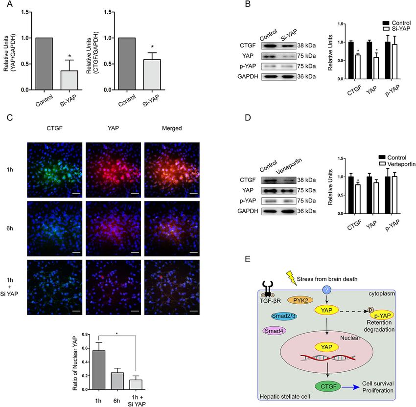
www.nature.com/scientificreports/
extended-criteria donors3. NAFLD represents a spectrum of diseases characterized by the excess accumulation
of triglyceride within the liver, ranging in severity from reversible simple hepatic steatosis to progressive non-
alcoholic steatohepatitis (NASH) and liver cirrhosis. As the prevalence of hepatic steatosis is increasing globally,
it is expected that a higher proportion of potential donors worldwide will have liver steatosis, thus influencing
the quality of grafts4. Hepatic steatosis, inflammation, and fibrosis constitute the major risk factors of impaired
liver graft function and predict liver-related mortality in patients with N AFLD5–7, which requires careful con-
sideration during graft evaluation. Few study has investigated the impact factors and the underlying mechanism
of liver steatosis under acute stress of brain death before organ p rocurement8.
Since NAFLD is usually a silent disease with few or no typical symptoms, accurate evaluation of liver steatosis
in potential donors is required. During liver graft procurement, assessment of steatosis through visual inspec-
tion has a positive predictive value of less than 20% for mild steatosis and only reaches up to 70% for severe
steatosis9. Ultrasonography, computed tomography, and magnetic resonance imaging are useful modalities to
detect NAFLD non-invasively; however, these assessments only provide qualitative information. Liver biopsy
and histopathological changes of the potential liver graft remain the gold standard for a diagnosis of NAFLD
and grading liver disease s everity10. However, liver biopsy is an invasive procedure with potential adverse effects.
Moreover, the prevalence of biopsy-proven NAFLD among potential donors was reported to range from 15 to
53%, which disqualified 3–21% of potential liver g rafts11. The interpretation of NAFLD, and consequently graft
usability, may be erroneous due to the heterogeneous distribution of hepatic steatosis and liver fibrosis. Steatosis
factors may cause cellular injury, inflammation, and fibrosis through oxidative stress, mitochondrial dysfunc-
tion, endoplasmic reticulum stress, iron accumulation, apoptosis, and production of adipocytokines, which have
been selected as clinical diagnosis and scoring biomarkers to assess the steatosis degree and NAFLD a ctivity12,13;
however, these markers have not been validated for graft evaluation.
Connective tissue growth factor (CTGF) expression in hepatic stellate cells (HSCs) and liver endothelial
cells correlates with the extent of acute liver injury, and upregulated CTGF expression was proposed as a cen-
tral pathway contributing to hepatic stellate cells (HSCs) activation during liver chromic inflammation and
fibrosis14,15. Most livers for transplantation are procured from brain-dead donors. The irreversible brain injury
causes hemodynamic instability and hormonal disturbance, which could trigger acute liver injury16 and thus
complicate the pathological process of NAFLD a ctivity17. Specifically, acute inflammation, steatosis, and fibrosis
caused by NAFLD might aggravate the activation of HSCs and lead to CTGF upregulation in the non-alcoholic
fatty liver from the brain-dead d onor14,15,17. The expression and regulation of hepatic CTGF in brain-dead donors
have not yet been studied.
To investigate the overall effect of brain death on intrahepatic gene expression, we applied RNA-sequencing
to investigate hepatic transcript level of non-steatotic liver from brain-dead donors. However, the results indi-
cated that CTGF expression was down-regulated in non-steatotic liver from brain-dead d onors18. Thereby, we
hypothesized that hepatic CTGF expression and its regulation after brain death might be different between non-
steatotic liver and non-alcoholic steatotic liver. To test this hypothesis, we investigated the expression pattern and
regulation mechanism of CTGF in non-steatotic liver and non-alcoholic steatotic liver from brain-dead donors
in human and rats, and explore the potential clinical application of CTGF.
Materials and methods
Animal models and study protocol. The animal experiments in this study conformed to the ARRIVE
(Animal Research: Reporting of In Vivo Experiments) guideline and were approved by the Institutional Animal
Ethics Committee of Zhengzhou University (No. 2019-KY-019). All methods were performed in accordance
with the relevant guidelines and regulations. The male Lewis rats were purchased from the Beijing Vital River
Laboratory Animal Technology Co., Ltd. (Beijing, China). All the 24 rats were housed in laminar flow cabinets
under specific pathogen-free conditions at room temperature with a 12 h light/dark cycle, with food and water
available ad libitum.
The model of NAFLD was established by feeding rats with the high fat diet19. The non-alcoholic steatotic
liver group (n = 10) was fed with a high-fat and high-cholesterol diet that consisted of 85% standard diet, 13%
lard oil and 2% cholesterol for 4 weeks. The non-steatotic liver group (n = 30) was fed with a standard diet before
establishment of brain death.
In the rats from non-alcoholic steatotic and non-steatotic liver groups, brain death was established by increas-
ing intracranial pressure and was further kept over a follow-up period20. To evaluate the dynamic change after
brain death, a number of 5 rats in the standard diet group were euthanized and liver samples were collected at
0 h, 1 h, 2 h, 4 h and 6 h after brain death. Five rats in non-alcoholic steatotic liver group were euthanized and
liver samples were collected before brain death and at 6 h after brain death, respectively (Supplementary Fig. 1).
Human samples. The human study was conducted under ethical approval of the Research Committee of
the Affiliated Hospital of Zhengzhou University (No. 2018-KY-73), and all patients or their family members
provided the informed consent. The human study was performed in accordance with the ethical principles of
Declaration of Helsinki21. All donors were from the intensive care unit of the hospital and were selected follow-
ing the national organ donation protocol22 (Supplementary Table 1 and Supplementary Fig. 1). No organs or
tissues were procured from prisoners. Human liver and blood samples for research purposes were collected from
brain-dead donors at the First Affiliated Hospital of Zhengzhou University.
All the patients in this study were grouped into two cohorts. In cohort 1, a number of 6 brain-dead donors
with non-steatosis and 6 brain-dead donors with non-alcoholic steatosis were enrolled to compare the intrahe-
patic CTGF expression. In cohort 2, 27 brain-dead donors with NAFLD and non-steatosis were used to evaluate
the correlation between CTGF expression and NAFLD activity in livers. The clinic-pathological characteristics
Scientific Reports | (2021) 11:3857 | https://doi.org/10.1038/s41598-021-83516-2 2
Vol:.(1234567890)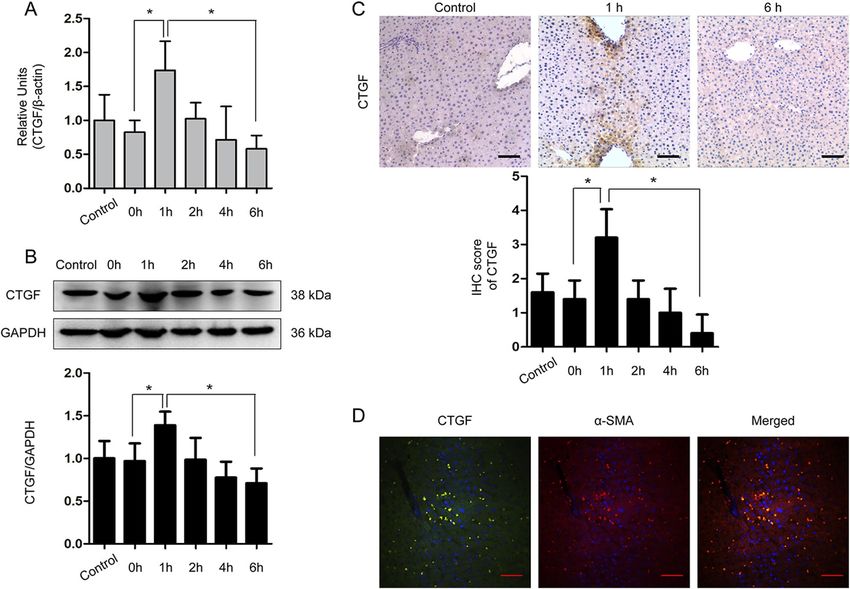
www.nature.com/scientificreports/
were summarized in Supplementary Table 1 and the diagnosis was confirmed from the pathology reports. Alco-
holic fatty liver disease and virus infection were ruled out in the cohorts.
Cell culture and an in vitro hypoxia‑reoxygenation model. Human HSC cell line, LX2, was obtained
from American Type Culture Collection (Manassas, USA) and cultured in DMEM with 1% penicillin/strepto-
mycin and 10% fetal bovine serum at 37 °C. LX2 cells were incubated under hypoxia (1% oxygen, 5% carbon
dioxide and 94% nitrogen) in the hypoxia chamber (Galaxy 48 R, Eppendorf, Hamburg, Germany) for 3 h,
followed by reoxygenation (hypoxia/ reoxygenation) under the normoxic conditions (21% oxygen, 5% carbon
dioxide and 74% nitrogen) for 6 h to mimic activation of HSCs under ischemia–reperfusion i njury23.
To evaluate the dynamic change after hypoxia/reoxygenation, LX2 cells were harvested at 0 h, 1 h, 2 h, 4 h and
6 h after normoxic conditions for RT-qPCR and Western blot analyses (Supplementary Fig. 1).
To investigate the roles of YAP in regulation of CTGF expression, YAP siRNA or YAP function inhibitor
(Verteporfin, 2 μM, s1786, Selleck Chemicals, Houston, USA) was incubated before hypoxia to observe the roles
of YAP in the regulation of CTGF expression LX2 cells.
siRNA transfection. YAP siRNA (5′ to 3′ CCACCAAGCUAGAUAAAGAdTdT) and control (catalog num-
ber AM17110) were produced by GenePharma (Shanghai, China). The transfection into the LX2 cells was car-
ried out with Lipofectamine RNAiMAX Reagent (Cat.13778-075, Invitrogen, Waltham, USA) in Opti-MEM
Medium (Gibco, Life Technologies, Rockville, USA) according to the manufacturer’s protocol.
Gene expression analysis and reverse transcription‑quantitative polymerase chain reaction
(RT‑qPCR). Total RNA was isolated from liver tissue samples or cells using RNAiso reagent (Takara, Shiga,
Japan). Specific primers for RT-qPCR used in the study (Supplementary Table 2) were designed and synthe-
sized by Invitrogen (Waltham, USA). Reverse transcription and RT-qPCR (RR047A and RR840A, Takara, Shiga,
Japan) were performed according to the manufacturer’s protocol.
Western blot. Primary antibodies used were anti-CTGF (1:1000, sc-101586, Santa Cruz, Dallas, USA),
p-YAP (1:1000, #13008, Cell Signaling Technology, Beverly, USA), YAP (1:1000, #14074, Cell Signaling Technol-
ogy, Beverly, USA), Smad2/Smad3 (1:1000, #5678, Cell Signaling Technology, Beverly, USA), p-Smad2/Smad3
(1:1000, #8828, Cell Signaling Technology, Beverly, USA), GAPDH (1:5000, 60004-1-Ig, Proteintech, Rosemont,
USA). Immunoreactivities were visualized by secondary horseradish peroxidase-conjugated rabbit (1:2000,
SA00001-2, Proteintech, Rosemont, USA), mouse (1:2000, SA00001-1, Proteintech, Rosemont, USA).
Hematoxylin–eosin (HE) staining and evaluation of NAFLD activity. Liver tissue were fixed in
4% paraformaldehyde in phosphate-buffered, embedded in paraffin wax, and stored at 4 °C. HE staining was
performed as before24, and degree of steatosis (macrovesicular steatosis) and NAFLD activity were evaluated
by the pathologist. Severity of steatosis was graded as non-steatotic liver donors (steatosis < 5%), mild steatosis
(steatosis 5%-20%), moderate steatosis (21%-40%) and severe steatosis (> 40% and fibrosis). The scoring system
of NAFLD activity was comprised of 4 histological features: steatosis (0–3), lobular inflammation (0–2), hepa-
tocellular ballooning (0–2), and fibrosis (0–4) evaluated semi-quantitatively25. NAFLD activity score of ≥ 5 cor-
responded to a high-activity NAFLD, a score of 3–4 corresponded to a moderate-activity NAFLD, and a score of
less than 3 corresponded to a non/low-activity NAFLD.
Immunohistochemistry (IHC) and immunofluorescence. Primary and secondary antibodies
against CTGF (1:50, sc-101586, Santa Cruz, Dallas, USA), α-SMA (1:100, Ab5694, Abcam, Cambridge, UK),
p-YAP(1:100, Ser127 , #13008, Cell Signaling Technology, Beverly, USA), YAP (1:100, #14074, Cell Signaling
Technology, Beverly, USA), transforming growth factor beta 1 (TGF-β1, 1:50, 21898-1-AP, Proteintech, Rose-
mont, USA), secondary against rabbit IHC kit (1:100; cat. no. SPN-9001; Beijing Zhongshan Golden Bridge Bio-
technology Co., Ltd.), against mouse IHC kit (1:100; cat. no. SPN-9002; Beijing Zhongshan Golden Bridge Bio-
technology Co., Ltd.), anti-Rabbit Cy3-conjugated Affinipure Goat (1:100, SA00009-1, Proteintech, Rosemont,
USA), anti-Mouse 488-conjugated Affinipure Goat (1:100, SA00009-2, Proteintech, Rosemont, USA) were used
in the immunostaining assays. Olympus IX71 microscope (Olympus Corporation,Tokyo, Japan) was used to
observe the slides.
Enzyme‑linked immunosorbent assay (ELISA). Human sera in cohort 2 were obtained, and the CTGF
levels were determined with human CTGF SimpleStep ELISA Kit (ab245716, Abcam, Cambridge, UK) accord-
ing to the manufacturer’s instructions.
Statistical analysis. Values are given as means with standard deviation (SD). Differences in groups were
analyzed by the two-tailed unpaired Student’s test or one-way ANOVA. The association analysis of CTGF expres-
sion with steatosis degree was employed with non-parametric (Spearman) correlation analysis. The statistical
tests were employed by using SPSS version 21.0 (IBM, Armonk, New York, USA), and the results for t test,
ANOVA and Spearman correlation were denoted as t, F and correlation coefficient. A probability level of less
than 5% (P < 0.05) was considered statistically significant.
Scientific Reports | (2021) 11:3857 | https://doi.org/10.1038/s41598-021-83516-2 3
Vol.:(0123456789)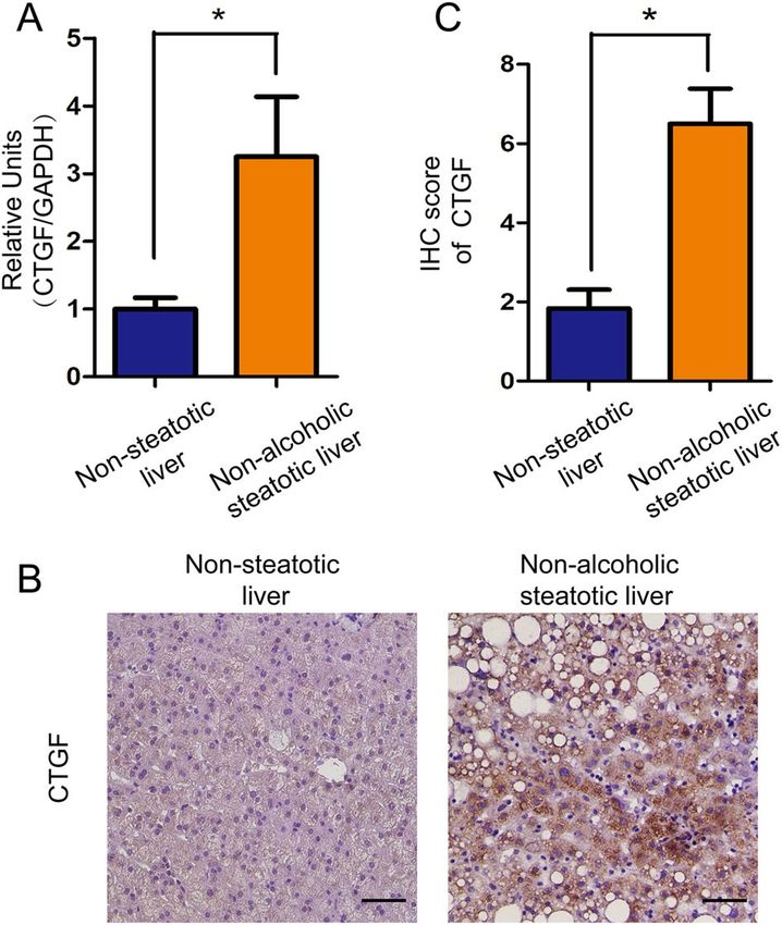
www.nature.com/scientificreports/
Figure 1. Expression of CTGF in the non-steatotic (n = 6) and non-alcoholic steatotic liver (n = 6) after brain
death in humans. (A) CTGF expression was down-regulated in the non-steatotic liver and up-regulated in the
non-alcoholic steatotic liver after brain death as determined by RT-qPCR (*P < 0.05). (B) Expression of CTGF in
the non-steatotic liver and in the non-alcoholic steatotic liver after brain death by IHC (Scale bar = 20 μm, ×200
magnification) and (C) its semi-quantitation (*P < 0.05).
Results
CTGF expression was down‑regulated in the non‑steatotic liver after brain death in
humans. Our previous study indicated that CTGF expression was downregulated in non-steatotic liver from
brain-dead donors by RNA-sequencing (Supplemental Fig. 2 and 3). To compare the expression of CTGF in
non-steatotic and non-alcoholic steatotic liver after brain death, we measured the CTGF in both mRNA and pro-
tein levels in non-steatotic and non-alcoholic steatotic liver from BDD donors. In human cohort 1, intrahepatic
CTGF mRNA and protein expression were significantly down-regulated in the non-steatotic liver, whereas it was
highly up-regulated in the non-alcoholic steatotic liver after brain death as determined by RT-qPCR (t = 2.483,
P = 0.032, Fig. 1A) and IHC (t = 4.641, P = 0.001) (Fig. 1B,C).
Time‑course analysis revealed intrahepatic CTGF expression in the non‑steatotic liver after
brain death. We further explored the dynamic expression of CTGF in the non-steatotic liver from a well-
described rat model of brain death. The CTGF expression significantly increased in the rats (n = 5 for each time
point), reaching the maximum at 1 h after induction of brain death, and then decreased gradually after 2 h until
becoming significantly down-regulated at 6 h after brain death as determined by RT-qPCR (F = 8.105, P = 0.001,
Fig. 2A), WB (F = 7.033, P = 0.001, Fig. 2B), and IHC (F = 10.950, P = 0.001, Fig. 2C).
To investigate the cellular origin of CTGF expression under brain death, hepatic cellular markers were
detected alongside CTGF in tissue samples collected at different time points after brain death induction by
double immunofluorescence. The results demonstrated that CTGF was co-expressed with α-SMA-positive cells
at 1 h after brain death (Fig. 2D), indicating that intrahepatic CTGF was mainly regulated by active HSCs in the
non-steatotic liver after brain death.
HSC undergoes activation and regulates tissue repair and after liver i njury26. Next we sought to determine if
CTGF expression associates with HSC activation. To this end, we cultured human HSC cell line LX2 and treated
the cells with a hypoxia/reoxygenation procedure to mimic the activation of HSCs in the microenvironment
of brain death. We found that CTGF expression was transiently up-regulated at 1 h after reoxygenation, and
then decreased at 2 h after reoxygenation as determined by RT-qPCR (F = 8.686, P = 0.001; Fig. 3A) and WB
(F = 6.913, P = 0.001; Fig. 3B,C), indicating a similar response and regulation of CTGF in HSCs as found in the
brain-dead rat model.
Intrahepatic CTGF expression in the non‑steatotic liver was regulated through YAP under
brain death. CTGF expression in activated HSCs are under regulation of either TGF-β pathway or Hippo/
YAP27,28. To clarify the mechanism of CTGF down-regulation in the non-steatotic liver after brain death, we used
Scientific Reports | (2021) 11:3857 | https://doi.org/10.1038/s41598-021-83516-2 4
Vol:.(1234567890)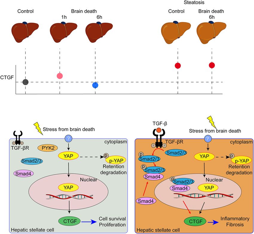
www.nature.com/scientificreports/
Figure 2. Expression dynamics of CTGF in the non-steatotic liver from the rat brain death model (n = 5
for each time point). Dynamic expression of CTGF in the non-steatotic liver from the rat brain death
model determined by (A) RT-qPCR, (B) WB, and (C) IHC (*P < 0.05 compared with 0 h). (D) Double
immunofluorescence of CTGF (green) and α-SMA (red) in the rat liver 1 h after brain death (scale bar = 20 μm,
×200 magnification). The shame-operated rats were regarded as control.
transcriptome analysis from the non-steatotic liver samples of human brain-dead donors18. Kyoto Encyclopedia
of Genes and Genomes (KEGG)29 enrichment analysis indicated that CTGF was correlated with the Hippo/YAP
pathway and not with the TGF-β pathway (Supplementary Fig. 2 and 3) in the non-steatotic liver under brain
death.
As described above, we found that CTGF expression was significantly down-regulated in the non-steatotic
liver under brain death. Thus, we proposed that the down-regulation of CTGF could be resulted from inactivation
of Hippo/YAP pathway in HSCs cells of the non-steatotic liver of BBDs. To assess this posibility, we measured
YAP expression in cultured LX2 cells under hypoxia/reoxygenation treatment. We found that YAP expression
was up-regulated cultured under hypoxia/reoxygenation at 1 h after reoxygenation, and was then subsequently
down-regulated after 2 h, in agreement with the pattern of CTGF expression (F = 23.900, P = 0.001; Fig. 3B,D).
In contrast, the p-YAP expression level decreased at 1 h, and increased at 4 h and 6 h after reoxygenation as
determined by WB (F = 16.820, P = 0.001; Fig. 3B,E). However, the level of p-SMAD2/3 did not increase during
this process (Fig. 3B).
To further verify the underlying regulation mechanism, we used siRNA technology to silence YAP mRNA
in LX2 cells. After knocking down YAP expression, CTGF expression was significantly down-regulated at 1 h
after reoxygenation as determined by RT-qPCR (t = 5.517, P = 0.031; Fig. 4A) and WB (t = 11.776, P = 0.001;
Fig. 4B). Immunofluorescence analysis further showed that the percentage of nuclear YAP was higher at 1 h,
and then decreased at 6 h after reoxygenation (t = 5.317, P = 0.002; Fig. 4C), with a further decrease of nuclear
YAP observed in siRNA-treated LX2 cells at 1 h after reoxygenation (t = 7.243, P = 0.001; Fig. 4C). This effect was
confirmed after treatment with the YAP-specific inhibitor verteporfin under hypoxia/reoxygenation, in which
CTGF expression down-regulated at 1 h after reoxygenation as determined by WB (t = 3.195, P = 0.019; Fig. 4D).
These data suggested that CTGF expression HSCs of non-steotic liver is dependent on Hippo/YAP pathway.
The proposed mechanism of regulation of CTGF in the non-steatotic liver after brain death is schematically
illustrated in Fig. 4E30.
Intrahepatic CTGF expression in the non‑alcoholic steatotic liver after brain death might be
dependent on the TGF‑β pathway. IHC analysis showed that CTGF was highly overexpressed in the
Scientific Reports | (2021) 11:3857 | https://doi.org/10.1038/s41598-021-83516-2 5
Vol.:(0123456789)www.nature.com/scientificreports/
Figure 3. LX2 cells treated with hypoxia for 3 h and reoxygenation for different time with untreated LX2 cells as
control. (A) Dynamics in the expression of CTGF mRNA in LX2 cells under hypoxia/reoxygenation (*P < 0.05).
(B) Dynamic expressions of CTGF, YAP, p-YAP and p-SMAD2/3 in LX2 cells under hypoxia and reoxygenation
culture conditions by WB and their quantifications (C–E) (*P < 0.05).
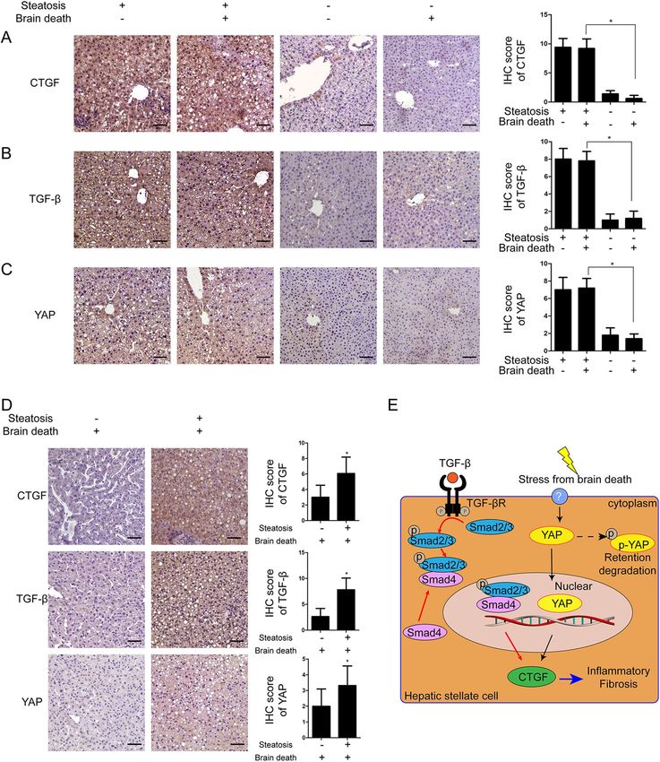
steatotic liver both before and after brain death in rats without a significant difference (n = 5 for each group,
t = 11.103, P = 0.001; Fig. 5A). Furthermore, TGF-β and YAP were also overexpressed in the non-alcoholic stea-
totic liver after brain death in rats (n = 5 for each group, t = 10.707 and 4.333, respectively, P = 0.001 and 0.003;
Fig. 5B,C).
In humans, CTGF, TGF-β and YAP were overexpressed in the non-alcoholic steatotic liver after brain death
as determined by IHC (Fig. 5D). These results supported that overexpression of CTGF in the non-alcoholic
steatotic liver, aggraved by brain death14,15,17, might be mainly mediated through the TGF-β pathway (Fig. 5E)31.
Instead, the expressions of TGF-β in the non-steatotic liver in rats were not significantly changed after brain
death by RT-qPCR (n = 5 for each group, F = 0.380, P = 0.858) (Supplementary Fig. 4) and IHC (n = 5 for each
group, t = 0.408, P = 0.694) (Fig. 5, Supplementary Fig. 4).
CTGF expression was correlated with NALFD activity in the livers of brain‑dead donors. Given
the different expression and regulation patterns of CTGF between the non-steatotic and non-alcoholic stea-
totic liver in rats under brain death, we further explored whether CTGF expression might predict the degree
of NAFLD activity under brain death using samples from human brain-dead donors in cohort 2. The serum
CTGF concentrations determined by ELISA and intrahepatic CTGF expression levels determined by IHC with
semi-quantitative immunoreactivity scores were positively correlated (Spearman correlation coefficients = 0.442,
P = 0.021; Fig. 6A). In addition, the serum CTGF concentration was positively correlated with the NAFLD activ-
ity score (Spearman correlation coefficients = 0.499, P = 0.008).
The concentrations of CTGF in cases of moderate (NAFLD score of 3–4) and high (NAFLD score ≥ 5) NAFLD
activity (n = 16) were higher than those in cases with no or low NAFLD activity (NAFLD score < 3) (n = 11)
(t = 3.971, P = 0.001; Fig. 6B). The area under the receiving operator characteristic curve for the prediction of
moderate/high-activity NAFLD (NAFLD score ≥ 3) was 0.903 (95% confidence interval 0.791–1.000). A cut-off
value of 106.93 μg/L, with a sensitivity of 87.5% and a specificity of 81.8%, had a positive predictive value of
87.5% and negative predictive value of 81.8%.
Discussion
The severity of steatosis of a liver graft is typically evaluated according to histopathological changes from liver
biopsy during graft p rocurement10. Liver grafts with mild steatosis can be safely used for liver transplantation;
however, livers with severe (> 60%) steatosis and NASH are usually discarded owing to the increased risk of pri-
mary non-function after transplantation, and those with moderate (40–60%) steatosis have also been associated
with decreased graft function and graft survival if transplanted10. Thus, identification of a non-invasive biomarker
Scientific Reports | (2021) 11:3857 | https://doi.org/10.1038/s41598-021-83516-2 6
Vol:.(1234567890)www.nature.com/scientificreports/
Figure 4. Regulation of CTGF through YAP in LX2 cells treated with Si-RNA or YAP inhibitor Verteporfin
under hypoxia for 3 h and reoxygenation for 1 h. LX2 cells treated with vehicle of Si-RNA or Verteporfin were
regarded as control. (A) Changes of YAP and CTGF mRNAs in LX2 cells after YAP-siRNA treatment under
hypoxia/reoxygenation determined by RT-qPCR (*P < 0.05 between siRNA and control). (B) Changes of CTGF,
YAP, and p-YAP protein levels after YAP-siRNA treatment in LX2 cells determined by WB (*P < 0.05 between
siRNA and control). (C) Immunofluorescence staining of CTGF (green) and YAP (red) in LX2 cells under
hypoxia/reoxygenation; DAPI (blue) was used for nuclear staining (scale bar = 20 μm, ×200 magnification) and
semi-quantitation of nuclear YAP (*P < 0.05). (D) Changes of CTGF, YAP, and p-YAP expression in LX2 cells
after treatment of the YAP inhibitor Verteporfin under hypoxia/reoxygenation (*P < 0.05). (E) Schematic of the
proposed mechanism of CTGF regulation in the non-steatotic liver after brain death.
that can provide reliably good accuracy to predict the steatosis degree, NAFLD activity, and inflammation-related
graft injury is urgently needed for improving liver graft assessment from a brain-dead d onor5,6.
18
The findings from our previous study and the current study demonstrated that CTGF expression after
brain death was down-regulated in the non-steatotic liver but was overexpressed in the non-alcoholic steatotic
liver of rats and humans. The clinical diagnosis of brain death requires reconfirmation at 6–12 h after the first
diagnosis. The acute-phase response during this period may provide biological information to elucidate the
regulation mechanism of potential biomarkers, which have thus far been neglected. Using the rat model of brain
death, we explored the dynamic expression of CTGF in the non-steatotic liver after the induction of brain death.
CTGF levels significantly increased initially after brain death, reaching the maximum at 1 h, and then gradually
Scientific Reports | (2021) 11:3857 | https://doi.org/10.1038/s41598-021-83516-2 7
Vol.:(0123456789)www.nature.com/scientificreports/
Figure 5. Expression of CTGF, YAP and TGF-β in non-steatotic liver and non-alcoholic steatotic liver under
brain death. Expression of (A) CTGF, (B) TGF-β, and (C) YAP in the non-steatotic liver and non-alcoholic
steatotic liver before and after brain dead in rats; n = 5 per group, scale bar = 20 μm, ×200 magnification
(*P < 0.05 between the non-steatotic liver and non-alcoholic steatotic liver after brain death). (D) Expression
of CTGF, TGF-βand YAP in human non-steatotic and non-alcoholic steatotic liver tissues (scale bar = 20 μm,
×200 magnification, n = 6). (E) Schematic of the proposed mechanism of CTGF regulation in the non-alcoholic
steatotic liver after brain death.
Scientific Reports | (2021) 11:3857 | https://doi.org/10.1038/s41598-021-83516-2 8
Vol:.(1234567890)www.nature.com/scientificreports/
Figure 6. Correlation between CTGF expression and NAFLD activity in humans. (A) Correlation between
systemic CTGF levels and semi-quantitative immunoreactivity scores of intrahepatic CTGF in cohort 2 (n = 27).
(B) Systemic CTGF levels was higher in moderate and high NAFLD activity group (NAFLD activity score ≥ 3,
n = 16) compared with no or no or low NAFLD activity group (NAFLD activity score ˂3, n = 11) (*P < 0.05).
decreased to become significantly down-regulated at 6 h after brain death induction. By contrast, there was no
significant difference in CTGF expression in the steatotic liver before and after brain death in rats.
The further correlation analysis based on the human samples showed that NAFLD activity in the non-alco-
holic steatotic liver was positively correlated with donor CTGF levels. Thus, measurement of the blood CTGF
level by ELISA before liver graft procurement would provide a rapid method to reliably assess the steatosis grade
and NAFLD activity from a potential donor with NAFLD. We found that a brain-dead donor with a CTGF blood
level above 106.93 μg/L is a reliable cut-off to indicate the need for further testing during graft assessment. How-
ever, this cut-off value should be validated with additional clinical data.
Transcriptome and KEGG signaling analysis revealed that the Hippo/YAP signaling pathway might mediate
the acute brain death-related injury in the non-steatotic liver through down-regulation of CTGF. This potential
role of Hippo/YAP on CTGF regulation was further verified by treatment of YAP siRNA or a YAP inhibitor under
in vitro hypoxia/reoxygenation in LX2 cells. Moreover, TGF-β overexpression was found after brain death in the
non-alcoholic steatotic liver, confirming previous reports showing that the grade of NAFLD activity, including
hepatic steatosis, inflammation and fibrosis, is correlated with TGF-β expression level5,6, and HSCs can medi-
ate the progression of NAFLD through TGF-β32–34. Overall, these findings indicate that the YAP-CTGF axis in
HSCs may be a key player modulating the acute liver injury caused by brain death in the non-steatotic liver,
and TGF-β/CTGF may account for the major mechanism contributing to moderate/high NAFLD severity after
brain death35,36. Thus, our findings suggested that in the brain death-related acute response, regulation of CTGF
expression in HSCs is mediated by YAP in the non-steatotic liver, while in the C Cl4-induced liver fibrosis and
the non-alcoholic steatotic liver is still dominated by TGF-β or TGF-β induced proline-rich tyrosine kinase 2
pathway35,37. The effect of YAP in HSC activation and liver injury was evidenced by a recent study claiming that
activation of YAP attenuates hepatic damage in liver ischemia–reperfusion injury23. The down regulation of
YAP-CTGF pathway in non-steaotic liver under brain death provides an interfering target for perservation of
liver graft of BDDs. Pharmacological activation of Hippo/YAP pathway may increase the expression of CTGF,
which has protective function for liver injury.
One of the limitations of this study is the lack of genetic knockout animal models or in vivo virus-mediated
gene transduction, which could provide more explicit causal evidence. However, brain death comprises a com-
plex pathophysiological process involving a transient catecholamine storm after cerebral injury, leading to a
subsequent catecholamine d eficit38. Therefore, we employed the well-described brain death animal model with
immune-component rodents without further gene interference, given that the effect of knockout or gene trans-
duction on brain death in rats is still unclear. An additional limitation of the human study is the small number
of patients, making it difficult to reliably detect CTGF expression and its correlation with NAFLD activity. Since
few study reported identification of a non-invasive biomarker from the extended-criteria donor with severe
(> 60%) steatosis and NASH, we managed to identify a non-invasive biomarker that may provide reliably good
accuracy to predict the steatosis degree and NAFLD activity. The further studies with more patients are warranted
to evaluate the clinical prognosis potential based on the current findings.
In conclusion, CTGF expression and regulation differ between nonsteatosis and nonalcoholic steatosis liv-
ers from BDDs. Down-regulation of CTGF could be considered as a potential biomarker to predict a non/mild
steatotic liver from NAFLD donors, and CTGF up-regulation could indicate a risk of a moderate/severe steatotic
liver that requires further examination prior to graft selection (Fig. 7).
Scientific Reports | (2021) 11:3857 | https://doi.org/10.1038/s41598-021-83516-2 9
Vol.:(0123456789)www.nature.com/scientificreports/
Figure 7. CTGF expression in hepatic stellate cells and its regulation in the non-steatotic liver and the non-
alcoholic steatotic liver after brain death.
Received: 29 November 2020; Accepted: 4 February 2021
References
1. Durand, F. et al. Report of the Paris consensus meeting on expanded criteria donors in liver transplantation. Liver Transplant. 14,
1694–1707. https://doi.org/10.1002/lt.21668 (2008).
2. Zhang, Q. Y., Zhang, Q. F. & Zhang, D. Z. The impact of steatosis on the outcome of liver transplantation: a meta-analysis. Biomed.
Res. Int. 2019, 3962785. https://doi.org/10.1155/2019/3962785 (2019).
3. McCormack, L., Dutkowski, P., El-Badry, A. M. & Clavien, P. A. Liver transplantation using fatty livers: always feasible?. J. Hepatol.
54, 1055–1062. https://doi.org/10.1016/j.jhep.2010.11.004 (2011).
4. Yu, Y., Cai, J., She, Z. & Li, H. Insights into the epidemiology, pathogenesis, and therapeutics of nonalcoholic fatty liver diseases.
Adv. Sci. 6, 20 (2018).
5. Perkins, J. D. Saying, “Yes” to obese living liver donors: short-term intensive treatment for donors with hepatic steatosis in living-
donor liver transplantation. Liver Transplant. 12, 1012–1013. https://doi.org/10.1002/lt.20807 (2006).
6. Dulai, P. S. et al. Increased risk of mortality by fibrosis stage in nonalcoholic fatty liver disease: systematic review and meta-analysis.
Hepatology 65, 1557–1565. https://doi.org/10.1002/hep.29085 (2017).
7. Fujii, Y. et al. Outcome of living-donor liver transplantation using grafts from donors treated for fatty liver. Ann. Transplant. 25,
e920677. https://doi.org/10.12659/AOT.920677 (2020).
8. Kyoung, K. H., Lee, S. G., Hwang, S., Kim, K. H. & Hong, S. K. Liver steatosis in brain-dead donors: progression pattern and
affecting factors. Transplant. Proc. 52, 1318–1324. https://doi.org/10.1016/j.transproceed.2020.02.161 (2020).
9. Jun, M. J. et al. Clinical implications of preoperative and intraoperative liver biopsies for evaluating donor steatosis in living related
liver transplantation. Liver Transplant. 20, 437–445. https://doi.org/10.1002/lt.23832 (2014).
10. Bedossa, P., Dargere, D. & Paradis, V. Sampling variability of liver fibrosis in chronic hepatitis C. Hepatology 38, 1449–1457. https
://doi.org/10.1016/j.hep.2003.09.022 (2003).
11. Nocito, A., El-Badry, A. M. & Clavien, P. A. When is steatosis too much for transplantation?. J. Hepatol. 45, 494–499. https://doi.
org/10.1016/j.jhep.2006.07.017 (2006).
12. Castera, L., Friedrich-Rust, M. & Loomba, R. Noninvasive assessment of liver disease in patients with nonalcoholic fatty liver
disease. Gastroenterology 156, 1264–1281 (2019).
13. Baselli, G. A. et al. Liver transcriptomics highlights interleukin-32 as novel NAFLD-related cytokine and candidate biomarker.
Gut 30, 2019–319226 (2020).
14. Weiskirchen, R. CCN proteins in normal and injured liver. Front. Biosci. (Landmark Ed) 16, 1939–1961 (2011).
15. Blom, I. E., Goldschmeding, R. & Leask, A. Gene regulation of connective tissue growth factor: new targets for antifibrotic therapy?.
Matrix Biol. 21, 473–482 (2002).
Scientific Reports | (2021) 11:3857 | https://doi.org/10.1038/s41598-021-83516-2 10
Vol:.(1234567890)www.nature.com/scientificreports/
16. Chen, S. et al. Donor brain death leads to a worse ischemia-reperfusion injury and biliary injury after liver transplantation in rats.
Transplant. Proc. 52, 373–382. https://doi.org/10.1016/j.transproceed.2019.10.012 (2020).
17. Xu, J. et al. The Impact of Ischemia/Reperfusion Injury on Liver Allografts from Deceased after Cardiac Death versus Deceased
after Brain Death Donors. PLoS ONE 11, e0148815 (2016).
18. Yang, D., Shi, J., Guo, W. & Zhang, S.-J. Value of CTGF in assessment of liver graft with non-alcoholic fatty liver disease after brain
death. Transplantation 104, S256. https://doi.org/10.1097/01.tp.0000699768.03050.26 (2020).
19. Xu, Z. J., Fan, J. G., Ding, X. D., Qiao, L. & Wang, G. L. Characterization of high-fat, diet-induced, non-alcoholic steatohepatitis
with fibrosis in rats. Dig. Dis. Sci. 55, 931–940 (2010).
20. Zhang, S. et al. Modified brain death model for rats. Exp. Clin. Transplant. 12, 469–473 (2014).
21. World Medical, A. World Medical Association Declaration of Helsinki: ethical principles for medical research involving human
subjects. JAMA 310, 2191–2194. https://doi.org/10.1001/jama.2013.281053 (2013).
22. Huang, J. The, “Chinese Mode” of organ donation and transplantation. Hepatobiliary Surg. Nutr. 6(4), 212–214. https://doi.
org/10.21037/hbsn.2017.07.08 (2017).
23. Liu, Y. et al. Activation of YAP attenuates hepatic damage and fibrosis in liver ischemia-reperfusion injury. J. Hepatol. 71, 719–730
(2019).
24. Shi, J. H., Hammarstrom, C., Grzyb, K. & Line, P. D. Experimental evaluation of liver regeneration patterns and liver function
following ALPPS. BJS Open 1, 84–96. https://doi.org/10.1002/bjs5.18 (2017).
25. Kleiner, D. E. et al. Design and validation of a histological scoring system for nonalcoholic fatty liver disease. Hepatology 41,
1313–1321 (2005).
26. Ding, N. et al. A vitamin D receptor/SMAD genomic circuit gates hepatic fibrotic response. Cell 153, 601–613. https://doi.
org/10.1016/j.cell.2013.03.028 (2013).
27. Williams, E. J., Gaca, M. D., Brigstock, D. R., Arthur, M. J. & Benyon, R. C. Increased expression of connective tissue growth factor
in fibrotic human liver and in activated hepatic stellate cells. J. Hepatol. 32, 754–761. https: //doi.org/10.1016/s0168- 8278(00)80244
-5 (2000).
28. Mannaerts, I. et al. The Hippo pathway effector YAP controls mouse hepatic stellate cell activation. J. Hepatol. 63, 679–688. https
://doi.org/10.1016/j.jhep.2015.04.011 (2015).
29. Kanehisa, M. & Goto, S. KEGG: kyoto encyclopedia of genes and genomes. Nucleic Acids Res. 28, 27–30. https://doi.org/10.1093/
nar/28.1.27 (2000).
30. Zhang, Q. et al. Yes-associated protein (YAP) and transcriptional coactivator with PDZ-binding motif (TAZ) mediate cell density-
dependent proinflammatory responses. J. Biol. Chem. 293, 18071–18085 (2018).
31. Gressner, O. A. & Gressner, A. M. Connective tissue growth factor: a fibrogenic master switch in fibrotic liver diseases. Liver Int.
28, 1065–1079 (2008).
32. Nati, M. et al. The role of immune cells in metabolism-related liver inflammation and development of non-alcoholic steatohepatitis
(NASH). Rev. Endocr. Metab. Disord. 17, 29–39. https://doi.org/10.1007/s11154-016-9339-2 (2016).
33. Zhang, C. Y., Yuan, W. G., He, P., Lei, J. H. & Wang, C. X. Liver fibrosis and hepatic stellate cells: etiology, pathological hallmarks
and therapeutic targets. World J. Gastroenterol. 22, 10512–10522. https://doi.org/10.3748/wjg.v22.i48.10512 (2016).
34. Heymann, F. & Tacke, F. Immunology in the liver—from homeostasis to disease. Nat. Rev. Gastroenterol. Hepatol. 13, 88–110. https
://doi.org/10.1038/nrgastro.2015.200 (2016).
35. Yoshida, K., Murata, M., Yamaguchi, T. & Matsuzaki, K. TGF-beta/Smad signaling during hepatic fibro-carcinogenesis (review).
Int. J. Oncol. 45, 1363–1371. https://doi.org/10.3892/ijo.2014.2552 (2014).
36. Schmitt-Graff, A., Kruger, S., Bochard, F., Gabbiani, G. & Denk, H. Modulation of alpha smooth muscle actin and desmin expres-
sion in perisinusoidal cells of normal and diseased human livers. Am. J. Pathol. 138, 1233–1242 (1991).
37. Kim, J. et al. Proline-rich tyrosine kinase 2 mediates transforming growth factor-beta-induced hepatic stellate cell activation and
liver fibrosis. Sci. Rep 10, 21018. https://doi.org/10.1038/s41598-020-78056-0 (2020).
38. Perez Lopez, S. et al. Brain death effects on catecholamine levels and subsequent cardiac damage assessed in organ donors. J. Heart
Lung Transplant. 28, 815–820 (2009).
Acknowledgements
The authors thank Dr. Hong-Bo Fang (Henan Key Laboratory of Digestive Organ transplantation, the First
Affiliated Hospital of Zhengzhou University, Zhengzhou, China) for technical assistance, and Dr. Feng Wang and
Mrs. Ai-Hua Yan (Department of Pathology, the First Affiliated Hospital of Zhengzhou University, Zhengzhou,
China) for the pathological evaluation.
Author contributions
S.-J.Z., D.-J.Y., J.-H.S., Z.-P.X., W.-Z.G. and M.S.A. participated in research design of this study. D.-J.Y. and J.-H.S.
performed the acquisition and analysis of data. D.-J.Y. and J.-H.S. participated in the writing of the paper. S.-J.Z.
supervised and coordinated this research. All authors discussed and interpreted the results of this manuscript.
All authors read the final version of the manuscript.
Funding
The National Natural Science Foundation of China (NoS. U1604282, 81971881), the Medical Science and Tech-
nology Program of Henan, China (No. SBGJ2018023) and the Hepatobiliary Research Foundation of Henan
Digestive Disease Association, China (No. GDXZ2019004).
Competing interests
The authors declare no competing interests.
Additional information
Supplementary Information The online version contains supplementary material available at https://doi.
org/10.1038/s41598-021-83516-2.
Correspondence and requests for materials should be addressed to S.-J.Z.
Reprints and permissions information is available at www.nature.com/reprints.
Publisher’s note Springer Nature remains neutral with regard to jurisdictional claims in published maps and
institutional affiliations.
Scientific Reports | (2021) 11:3857 | https://doi.org/10.1038/s41598-021-83516-2 11
Vol.:(0123456789)www.nature.com/scientificreports/
Open Access This article is licensed under a Creative Commons Attribution 4.0 International
License, which permits use, sharing, adaptation, distribution and reproduction in any medium or
format, as long as you give appropriate credit to the original author(s) and the source, provide a link to the
Creative Commons licence, and indicate if changes were made. The images or other third party material in this
article are included in the article’s Creative Commons licence, unless indicated otherwise in a credit line to the
material. If material is not included in the article’s Creative Commons licence and your intended use is not
permitted by statutory regulation or exceeds the permitted use, you will need to obtain permission directly from
the copyright holder. To view a copy of this licence, visit http://creativecommons.org/licenses/by/4.0/.
© The Author(s) 2021
Scientific Reports | (2021) 11:3857 | https://doi.org/10.1038/s41598-021-83516-2 12
Vol:.(1234567890)You can also read