Upregulated miR 411 5p levels promote lymph node metastasis by targeting RYBP in head and neck squamous cell carcinoma
←
→
Page content transcription
If your browser does not render page correctly, please read the page content below
INTERNATIONAL JOURNAL OF MOlecular medicine 47: 36, 2021

        Upregulated miR‑411‑5p levels promote lymph node metastasis
        by targeting RYBP in head and neck squamous cell carcinoma
                    CHI ZHANG1,2*, HONGFEI WANG1,2*, MIAO DENG1,2, LIHONG HE1,2, FAN PING1,2,
                            YUAN HE1,2, ZHAONA FAN1,2, BIN CHENG1,2 and JUAN XIA1,2

    1
     Department of Oral Medicine, Hospital of Stomatology, Sun Yat‑Sen University; 2Guangdong Provincial Key Laboratory of
       Stomatology, Guanghua School of Stomatology, Sun Yat‑Sen University, Guangzhou, Guangdong 510055, P.R. China

                                  Received September 22, 2020; Accepted December 15, 2020

                                                 DOI: 10.3892/ijmm.2021.4869

Abstract. Metastasis is the primary cause of the high mortality   Introduction
rates in head and neck squamous cell carcinoma (HNSCC).
MicroRNA (miR)‑411‑5p has been discovered to serve an             Head and neck squamous cell carcinoma (HNSCC) is the
important role in cancer metastases. However, to the best of      sixth most common type of cancer worldwide and has a
our knowledge, the association between miR‑411‑5p expres‑         poor 5‑year survival rate (1). According to statistics from the
sion levels and HNSCC metastasis has not been thoroughly          American Cancer Society, an estimated 53,000 new cases of
investigated. The present study aimed to research the func‑       HNSCC were diagnosed in the United States in 2019 (2) and
tion of miR‑411‑5p in HNSCC metastasis. The results of the        globally, 600,000 new cases of HNSCC are diagnosed annu‑
present study revealed that miR‑411‑5p expression levels were     ally (1). Although there has been significant advancements in
upregulated in patients with HNSCC with lymph node metas‑         the diagnosis and treatment methods of HNSCC over the past
tasis and the upregulated expression levels of miR‑411‑5p were    few years, the 5‑year survival rate for HNSCC has not signifi‑
positively associated with the metastatic potential of HNSCC.     cantly improved. Cervical lymph node metastasis (LNM) is
Moreover, miR‑411‑5p promoted HNSCC cell migration,               an important and independent prognostic factor affecting the
invasion and epithelial‑mesenchymal transition (EMT). The         treatment of HNSCC, and is considered to be the main cause
results of the dual‑luciferase reporter assays identified RING1   of the high mortality of HNSCC (3). Therefore, an improved
and YY1 binding protein (RYBP) as a functional downstream         understanding of the mechanisms of metastasis and the iden‑
target gene for miR‑411‑5p. Therefore, whether miR‑411‑5p         tification of novel biomarkers are urgently required to develop
downregulated the expression levels of RYBP in HNSCC cells        effective therapeutic strategies for HNSCC.
was subsequently investigated. Notably, the silencing of RYBP          MicroRNAs (miRNAs/miRs) are a class of short
expression restored the stimulatory effects of miR‑411‑5p         non‑coding RNAs of 18‑24 nucleotides in length, which regu‑
on HNSCC cell migration, invasion and EMT. In addition,           late gene expression by binding to the 3'‑untranslated region
the mRNA expression levels of miR‑411‑5p and RYBP were            (UTR) of target mRNAs to inhibit protein translation and/or
found to be inversely correlated in HNSCC samples. In             degrade the mRNA (4,5). An increasing number of studies
conclusion, the results of the present study indicated that the   have reported that miRNAs play important roles as oncogenes
miR‑411‑5p‑mediated downregulation of RYBP expression             or tumor suppressor by regulating the biological behaviors
levels may exert an important role in HNSCC metastasis and        of cancer cells (6‑10). Moreover, accumulating studies have
may provide a novel target for the treatment of HNSCC.            reported that numerous miRNAs are abnormally expressed in
                                                                  HNSCC tissues, which affects the cell migration and invasion
                                                                  of HNSCC (11‑13).
                                                                       miR‑411 belongs to the 14q32.31 miRNA cluster (14).
Correspondence to: Professor Bin Cheng or Professor Juan          Previous studies have reported that miR‑411‑5p inhibits the
Xia, Department of Oral Medicine, Hospital of Stomatology,        metastasis of breast cancer cells (15) and the overexpression
Sun Yat‑Sen University, 56 Lingyuan West Road, Guangzhou,         of miR‑411 suppresses renal cell cancer migration (16). In
Guangdong 510055, P.R. China                                      non‑small cell lung cancer (NSCLC), miR‑411‑5p/3p can
E‑mail: chengbin@mail.sysu.edu.cn
                                                                  promote NSCLC cell proliferation, tumor growth and metas‑
E‑mail: xiajuan@mail.sysu.edu.cn
                                                                  tasis both in vitro and in vivo (17). However, these studies
*
    Contributed equally                                           mainly focused on the differences in miR‑411‑5p expression
                                                                  levels between cancer tissues and paracancerous tissues. To
Key words: head and neck squamous cell carcinoma, metastasis,     further understand the effect of miR‑411‑5p on metastasis
epithelial mesenchymal transition, microRNA‑411‑5p, RING1 and     in HNSCC, the present study investigated the differences
YY1 binding protein                                               in miR‑411‑5p expression levels between patients with and
                                                                  without LNM, and further analyzed the mechanism of action
                                                                  of miR‑411‑5p in regulating HNSCC metastasis.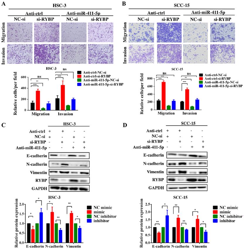
2 ZHANG et al: miR-411-5p PROMOTES LYMPH NODE METASTASIS BY TARGETING RYBP
Ring1 and YY1 binding protein (RYBP) is a member of negative controls (NCs; all Guangzhou RiboBio Co., Ltd.)
Polycomb group (PcG) proteins (18). It has been demonstrated using Lipofectamine ® 3000 (Invitrogen; Thermo Fisher
that the dysregulation of RYBP leads to a poorer prognosis in Scientific, Inc.), according to the manufacturer's protocols. The
hepatocellular carcinoma (19) and RYBP has been found to sequences were as follows: miR‑411‑5p mimic, 5'‑UAGUAG
act as a tumor suppressor in different types of cancer (20‑22). ACCGUAUAGCGUACG‑3'; miR‑411‑5p inhibitor, 5'‑CGU
From previous research, it is known that different miRNAs ACGCUAUACGGUCUACUA‑3'; and si‑RYBP, 5'‑ACAGCA
can regulate RYBP expression in tumorigenesis in melanoma TACAGTC TGCAAA‑3'. Lentiviruses encoding a reverse
and gastric cancer (23,24). complementary sequence of miR‑411‑5p (Anti‑miR‑411‑5p)
Thus, it was hypothesized that miR‑411‑5p can affect and an empty vector (Anti‑ctrl) were constructed by Shanghai
metastasis and epithelial‑mesenchymal transition (EMT) by GeneChem Co., Ltd. A total of 1x10⁵ cells HSC‑3 and SCC‑15
regulating RYBP expression in HNSCC cells. The present cells were infected with the lentiviral vectors (1x10⁸ TU/ml),
study investigated the effect of miR‑411‑5p on metastasis and and stable cell lines were selected following treatment with
EMT in vitro and in vivo, and further clarify the potential 2 µg/ml puromycin for 2 weeks.
mechanisms of miR‑411‑5p in the development of HNSCC
metastasis. The results of the present study may provide a RNA extraction and reverse transcription‑quantitative PCR
novel prognostic and therapeutic strategy to treat HNSCC (RT‑qPCR). Total RNA was extracted from tissues or cultured
metastasis. cells using TRIzol® reagent (Invitrogen; Thermo Fisher
Scientific, Inc.). To analyze the expression levels of miR‑411‑5p,
Materials and methods Bulge‑Loop™ miRNA qRT‑PCR Primer sets were synthe‑
sized by Guangzhou RiboBio Co., Ltd. Total RNA was reverse
The Cancer Genome Atlas (TCGA) analysis. The miRNA transcribed into cDNA using the PrimeScript RT Master Mix
expression profiles of TGCA‑HNSC dataset and the clinical (Takara Bio, Inc.), according to the manufacturer's protocols.
information were obtained from TCGA database (https://portal. qPCR was subsequently performed using the LightCycler ®
gdc.cancer.gov/) (25). A total of 528 patients with HNSCC 480 SYBR Green I Master kit (Roche Diagnostics), according
from TCGA database were selected for analysis in the present to the manufacturer's protocols. The following thermocycling
study. The exclusion criteria were as follows: i) Incomplete conditions were used for qPCR: Initial denaturation at 95˚C for
information on LNM; ii) incomplete patient clinical data; 5 min; followed by 40 cycles of denaturation for 10 sec at 95˚C,
and iii) the miR‑411‑5p expression data were incomplete. annealing for 20 sec at 60˚C and extension for 20 sec at 72˚C;
Following the exclusion of patients, the miR‑411‑5p expression and a final 10 min extension at 72˚C. The relative expres‑
levels and clinical information from 378 patients with HNSCC sion levels were calculated using the 2‑ΔΔCq method (26). The
were used in the present study. following primers were used for the qPCR: RYBP forward,
5'‑GGGGTGGTGGGGTGGCATACT‑3' and reverse, 5'‑CGC
Patient studies. A total of 52 tissues from patients with AGACGAAGGGTTT TGG GAT T‑3'; and GAPDH forward,
HNSCC were collected from The Hospital of Stomatology, 5'‑GCACCGTCAAGGCTGAGAAC‑3' and reverse, 5'‑TGG
Sun Yat‑Sen University (Guangzhou, China) between October TGAAGACGCCAGTGGA‑3'. GAPDH and U6 were used as
2017 and June 2018. No patient had received radiotherapy or the internal controls for RYBP and miR‑411‑5p, respectively.
chemotherapy before the biopsy, and all patients were diag‑ GAPDH and RYBP were purchased from Sangon Biotech
nosed with HNSCC. The tissue samples were frozen at ‑80˚C Co., Ltd., while U6 and miR‑411‑5p were purchased from
until required for use in further experiments. The histology and Guangzhou RiboBio Co., Ltd.
pathology of all samples was examined by two independent
pathologists. Written informed consent was provided from all Transwell assay. Cell migration and invasion assays were
patients prior to participation and the study was approved by performed using Transwell chambers (8‑mm pore size;
the Ethics Committee of Sun Yat‑Sen University [approval Corning, Inc.). Briefly, for the migration assay, HSC‑3 (3x10⁴)
no. ERC‑(2017)‑32]. or SCC‑15 (8x10 4) cells were plated in serum‑free medium
into the upper chambers of the Transwell plates. For the inva‑
Cell lines and culture. The human HNSCC cell lines, sion assay, HSC‑3 (6x10⁴) or SCC‑15 (2x10⁵) cells were seeded
HSC‑3 and SCC‑15, were purchased from the American into the upper chambers with Transwell plates precoated with
Type Culture Collection. HSC‑3 cells were cultured in Matrigel for 6 h at 37˚C. The lower chambers were filled
DMEM supplemented with 10% FBS, 100 IU/ml penicillin with medium supplemented with 10% FBS. Following incu‑
and 100 µg/ml streptomycin. SCC‑15 cells were cultured in bation for 24 h at 37˚C, the medium was removed and the
DMEM/F12 (1:1) supplemented with 10% FBS, 100 IU/ml cells were fixed with 4% formaldehyde for 20 min at room
penicillin and 100 µg/ml streptomycin. All culture reagents temperature. Cells that had not migrated or invaded through
were purchased from Gibco (Thermo Fisher Scientific, Inc.). the membrane were gently removed using a cotton swab. The
All cells were maintained at 37˚C in a humidified atmo‑ fixed cells were subsequently stained with 0.4% crystal violet
sphere with 5% CO2. for 15 min at room temperature and observed under a light
microscope (Zeiss GmbH). A total of five representative fields
Cell transfection and lentiviral vector construction. A total of at x100 magnification were randomly imaged. ImageJ v1.8.0
1x10⁵ cells were transiently transfected with 50 nM chemically (National Institutes of Health) was used for quantification,
synthesized miR‑411‑5p mimic, 100 nM miR‑411‑5p inhibitor, and the average counts of the five images represented the
50 nM small interfering RNA (si)‑RYBP and the corresponding migrated cells.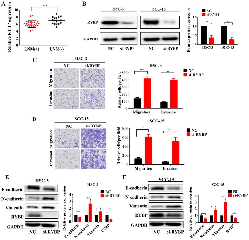
INTERNATIONAL JOURNAL OF MOlecular medicine 47: 36, 2021                                         3

Cell Counting Kit‑8 (CCK‑8) assays. The proliferation of          at room temperature. Protein bands were visualized using
HNSCC cells transfected with the miR‑411‑5p mimic or              Immobilon Western Chemiluminescent HRP substrate (EMD
miR‑411‑5p inhibitor was determined using a CCK‑8 assay           Millipore) and an ImageQuant™ LAS 4000 mini‑imaging
(Telenbiotech). Briefly, 2x10³ cells were seeded into a 96‑well   system (Cytiva). Semi‑quantification was performed using
plate and cultured for 24 h, prior to transfection with the       ImageJ (v1.8.0).
miR‑411‑5p mimic or inhibitor. Following incubation for 24,
48, 72, 96 or 120 h, CCK‑8 reagent was added to the culture       Immunohistochemistry (IHC). IHC staining was performed
medium for 2 h. The absorbance of each well was measured          with paraffin‑embedded sections. Briefly, the lymph nodes
at a wavelength of 450 nm. All experiments were performed         were immediately fixed in 4% paraformaldehyde for 24 h and
in triplicate.                                                    soaked in ethanol (70 to 100%), following which the sections
                                                                  (4 µm thick) were embedded in paraffin. The tissue sections
Colony formation assay. For the colony formation assay,           were incubated in 10% goat serum blocking solution (Wuhan
5x10² cells were seeded into 6‑well plates in triplicate 24 h     Boster Biological Technology, Ltd.) for 1 h at room tempera‑
post‑transfection. Following the culture of cells for 10 days,    ture, followed by incubation with an anti‑pan‑cytokeratin
the visible colonies were stained with 0.4% crystal violet for    primary antibody (1:200; cat. no. ab9377; Abcam) overnight
10 min at room temperature. Colonies with diameters >1 mm         at 4˚C. Sections were then incubated with a secondary
were defined as colonies.                                         antibody for 30 min at room temperature according to the
                                                                  instructions of the GT Vision™ and Detection System/MoRb
Dual‑luciferase reporter assay. To further analyze the            kit (Gene Tech), which contained the secondary antibody and
molecular mechanism of miR‑411‑5p‑mediated metastasis in          DAB, followed by staining with DAB and counterstaining
HNSCC, miRDB (http://www.mirdb.org/miRDB), miRTar‑                with hematoxylin for 1 min at room temperature. Then, two
Base (http://mirtarbase.mbc.nctu.edu.tw/php/) and TargetScan      senior pathologists blinded to the data assessed and scored the
7.1 (http://www.targetscan.org/vert_71) databases were used.      IHC results. The extent of staining was scored according to
The potential miR‑411‑5p binding site in RYBP 3'‑UTR              the percentage of positively stained cells: i) 1, 70%
org/vert_71). For luciferase assays, wild‑type (Wt) or Mutant     of cells. The staining intensity was graded as follows: i) 0, no
(Mut) RYBP 3'‑UTR sequences were designed, synthe‑                staining; ii) 1, weak staining (light yellow); iii) 2, moderate
sized and cloned into a pMIR‑REPORT™ vector by OBiO               staining (yellow brown); and iv) 3, strong staining (brown), as
Technology (Shanghai) Co., Ltd. to generate RYBP‑3'‑UTR‑Wt        previously described (9). The staining index was calculated
and RYBP‑3'‑UTR‑mut vectors, respectively. Then, HNSCC            by multiplying the percentage of positive cells and staining
cells (4x10⁴) were co‑transfected with 100 nM miR‑411‑5p          intensity. Five random fields per sample were analyzed using
mimics or NC and RYBP‑3'‑UTR‑Wt or RYBP‑3'‑UTR‑mut                a light microscope (magnification, x100) to visualize sections.
vectors using Lipofectamine 3000 reagent. Following 48 h
of transfection, the relative luciferase activity was measured    In vivo xenograft experiments. All animal experiments
using a Dual‑Luciferase Reporter Assay System (Promega            were approved by the Ethical Committee of Sun Yat‑Sen
Corporation), according to the manufacturer's protocols,          University (approval no. SYSU‑IACUC‑2020‑000562) and
and was compared with the Renilla luciferase activity of the      were performed in accordance with the guidelines for animal
samples. The assays were performed in triplicate.                 care and protection (27). A total of 14 BALB/c nude mice
                                                                  (female; age, 4‑6 weeks old; weight, 18‑20 g) were obtained
Western blotting. Total protein was extracted from cells using    from GemPharmatech Co., Ltd., and were housed in specific
RIPA lysis buffer (Beyotime Institute of Biotechnology)           pathogen‑free conditions at 25˚C and 50% humidity, with a
supplemented with protease and phosphatase inhibitors.            12‑h light/dark cycle and free access to food and water. The
Total protein was quantified using a BCA kit (Wuhan Boster        mice were randomly divided into 2 groups (n=7). A total of
Biological Technology Co., Ltd.), according to the manufac‑       2x10⁶ SCC‑15 cells with or without miR‑411‑5p expression
turer's protocols, and 20 µg protein/lane was separated via       knockdown were suspended in 100 µl PBS, and then injected
SDS‑PAGE on a 10% gel. The separated proteins were subse‑         into the tongue of the nude mice following anesthesia by isoflu‑
quently transferred onto PVDF membranes (EMD Millipore)           rane [4% (0.5 l/min) for induction and 2% for maintenance].
and blocked with 5% non‑fat milk for 1 h at room tempera‑         Following 8 weeks, the maximum tumor diameter in the mice
ture. The membranes were then incubated with the following        was recorded as 5.65 mm and the maximum tumor volume was
primary antibodies: Anti‑E‑cadherin (1:1,000; cat. no. 14472;     62.40 mm3, all mice were sacrificed using cervical dislocation
Cell Signaling Technology, Inc.), anti‑N‑cadherin (1:1,000;       and death was confirmed by the absence of breathing, and the
cat. no. 13116; Cell Signaling Technology, Inc.), anti‑vimentin   lack of a heartbeat and corneal reflex. Finally, the cervical
(1:1,000; cat. no. 5741; Cell Signaling Technology, Inc.),        lymph nodes were collected and analyzed using IHC analysis.
anti‑GAPDH (1:1,000; cat. no. 5174; Cell Signaling Technology,
Inc.) and anti‑RYBP (1:500; cat. no. sc‑374235; Santa Cruz        Statistical analysis. Statistical analysis was performed using
Biotechnology, Inc.). Following the primary antibody incuba‑      SPSS version 20.0 software (IBM Corp.) and GraphPad
tion, the membrane was washed three times with TBS with           Prism 6.0 software (GraphPad Software, Inc.). Statistical
2% Tween‑20 and incubated with HRP‑conjugated anti‑rabbit         differences between two groups were determined using an
(cat. no. 7074) or anti‑mouse (cat. no. 7076) secondary anti‑     unpaired Student's t‑test, while multiple group comparisons
bodies (1:5,000; Cell Signaling Technology, Inc.) for 1 h         were performed using a one‑way ANOVA, followed by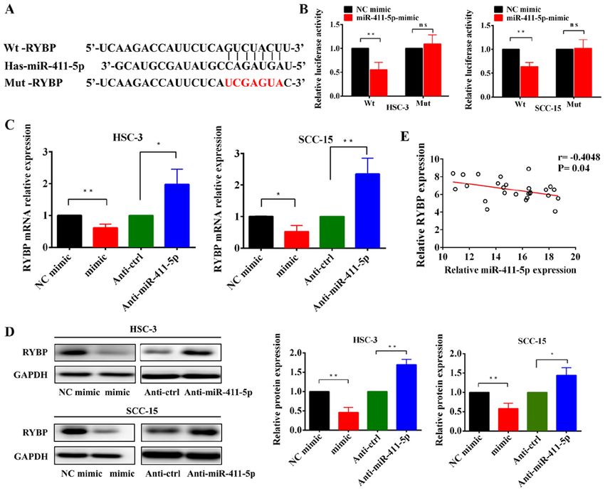
4                       ZHANG et al: miR-411-5p PROMOTES LYMPH NODE METASTASIS BY TARGETING RYBP

Table I. Association between the expression of miR‑411‑5p and clinicopathological characteristics in HNSCC clinical sample.

                                                       miR‑411‑5p expression, n
	--------------------------------------------------
Clinicopathological characteristics Number of cases, n  High             Low                                              P‑value

Age, years
 ≥55             26 11 15 0.406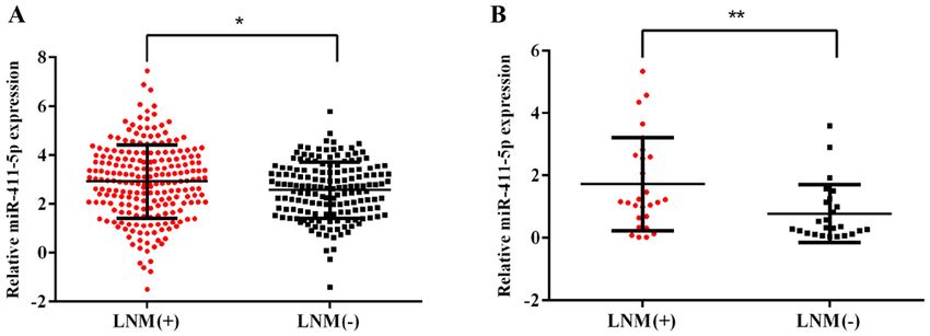
INTERNATIONAL JOURNAL OF MOlecular medicine 47: 36, 2021                                                            5

Figure 1. miR‑411‑5p expression levels are upregulated in patients with HNSCC with LNM. (A) Expression levels of miR‑411‑5p in patients with HNSCC with
or without LNM from The Cancer Genome Atlas database. (B) Expression levels of miR‑411‑5p in patients with HNSCC with or without LNM were analyzed
using reverse transcription‑quantitative PCR. LNM(+), patients with LNM; LNM(‑), patients without LNM. *P
6                           ZHANG et al: miR-411-5p PROMOTES LYMPH NODE METASTASIS BY TARGETING RYBP

Figure 3. miR‑411‑5p promotes HNSCC cell migration, invasion and EMT in vitro and metastasis in vivo. (A and B) Transfection efficiency of miR‑411‑5p was
verified using reverse transcription‑quantitative PCR. (C) Effects of miR‑411‑5p mimics and inhibitors on the invasion and migration of HNSCC cells were
analyzed using Transwell assays. (D) Representative staining of metastatic tumor cells in the cervical lymph nodes of mice following the injection of 2x10⁶
SCC‑15 cells with or without miR‑411‑5p inhibitor using anti‑pan‑cytokeratin. (E) Metastatic ratio of cervical lymph nodes. (F) Overexpression of miR‑411‑5p
downregulated the expression levels of E‑cadherin, and upregulated N‑cadherin and vimentin expression levels in HNSCC cells. By contrast, the knockdown
of miR‑411‑5p upregulated the expression levels of E‑cadherin, and downregulated vimentin and N‑cadherin expression levels. The expression levels of the
EMT‑related markers, E‑cadherin, N‑cadherin and vimentin were analyzed using western blotting. GAPDH was used as the internal loading control. Scale
bar, 100 µm. *P
INTERNATIONAL JOURNAL OF MOlecular medicine 47: 36, 2021                                                          7

Figure 4. RYBP is a direct target gene of miR‑411‑5p in HNSCC cells. (A) Predicted and mut binding sites of miR‑411‑5p in the 3'‑untranslated region of
RYBP. (B) Relative luciferase activity was analyzed in HSC‑3 and SCC‑15 cells transfected with miR‑411‑5p mimic or NC and Wt or Mut reporter genes.
(C and D) Reverse transcription‑quantitative PCR and western blotting of RYBP mRNA and protein expression levels, respectively, following the overexpres‑
sion and knockdown of miR‑411‑5p in HNSCC cells. (E) Spearman's correlation analysis was used to determine the correlation between RYBP and miR‑411‑5p
expression levels in HNSCC tissues. *P
8                          ZHANG et al: miR-411-5p PROMOTES LYMPH NODE METASTASIS BY TARGETING RYBP

Figure 5. Knockdown of RYBP promotes HNSCC cell migration, invasion and epithelial‑mesenchymal transition. (A) Expression levels of RYBP in patients
with HNSCC with or without LNM were analyzed using reverse transcription‑quantitative PCR. (B) Western blotting of the protein expression levels of RYBP
following the silencing of RYBP in HNSCC cells. (C and D) Effects of the silencing of RYBP on HNSCC cell invasion and migration. (E and F) Western
blotting of the effect of RYBP on the expression levels of E‑cadherin, N‑cadherin and vimentin. GAPDH was used as the internal loading control. Scale
bar, 100 µm. *P
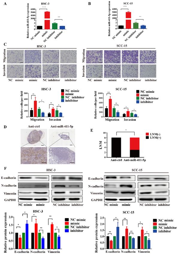
INTERNATIONAL JOURNAL OF MOlecular medicine 47: 36, 2021                                                          9

Figure 6. RYBP, as a direct target, may be responsible for miR‑411‑5p‑induced head and neck squamous cell carcinoma cell invasion, migration and epithe‑
lial‑mesenchymal transition. (A and B) Transwell assays were performed following the co‑transfection of HSC‑3 and SCC‑15 cells with the miR‑411‑5p
inhibitor and si‑RYBP. (C and D) Western blotting of E‑cadherin, N‑cadherin and vimentin expression levels following the co‑transfection of HSC‑3 and
SCC‑15 cells with the miR‑411‑5p inhibitor and si‑RYBP. GAPDH was used as the internal loading control. Scale bar, 100 µm. *P
10                     ZHANG et al: miR-411-5p PROMOTES LYMPH NODE METASTASIS BY TARGETING RYBP

are well‑studied in the context of EMT plasticity (36). For       Authors' contributions
example, Lin et al (37) reported that the downregulation of
miR‑639 expression levels was associated with TGF‑β‑induced       JX and BC conceived and designed the experiments. CZ and
EMT in tongue oral squamous cell carcinoma (OSCC), which          HFW performed the experiments and collected important
may represent a possible target for the treatment of OSCC         background information. MD, LHH and FP were involved in
metastasis. Another previous study reported that the miR‑200      data acquisition and analysis. YH and ZNF drafted the manu‑
family suppressed the EMT process and the metastatic ability      script and acquired data. JX and BC confirm the authenticity
of tongue OSCC cells (38). The findings of the present study      of all the raw data. All authors read and approved the final
demonstrated that miR‑411‑5p promoted EMT, indicating that        manuscript.
miR‑411‑5p may affect the metastatic ability of HNSCC by
promoting EMT.                                                    Ethics approval and consent to participate
    miRNAs are known to regulate gene expression by
binding to the 3'‑UTR of target mRNAs and either inhibiting       The study was approved by the Ethics Committee of Sun
their translation or inducing the degradation of the mRNA (4).    Yat‑Sen University (Guangzhou, China). Informed consent
To further understand the mechanism of action of miR‑411‑5p       was obtained from all participants. All included patients
in HNSCC, bioinformatics analysis was performed. The results      fulfilled criteria and completed the study.
revealed that miR‑411‑5p bound to the 3'‑UTR of RYBP.
Dual‑luciferase reporter assays validated that RYBP was a         Patient consent for publication
direct downstream target of miR‑411‑5p. Further analysis
confirmed that the overexpression of miR‑411‑5p downregu‑         All participants provided written informed consent.
lated RYBP expression levels, whereas the knockdown of
miR‑411‑5p upregulated RYBP expression levels at both the         Competing interests
mRNA and protein levels. RYBP is a member of the PcG of
proteins (18). Dinglin et al (39) previously reported that RYBP   The authors declare that they have no competing interests.
inhibited lung cancer metastasis by reversing EMT. Another
previous study reported that RYBP expression levels were          References
negatively correlated with the upregulated expression levels
of the EMT‑related transcription factors, zinc finger E‑box        1. Leemans CR, Braakhuis BJ and Brakenhoff RH: The molecular
                                                                      biology of head and neck cancer. Nat Rev Cancer 11: 9‑22, 2011.
binding homeobox (ZEB)1 and ZEB2 (22). The present study           2. Siegel RL, Miller KD and Jemal A: Cancer statistics 2019. CA
revealed that RYBP expression levels were downregulated in            Cancer J Clin 69: 7‑34, 2019.
patients with HNSCC with LNM. The genetic silencing of             3. Inglehart RC, Scanlon CS and Silva NJ: Reviewing and recon‑
                                                                      sidering invasion assays in head and neck cancer. Oral Oncol 12:
RYBP promoted the metastasis, invasion and EMT of HNSCC               1137‑1143, 2014.
cells. In addition, the knockdown of RYBP expression restored      4. Shukla GC, Singh J and Barik S: MicroRNAs: Processing, matu‑
the stimulatory effects of miR‑411‑5p on HNSCC cell migra‑            ration, target recognition and regulatory functions. Mol Cellular
                                                                      Pharmacol 3: 83‑92, 2011.
tion, invasion and EMT, thus supporting the contribution           5. Calin GA and Croce CM: MicroRNA signatures in human
of miR‑411‑5p‑regulated RYBP in HNSCC metastasis and                  cancers. Nat Rev Cancer 6: 857‑866, 2006.
EMT. These results suggested that miR‑411‑5p may have the          6. Hui ABY, Lenarduzzi M, Krushel T, Waldron L, Pintilie M,
                                                                      Shi W, Perez‑Ordonez B, Jurisica I, O'Sullivan B, Waldron J, et al:
potential to be a prognostic marker and potential predictor of        Comprehensive MicroRNA profiling for head and neck squa‑
cervical LNM in HNSCC.                                                mous cell carcinomas. Clin Cancer Res 16: 1129‑1139, 2010.
    In conclusion, the findings of the present study suggested     7. Liu X, Bi L, Wang Q, Wen M, Li C, Ren Y, Jiao Q, Mao JH,
                                                                      Wang C, Wei G and Wang Y: MiR‑1204 targets VDR to promotes
that the upregulated expression levels of miR‑411‑5p were             epithelial‑mesenchymal transition and metastasis in breast
associated with a poor prognosis in patients with HNSCC               cancer. Oncogene 37: 3426‑3439, 2018.
with LNM. Therefore, the miR‑411‑5p/RYBP axis may serve            8. Shiiba M, Uzawa K and Tanzawa H: MicroRNAs in head and
                                                                      neck squamous cell carcinoma (HNSCC) and oral squamous cell
a significant role in promoting HNSCC metastasis and EMT.             carcinoma (OSCC). Cancers (Basel) 2: 653‑669, 2010.
                                                                   9. Li Y, He Q, Wen X, Hong X, Yang X, Tang X, Zhang P, Lei Y,
Acknowledgements                                                      Sun Y, Zhang J, et al: EZH2‑DNMT1‑mediated epigenetic
                                                                      silencing of miR‑142‑3p promotes metastasis through targeting
                                                                      ZEB2 in nasopharyngeal carcinoma. Cell Death Differ 26:
Not applicable.                                                       1089‑1106, 2019.
                                                                  10. Hayes J, Peruzzi PP and Lawler S: MicroRNAs in cancer: Biomarkers,
                                                                      functions and therapy. Trends Mol Med 20: 460‑469, 2014.
Funding                                                           11. Dong Y, Zheng Y, Wang C, Ding X, Du Y, Liu L, Zhang W,
                                                                      Zhang W, Zhong Y, Wu Y and Song X: MiR‑876‑5p modulates
This study was supported by the National Nature Science               head and neck squamous cell carcinoma metastasis and invasion
                                                                      by targeting vimentin. Cancer Cell Int 18: 121, 2018.
Foundation of China (grant no. 81870769) and Guangdong            12. Shiah SG, Hsiao JR, Chang WM, Chen YW, Jin YT, Wong TY,
Financial Fund for High‑Caliber Hospital Construction (grant          Huang JS, Tsai ST, Hsu YM, Chou ST, et al: Downregulated
no. 174‑2018‑XMZC‑0001‑03‑0125/D‑05).                                 miR329 and miR410 promote the proliferation and invasion
                                                                      of oral squamous cell carcinoma by targeting Wnt‑7b. Cancer
                                                                      Res 74: 7560‑7572, 2014.
Availability of data and materials                                13. de Ca r va l ho AC, Scapulatempo ‑Neto C, Ma ia DC,
                                                                      Evangelista AF, Morini MA, Carvalho AL and Vettore AL:
                                                                      Accuracy of microRNAs as markers for the detection of neck
All data generated or analyzed during this study are included         lymph node metastases in patients with head and neck squamous
in this published article.                                            cell carcinoma. BMC Med 13: 108, 2015.
INTERNATIONAL JOURNAL OF MOlecular medicine 47: 36, 2021 11
14. Nadal E, Zhong J, Lin J, Reddy RM, Ramnath N, Orringer MB, 28. Loberg RD, Bradley DA, Tomlins SA, Chinnaiyan AM and
Chang AC, Beer DG and Chen G: A MicroRNA cluster at 14q32 Pienta KJ: The lethal phenotype of cancer: The molecular basis
drives aggressive lung adenocarcinoma. Clin Cancer Res 20: of death due to malignancy. CA Cancer J Clin 57: 225‑241, 2007.
3107‑3117, 2014. 29. Xu N, Yang W, Liu Y, Yan F and Yu Z: MicroRNA‑411 promoted
15. Zhang Y, Xu G, Liu G, Ye Y, Zhang C, Fan C, Wang H, Cai H, the osteosarcoma progression by suppressing MTSS1 expression.
Xiao R, Huang Z and Luo Q: MiR‑411‑5p inhibits proliferation Environ Sci Pollutr Res Int 25: 12064‑12071, 2018.
and metastasis of breast cancer cell via targeting GRB2. Biochem 30. Zhao J, Xu J and Zhang R: MicroRNA‑411 inhibits malignant
Bioph Res Commun 476: 607‑613, 2016. biological behaviours of colorectal cancer cells by directly
16. Zhang X, Zhang M, Cheng J, Lv Z, Wang F and Cai Z: MiR‑411 targeting PIK3R3. Oncol Rep 39: 633‑642, 2018.
functions as a tumor suppressor in renal cell cancer. Int J Biol 31. Jin H, Sun W, Zhang Y, Yan H, Liufu H, Wang S, Chen C, Gu J,
Markers 32: e454‑e460, 2017. Hua X, Zhou L, et al: MicroRNA‑411 downregulation enhances
17. Zhang C, Wang H, Liu X, Hu Y, Ding L, Zhang X, Sun Q and tumor growth by upregulating MLLT11 expression in human
Li Y: Oncogenic microRNA‑411 promotes lung carcinogenesis bladder cancer. Mol Ther Nucleic Acids 11: 312‑322, 2018.
by directly targeting suppressor genes SPRY4 and TXNIP. 32. Liu Y, Liu T, Jin H, Yin L, Yu H and Bi J: MiR‑411 suppresses the
Oncogene 38: 1892‑1904, 2019. development of bladder cancer by regulating ZnT1. Onco Targets
18. Garcia E, Marcos‑Gutierrez C, del Mar Lorente M, Moreno JC Ther 11: 8695‑8704, 2018.
and Vidal M: RYBP, a new repressor protein that interacts with 33. Bai TL, Liu YB and Li BH: MiR‑411 inhibits gastric cancer
components of the mammalian Polycomb complex, and with the proliferation and migration through targeting SETD6. Eur Rev
transcription factor YY1. EMBO J 18: 3404‑3418, 1999. Med Pharmaco 23: 3344‑3350, 2019.
19. Zhao Q, Cai W, Zhang X, Tian S, Zhang J, Li H, Hou C, Ma X, 34. Gonzalez DM and Medici D: Signaling mechanisms of the
Chen H, Huang B and Chen D: RYBP expression is regulated epithelial‑mesenchymal transition. Sci Signal 7: re8, 2014.
by KLF4 and Sp1 and is related to hepatocellular carcinoma 35. Lamouille S, Xu J and Derynck R: Molecular mechanisms of
prognosis. J Biol Chem 292: 2143‑2158, 2017. epithelial‑mesenchymal transition. Nat Rev Mol Cell Bio 15:
20. Zhou H, Li J, Zhang Z, Ye R, Shao N, Cheang T and Wang S: 178‑196, 2014.
RING1 and YY1 binding protein suppresses breast cancer 36. Domingues CSDC, Serambeque BP, Laranjo Cândido MS,
growth and metastasis. Int J Oncol 49: 2442‑2452, 2016. Marto CMM, Veiga FJB, Sarmento Antunes Cruz Ribeiro AB,
21. Voruganti S, Xu F, Qin JJ, Guo Y, Sarkar S, Gao M, Zheng Z, Figueiras A R R, Botelho MF R and Dourado MA R F:
Wang MH, Zhou J, Qian B, et al: RYBP predicts survival of Epithelial‑mesenchymal transition and microRNAs: Challenges
patients with non‑small cell lung cancer and regulates tumor and future perspectives in oral cancer. Head Neck 40: 2304‑2313,
cell growth and the response to chemotherapy. Cancer Lett 369: 2018.
386‑395, 2015. 37. Lin Z, Sun L, Chen W, Liu B, Wang Y, Fan S, Li Y and Li J:
22. Zhu X, Yan M, Luo W, Liu W, Ren Y, Bei C, Tang G, Chen R and MiR‑639 regulates transforming growth factor beta‑induced
Tan S: Expression and clinical significanceof PcG‑associated epithelial‑mesenchymal transition in human tongue cancer cells
protein RYBP in hepatocellular carcinoma. Oncol Lett 13: by targeting FOXC1. Cancer Sci 105: 1288‑1298, 2014.
141‑150, 2017. 38. Tamagawa S, Beder LB, Hotomi M, Gunduz M, Yata K,
23. Luan PB, Jia XZ and Yao J: MiR‑769‑5p functions as an onco‑ Grenman R and Yamanaka N: Role of miR‑200c/miR‑141 in the
gene by down‑regulating RYBP expression in gastric cancer. Eur regulation of epithelial‑mesenchymal transition and migration
Rev Med Pharmacol Sci 24: 6699‑6706, 2020. in head and neck squamous cell carcinoma. Int J Mol Med 33:
24. Zhao G, Li Q, Wang A and Jiao J: YY1 regulates melanoma 879‑886, 2014.
tumorigenesis through a miR‑9 ~ RYBP axis. J Exp Clin Cancer 39. Dinglin X, Ding L, Li Q, Liu Y, Zhang J and Yao H: RYBP
Res 34: 66, 2015. inhibits progression and metastasis of lung cancer by suppressing
25. Pérez Sayáns M, Chamorro Petronacci CM, Lorenzo Pouso AI, EGFR signaling and epithelial‑mesenchymal transition. Transl
Padín Iruegas E, Blanco Carrión A, Suárez Peñaranda JM and Oncol 10: 280‑287, 2017.
García García A: Comprehensive genomic review of TCGA head
and neck squamous cell carcinomas (HNSCC). J Clin Med 8:
1896, 2019. This work is licensed under a Creative Commons
26. Livak KJ and Schmittgen TD: Analysis of relative gene expres‑
Attribution-NonCommercial-NoDerivatives 4.0
sion data using real‑time quantitative PCR and the 2(‑Delta Delta
C(T)) method. Methods 25: 402‑408, 2001. International (CC BY-NC-ND 4.0) License.
27. National Research Council (US) Committee for the Update of
the Guide for the Care and Use of Laboratory Animals: Guide
for the care and use of laboratory animals. 8th edition. National
Academies Press (US), Washington, DC, 2011.You can also read