A Differential Pattern of Batokine Expression in Perivascular Adipose Tissue Depots From Mice
←
→
Page content transcription
If your browser does not render page correctly, please read the page content below
BRIEF RESEARCH REPORT
                                                                                                                                                  published: 04 August 2021
                                                                                                                                           doi: 10.3389/fphys.2021.714530

                                                A Differential Pattern of Batokine
                                                Expression in Perivascular Adipose
                                                Tissue Depots From Mice
                                                Alberto Mestres-Arenas1, Joan Villarroya1,2,3, Marta Giralt1,2,3, Francesc Villarroya1,2,3 and
                                                Marion Peyrou1,2,3*
                                                1
                                                 Departament de Bioquímica i Biomedicina Molecular and Institut de Biomedicina, Universitat de Barcelona, Barcelona,
                                                Spain, 2Centro de Investigación Biomédica en Red “Fisiopatología de la Obesidad y Nutrición”, Madrid, Spain, 3Institut de
                                                Recerca Hospital Sant Joan de Déu, Barcelona, Spain

                                                Depending on its anatomical placement, perivascular adipose tissue (PVAT) has been
                                                found to possess features more (e.g., aortic thoracic) or less (e.g., aortic abdominal) similar
                                                to brown/beige adipose tissue in mice, whereas PVAT surrounding the mesenteric arteries
                                                and the caudal part of abdominal aorta is similar to white fat. PVAT is thought to influence
                                                vascular function through the effects of adipose-secreted molecules on vessels. Brown
                            Edited by:          adipose tissue was recently shown to play differential secretory role via secretion of the
                           Paula Oliver,        so-called batokines but the involvement of differential batokine production in PVAT brown/
     University of the Balearic Islands,
                                   Spain
                                                beige plasticity was unclear. The current study characterizes for the first time the expression
                       Reviewed by:
                                                of batokines at aortic thoracic PVAT (tPVAT) and aortic abdominal PVAT (aPVAT) in
               Timothy P. Fitzgibbons,          comparison with typical brown and white adipose depots, in basal and thermogenically
 University of Massachusetts Medical
                                                activated conditions. We found that both PVAT depots increased their expression of genes
                School, United States
                   Andres Contreras,            encoding the batokines bone morphogenetic protein-8b (BMP8B), fibroblast growth
            Michigan State University,          factor-21 (FGF21), and kininogen-2 (KNG2) in response to cold, indicating that, under
                        United States
                                                cold-induced thermogenic activation, both thoracic aorta and abdominal aorta would
                 *Correspondence:
                      Marion Peyrou
                                                experience intense local exposure to these PVAT-secreted batokines. In contrast, the
           peyrou.marion@gmail.com              gene expression levels of growth/differentiation factor-15 and vascular endothelial growth
                                                factor-A were induced only in tPVAT. Under short-term high-fat diet-induced thermogenic
                   Specialty section:
         This article was submitted to          activation, the thoracic aorta would be specifically exposed to a local increase in PVAT-
                Integrative Physiology,         originating BMP8B, FGF21, and KNG2. Our data support the notion that acquisition of
               a section of the journal
                                                a brown/beige phenotype in PVAT is associated with upregulation of batokines, mainly
               Frontiers in Physiology
                                                BMP8B, FGF21, and KNG2, that can differentially target the vascular system.
           Received: 25 May 2021
           Accepted: 13 July 2021               Keywords: brown adipose tissue, white adipose tissue, thoracic perivascular adipose tissue, abdominal perivascular
         Published: 04 August 2021              adipose tissue, batokine
                              Citation:
      Mestres-Arenas A, Villarroya J,
             Giralt M, Villarroya F and
Peyrou M (2021) A Differential Pattern
                                                INTRODUCTION
            of Batokine Expression in
 Perivascular Adipose Tissue Depots
                                                Two types of adipose tissues have been traditionally distinguished in mammals: white adipose
                            From Mice.          tissues (WAT) and brown adipose tissues (BAT). WAT, which is composed mostly of white
           Front. Physiol. 12:714530.           adipocytes containing unilocular fat vacuoles, is the main site of metabolic energy storage in
    doi: 10.3389/fphys.2021.714530              the body. BAT, meanwhile, is composed of brown adipocytes that contain multilocular lipid

Frontiers in Physiology | www.frontiersin.org                                          1                                            August 2021 | Volume 12 | Article 714530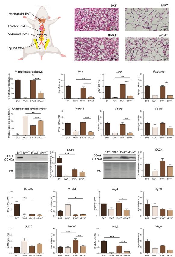
Mestres-Arenas et al.                                                                                                   Batokine Expression in PVAT

droplets and large amounts of mitochondria harboring the                   to mild cold (16°C) appear to express Ucp1 more intensely
uncoupling protein-1 (UCP1) that enables the tissue to mediate             in tPVAT, like in BAT (Chang et al., 2012), and Reynés et al.
adaptive thermogenesis (Cannon and Nedergaard, 2004). WAT                  (2019) reported that cold exposure in ferrets (an animal model
is largely present at subcutaneous and visceral sites in                   whose adipose tissue features and plasticity resemble those
experimental rodent models as well as in humans. In contrast,              humans more closely than do those of rats and mice) induces
BAT is present at specific depots in the interscapular, subscapular,       thermogenic activation and represses pro-inflammatory immune
cervical, and other regions of rodents, whereas in humans, it              pathways in peri-aortic tPVAT.
is mostly present in the interscapular region of neonates, and                 Increased PVAT fat accumulation (which is reminiscent of
mainly subscapular depots of adults (Cinti, 2001; Nedergaard               enhanced acquisition of white adipocyte-like morphology) is
et al., 2007). In recent years, a remarkable white-to-brown                associated with cardiovascular risk (Montani et al., 2004), while
plasticity has been recognized under circumstances of enhanced             increased browning at PVAT has been proposed to have protective
thermogenic stimulus (e.g., a cold environment): Anatomical                effects (Aldiss et al., 2017). It is unknown how common it is
sites typically containing WAT (mostly subcutaneous) may                   for obesity phenotypes maintaining a healthy state to specifically
become enriched in brown adipocyte-like cells that are termed              influence PVAT functions. In general, the importance of PVAT
“beige” adipocytes. These beige adipocytes are thermogenic                 for vascular health has been hypothesized to be associated
and contain UCP1, but they are derived from a different cell               with its secretory properties and the effects of adipocyte-derived
lineage than the “classical” brown adipocytes present at                   factors on adjacent endothelial cells. Thus, enhanced secretion
anatomically defined BAT depots (Giralt and Villarroya, 2013;              of pro-inflammatory factors, which is typical of hypertrophied
Chouchani and Kajimura, 2019). Whereas hypertrophied WAT,                  white adipocytes, is considered to occur in high-fat-containing
as in obesity, is associated with multiple diseases ranging from           PVAT depots under conditions, such as obesity, thereby
type II diabetes and cardiovascular disease to cancer, high                contributing to vascular damage (Oikonomou and Antoniades,
BAT activity is positively correlated with cardio-metabolic health         2017). However, we know little about how the brown/beige
(Becher et al., 2021).                                                     phenotype of PVAT at distinct anatomical sites and under
    In addition to the roles played by WAT and BAT in the                  distinct environmental conditions can help determine the pattern
energy balance (storage vs. expenditure, respectively), the two            of batokine secretion and downstream influences on
types of adipose tissue are now recognized as playing different            vascular functions.
secretory roles. That of WAT has been extensively studied since                Some batokines, such as kininogen-2 (KNG2) and possibly
the initial discovery of leptin, and over a hundred of “white”             fibroblast growth factor-21 (FGF21), have been shown to
adipokines have been identified (Giralt et al., 2016). However,            influence the vascular system in experimental settings unrelated
it was only recently that BAT was shown to have a relevant                 to a brown/beige adipose origin (Domouzoglou et al., 2015;
secretory activity distinct from that of WAT (Villarroya et al.,           Hamid et al., 2020). Vascular endothelial growth factor-A
2017a). The pattern of BAT-derived secreted factors, termed                (VEGFA) and bone morphogenetic protein-8b (BMP8B) secreted
“batokines,” appears to mediate autocrine, paracrine, and even             specifically from brown adipocytes were shown to promote
endocrine effects distinct from those mediated by the WAT                  vascularization at BAT depots (Tonello et al., 1999; Xue et al.,
secretome (Villarroya et al., 2017b, 2019).                                2009; Pellegrinelli et al., 2018). However, the local secretory
    Perivascular adipose tissue (PVAT) is generally defined as             capacity of brown/beige-like PVAT depots has not been analyzed
a layer of adipose tissue that surrounds large vessels, such as            to date. The objective of our study was to characterize for
the aortic artery. In rodents, histological (massive presence of           the first time the expression of batokines at two distinct PVAT
multilocular adipocytes) and molecular (expression of Ucp1                 sites, aortic tPVAT and aPVAT, in comparison with typical
and other genes) characterization of PVAT has indicated that               BAT and WAT depots, under basal conditions and conditions
the aortic thoracic PVAT (tPVAT) resembles BAT, whereas                    of cold-induced and diet-induced thermogenic activation.
PVAT around the abdominal aorta (aPVAT) exhibits a mixture
of multilocular Ucp1-expressing adipocytes together with typical
unilocular white adipocytes (Fitzgibbons et al., 2011). In humans,         MATERIALS AND METHODS
tPVAT also contains brown adipocyte-like cells, while aPVAT
resembles more typical WAT (Chatterjee et al., 2009). PET                  Animal Procedures
scan-based functional data and histology analyses indicate that            Experiments were conducted on 9- to 11-week-old male wild-
PVAT at human thoracic aorta resembles BAT (Wei et al.,                    type FVB/NJ mice, which were maintained at room temperature
2015). Despite these findings under basal conditions, however,             (21°C) or exposed to cold (4°C) for 1 week. Cold-exposed
we know little about the plasticity of PVAT in response to                 animals were housed in pairs. Total body weight and food
thermogenic challenges known to elicit BAT activation and                  consumption were monitored daily. Mice were maintained on
WAT browning. Li et al. (2017) reported that cold exposure                 a standard rodent diet (10% Kcal fat; Teklad Diet, Envigo) or,
of rats induced Ucp1 expression in both tPVAT and aPVAT,                   where indicated, fed a high-fat diet (HFD) for 4 weeks (#TD.06415,
as it does in interscapular BAT and inguinal WAT (iWAT).                   45% Kcal fat, Teklad Custom Diet, Envigo). Body weight and
Consistently, metabolic activation of tPVAT was noted in rats              energy intake of FVB/NJ mice before and after the different
treated with a β3-adrenergic agonist, similar to what occurs               experimental settings are available in Supplementary Table 1.
in interscapular BAT (Mirbolooki et al., 2011). Mice exposed               For all three experimental groups (control, cold-exposed, and

Frontiers in Physiology | www.frontiersin.org                          2                                    August 2021 | Volume 12 | Article 714530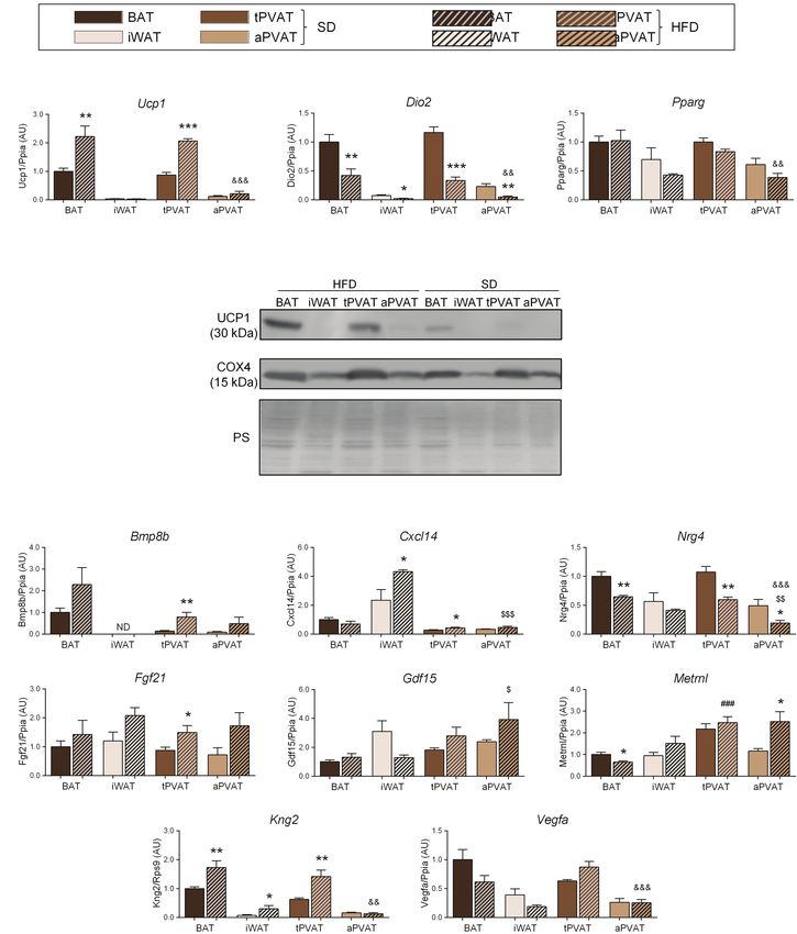
Mestres-Arenas et al.                                                                                                 Batokine Expression in PVAT

HFD-fed individuals), mice were maintained under controlled            Western Blotting
humidity and a 12-h light/dark cycle (8:00 am–8:00 pm). For            Tissue samples for protein analysis were homogenized in RIPA
sample collection, animals were sacrificed by decapitation.            buffer containing 50 mM Tris-HCl (pH 7.4), 150 mM NaCl,
Interscapular BAT, iWAT, tPVAT, and aPVAT depots were                  0.1 mM EDTA, 0.1 mM EGTA, 1% NP-40, 0.5% sodium
dissected (see Figure 1A) and weighed, and a piece was shock-          deoxycholate, 0.1% sodium dodecyl sulfate, 2 mM sodium
frozen in liquid nitrogen and stored at −80°C for future RNA           orthovanadate, 10 mM β-glycerophosphate, 5 mM sodium fluoride,
and protein analyses. All experimental procedures performed            1% phenylmethylsulfonyl fluoride, and a protease inhibitor cocktail
were authorized by the Institutional Animal Care and Use               (cOmplete-Mini, Roche). The protein concentration of each extract
Committee of the University of Barcelona.                              was determined by the BCA method (Thermo Fisher Scientific).
                                                                       Total protein (20 μg/lane) was resolved by SDS-PAGE in homemade
Histological Analysis                                                  12% acrylamide/bisacrylamide gels and transferred to PVDF
BAT, iWAT, tPVAT, and aPVAT samples were collected,                    membranes (GE Healthcare). The membranes were incubated
immediately fixed for 24 h in 4% paraformaldehyde, and then            with primary antibodies specific for UCP1 (ab10983, Abcam)
preserved in 70% ethanol. For analysis, the samples were               or cytochrome oxidase subunit IV (COX4; A-21342, Molecular
dehydrated, embedded in paraffin blocks, cut into 5-μm-thick           Probes) diluted 1:1,000 in 5% BSA, followed by an incubation
sections, and stained with hematoxylin and eosin. The stained          with the corresponding secondary antibodies HRP-conjugated
sections were visualized with an Olympus BX61 light microscope         anti-rabbit IgG (sc-2004, Santa Cruz) or anti-mouse IgG (170–6,516,
at 20X and 4X. The percentage of multilocular adipocyte area           Bio-Rad) diluted 1:3,000 in 5% milk. Signals were detected by
and the mean unilocular adipocyte diameter were measured               chemiluminescence after addition of the Immobilon Western
from digitalized images using the Fiji/ImageJ software.                HRP substrate (Millipore), and digitalized images were quantified
Multilocular adipocyte cell limits were manually drawn to              using Fiji/ImageJ software relative to total protein Ponceau staining.
measure area, then divided by the total area to obtain the
percentage of multilocular adipocytes. For unilocular adipocyte,       Statistical Analysis
size measurements were obtained by manual drawing after                Data are expressed as mean ± standard error of the mean with
initial setting of image scale. Results are average of at least        respect to BAT under the control condition, which was adjusted
20 representative measurements per image in each of the 8–10           to have a mean = 1. Exact number of replicates is given in
independent sections analyzed.                                         each figure legend. Putative outliers were detected and removed
                                                                       prior to statistical analyses using the Grubbs’ test, and the Shapiro-
Total RNA Isolation                                                    Wilk test was applied to establish the normality of datasets. A
Total RNA was extracted from tissue homogenates using a                statistically significant difference between two groups was assessed
column-affinity-based methodology. A NucleoSpin RNA kit was            by two-tailed unpaired Student’s t-test. For datasets that did not
utilized for BAT and iWAT samples and a NuceloSpin RNA                 follow a normal distribution (Cxcl14 transcript in basal conditions),
XS kit was used for both perivascular samples (all from                differences were evaluated using a two-tailed unpaired
Macherey-Nagel), following the supplier’s recommendations.             nonparametric Mann-Whitney’s U-test. One-way ANOVA followed
Final RNA yield was quantified using a NanoDrop ND-100                 by Tukey Kramer’s multiple comparisons post-hoc test was used
spectrophotometer (NanoDrop Technologies).                             to compare three or more groups. Statistical analyses were
                                                                       performed with GraphPad Prism 8 (GraphPad Software Inc.)
Reverse Transcription Quantitative                                     and the programming environment R. In all cases, the statistical
Real-Time Polymerase Chain Reaction                                    significance threshold was set at p < 0.05.
Analysis
Reverse transcription was performed with 500 ng of total RNA
employing a High Capacity RNA-to-cDNA kit (Applied                     RESULTS
Biosystems) according to the manufacturer’s instructions.
Platinum Quantitative PCR SuperMix-UDG with ROX reagent                Figure 1B shows the microscopic morphology of tPVAT and
or SYBR Select Master Mix (both from Thermo Fisher Scientific)         aPVAT in comparison with interscapular BAT (an adipose depot
was used as the master mix in a final reaction volume of               representative of classical BAT) and iWAT (as characteristic of
20 μl that contained specific TaqMan or SYBR Green probes              subcutaneous WAT, a depot highly susceptible to browning).
(see Supplementary Table 2) for each gene of interest. An              Under basal conditions, in which mice were maintained at 21°C
ABI 7500 Real-Time PCR System (Thermo Fisher Scientific)               and fed a standard diet, the microscopic morphology of tPVAT
was used to perform reverse transcription quantitative real-           closely resembled that of BAT, with numerous multilocular
time polymerase chain reaction (RT-qPCR) amplification. The            adipocytes (more than 90%; Figure 1C). In contrast, the microscopic
relative expression levels of target genes in all samples were         appearance of aPVAT was characterized by the presence of both
normalized to those of the Ppia or Rps9 housekeeping genes             unilocular and multilocular adipocytes. The percentage of
using the 2−AACt method (Livak and Schmittgen, 2001). For              multilocular adipocytes in aPVAT was much higher than in iWAT
more information regarding RT-qPCR protocol, see MIQE                  (Figure 1C) and the unilocular adipocytes were smaller in aPVAT
guidelines in Supplementary Table 3.                                   than in iWAT (Figure 1D). Supplementary Figure 2 shows low

Frontiers in Physiology | www.frontiersin.org                      3                                      August 2021 | Volume 12 | Article 714530
Mestres-Arenas et al. Batokine Expression in PVAT
A B
C E
D
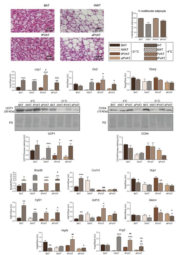
F
G
FIGURE 1 | Characterization of perivascular adipose depots in mice under basal conditions. (A) Schematic representation of the anatomical placement of
interscapular brown adipose tissues (BAT), inguinal WAT (iWAT), thoracic PVAT (tPVAT), and abdominal PVAT (aPVAT) depots in mice. (B) Representative optical
(Continued)
Frontiers in Physiology | www.frontiersin.org 4 August 2021 | Volume 12 | Article 714530Mestres-Arenas et al. Batokine Expression in PVAT
FIGURE 1 | microscopic pictures of hematoxylin-eosin stained sections of adipose tissue samples. (C, D) Quantitative assessment of cell morphology in adipose
tissue depots. (E) Relative transcript levels of genes encoding components of the thermogenic and adipogenic machinery. (F) Relative levels of uncoupling protein-1
(UCP1) and cytochrome oxidase subunit IV (COX4) proteins (left, representative immunoblots; right, quantitative measurements). (G) Relative transcript levels of
batokine-encoding genes. Bars correspond to means ± standard error of the mean (SEM) values from 4 to 6 mice. Statistical differences (*p < 0.05, **p < 0.01, and
***p < 0.001) are shown for comparisons of BAT vs. tPVAT, iWAT vs. aPVAT, and tPVAT vs. aPVAT depot pairs. ND, non-detectable.
magnification pictures of the adipose depots indicating the aPVAT shows a remarkable heterogeneity with areas showing
considerably homogeneous extent of morphological phenotype. predominant brown-like adipocytes, whereas in other areas, most
The gene expression pattern reinforced the close similarity of cells remained with a white phenotype, rather similarly to what
tPVAT and BAT, as the whole set of transcripts for biomarkers happens in iWAT. Cold-induced Ucp1 mRNA expression was
of the brown/beige phenotype (i.e., Ucp1, Dio2, Ppargc1a, Prdm16, intense and fairly similar in tPVAT, aPVAT, BAT, and iWAT,
and Ppara) showed essentially equal expression levels in tPVAT with the highest cold-induced increase in Ucp1 transcript levels
relative to BAT (Figure 1E). Conversely, these transcripts were seen in tPVAT (Figure 2C). A similar pattern of cold-induced
poorly expressed in aPVAT, where they were found in a range expression was found at the distinct adipose depots for Dio2,
similar to that seen in iWAT, with the exception of a mild trend another marker of brown/beige activation. The expression level
for higher expression of Ucp1 in aPVAT. This observation may of the general non-thermogenesis-related adipogenesis gene, Pparg,
be due to less brown/beige adipocytes abundance in aPVAT. was unaltered at any of the adipose depots in response to cold.
Meanwhile, the expression of Pparg, a gene commonly found Parallel cold-induced increases were seen in the relative UCP1
in both brown/beige and white adipocyte phenotypes, was similar protein levels at the four adipose depots, confirming our transcript-
across all four adipose depots (Figure 1E). level findings, whereas COX4 protein levels were insensitive to
We complemented this analysis by determining the protein cold-induced stimulation (Figure 2D).
levels of UCP1 and COX4, which is an indicator of the Regarding the expression of batokine-encoding genes, the
mitochondrial enrichment typical of brown/beige adipocytes. transcript levels of Bmp8b, Cxcl14, Fgf21, and Gdf15 were
UCP1 protein was detected in tPVAT, but at a lower abundance increased in BAT in response to 1-week cold exposure
than in BAT. In addition, while UCP1 protein level was very (Figure 2E), in accordance with the previous reports (Hondares
low in iWAT, aPVAT level appeared intermediate between PVAT et al., 2011; Whittle et al., 2012; Cereijo et al., 2018; Campderrós
and iWAT (Figure 1F). The differences were milder for COX4, et al., 2019). We found that 1 week of cold significantly reduced
but the highest levels were also found in BAT and tPVAT, Vegfa mRNA in BAT, which is consistent with the previous
and the lowest in iWAT and aPVAT (Figure 1F). data reporting that short-term, but not long-term, cold exposures
We next analyzed the expression patterns of batokine-encoding induce Vegfa gene expression in BAT (Asano et al., 1997).
genes in the distinct adipose depots (Figure 1G). Bmp8b and Kng2 Enhanced expression levels of Bmp8b, Fgf21, and Kng2 were
exhibited an expression pattern resembling that of the brown/beige found in iWAT in association with cold-induced browning,
marker genes: Their expression levels were maximal in BAT, minimal also as previously reported (Fisher et al., 2012; Peyrou et al.,
in iWAT, relatively high in tPVAT (especially for Kng2), and low (but 2020). The pattern of cold-induced expression changes in
still above the minimal level in iWAT) in aPVAT. The expression batokine-encoding genes were distinct according to the type
levels of Vegfa and Nrg4 followed an analogous but less intense pattern: of PVAT: Bmp8b, Fgf21, and Kng2 were strongly induced in
They were higher in BAT and tPVAT and lower in iWAT and aPVAT response to cold both in tPVAT and aPVAT, whereas Gdf15
(Figure 1G). On the other hand, Cxcl14 expression followed a totally was only induced in tPVAT (as also seen in BAT). The Cxcl14
distinct pattern, with the highest expression found in iWAT and the transcript was not altered at any site other than BAT, while
lowest in both PVAT depots. Metrnl also followed a distinct expression the expression levels of Nrg4, Vegfa, and Metrnl mRNAs were
profile, characterized by similar levels in the various adipose depots not induced by cold. Indeed, the opposite was observed as:
with the exception of tPVAT, in which Metrnl was significantly increased. Nrg4 expression was downregulated by cold in both BAT and
Finally, no significant difference was found among the four adipose tPVAT, Vegfa was decreased in BAT, and Metrnl expression
depots for Fgf21 or Gdf15 (Figure 1G). was reduced in all adipose depots tested, except for BAT.
We then set out to determine the plasticity of mouse PVAT We next expanded our study to a second distinct inducer
depots in response to a browning-inducing stimulus (exposure of adipose tissue plasticity. For this purpose, we treated mice
of mice to a cold environment for 1 week) and assess the extent with a HFD for 1 month, which was reported to represent a
to which such plasticity influences batokine expression. As expected, relatively short experimental design favoring diet-induced
mice maintained at 4°C for 7 days exhibited a massive browning thermogenesis and UCP1 induction at BAT (Fromme and
of iWAT, as assessed by cell morphology. Moreover, a large Klingenspor, 2011). Consistent with these reports, HFD upregulated
percentage of adipocytes acquired a multilocular morphology, Ucp1 transcript (Figure 3A) and UCP1 protein (Figure 3B)
which was not seen under the control situation at 21°C (Figure 2A). levels in BAT but not in iWAT. Notably, an identical pattern
aPVAT experienced a similar but slightly more intense response of changes was observed for tPVAT relative to BAT, while aPVAT
to cold, with more than 75% of aPVAT cells acquiring a multilocular behaved like iWAT. Expression of Dio2, which is an indicator
appearance after cold exposure (Figure 2B). Low magnification of cold-induced thermogenesis in BAT and iWAT, was
pictures (Supplementary Figure 2) indicated that, after cold, downregulated in response to HFD in all four depots (Figure 3A),
Frontiers in Physiology | www.frontiersin.org 5 August 2021 | Volume 12 | Article 714530Mestres-Arenas et al. Batokine Expression in PVAT
A B
C
D
E
FIGURE 2 | Effects of 1-week cold (4°C) exposure on perivascular adipose depots in mice. (A) Representative optical microscopic pictures of hematoxylin-eosin stained
sections of adipose tissue samples. (B) Quantitative assessment of cell morphology in adipose tissue depots. (C) Relative transcript levels of genes encoding components
(Continued)
Frontiers in Physiology | www.frontiersin.org 6 August 2021 | Volume 12 | Article 714530Mestres-Arenas et al. Batokine Expression in PVAT
FIGURE 2 | of the thermogenic and adipogenic machinery. (D) Relative levels of UCP1 and COX4 proteins (top, representative immunoblots; bottom, quantitative
measurements). (E) Relative transcript levels of batokine-encoding genes. Bars correspond to means ± SEM values from 3 to 6 mice. Statistical differences (*p < 0.05,
**p < 0.01, and ***p < 0.001) are shown for the cold effect at each adipose depot. Comparison among depots in the cold condition is shown for BAT vs. tPVAT (#p < 0.05,
##
p < 0.01, and ###p < 0.001), iWAT vs. aPVAT ($p < 0.05 and $$$p < 0.001), and tPVAT vs. aPVAT (&p < 0.05, &&p < 0.01, and &&&p < 0.001). ND, non-detectable.
whereas HFD feeding did not cause remarkable changes in Pparg Under basal conditions, several batokines (Bmp8b, Nrg4,
expression in any of the analyzed adipose tissues. Kng2, and Vegfa) exhibited mRNA expression patterns in tPVAT
The transcript expression levels of the batokines Bmp8b and and aPVAT that were similar to those in BAT and iWAT,
Fgf21 trended higher in response to HFD at all adipose depots, respectively. This suggests that, at the most common environmental
except for Bmp8b expression in iWAT, which was undetectable. temperature conditions for rodent models (21°C), the thoracic
Whereas changes due to HFD were statistically significant only section of the aortic artery is highly exposed to the local
at tPVAT, ANOVA of the whole set of adipose depots revealed secretion of these factors by PVAT, whereas the abdominal
that the “diet factor” is associated with overall significant aorta has much less such exposure. Additionally, our findings
increases in the transcript levels of Bmp8b (p = 0.0092) and suggest that PVAT shows a highly sensitive ability to modify
Fgf21 (p = 0.0017). Likewise, a significant HFD-induced increase its batokine expression profile, following a distinct pattern
in BAT and tPVAT, but not in aPVAT, was found for Kng2. according to the PVAT depot and for each specific batokine.
Cxcl14 expression was induced by HFD in iWAT and tPVAT, For example, tPVAT and aPVAT increased their expression
whereas Metrnl was induced by HFD only in aPVAT. Nrg4 levels of Bmp8b, Fgf21, and Kng2 in response to cold, whereas
expression was down-regulated in response to HFD at all of Gdf15 and Vegfa were induced in tPVAT but not in aPVAT.
the analyzed adipose depots, although not to a significant Nevertheless, there is a remarkable capacity of aPVAT, which
degree in iWAT, whereas Vegfa expression was downregulated possesses low-level expression of most batokines under basal
by HFD in BAT and iWAT, but not in either PVAT (Figure 3C). condition, to induce the expression of Bmp8b, Fgf21, and Kng2
transcripts in response to cold. In response to a short-term
HFD, a milder thermogenic stimulus, the induction of batokine
DISCUSSION gene expression was more restricted to tPVAT, and basically
concerned the transcripts for Bmp8b, Fgf21, and Kng2. Based
Microscopy morphology (percentage of multilocular adipocytes), on our findings, we hypothesized that under cold conditions,
gene expression, and relative protein abundance of brown/ both the thoracic and the abdominal aorta experience intense
beige phenotype markers under basal conditions indicated that local exposure to these PVAT-synthesized and secreted batokines.
there was a strong resemblance between tPVAT and classical Under short-term HFD, in contrast, only the thoracic aorta is
BAT, and also a close similarity between aPVAT and iWAT. exposed to local increases in PVAT-originating BMP8B, FGF21,
This is in agreement with the previous reports in mice and and KNG2.
other rodent models (Fitzgibbons et al., 2011; Chang et al., Several indirect lines of evidence suggest that batokines
2012; Reynés et al., 2019). Further, we found that tPVAT expressed in BAT/beige-activated PVAT may exert relevant
responded to cold-induced thermogenesis by eliciting a pattern effects on vascular function. In this context, classical BAT-secreted
of thermogenesis-related gene expression similar to that observed BMP8B has been shown to promote vascularization and induce
in BAT, whereas aPVAT resembled iWAT in that it showed a the expression of angiogenic factors (Pellegrinelli et al., 2018).
strong capacity for browning in response to cold stimulus. Thus, we can expect that local secretion of BMP8B by brown/
These findings indicate a strong plasticity capacity of mouse beige-activated PVAT could exert analogous effects on attached
PVAT to acquire a phenotype of activated BAT (tPVAT) and large vessels. Published findings suggest that kininogens, which
beige (aPVAT) adipose tissues in response to cold and thus are key components of the kinin-kallikrein system, play a role
confirm the previous reports in rodents (Chang et al., 2012; in the cardiovascular system, such as preventing hypertension
Li et al., 2017) and in other animal models (Reynés et al., and ischemia (Hamid et al., 2020). Gdf15 and Vegfa appear
2019). Meanwhile, we showed that short-term feeding of a to be particularly induced in tPVAT in response to cold. GDF15
HFD induced signs of thermogenic activation (induction of has been reported to modulate vascular contraction and relaxation
Ucp1 expression) in BAT and tPVAT, indicating that their responses in an endothelium-dependent fashion (Mazagova
resemblance is not limited to the cold response, but also et al., 2013), whereas VEGFA is a very well-known angiogenic
concerns diet-induced thermogenic activation. We saw in our factor. Overall, these findings indicate that the acquisition of
study that the extent of Ucp1 mRNA expression and UCP1 an active BAT-like phenotype and browning features in PVAT
protein levels at PVAT depots under distinct conditions is not depots may result in a distinct pattern of batokine secretion,
always concordant. There are several reports indicating specific implying a differential functional impact on vascular cells
regulation of Ucp1 mRNA translation to UCP1 protein as part distinct from the standard adipokine secretion elicited from
of the thermogenic machinery of brown adipocytes and in conventional white fat. Thus, it is tempting to speculate that
response to thermogenic stimuli (Dai et al., 2015; Chen et al., enhanced PVAT batokine secretion would play a cardiovascular-
2018). Thus, it cannot be ruled out that translation efficiency protective role, especially since Chang et al. (2012) showed
of Ucp1 transcripts was different in BAT relative to PVAT depots. that mild cold-induced PVAT activation attenuates age-dependent
Frontiers in Physiology | www.frontiersin.org 7 August 2021 | Volume 12 | Article 714530Mestres-Arenas et al. Batokine Expression in PVAT
A
B
C
FIGURE 3 | Effect of HFD feeding for 1 month on perivascular adipose depots in mice. (A) Relative transcript levels of genes encoding components of the
thermogenic and adipogenic machinery. (B) Representative immunoblots of UCP1 and COX4 proteins. (C) Relative transcript levels of batokine-encoding genes.
Bars correspond to means ± SEM from 5 to 6 mice. Significant differences (*p < 0.05, **p < 0.01, and ***p < 0.001) are shown for HFD feeding effect at each
adipose depot. Comparison among depots in the HFD-fed condition is shown for BAT vs. tPVAT (###p < 0.001), iWAT vs. aPVAT ($$p < 0.01 and $$$p < 0.001), and
tPVAT vs. aPVAT (&&p < 0.01 and &&&p < 0.001). ND, non-detectable.
and obesity-induced endothelial dysfunction in mice. However, We know far less about human PVAT compared to that in
the fact that our study is based on transcriptomic data is a rodent models and there are indications that human PVAT
limitation, and further research is needed to confirm regulated has somewhat distinct features relative to mice. In spite of
production of batokines by PVAT depots as suggested by that, recent studies reported that human tPVAT shows some
transcript-based data. BAT/beige features (for instance Ucp1 expression; Wei et al.,
Frontiers in Physiology | www.frontiersin.org 8 August 2021 | Volume 12 | Article 714530Mestres-Arenas et al. Batokine Expression in PVAT
2015; Vargas et al., 2018; Hamid et al., 2020), whereas aPVAT ETHICS STATEMENT
resembles WAT. The plasticity of human PVAT depots in
response to thermogenic stimuli is unknown, but a recent The animal study was reviewed and approved by Institutional
study based on autopsies of Siberian adults indicated that Animal Care and Use Committee of the University of
Barcelona (approval code 9292).
almost one half the individuals assessed exhibited multilocular
adipocytes, paucilocular adipocytes, and UCP1 expression in
mediastinal peri-aortic PVAT (Efremova et al., 2020).
AUTHOR CONTRIBUTIONS
MP, MG, and FV designed the study. AM-A and JV obtained
CONCLUSION the samples. AM-A performed the analytical procedures. MP
and FV wrote the manuscript. All authors discussed the data
We herein report that PVAT batokine expression is generally and approved the final version.
concordant with the distinct resemblances of tPVAT and aPVAT
to BAT and iWAT, respectively. Moreover, there is a remarkable
plasticity of PVAT depots to acquire features of activated BAT FUNDING
(tPVAT) and beige (aPVAT) adipose tissues in response to
thermogenic stimuli, and this is paralleled by a distinct expression This study has been supported by the grants SAF2017-85722-R
and PID2020-114112RB-I00 Ministerio de Ciencia, Innovación
pattern of batokines. Further research is warranted to determine
y Universidades, Spain and PI17-00420 and PI20-00106 from
how the individual batokines released by PVAT depots of
Instituto de Salud Carlos III, Spain co-financed by the European
distinct BAT/beige phenotypes specifically impact adjacent Regional Development Fund.
vascular structures at upper and lower sites of main vessels.
SUPPLEMENTARY MATERIAL
DATA AVAILABILITY STATEMENT
The Supplementary Material for this article can be found online
The raw data supporting the conclusions of this article will at https://www.frontiersin.org/articles/10.3389/fphys.2021.714530/
be made available by the authors, without undue reservation. full#supplementary-material
adipocytes: influence of high-fat feeding. Circ. Res. 104, 541–549. doi: 10.1161/
REFERENCES CIRCRESAHA.108.182998
Chen, H. F., Hsu, C. M., and Huang, Y. S. (2018). CPEB2-dependent translation
Aldiss, P., Davies, G., Woods, R., Budge, H., Sacks, H. S., and Symonds, M. E.
of long 3'-UTR Ucp1 mRNA promotes thermogenesis in brown adipose
(2017). ‘Browning’ the cardiac and peri-vascular adipose tissues to modulate
tissue. EMBO J. 37:e99071. doi: 10.15252/embj.201899071
cardiovascular risk. Int. J. Cardiol. 228, 265–274. doi: 10.1016/j.ijcard.2016.
Chouchani, E. T., and Kajimura, S. (2019). Metabolic adaptation and maladaptation
11.074
in adipose tissue. Nat. Metab. 1, 189–200. doi: 10.1038/s42255-018-0021-8
Asano, A., Morimatsu, M., Nikami, H., Yoshida, T., and Saito, M. (1997).
Cinti, S. (2001). The Adipose Organ. Milano, Italy: EditriceKurtis.
Adrenergic activation of vascular endothelial growth factor mRNA expression
Dai, N., Zhao, L., Wrighting, D., Krämer, D., Majithia, A., Wang, Y., et al.
in rat brown adipose tissue: implication in cold-induced angiogenesis. Biochem. (2015). IGF2BP2/IMP2-deficient mice resist obesity through enhanced
J. 328, 179–183. doi: 10.1042/bj3280179 translation of Ucp1 mRNA and other mRNAs encoding mitochondrial
Becher, T., Palanisamy, S., Kramer, D. J., Eljalby, M., Marx, S. J., Wibmer, A. G., proteins. Cell Metab. 21, 609–621. doi: 10.1016/j.cmet.2015.03.006
et al. (2021). Brown adipose tissue is associated with cardiometabolic health. Domouzoglou, E. M., Naka, K. K., Vlahos, A. P., Papafaklis, M. I., Michalis, L. K.,
Nat. Med. 27, 58–65. doi: 10.1038/s41591-020-1126-7 Tsatsoulis, A., et al. (2015). Fibroblast growth factors in cardiovascular
Campderrós, L., Moure, R., Cairó, M., Gavaldà-Navarro, A., Quesada-López, T., disease: the emerging role of FGF21. Am. J. Physiol. Heart Circ. Physiol.
Cereijo, R., et al. (2019). Brown adipocytes secrete GDF15 in response 309, H1029–H1038. doi: 10.1152/ajpheart.00527.2015
to thermogenic activation. Obesity 27, 1606–1616. doi: 10.1002/oby. Efremova, A., Senzacqua, M., Venema, W., Isakov, E., Di Vincenzo, A.,
22584 Zingaretti, M. C., et al. (2020). A large proportion of mediastinal and
Cannon, B., and Nedergaard, J. (2004). Brown adipose tissue: function and perirenal visceral fat of Siberian adult people is formed by UCP1
physiological significance. Physiol. Rev. 84, 277–359. doi: 10.1152/physrev. immunoreactive multilocular and paucilocular adipocytes. J. Physiol. Biochem.
00015.2003 76, 185–192. doi: 10.1007/s13105-019-00721-4
Cereijo, R., Gavaldà-Navarro, A., Cairó, M., Quesada-López, T., Villarroya, J., Fisher, F. M., Kleiner, S., Douris, N., Fox, E. C., Mepani, R. J., Verdeguer, F.,
Morón-Ros, S., et al. (2018). CXCL14, a brown adipokine that mediates et al. (2012). FGF21 regulates PGC-1α and browning of white adipose
brown-fat-to-macrophage communication in thermogenic adaptation. Cell tissues in adaptive thermogenesis. Genes Dev. 26, 271–281. doi: 10.1101/
Metab. 28, 750–763. doi: 10.1016/j.cmet.2018.07.015 gad.177857.111
Chang, L., Villacorta, L., Li, R., Hamblin, M., Xu, W., Dou, C., et al. (2012). Fitzgibbons, T. P., Kogan, S., Aouadi, M., Hendricks, G. M., Straubhaar, J.,
Loss of perivascular adipose tissue on peroxisome proliferator-activated and Czech, M. P. (2011). Similarity of mouse perivascular and brown adipose
receptor-γ deletion in smooth muscle cells impairs intravascular thermoregulation tissues and their resistance to diet-induced inflammation. Am. J. Physiol.
and enhances atherosclerosis. Circulation 126, 1067–1078. doi: 10.1161/ Heart Circ. Physiol. 301, H1425–H1437. doi: 10.1152/ajpheart.00376.2011
CIRCULATIONAHA.112.104489 Fromme, T., and Klingenspor, M. (2011). Uncoupling protein 1 expression and
Chatterjee, T. K., Stoll, L. L., Denning, G. M., Harrelson, A., Blomkalns, A. L., high-fat diets. Am. J. Physiol. Regul. Integr. Comp. Physiol. 300, R1–R8. doi:
Idelman, G., et al. (2009). Proinflammatory phenotype of perivascular 10.1152/ajpregu.00411.2010
Frontiers in Physiology | www.frontiersin.org 9 August 2021 | Volume 12 | Article 714530Mestres-Arenas et al. Batokine Expression in PVAT
Giralt, M., Cereijo, R., and Villarroya, F. (2016). Adipokines and the endocrine Reynés, B., van Schothorst, E. M., Keijer, J., Ceresi, E., Oliver, P., and Palou, A.
role of adipose tissues. Handb. Exp. Pharmacol. 233, 265–282. doi: (2019). Cold induced depot-specific browning in ferret aortic perivascular
10.1007/164_2015_6 adipose tissue. Front. Physiol. 10:1171. doi: 10.3389/fphys.2019.01171
Giralt, M., and Villarroya, F. (2013). White, brown, beige/brite: different adipose Tonello, C., Giordano, A., Cozzi, V., Cinti, S., Stock, M. J., Carruba, M. O.,
cells for different functions? Endocrinology 154, 2992–3000. doi: 10.1210/ et al. (1999). Role of sympathetic activity in controlling the expression of
en.2013-1403 vascular endothelial growth factor in brown fat cells of lean and genetically
Hamid, S., Rhaleb, I. A., Kassem, K. M., and Rhaleb, N. E. (2020). Role of obese rats. FEBS Lett. 442, 167–172. doi: 10.1016/S0014-5793(98)01627-5
kinins in hypertension and heart failure. Pharmaceuticals 13:347. doi: 10.3390/ Vargas, D., López, C., Acero, E., Benitez, E., Wintaco, A., Camacho, J., et al.
ph13110347 (2018). Thermogenic capacity of human periaortic adipose tissue is transformed
Hondares, E., Iglesias, R., Giralt, A., Gonzalez, F. J., Giralt, M., Mampel, T., by body weight. PLoS One 13:e0194269. doi: 10.1371/journal.pone.0194269
et al. (2011). Thermogenic activation induces FGF21 expression and release Villarroya, J., Cereijo, R., Gavaldà-Navarro, A., Peyrou, M., Giralt, M., and
in brown adipose tissue. J. Biol. Chem. 286, 12983–12990. doi: 10.1074/jbc. Villarroya, F. (2019). New insights into the secretory functions of brown
M110.215889 adipose tissue. J. Endocrinol. 243, R19–R27. doi: 10.1530/JOE-19-0295
Li, R. M., Chen, S. Q., Zeng, N. X., Zheng, S. H., Guan, L., Liu, H. M., et al. Villarroya, F., Cereijo, R., Villarroya, J., and Giralt, M. (2017a). Brown adipose
(2017). Browning of abdominal aorta perivascular adipose tissue inhibits tissue as a secretory organ. Nat. Rev. Endocrinol. 13, 26–35. doi: 10.1038/
adipose tissue inflammation. Metab. Syndr. Relat. Disord. 15, 450–457. doi: nrendo.2016.136
10.1089/met.2017.0074 Villarroya, F., Gavaldà-Navarro, A., Peyrou, M., Villarroya, J., and Giralt, M.
Livak, K. J., and Schmittgen, T. D. (2001). Analysis of relative gene expression (2017b). The lives and times of brown adipokines. Trends Endocrinol. Metab.
data using real-time quantitative PCR and the 2(−Delta Delta C(T)) method. 28, 855–867. doi: 10.1016/j.tem.2017.10.005
Methods 25, 402–408. doi: 10.1006/meth.2001.1262 Wei, H., Chiba, S., Moriwaki, C., Kitamura, H., and Ina, K. (2015). A clinical
Mazagova, M., Buikema, H., Landheer, S. W., Vavrinec, P., Buiten, A. V., approach to brown adipose tissue in the Para-aortic area of the human
Henning, R. H., et al. (2013). Growth differentiation factor 15 impairs aortic thorax. PLoS One 10:e0122594. doi: 10.1371/journal.pone.0122594
contractile and relaxing function through altered caveolar signaling of the Whittle, A. J., Carobbio, S., Martins, L., Slawik, M., Hondares, E., Vázquez, M. J.,
endothelium. Am. J. Physiol. Heart Circ. Physiol. 304, H709–H718. doi: et al. (2012). BMP8B increases brown adipose tissue thermogenesis through
10.1152/ajpheart.00543.2012 both central and peripheral actions. Cell 149, 871–885. doi: 10.1016/j.
Mirbolooki, M. R., Constantinescu, C. C., Pan, M. L., and Mukherjee, J. (2011). cell.2012.02.066
Quantitative assessment of brown adipose tissue metabolic activity and Xue, Y., Petrovic, N., Cao, R., Larsson, O., Lim, S., Chen, S., et al. (2009).
volume using 18F-FDG PET/CT and β3-adrenergic receptor activation. Hypoxia-independent angiogenesis in adipose tissues during cold acclimation.
EJNMMI Res. 1:30. doi: 10.1186/2191-219X-1-30 Cell Metab. 9, 99–109. doi: 10.1016/j.cmet.2008.11.009
Montani, J. P., Carroll, J. F., Dwyer, T. M., Antic, V., Yang, Z., and Dulloo, A. G.
(2004). Ectopic fat storage in heart, blood vessels and kidneys in the Conflict of Interest: The authors declare that the research was conducted in
pathogenesis of cardiovascular diseases. Int. J. Obes. Relat. Metab. Disord. the absence of any commercial or financial relationships that could be construed
28(Suppl. 4), S58–S65. doi: 10.1038/sj.ijo.0802858 as a potential conflict of interest.
Nedergaard, J., Bengtsson, T., and Cannon, B. (2007). Unexpected evidence
for active brown adipose tissue in adult humans. Am. J. Physiol. Endocrinol. Publisher’s Note: All claims expressed in this article are solely those of the
Metab. 293, E444–E452. doi: 10.1152/ajpendo.00691.2006 authors and do not necessarily represent those of their affiliated organizations,
Oikonomou, E. K., and Antoniades, C. (2017). Immunometabolic regulation or those of the publisher, the editors and the reviewers. Any product that may
of vascular redox state: The role of adipose tissue. Antioxid. Redox Signal be evaluated in this article, or claim that may be made by its manufacturer, is
29, 313–336. doi: 10.1089/ars.2017.7017 not guaranteed or endorsed by the publisher.
Pellegrinelli, V., Peirce, V. J., Howard, L., Virtue, S., Türei, D., Senzacqua, M.,
et al. (2018). Adipocyte-secreted BMP8b mediates adrenergic-induced Copyright © 2021 Mestres-Arenas, Villarroya, Giralt, Villarroya and Peyrou. This
remodeling of the neuro-vascular network in adipose tissue. Nat. Commun. is an open-access article distributed under the terms of the Creative Commons
9:4974. doi: 10.1038/s41467-018-07453-x Attribution License (CC BY). The use, distribution or reproduction in other forums
Peyrou, M., Cereijo, R., Quesada-López, T., Campderrós, L., Gavaldà-Navarro, A., is permitted, provided the original author(s) and the copyright owner(s) are credited
Liñares-Pose, L., et al. (2020). The kallikrein-kinin pathway as a mechanism and that the original publication in this journal is cited, in accordance with accepted
for auto-control of brown adipose tissue activity. Nat. Commun. 11:2132. academic practice. No use, distribution or reproduction is permitted which does not
doi: 10.1038/s41467-020-16009-x comply with these terms.
Frontiers in Physiology | www.frontiersin.org 10 August 2021 | Volume 12 | Article 714530You can also read