Protein Tyrosine Kinase 7 Regulates EGFR/Akt Signaling Pathway and Correlates With Malignant Progression in Triple-Negative Breast Cancer
←
→
Page content transcription
If your browser does not render page correctly, please read the page content below
ORIGINAL RESEARCH
                                                                                                                                                    published: 22 July 2021
                                                                                                                                            doi: 10.3389/fonc.2021.699889

                                              Protein Tyrosine Kinase 7 Regulates
                                              EGFR/Akt Signaling Pathway
                                              and Correlates With Malignant
                                              Progression in Triple-Negative
                                              Breast Cancer
                                              Nai-Peng Cui 1,2†, Shu Qiao 1†, Shan Jiang 1†, Jin-Lin Hu 1, Ting-Ting Wang 1,
                            Edited by:        Wen-Wen Liu 2,3, Yan Qin 2,3, Ya-Nan Wang 4, Li-Shuang Zheng 3, Jin-Chao Zhang 5,
                          Faqing Tang,        Yong-Ping Ma 6*, Bao-Ping Chen 1* and Jian-Hong Shi 2,3*
         Central South University, China
                                              1 Department of Breast Surgery, Hebei Key Laboratory of Cancer Radiotherapy and Chemotherapy, Affiliated Hospital of
                     Reviewed by:
                                              Hebei University, Baoding, China, 2 Institute of Life Science and Green Development, Hebei University, Baoding, China,
                    Elena Gershtein,          3 Central Laboratory, Hebei Key Laboratory of Cancer Radiotherapy and Chemotherapy, Affiliated Hospital of Hebei
Russian Cancer Research Center NN
                                              University, Baoding, China, 4 Department of Pathology, Affiliated Hospital of Hebei University, Baoding, China, 5 College of
                     Blokhin, Russia
                                              Chemistry and Environmental Science, Key Laboratory of Analytical Science and Technology of Hebei Province, and MOE
                 Elena Provenzano,
                                              Key Laboratory of Medicinal Chemistry and Molecular Diagnostics, Hebei University, Baoding, China, 6 Department of
   Cambridge Biomedical Research
                                              Stomatology, Baoding Second Hospital, Baoding, China
     Centre (NIHR), United Kingdom

                     *Correspondence:
                          Jian-Hong Shi       Purpose: Triple-negative breast cancer (TNBC), the most aggressive subtype of breast
               shijianhong@hbu.edu.cn         cancer, is associated with high invasiveness, high metastatic occurrence and poor
                         Bao-Ping Chen
                     cbp1962@126.com
                                              prognosis. Protein tyrosine kinase 7 (PTK7) plays an important role in multiple cancers.
                          Yong-Ping Ma        However, the role of PTK7 in TNBC has not been well addressed. This study was
                 3201165077@qq.com
                                              performed to evaluate the role of PTK7 in the progression of TNBC.
     †
      These authors have contributed
                 equally to this work         Methods: Correlation of PTK7 expression with clinicopathological parameters was
                                              assessed using tissue microarray immunohistochemistry (IHC) staining in 280 patients
                     Specialty section:       with breast cancer. PTK7 expression in TNBC (MDA-MB-468, MDA-MB-436 and MDA-
           This article was submitted to
                          Breast Cancer,
                                              MB-231) and non-TNBC (MCF7 and SK-BR-3) breast cancer cell lines were examined
                 a section of the journal     using immunoblotting assay. PTK7 correlated genes in invasive breast carcinoma were
                   Frontiers in Oncology
                                              analyzed using cBioPortal breast cancer datasets including 1,904 patients. PTK7
              Received: 24 April 2021
                                              overexpressed or knockdown TNBC cell lines (MDA-MB-468 and MDA-MB-436) were
              Accepted: 07 July 2021
              Published: 22 July 2021         used to analyze the potential roles of PTK7 in TNBC metastasis and tumor progression. A
                             Citation:        TNBC tumor bearing mouse model was established to further analyze the role of PTK7 in
    Cui N-P, Qiao S, Jiang S, Hu J-L,         TNBC tumorigenicity in vivo.
Wang T-T, Liu W-W, Qin Y, Wang Y-N,
      Zheng L-S, Zhang J-C, Ma Y-P,           Results: PTK7 is highly expressed in breast cancer and correlates with worse prognosis
        Chen B-P and Shi J-H (2021)           and associates with tumor metastasis and progression in TNBC. Co-expression analysis
 Protein Tyrosine Kinase 7 Regulates
    EGFR/Akt Signaling Pathway and            and gain- or loss-of-function of PTK7 in TNBC cell lines revealed that PTK7 participates in
Correlates With Malignant Progression         EGFR/Akt signaling regulation and associated with extracellular matrix organization and
    in Triple-Negative Breast Cancer.
             Front. Oncol. 11:699889.
                                              migration genes in breast cancer, including COL1A1, FN1, WNT5B, MMP11, MMP14 and
      doi: 10.3389/fonc.2021.699889           SDC1. Gain- or loss-of-function experiments of PTK7 suggested that PTK7 promotes

Frontiers in Oncology | www.frontiersin.org                                           1                                            July 2021 | Volume 11 | Article 699889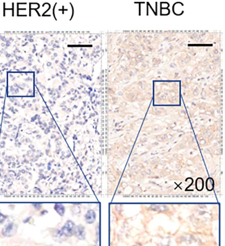
Cui et al.                                                                                                                   PTK7 Regulates EGFR/Akt in TNBC

                                              proliferation and migration in TNBC cell lines. PTK7 knockdown MDA-MB-468 cell
                                              bearing mouse model further demonstrated that PTK7-deficiency inhibits TNBC tumor
                                              progression in vivo.
                                              Conclusion: This study identified PTK7 as a potential marker of worse prognosis in TNBC
                                              and revealed PTK7 promotes TNBC metastasis and progression via EGFR/Akt signaling
                                              pathway.
                                              Keywords: PTK7, triple-negative breast cancer (TNBC), migration, progression, EGFR

INTRODUCTION                                                                           and lymph node metastasis (23). However, Gartner and
                                                                                       colleagues found elevated PTK7 mRNA expression level in
Triple-negative breast cancer (TNBC) is the most aggressive                            TNBC cell lines and PTK7 overexpression in metastatic lymph
subtype of breast cancer characterized by high invasiveness,                           node predicts shorter disease-free survival (DFS) in breast cancer
metastasis and heterogeneous clinical behavior (1–3). Due to                           patients (24). The controversy of PTK7 function in breast cancer
lacking expression of estrogen receptor (ER), progesterone                             may be due to its multiple molecular subtypes and heterogeneity.
receptor (PR), or human epidermal growth factor receptor 2                                Although some lines of evidence revealed the important role
(HER2), TNBC patients are not sensitive to endocrine therapy or                        of PTK7 in tumor progression, the molecular functions of PTK7
HER2-targeted therapy (4, 5). Resistance to conventional                               in metastasis and motility in TNBC remains elusive. Here we
systemic radiotherapy and chemotherapy and high occurrence                             demonstrate that PTK7 were predominantly upregulated in
of post-chemotherapy metastasis make it urgent to develop new                          breast cancer tissues. Expression levels of PTK7 predict a poor
TNBC treatment strategies (6–8). Therefore, the importance of                          outcome and an increased risk for cancer metastasis in TNBC
understanding the molecular biology of TNBC has gained                                 patients. Moreover, PTK7 regulates tumor metastasis and
considerable attention.                                                                collagen fibril organization via EGFR-Akt pathway.
   Protein tyrosine kinase 7 (PTK7), a member of the receptor
tyrosine kinase (RTK) superfamily, is a catalytically inactive RTK
that plays a role in multiple cellular processes including polarity
and adhesion (9–12). PTK7 interacts with Wnt3a and Wnt8 and                            MATERIAL AND METHODS
acts as an important regulator of both non-canonical and
canonical Wnt signaling in multiple developmental contexts
                                                                                       Plasmid Constructs and Reagents
                                                                                       Antibodies for PTK7 (25618, 1:1,000) and phosphor-Akt (S473)
(13, 14). PTK7 activates AP-1 and NF-kB signaling and
                                                                                       (4060, 1:1,000) were purchased from Cell Signaling Technology
upregulates matrix metalloproteinase-9 (MMP9) which results
                                                                                       (Danvers, MA, USA). Antibody for b-actin (AC026, 1:20,000) was
in increasing invasive properties of esophageal squamous cell
                                                                                       from Abclonal (Wuhan, Hubei, China). Antibody for Tubulin
carcinoma cells (15). PTK7 binds and activates FGFR1 and
                                                                                       (10068-1-AP, 1:1,000) was from Proteintech (Chicago, IL, USA).
increases tumorigenicity (16). Furthermore, PTK7 regulates the
                                                                                       Antibody for EGFR (1114-1, 1:1,000) was from Epitomics
activity of kinase insert domain receptor (KDR) and thereby
                                                                                       (Burlingame, CA, USA). Antibody for phosphor-EGFR (Y1173)
participates in VEGF induced tumor angiogenesis (17).
                                                                                       (ET1610-4, 1:1,000) was from HuaBio (Hangzhou, Zhejiang,
   The expression and function of PTK7 have been investigated
                                                                                       China). Antibody for Akt (B-1, 1:1,000) was from Santa Cruz
in several human cancers, although controversial results have
                                                                                       Biotechnology (Santa Cruz, CA, USA). The human PTK7
been obtained (18–24). PTK7 is highly expressed and plays an
                                                                                       expression plasmid was from Addgene (Watertown, MA, USA).
oncogenic role in lung adenocarcinoma (18). PTK7 is
                                                                                       LV3 lentiviral vectors encoding shRNAs silencing PTK7 or a
overexpressed and contributes to thyroid (19) and cervical (22)
                                                                                       nonsilencing control shRNA (shNC) were purchased from
cancer progression. A bioinformatics analysis reported that
                                                                                       GenePharma (Suzhou, Jiangsu, China). The sequences of PTK7
PTK7 is highly expressed in stage I-IV hepatocellular
                                                                                       shRNAs: shPTK7#1: 5’-GGATGATGTCACTGGAGAAGA-3’;
carcinoma (HCC) and considered as an independent
                                                                                       shPTK7#2: 5’-GGAGGGAGTTGGAGATGTTTG-3’. For gene
prognostic marker for reduced overall survival (21). Another
                                                                                       silencing, 293T cells were transfected with lentiviral vectors
investigation of PTK7 expression in 79 consecutive invasive
                                                                                       together with packaging plasmids and packaged lentiviral
breast cancer tissues by immunohistochemistry found that
                                                                                       particles were prepared and used to infect indicated cells
PTK7 expression level negatively associates with tumor grade
                                                                                       followed by puromycin selection.

Abbreviations: BP, Biological Process; ER, estrogen receptor; GO, Gene Ontology;       Patients and Tissue Microarray
HER2, human epidermal growth factor receptor 2; IHC, Immunohistochemistry;             Two tissue microarrays containing 280 cases of breast cancer tissues
KDR, kinase insert domain receptor; KEGG, Kyoto Encyclopedia of Genes and
Genomes; MMP9, matrix metalloproteinase-9; PR, progesterone receptor; PTK7,
                                                                                       collected from 2006 to 2016 with overall survival time (3- to 11-year
protein tyrosine kinase 7; RFS, recurrence-free survival; RPTK, receptor protein       follow-up, mean follow-up time was 106 months) were purchased
tyrosine kinase; TNBC, triple-negative breast cancer.                                  from BioChip (Shanghai, China). The breast cancers were divided

Frontiers in Oncology | www.frontiersin.org                                        2                                     July 2021 | Volume 11 | Article 699889
Cui et al.                                                                                                                          PTK7 Regulates EGFR/Akt in TNBC

into the four intrinsic subtypes, Luminal A, Luminal B, HER2(+)                         IHC Staining
and TNBC, based on immunohistochemistry (IHC) results for ER,                           Tissue microarrays were treated with heat-induced antigen-
PR, HER2 and Ki67 provided by BioChip. ER, PR and HER2                                  retrieval procedures and IHC staining was performed using the
positivity was defined using 2018 ASCO/CAP guidelines. ER and                            avidin–biotin complex method. The tissue sections were blocked
PR positivity was defined as ER ≥ 1%, PR ≥ 1%, respectively. For                         with 10% goat serum and incubated with anti-PTK7 antibody
HER2, IHC 3+ or IHC 2+/ISH+ was defined as HER2 positive. ER                             (25618; 1:1,000 dilution; Cell Signaling Technology) at 4°C
positive, PR ≥ 20% and Ki67 < 15% was defined as Luminal A. ER                           overnight. Then, the slides were washed three times using PBS
positive, PR < 20% and Ki67 > 30% was defined as Luminal B. All                          followed by biotinylated-secondary antibody incubation for 2
the patients provided informed consent. The study was approved by                       hours at room temperature. The slides were washed three times
Institutional Review Board of Hebei University Affiliated Hospital.                      and incubated with streptavidin/HRP. DAB peroxidase substrate
The patient information and histological features were displayed in                     was utilized for visualization. The IHC staining was assessed by
Tables 1 and 2. The analysis of clinicopathological features were                       two pathologists who were blinded to clinical information. PTK7
based on 280 breast cancer cases or 49 TNBC cases where indicated,                      IHC score was assessed according to the staining intensity (no
excluding a few cases because of missing data.                                          staining = 0; weak staining = 1, moderate staining = 2 and strong
                                                                                        staining = 3) and the percentage of stained cells (0–4% = 0, 5%–
                                                                                        25% = 1, 26%–50% = 2, 51%–75% = 3 and 76%–100% = 4). IHC
TABLE 1 | Patient characteristics.                                                      score = stained cell percentage score × staining intensity score.
Variable                                                      No. of Patients (%)
                                                                                        PTK7 protein expression was divided into low expression (IHC
                                                                                        score 0~4), medium expression (IHC score 4~8) and high
No. of BC patients                                                 280 (100)            expression (IHC score 8~12) according to the IHC score.
Age: Median [range]                                                59 [29-88]
Molecular typing
  Luminal A                                                        96 (37.5)            PTK7 Gene Expression Profiling
  Luminal B                                                        37 (14.5)
                                                                                        GEPIA: Gene Expression Profiling Interactive Analysis system
  HER2(+)                                                          74 (28.9)
  TNBC                                                             49 (19.1)            (http://gepia.cancer-pku.cn/), a newly developed interactive web
TNM stage                                                                               server for analyzing the RNA sequencing expression data was
  I                                                                57 (20.4)            used to analyze PTK7 expression in breast invasive carcinoma
  II                                                               138 (49.3)           (n = 1,085) and matched normal breast tissues (TCGA normal
  III                                                              81 (28.9)
                                                                                        and GTEx dataset, n = 291). PTK7 expression according to
Lymphatic metastasis
  Negative                                                         143 (51.3)           triple-negative status using Breast Cancer Gene-Expression
  Positive                                                         136 (48.7)           Miner v4.3 system (http://bcgenex.centregauducheau.fr/BC-
Distant metastasis                                                                      GEM/). TNBC (n = 572) and non-TNBC breast cancer (n =
  Negative                                                         280 (100)            6,739) DNA microarray data were selected. For PTK7 genetic
  Positive                                                           0 (0)
Prognosis
                                                                                        alteration analysis in invasive breast carcinoma, cBioPortal for
  Survival                                                         208 (74.3)           Cancer Genomics (http://www.cbioportal.org/) breast cancer
  Death                                                            72 (25.7)            datasets were used which includes 1,904 patients with Agilent

TABLE 2 | Molecular subtyping and clinical characteristics.

Variable                                                                                              Molecular subtyping

                                                      Luminal A                          Luminal B                          HER2(+)                          TNBC

No. of subtyping patients: n (%)                       96 (100)                           37 (100)                           74 (100)                       49 (100)
Age: Median [range]                                   61 [37-88]                         66 [41-88]                         57 [33-87]                     57 [32-84]
TNM stage: n (%)
  I                                                    21 (22.1)                          7 (18.9)                          16 (22.2)                        12 (25)
  II                                                   46 (48.4)                          24 (64.9)                         33 (45.8)                       18 (37.5)
  III                                                  28 (29.5)                          6 (16.2)                          23 (32.0)                       18 (37.5)
Lymphatic metastasis: n (%)
  Negative                                             47 (51.6)                          20 (58.8)                         36 (50.7)                       26 (53.1)
  Positive                                             44 (48.4)                          14 (41.2)                         35 (49.3)                       23 (46.9)
Prognosis: n (%)
  Survival                                             79 (82.3)                          34 (91.9)                         52 (70.3)                       30 (61.2)
  Death                                                17 (17.7)                           3 (8.1)                          22 (29.7)                       19 (38.8)
PTK7 expression
  IHC score: Mean ± s.d.                             5.06 ± 2.42                        5.12 ± 2.39                     6.20 ± 2.41                       7.46 ± 2.68
  Low PTK7 level: n (%)                               36 (37.5)                          13 (35.1)                       20 (27.0)                          6 (12.2)
  Medium PTK7 level: n (%)                            41 (42.7)                          14 (37.8)                       25 (33.8)                         12 (24.5)
  High PTK7 level: n (%)                              19 (19.8)                          10 (27.0)                       29 (39.2)                         31 (63.3)

Frontiers in Oncology | www.frontiersin.org                                         3                                           July 2021 | Volume 11 | Article 699889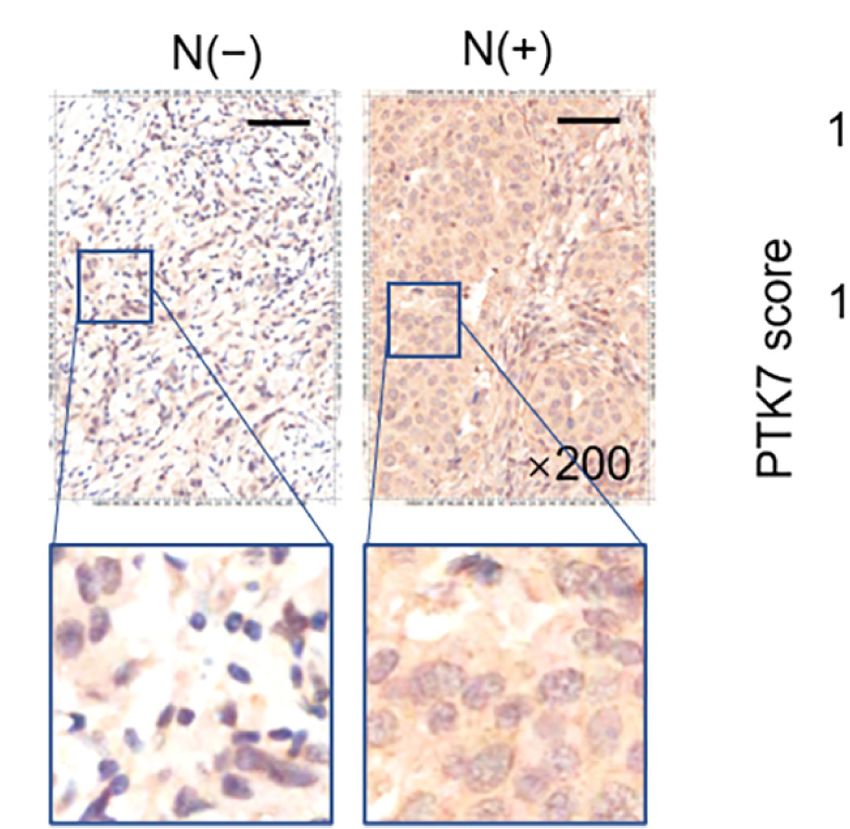
Cui et al.                                                                                                    PTK7 Regulates EGFR/Akt in TNBC

microarray data (METABRIC, Nature 2012 & Nat                             stable cell lines. Immunoblotting assays were performed to
Commun 2016).                                                            examine the silencing efficiency.

Recurrence-Free Survival (RFS) Assay by                                  Immunoblot Assay
Kaplan-Meier Plotter                                                     Total cell lysates were prepared using RIPA buffer (50 mM Tris-
The prognostic value of PTK7 mRNA expression was evaluated               HCl, pH7.4, 150 mM NaCl, 1% NP-40, 0.5% Na-deoxycholate, 1
using an online database, Kaplan-Meier Plotter (http://www.              mM EDTA, 1 mM PMSF, 1 mM DTT, protease inhibitor
kmplot.com/). To analyze RFS of patients with Luminal A,                 cocktail). Cell lysates were separated by SDS-PAGE, transferred
Luminal B, HER2(+) and TNBC subtypes of breast cancer,                   to PVDF membranes, blocked with 5% non-fat milk and
patients were divided into two groups (high versus low                   incubated with a specific primary antibody. The membranes
expression) and assessed by a Kaplan-Meier survival plot, with           were then washed and incubated with HRP-conjugated
the hazard ratio (HR) with 95% confidence intervals (CIs) and             secondary antibody and visualized by chemiluminescent
log rank P-value.                                                        detection (ECL, Roche Diagnostics, Penzberg, Germany) and
                                                                         exposure to X-ray film (Thermo Fisher Scientific, Waltham,
KEGG, GO and PTK7 Correlated-                                            MA, USA). The experiment was repeated independently 3 times.
Gene Analysis                                                            Actin Cytoskeleton Staining
PTK7 correlated genes were investigated using breast cancer              Cells were fixed in 4% paraformaldehyde at room temperature
datasets including 1,904 patients with Agilent microarray data           for 10 min followed by permeabilization with 0.1% Triton X-100.
(http://www.cbioportal.org/). Positively- (Spearman’s                    Cells were incubated with TRITC-tagged phalloidin in the dark
correlation > 0.3, P < 0.01) and negatively- (Spearman’s                 at room temperature for 30 min and stained with 4’,6-diamidino-
correlation < -0.3, P < 0.01) correlated genes were selected as          2-phenylindole (DAPI) for 3 min to visualize nuclear. Confocal
candidate PTK7 correlated genes. PTK7 correlated genes were              microscopy was performed with the Confocal Laser Scanning
analyzed using Kyoto Encyclopedia of Genes and Genomes                   Microscope Systems (FV3000, Olympus, Shinjuku, Japan). The
(KEGG) by DAVID: Functional Annotation Tools (https://                   experiment was repeated independently 3 times.
david.ncifcrf.gov/tools.jsp) and Gene Ontology (GO) was
performed using DAVID: Functional Annotation Tools                       Cell Proliferation Assays
(https://david.ncifcrf.gov/tools.jsp). Pair-wise gene correlation        For MTT assay, 1×10⁴ cells were seeded into 96-well plates and
of PTK7 with EGFR, COL1A1, FN1, WNT5B, MMP11,                            cultured for 0, 24, 48 or 72 hours. Before detection, each well was
MMP14 and SDC1 in breast invasive carcinoma were analyzed                added with 20 mL MTT reagent (0.5 mg/mL in PBS) followed by
using GEPIA Correlation Analysis tools (http://gepia.cancer-             an additional 2 hours incubation. The medium was removed and
pku.cn/detail.php?clicktag=correlation).                                 purple-blue MTT formazan precipitate was dissolved in 100 mL
                                                                         DMSO for 10 min at room temperature. The absorbance was
Cell Culture                                                             measured at 490 nm using a BioTek Epoch Spectrophomometer
Human TNBC cell lines MDA-MB-436, MDA-MB-468, MDA-                       (BioTek, Winooski, VT, USA). For colony formation, a single-cell
MB-231, MCF7 and SK-BR-3 were obtained from Cell Resource                suspension was prepared and cells were seeded into a 6-well plate
Center (IBMS, CAMS/PUMC, Beijing, China). Human embryo                   in a concentration at 750 cells/mL and incubated for 2 weeks. Cells
kidney 293T cell line was obtained from Cell Resource Center of          were stained with crystal violet and colony formation was
Shanghai Institutes for Biological Sciences, Chinese Academy of          photographed under a phase-contrast microscope and colony
Science, China. MDA-MB-436, MDA-MB-468 and MDA-MB-                       numbers and diameters were measured. All the experiments
231 were cultured in RPMI-1640 medium supplemented with                  were repeated independently 3 times.
10% FBS, 100 U/mL of penicillin, and 100 mg/mL of
streptomycin. HEK293T, MCF7 and SK-BR-3 cells were                       Cell Invasion Assay
maintained in Dulbecco’s Modified Eagle’s Medium (DMEM)                   Cell invasion assay were performed using a modified Boyden
with 10% FBS, 100 U/mL of penicillin, and 100 mg/mL of                   transwell system. The transwell permeable supports chambers
streptomycin. All cell lines were cultured in a humidified                (Corning Incorporated, Corning, NY, USA) with 8-mm pore size
atmosphere of 5% CO2, 95% air at 37°C.                                   were pre-coated with 10 mg/L Matrigel overnight at 4°C and
                                                                         1×10⁵ cells were seeded to the upper chamber of the transwell
Gene Silencing                                                           system and incubated at 37°C for 24 hours. Cells remaining on
For gene silencing, HEK293T cells were transfected with LV3              the upper chamber were mechanically erased with a cotton swab
lentiviral vectors encoding specific shRNAs targeting PTK7                and the cells migrated to the lower surface of the filter were
(shPTK7#1 and shPTK7#2) or control shRNAs (shNC) along                   stained with crystal violet and counted under the microscope.
with packaging plasmids psPAX2 and pMD2.G. The supernatant               The experiment was repeated independently 3 times.
was collected at 48 hours after transfection and filtered through a
0.45 mm polysulfone filter for lentiviral particles preparation.          Tumor Xenograft
MDA-MB-436 and MDA-MB-468 cells were than transduced                     Male BALB/c-nu mice at 4-5 weeks old were used to establish
with the packaged virus and selected by puromycin to establish           TNBC mouse model in vivo. All the mice were purchased from

Frontiers in Oncology | www.frontiersin.org                          4                                    July 2021 | Volume 11 | Article 699889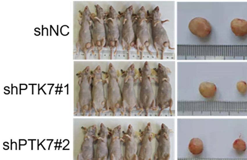
Cui et al. PTK7 Regulates EGFR/Akt in TNBC
the Beijing HFK Bioscience Co., Ltd (Beijing, China) and housed RESULTS
in a specific pathogen-free environment at Hebei University
Laboratory Animal Research Center. All experiments were PTK7 Is Highly Expressed and Correlates
approved by the Animal Research Ethics Committee of the With Worse Prognosis in TNBC
authors’ institution. Briefly, MDA-MB-468 cells (5×10⁵) were To explore the potential role of PTK7 in breast cancer, we
injected s.c. into the right mammary fat pad of nude mice. Each analyzed PTK7 expression in breast cancer using an RNA-Seq
group consisted of six mice. The challenged mice were datasets GEPIA: Gene Expression Profiling Interactive Analysis
monitored every 2 days for tumor growth. The tumor volume system (http://gepia.cancer-pku.cn/) and found that PTK7
was estimated according to the formula: Volume = 0.5 × a × b2, transcription levels are significantly higher in breast invasive
where a and b represent the largest and smallest diameters, carcinoma (BRCA) tissues (n = 1,085) than that in matched non-
respectively. All the mice were sacrificed 61 days after injection tumor tissues (n = 291), suggesting a potential role of PTK7 in
and the tumors were weighted, measured and photographed. The breast cancer (Figure 1A).
experiment was repeated independently 2 times. To further investigate the clinical relevance of PTK7, we
evaluated breast cancer tissue samples from 280 human
subjects (Table 1) and performed IHC staining against PTK7
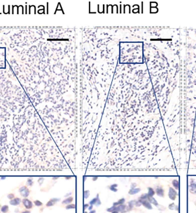
(Figure 1B). IHC staining showed that PTK7 was expressed both
Statistical Analysis in the cytosol and the nucleus of breast cancer cells (Figure 1C).
Statistical analysis was performed using GraphPad Prism The samples were divided into four subtypes, Luminal A,
Software 8.0 (GraphPad Software, San Diego, CA, USA). Two- Luminal B, HER2(+) and TNBC, based on ER, PR, HER2 and
tailed Student’s t tests or one-way ANOVA according to the Ki67 expression. Interestingly, PTK7 expression was distinctively
number of groups compared. P-values < 0.05 were considered higher in TNBC subtype than that in Luminal A, Luminal B and
significant and the level of significance expressed as follows: HER2(+) molecular subtypes (Figures 1C, D). Next, three TNBC
*, P < 0.05; **, P < 0.01; ***, P < 0.001; ****, P < 0.0001. cell lines (MDA-MB-468, MDA-MB-436 and MDA-MB-231),
A C
B D
FIGURE 1 | PTK7 expression is upregulated in breast cancer. (A) Box plots of PTK7 expression in breast invasive carcinoma (BRCA) using GEPIA: Gene
Expression Profiling Interactive Analysis system (http://gepia.cancer-pku.cn/). BRCA tumor (T) and non-tumor (N) TCGA normal and GTEx dataset included 1,085
tumor cases (T) and 291 non-tumor cases (N) was selected to observe the expression of PTK7. (B) IHC staining of PTK7 expression in breast cancer tissue
microarray. (C) Representative images from PTK7 IHC staining in Luminal A, Luminal B, HER2(+) and TNBC subtypes of breast cancer tissues. Magnification, ×200;
scale bars, 100 mm. (D) Scatter dot plots of PTK7 expression in tumors with different molecular subtypes. Data were analyzed using one-way ANOVA and Tukey’s
multiple comparisons test and are shown as mean ± s.d. *P < 0.05, **P < 0.01, ****P < 0.0001. ns, no significance.
Frontiers in Oncology | www.frontiersin.org 5 July 2021 | Volume 11 | Article 699889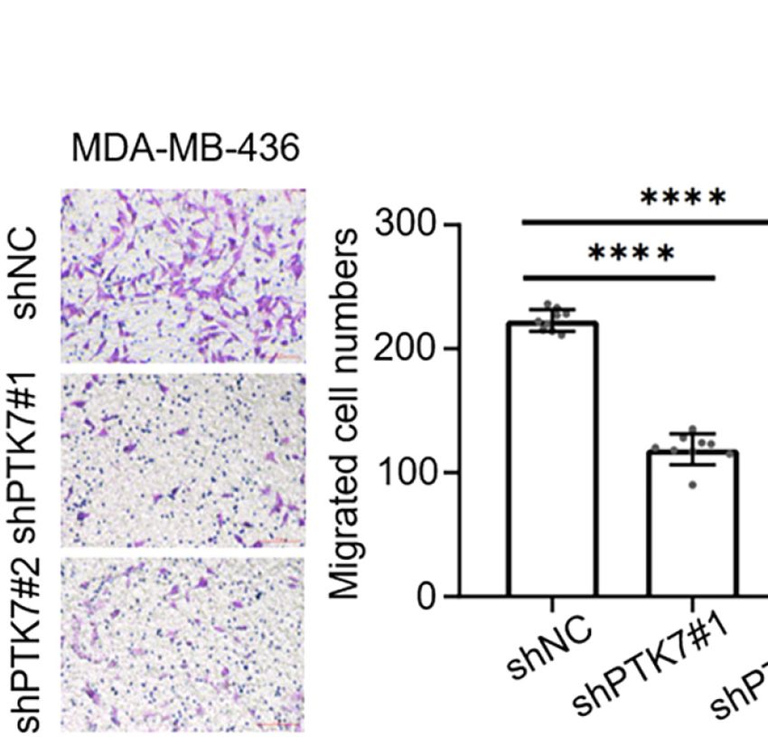
Cui et al.                                                                                                                      PTK7 Regulates EGFR/Akt in TNBC

ER(+) breast cancer cell line (MCF7) and HER2(+) breast cancer                      divided into two groups based on PTK7 expression and no
cell line (SK-BR-3) were used to analyze PTK7 expression and                        significant difference was found Luminal A, Luminal B and
the result showed significantly higher PTK7 levels in TNBC cells                     HER2 subtypes of breast cancer (Supplementary Figure S1C).
than that in MCF7 and SK-BR-3 cells (data not shown).                               Interestingly, a significantly worse RFS was found in PTK7 high
    PTK7 genetic alteration and expression levels were further                      expressed TNBC (Supplementary Figure S1C). These data
analyzed using online database in different molecular subtypes of                   indicated that PTK7 plays an important role in TNBC and
breast cancer. TNBC (n = 572) and non-TNBC breast cancer (n =                       correlated with breast cancer prognosis.
6,739) DNA microarray data were selected from Breast Cancer
Gene-Expression Miner v4.3 system (http://bcgenex.                                  Elevated PTK7 Is Associated With Tumor
centregauducheau.fr/BC-GEM/) and PTK7 expressions were                              Growth and Metastasis in TNBC
higher in TNBC than that in non-TNBC (Supplementary Figure                          Next, we selected all the TNBC tissue samples (n = 49) from 280
S1A). PTK7 genetic alterations in invasive breast carcinoma were                    subjects of breast cancer tissue microarray (Table 1) and divided
analyzed using cBioPortal for Cancer Genomics (http://www.                          them into groups based on TNM stages and lymph node metastasis.
cbioportal.org/) breast cancer datasets and the results revealed                    PTK7 expression was significantly higher in TNM II and TNM III
that PTK7 genetic amplification exists in 1.6% cases (n = 30) of                     groups than that in TNM I group (Figures 3A, B). Moreover,
invasive breast carcinoma patients (n = 1,904), most of which are                   elevated PTK7 was observed in TNBC with lymph node metastasis
ER(-), PR(-) and HER2(-) (TNBC, n = 22) (Supplementary                              (Figures 3D, E). When dividing TNBC tumor samples into groups
Figure S1B).                                                                        based on PTK7 IHC staining score, the percentage of high PTK7
    PTK7 expression was qualified as low, medium and high                            expression samples was significantly higher in TNBC with TNM
according to IHC score and a follow-up analysis of patient                          stage III and lymph node metastasis groups (Figures 3C, F). These
overall survival showed that higher expression of PTK7 in                           data therefore collectively suggested that PTK7 participates in tumor
TNBC breast cancer tissue correlated with a worse outcome                           metastasis in TNBC.
(Figure 2D). However, there was no statistical difference in
Luminal A, Luminal B and HER2(+) subtypes (Figures 2A–C).                           PTK7 Upregulates EGFR/Akt
Next, we performed RFS analysis using online database Kaplan-                       Signaling Activation
Meier Plotter (http://www.kmplot.com/) to assess the effect of                      We next analyzed PTK7 co-expression genes using breast cancer
PTK7 on breast cancer prognosis. Breast cancer samples were                         datasets including 1,904 patients with Agilent microarray data

                                  A                                                 B

                                  C                                                 D

 FIGURE 2 | PTK7 upregulation is associated with poor patient survival in TNBC. Breast cancer samples were divided into groups based on PTK7 expression [low
 expression (IHC score 0~4), medium expression (IHC score 4~8) and high expression (IHC score 8~12)]. Kaplan-Meier overall survival curve analysis and two-sided
 log-rank tests were performed in Luminal A (A), Luminal B (B), HER2(+) (C) and TNBC (D) breast cancer molecular subtypes, respectively. Marks on graph lines
 represent censored samples.

Frontiers in Oncology | www.frontiersin.org                                     6                                          July 2021 | Volume 11 | Article 699889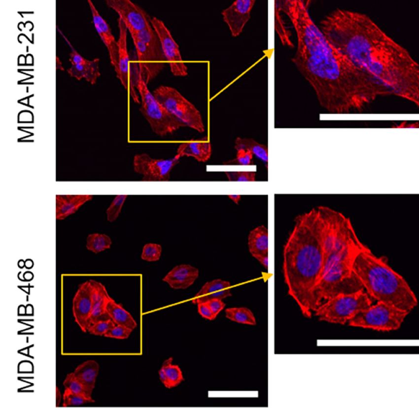
Cui et al.                                                                                                                       PTK7 Regulates EGFR/Akt in TNBC

                    A                                          B                            C

                    D                                 E                              F

 FIGURE 3 | Upregulation of PTK7 is related to metastasis and TNM stage in TNBC. (A) TNBC samples from 280 subjects of breast cancer tissue microarray were
 selected and divided into three groups based on TNM stages (AJCC staging). Representative images of IHC staining of PTK7 expression in the three groups (stage I,
 II and III) are shown. Magnification, ×200; scale bars, 100 mm. (B) Scatter dot plots of PTK7 scores in the three groups described in (A). Data were analyzed using
 one-way ANOVA and are shown as mean ± s.d. *P < 0.05. (C) The percentage of cases in the groups described in (A). Data were analyzed using Pearson’s c2 test.
 Light grey, low PTK7 level (IHC score 0~4); dark grey, medium PTK7 level (IHC score 5~8); black, high PTK7 level (IHC score 8~12). (D) TNBC samples were
 divided into two groups based on lymph node metastasis. Representative images of PTK7 staining in TNBC with or without lymph node metastasis are shown.
 Magnification, ×200; scale bars, 100 mm. (E) Scatter dot plots of PTK7 scores in the two groups described in (D). Data were analyzed using one-way ANOVA and
 are shown as mean ± s.d. *P < 0.05. (F) The percentage of cases in the groups described in (D). Data were analyzed using Pearson’s c2 test. Light grey, low PTK7
 level (IHC score 0~4); dark grey, medium PTK7 level (IHC score 5~8); black, high PTK7 level (IHC score 8~12). ns, no significance.

(http://www.cbioportal.org/). As shown in Figure 4A,                                 PTK7 Is Associated With Extracellular
Supplementary Figure S2 and Supplementary Table S1, 374                              Matrix Organization and Cytoskeleton
PTK7 positively-correlated genes (Spearman’s correlation > 0.3,                      Remodeling in Breast Cancer Cells
P < 0.01) and 289 PTK7-negatively-correlated genes (Spearman’s
correlation < -0.3, P < 0.01) was found. The functions of PTK7                       We further investigated Gene Ontology (GO) using DAVID:
positively-correlated genes were predicted by the analysis of                        Functional Annotation Tools (https://david.ncifcrf.gov/tools.jsp).
Kyoto Encyclopedia of Genes and Genomes (KEGG) by                                    Biological Process (BP) of PTK7 positively- and negatively-
DAVID: Functional Annotation Tools (https://david.ncifcrf.                           correlated genes showed that 16 biological processes, including
gov/tools.jsp) and 9 pathways related to the functions of PTK7                       extracellular matrix organization (GO:0030198), cell adhesion
alterations in invasive breast cancer were found (Figure 4B, right                   (GO:0007155), actin filament organization (GO:0007015) and
panel). PI3K/Akt signaling pathway (hsa04151) and actin                              positive regulation of cell migration (GO:0030335) were related
cytoskeleton regulation (hsa04810) were significantly enriched                        to PTK7 positively-correlated genes (Figure 5A). To further exam
in PTK7 positively-correlated genes and the associated genes are                     the molecular mechanism, pair-wise gene correlation analysis of
listed (Figure 4B, left panel). To further investigate function of                   PTK7 and key migration associated genes in breast cancer were
PTK7 in EGFR-PI3K-Akt signaling pathway in breast cancer, we                         analyzed using GEPIA correlation analysis tool. As shown in
performed PTK7 and EGFR pair-wise gene correlation analysis                          Figure 5B, PTK7 expression in breast cancer was significantly
using GEPIA (Figure 4C) and further confirmed EGFR                                    positively correlated with COL1A1, FN1, WNT5B, MMP11,
expression positively correlated with PTK7 (R = 0.42, P = 2e-                        MMP14 and SDC1 in breast cancer.
48). Then, wild type or PTK7-knockdown TNBC cells (MDA-                                  To further identify the potential role of PTK7 in TNBC
MB-468 and MDA-MB-231) were stimulated with EGF (500                                 cytoskeleton remodeling, MDA-MB-231 and MDA-MB-468
ng/ml) and phosphor-EGFR and phosphor-Akt levels were                                cells were transduced with shNC, shPTK7#1 or shPTK7#2. F-
significantly lower in PTK7-knockdown cells than that in                              actin filaments were stained with phalloidin and the result
control cells (Figures 4D, E), which suggested that PTK7                             showed that the actin filaments were recruited into thick and
regulates EGFR/Akt signaling pathway.                                                long actin bundles aligned along the long axis in shNC MDA-

Frontiers in Oncology | www.frontiersin.org                                      7                                          July 2021 | Volume 11 | Article 699889
Cui et al.                                                                                                                         PTK7 Regulates EGFR/Akt in TNBC

             A                                      B

             C                                  D                                                        E

 FIGURE 4 | PTK7 regulates EGFR-PI3K-Akt pathway in breast cancer. (A) PTK7 co-expression analysis in invasive breast carcinoma using breast cancer datasets
 including 1,904 patients with Agilent microarray data (http://www.cbioportal.org/) and 374 PTK7 positively- (Spearman’s correlation > 0.3, P < 0.01) and 289 PTK7
 negative- (Spearman’s correlation < -0.3, P < 0.01) correlated genes were selected and used as candidate genes in the following analysis. (B) PTK7 positively-
 correlated genes were analyzed using Kyoto Encyclopedia of Genes and Genomes (KEGG) by DAVID: Functional Annotation Tools (https://david.ncifcrf.gov/tools.
 jsp). PTK7 positively correlated genes enriched in PI3K-Akt signaling (hsa04151) and Regulation of actin cytoskeleton (hsa04810) pathways are listed in the frames,
 respectively. (C) PTK7 and EGFR pair-wise gene correlation in breast invasive carcinoma were analyzed using GEPIA Correlation Analysis tools (http://gepia.cancer-
 pku.cn/detail.php?clicktag=correlation). (D, E) MDA-MB-468 (D) and MDA-MB-231 (E) cells were transduced with a non-targeting control shRNA (shNC) or two
 different PTK7-specific shRNAs (shPTK71 and shPTK7#2). Cells were stimulated with EGF (500 ng/ml) for 0, 20 or 40 minutes and phospho-EGFR and phosphor-
 Akt levels were evaluated using immunoblotting assay.

MB-231 and MDA-MB-468 cells; PTK7-knockdown markedly                                  PTK7-Deficiency Inhibits TNBC Tumor
reduced thick stress fibers (Figure 5C).                                               Growth In Vivo
                                                                                      To further analyze the role of PTK7 in TNBC tumorigenicity in
PTK7 Promotes Proliferation and                                                       vivo, shNC, shPTK7#1 and shPTK7#2 stable transduced MDA-
Migration in TNBC Cell Lines                                                          MB-468 cells were used to establish TNBC tumor bearing mouse
To identify the consequences of PTK7 in TNBC progression,                             model. The challenged mice were monitored every two days and
human PTK7 overexpression or knockdown TNBC cell lines                                sacrificed at day 61 after injection (Figure 7A). PTK7 knockdown
MDA-MB-436 and MDA-MB-468 were used and MTT cell                                      dramatically inhibited tumor growth (Figures 7B, C). These results
proliferation assay were performed. As expected,                                      suggested that PTK7 is required for TNBC progression in vivo.
overexpression of PTK7 in MDA-MB-436 and MDA-MB-468
cells significantly promotes proliferation activity (Figures 6A, B)
and knockdown of PTK7 resulted in a decrease of cell growth                           DISCUSSION
(Figures 6C, D). Colony formation assay showed that both the
colony numbers and colony diameters significantly decreased in                         RTKs, a protein kinase family transducing extracellular signals
PTK7-knockdown cells (Figure 6E). Matrigel pre-coated Boyden                          across the cell membrane, were known to be grouped into 20
chamber was then used to analyze the roles of PTK7 in TNBC                            subfamilies and play pivotal roles in diverse cellular activities
cell migration and invasion. Knockdown of PTK7 in MDA-MB-                             including growth, differentiation, motility, and death (25–28).
436 and MDA-MB-468 cells exhibited decreased migration                                Many RTKs are involved in oncogenesis (29, 30). PTK7 is a
ability (Figures 6F, G), and overexpression of PTK7 promoted                          particular member of the RTK family that lacks detectable catalytic
transwell migration in TNBCs (Supplementary Figure S3).                               tyrosine kinase activity. Although PTK7 plays a role in multiple

Frontiers in Oncology | www.frontiersin.org                                       8                                           July 2021 | Volume 11 | Article 699889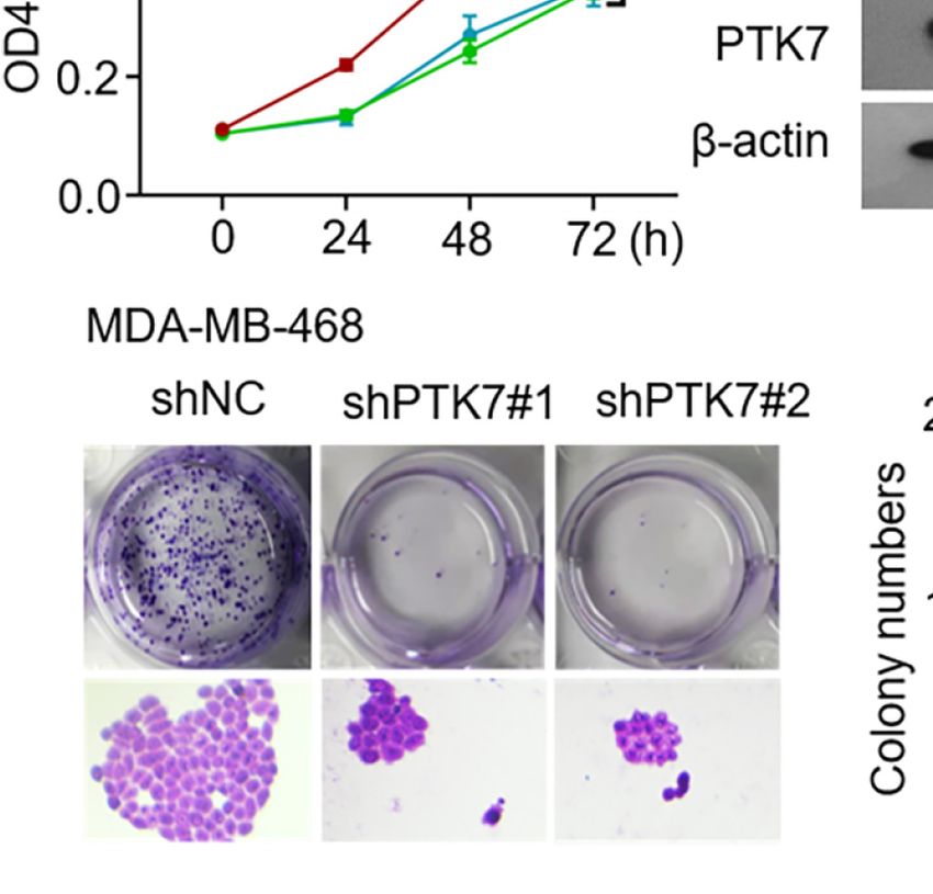
Cui et al.                                                                                                                           PTK7 Regulates EGFR/Akt in TNBC

             A

             B

             C

 FIGURE 5 | PTK7 associates with extracellular matrix organization and migration in breast cancer cells. (A) Gene Ontology (GO) was performed using DAVID:
 Functional Annotation Tools (https://david.ncifcrf.gov/tools.jsp) and Biological Process (BP) of PTK7 positively- and negatively-correlated genes were shown. (B) Pair-
 wise gene correlation of PTK7 with COL1A1, FN1, WNT5B, MMP11, MMP14 or SDC1 in breast invasive carcinoma were analyzed using GEPIA Correlation Analysis
 tools (http://gepia.cancer-pku.cn/detail.php?clicktag=correlation). (C) MDA-MB-231 and MDA-MB-468 cells were transduced with shNC, shPTK71 or shPTK72.
 Cells were stained for F-actin with TRITC-phalloidin. Pictures show the TRITC-tagged Phalloidin (red) and DAPI (purple). Presentative images are shown.
 Magnification, 400×; scale bars, 50 mm.

cellular processes during tumor progression, the definite role of                       (32). PTK7 expression in breast cancer predicts poor prognosis
PTK7 in breast cancer progression remains unclear.                                     (24). Recent evidence including 79 consecutive invasive breast
    A recent meta-analysis of the prognostic value of PTK7                             cancer tissues demonstrated that PTK7 expression is negatively
expression in human malignancies revealed that higher                                  associated with tumor grade and lymph node metastasis and may
expression of PTK7 significantly indicates worse prognosis in                           serve as a tumor suppressor in breast cancer (23).
human malignancies in 11 studies published with a total sample                             To reveal the clinical relevance of PTK7 in breast cancer, in
size of 2431 participants (31). The expression and function of                         the present study, we evaluated breast cancer tissue samples from
PTK7 in breast cancer have been well investigated, however,                            280 human subjects and performed tissue microarray IHC
controversial results were obtained. Several studies suggested                         staining against PTK7. There was no significant associate of
that PTK7 is highly expressed in TNBC cell lines and associates                        PTK7 expression with TNM stages from totally 280 breast cancer
with resistance to anthracycline-based chemotherapy in TNBC                            tissues. Interestingly, either correlations of PTK7 expression with

Frontiers in Oncology | www.frontiersin.org                                        9                                            July 2021 | Volume 11 | Article 699889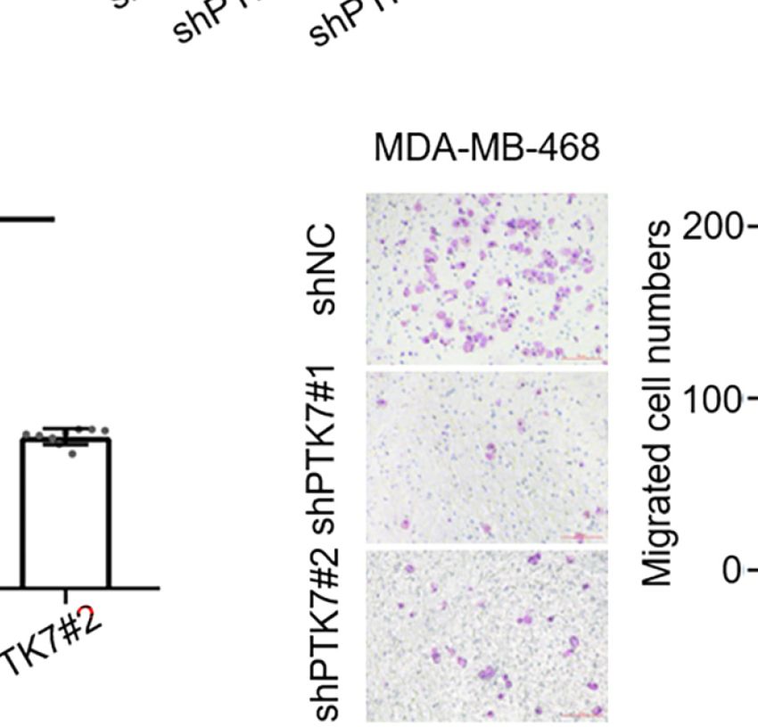
Cui et al.                                                                                                                     PTK7 Regulates EGFR/Akt in TNBC

             A                                                                  B

             C                                                                  D

             E

             F                                                            G

 FIGURE 6 | PTK7 participates in cell proliferation and migration in TNBC cell lines. (A, B) TNBC cell lines MDA-MB-436 (A) and MDA-MB-468 (B) were transfected
 with control vector pcDNA3 or PTK7 expression vector pcDNA3-PTK7 for 48 h and proliferation was evaluated using MTT assay. Data are shown as mean ± s.d.
 ****P < 0.0001. (C, D) MDA-MB-436 (C) and MDA-MB-468 (D) were transduced with a non-targeting control shRNA (shNC) or two different PTK7-specific shRNAs
 (shPTK71 and shPTK7#2). Cell proliferation was evaluated using MTT assay. Data are shown as mean ± s.d. ****P < 0.0001. (E) Colony formation assay was
 performed to determine proliferation in shNC-, shPTK7#1- or shPTK7#2-transduced MDA-MB-468 cells. Presentative images are shown (left) and colony numbers
 and colony diameters were shown as mean ± s.d. Magnification, ×100; ****P < 0.0001. (F, G) Transwell migration assay using Boyden chamber in shNC-,
 shPTK7#1- or shPTK7#2-transduced MDA-MB-436 (F) and MDA-MB-468 (G) cells was performed and photographed under a light microscope. Presentative
 images are shown (left) and migrated cells were counted and shown as mean ± s.d. Magnification, ×100; ****P < 0.0001.

clinicopathological parameters by tissue microarray IHC                                Compared with Luminal A, Luminal B and HER2(+) breast
staining or online RFS analysis by Kaplan-Meier Plotter                             cancer subtypes, patients with TNBC were always recognized to
(http://www.kmplot.com/) demonstrated that PTK7 expression                          have the worst overall survival data due to its rapid progression,
extraordinarily correlates with worse prognosis in ER/PR/HER2-                      high probabilities of early recurrence, and distant metastasis
negative (TNBC) breast cancer, which suggested a special                            resistant to standard treatment (33). According to the present
relationship of PTK7 expression with worse prognosis in                             data, TNBC with high PTK7 expression level predicts worse
TNBC. The function of PTK7 in breast cancer exhibits                                outcome. KEGG analysis and PTK7 gain- or loss-of-function
heterogeneity in multiple molecular subtypes may due to                             TNBC cell lines revealed that PTK7 regulates EGFR/Akt
different cell context and intracellular signaling mechanisms.                      signaling pathway. GO assay further demonstrated PTK7

Frontiers in Oncology | www.frontiersin.org                                    10                                         July 2021 | Volume 11 | Article 699889
Cui et al. PTK7 Regulates EGFR/Akt in TNBC
A
B C
FIGURE 7 | PTK7 promotes in vivo TNBC tumorigenicity. (A) Nude mice were injected into the mammary fat pad with shNC-, shPTK7#1- or shPTK7#2-transduced
MDA-MB-468 cells at 5×10⁵ cells per site. Mice were sacrificed and tumors were photographed 61 days after subcutaneous injection. (B) Tumor volume was
monitored every 2 days after injection and tumor growth curve is shown. Tumor volume was calculated by the formula: V = 1/2×a (length)×b2 (width). (C) The harvest
tumor weight was shown as mean ± s.d. ***P < 0.001; ****P < 0.0001.
participates in extracellular matrix organization and migration in novel therapies to fight against TNBC. To further define the
TNBC cells. A recent study revealed that PTK7 expression is independent predictive role and targeted therapy strategy of PTK7
associated with EGFR mutations and plays an oncogenic role in in TNBC, a larger sample of patients with TNBC treatment
lung adenocarcinomas (18). The role of PTK7-targeted antibody- information should be investigated.
drug conjugate has been investigated in several solid tumors,
including TNBC, and exhibits potential therapeutic activity (34–
36). In addition, our present data demonstrated that loss of PTK7
expression in TNBC cells results in a downregulated EGFR/Akt DATA AVAILABILITY STATEMENT
signaling and reduced tumor growth in MBA-MD-468 TNBC
cancer xenografts. These findings may have significant Publicly available datasets were analyzed in this study. This data
implicants for the treatment of TNBC via targeting PTK7. can be found here: GEPIA: Gene Expression Profiling Interactive
Taken together, this study identified PTK7 as a potential marker Analysis system (http://gepia.cancer-pku.cn/) cBioPortal for
of worse prognosis in TNBC. PTK7 promotes extracellular matrix Cancer Genomics (http://www.cbioportal.org/) Kaplan-Meier
organization and migration via EGFR/PI3K/Akt signaling pathway Plotter (http://www.kmplot.com/) DAVID: Functional
in TNBC. Strategies targeting PTK7 may inform the development of Annotation Tools (https://david.ncifcrf.gov/tools.jsp).
Frontiers in Oncology | www.frontiersin.org 11 July 2021 | Volume 11 | Article 699889Cui et al. PTK7 Regulates EGFR/Akt in TNBC
ETHICS STATEMENT FUNDING
The studies involving human participants were reviewed and This work was supported by the Natural Science Foundation of
approved by Ethics Committee of Affiliated Hospital of Hebei Hebei Province [grant number H2019201259, C2020201052,
University. The patients/participants provided their written H2020201295], the National Natural Science Foundation of
informed consent to participate in this study. The animal study China [grant Number 31971304], Hebei Province Foundation
was reviewed and approved by Animal Welfare and Ethical for the Returned Overseas Scholars [grant Number
Committee of Hebei University. C20200305], Government Foundation of Clinical Medicine
Talents Training Program of Hebei Province [grant number
361007], Medical Scientific Research Project of Hebei Province
AUTHOR CONTRIBUTIONS [grant number 20200170], Medical Science Foundation of
N-PC, SQ, SJ, and J-HS designed and carried out the experiments. Hebei University [grant number 2020A09] and Hebei
SQ and SJ analyzed the data. N-PC, SQ, YQ, Y-NW, and L-SZ University graduate innovation funding project [grant
performed immunohistochemical staining, tumor xenograft. SJ, number hbu2020ss081].
J-LH, T-TW, and W-WL performed cell culture, gene silencing,
immunoblotting, actin cytoskeleton staining, cell proliferation
assays, cell proliferation and invasion assays. J-HS, N-PC, SQ, SUPPLEMENTARY MATERIAL
SJ, and Y-NW analyzed and interpreted the data. J-HS, J-CZ,
Y-PM, and B-PC provided supervision and guidance. J-HS, Y-PM, The Supplementary Material for this article can be found online
and B-PC wrote the manuscript. All authors contributed to the at: https://www.frontiersin.org/articles/10.3389/fonc.2021.
article and approved the submitted version. 699889/full#supplementary-material
REFERENCES 12. Golubkov VS, Aleshin AE, Strongin AY. Potential Relation of Aberrant
Proteolysis of Human Protein Tyrosine Kinase 7 (PTK7) Chuzhoi by
1. Bao X, Shi R, Zhao T, Wang Y, Anastasov N, Rosemann M, et al. Integrated Membrane Type 1 Matrix Metalloproteinase (MT1-MMP) to Congenital
Analysis of Single-Cell RNA-Seq and Bulk RNA-Seq Unravels Tumour Defects. J Biol Chem (2011) 286(23):20970–6. doi: 10.1074/jbc.M111.237669
Heterogeneity Plus M2-Like Tumour-Associated Macrophage Infiltration 13. Peradziryi H, Kaplan NA, Podleschny M, Liu X, Wehner P, Borchers A, et al.
and Aggressiveness in TNBC. Cancer Immunol Immunother: CII (2021) 70 PTK7/Otk Interacts With Wnts and Inhibits Canonical Wnt Signalling.
(1):189–202. doi: 10.1007/s00262-020-02669-7 EMBO J (2011) 30(18):3729–40. doi: 10.1038/emboj.2011.236
2. Killock D. Pembrolizumab can Delay Progression of TNBC. Nat Rev Clin 14. Hayes M, Naito M, Daulat A, Angers S, Ciruna B. Ptk7 Promotes non-
Oncol (2021) 18(2):64. doi: 10.1038/s41571-020-00465-x Canonical Wnt/PCP-Mediated Morphogenesis and Inhibits Wnt/beta-
3. Sun X, Wang M, Wang M, Yu X, Guo J, Sun T, et al. Metabolic Catenin-Dependent Cell Fate Decisions During Vertebrate Development.
Reprogramming in Triple-Negative Breast Cancer. Front Oncol (2020) Development (2013) 140(8):1807–18. doi: 10.1242/dev.090183
10:428. doi: 10.3389/fonc.2020.00428 15. Shin WS, Hong Y, Lee HW, Lee ST. Catalytically Defective Receptor Protein
4. Avalos-Moreno M, Lopez-Tejada A, Blaya-Canovas JL, Cara-Lupianez FE, Tyrosine Kinase PTK7 Enhances Invasive Phenotype by Inducing MMP-9 Through
Gonzalez-Gonzalez A, Lorente JA, et al. Drug Repurposing for Triple- Activation of AP-1 and NF-kappaB in Esophageal Squamous Cell Carcinoma Cells.
Negative Breast Cancer. J Personalized Med (2020) 10(4):200. doi: 10.3390/ Oncotarget (2016) 7(45):73242–56. doi: 10.18632/oncotarget.12303
jpm10040200 16. Shin WS, Lee HW, Lee ST. Catalytically Inactive Receptor Tyrosine Kinase
5. Azim HA, Ghosn M, Oualla K, Kassem L. Personalized Treatment in PTK7 Activates FGFR1 Independent of FGF. FASEB J (2019) 33(11):12960–
Metastatic Triple-Negative Breast Cancer: The Outlook in 2020. Breast J 71. doi: 10.1096/fj.201900932R
(2020) 26(1):69–80. doi: 10.1111/tbj.13713 17. Shin WS, Na HW, Lee ST. Biphasic Effect of PTK7 on KDR Activity in
6. Wu S, Liu D, Li W, Song B, Chen C, Chen D, et al. Enhancing TNBC Chemo- Endothelial Cells and Angiogenesis. Biochim Biophys Acta (2015) 1853(10 Pt
Immunotherapy via Combination Reprogramming Tumor Immune A):2251–60. doi: 10.1016/j.bbamcr.2015.05.015
Microenvironment With Immunogenic Cell Death. Int J Pharm (2021) 18. Jiang W, He J, Lv B, Xi X, He G, He J. PTK7 Expression is Associated With Lymph
598:120333. doi: 10.1016/j.ijpharm.2021.120333 Node Metastasis, ALK and EGFR Mutations in Lung Adenocarcinomas. Histol
7. Vagia E, Mahalingam D, Cristofanilli M. The Landscape of Targeted Histopathol (2020) 35(5):489–95. doi: 10.14670/HH-18-183
Therapies in TNBC. Cancers (2020) 12(4):916. doi: 10.3390/cancers12040916 19. Duan F, Tang J, Kong FL, Zou HW, Ni BL, Yu JC. Identification of PTK7 as a
8. Massa C, Karn T, Denkert C, Schneeweiss A, Hanusch C, Blohmer JU, et al. Promising Therapeutic Target for Thyroid Cancer. Eur Rev Med Pharmacol
Differential Effect on Different Immune Subsets of Neoadjuvant Sci (2020) 24(12):6809–17. doi: 10.26355/eurrev_202006_21670
Chemotherapy in Patients With TNBC. J Immunother Cancer (2020) 8(2): 20. Bie J, Hu X, Yang M, Shi X, Zhang X, Wang Z. PTK7 Promotes the Malignant
e001261. doi: 10.1136/jitc-2020-001261 Properties of Cancer Stem-Like Cells in Esophageal Squamous Cell Lines.
9. Lu X, Borchers AG, Jolicoeur C, Rayburn H, Baker JC, Tessier-Lavigne M. Hum Cell (2020) 33(2):356–65. doi: 10.1007/s13577-019-00309-6
PTK7/CCK-4 Is a Novel Regulator of Planar Cell Polarity in Vertebrates. 21. Zou RC, Liang Y, Li LL, Tang JZ, Yang YP, Geng YC, et al. Bioinformatics
Nature (2004) 430(6995):93–8. doi: 10.1038/nature02677 Analysis Identifies Protein Tyrosine Kinase 7 (PTK7) as a Potential Prognostic
10. Lee J, Andreeva A, Sipe CW, Liu L, Cheng A, Lu X. PTK7 Regulates Myosin II and Therapeutic Biomarker in Stages I to IV Hepatocellular Carcinoma. Med
Activity to Orient Planar Polarity in the Mammalian Auditory Epithelium. Sci Monit (2019) 25:8618–27. doi: 10.12659/MSM.917142
Curr Biol (2012) 22(11):956–66. doi: 10.1016/j.cub.2012.03.068 22. Sun JJ, Li HL, Guo SJ, Ma H, Liu SJ, Liu D, et al. The Increased PTK7
11. Golubkov VS, Chekanov AV, Cieplak P, Aleshin AE, Chernov AV, Zhu W, Expression Is a Malignant Factor in Cervical Cancer. Dis Markers (2019)
et al. The Wnt/planar Cell Polarity Protein-Tyrosine Kinase-7 (PTK7) is a 2019:5380197. doi: 10.1155/2019/5380197
Highly Efficient Proteolytic Target of Membrane Type-1 Matrix 23. Sun J, Zhou Q, Tao Y, Chen J, Wang J. Loss of Expression of Protein Tyrosine
Metalloproteinase: Implications in Cancer and Embryogenesis. J Biol Chem Kinase 7 in Invasive Ductal Breast Cancers. Int J Clin Exp Pathol (2019) 12
(2010) 285(46):35740–9. doi: 10.1074/jbc.M110.165159 (3):1052–9.
Frontiers in Oncology | www.frontiersin.org 12 July 2021 | Volume 11 | Article 699889Cui et al. PTK7 Regulates EGFR/Akt in TNBC
24. Gartner S, Gunesch A, Knyazeva T, Wolf P, Hogel B, Eiermann W, et al. PTK 7 is a 33. Wu SY, Wang H, Shao ZM, Jiang YZ. Triple-Negative Breast Cancer: New
Transforming Gene and Prognostic Marker for Breast Cancer and Nodal Metastasis Treatment Strategies in the Era of Precision Medicine. Sci China Life Sci
Involvement. PloS One (2014) 9(1):e84472. doi: 10.1371/journal.pone.0084472 (2021) 64(3):372–88. doi: 10.1007/s11427-020-1714-8
25. Xu P, Chu J, Li Y, Wang Y, He Y, Qi C, et al. Novel Promising 4- 34. Messerli SM, Hoffman MM, Gnimpieba EZ, Bhardwaj RD. Therapeutic
Anilinoquinazoline-Based Derivatives as Multi-Target RTKs Inhibitors: Targeting of PTK7 is Cytotoxic in Atypical Teratoid Rhabdoid Tumors.
Design, Molecular Docking, Synthesis, and Antitumor Activities. Vitro Vivo Mol Cancer Res (2017) 15(8):973–83. doi: 10.1158/1541-7786.MCR-16-0432
Bioorg Med Chem (2019) 27(20):114938. doi: 10.1016/j.bmc.2019.06.001 35. Maitland ML, Sachdev JC, Sharma MR, Moreno V, Boni V, Kummar S, et al.
26. Majumder P, Roy K, Bagh S, Mukhopadhyay D. Receptor Tyrosine Kinases (RTKs) First-In-Human Study of PF-06647020 (Cofetuzumab Pelidotin), an
Consociate in Regulatory Clusters in Alzheimer’s Disease and Type 2 Diabetes. Mol Antibody-Drug Conjugate Targeting Protein Tyrosine Kinase 7 (PTK7), in
Cell Biochem (2019) 459(1-2):171–82. doi: 10.1007/s11010-019-03560-5 Advanced Solid Tumors. Clin Cancer Res (2021). doi: 10.1158/1078-
27. Di Liberto V, Mudo G, Belluardo N. Crosstalk Between Receptor Tyrosine Kinases 0432.CCR-20-3757
(RTKs) and G Protein-Coupled Receptors (GPCR) in the Brain: Focus on 36. Damelin M, Bankovich A, Bernstein J, Lucas J, Chen L, Williams S, et al. A
Heteroreceptor Complexes and Related Functional Neurotrophic Effects. PTK7-Targeted Antibody-Drug Conjugate Reduces Tumor-Initiating Cells
Neuropharmacology (2019) 152:67–77. doi: 10.1016/j.neuropharm.2018.11.018 and Induces Sustained Tumor Regressions. Sci Transl Med (2017) 9(372).
28. Shan Y, Wang B, Zhang J. New Strategies in Achieving Antiangiogenic Effect: doi: 10.1126/scitranslmed.aag2611
Multiplex Inhibitors Suppressing Compensatory Activations of RTKs. Med
Res Rev (2018) 38(5):1674–705. doi: 10.1002/med.21517 Conflict of Interest: The authors declare that the research was conducted in the
29. Nadhan R, Srinivas P, Pillai MR. RTKs in Pathobiology of Head and Neck absence of any commercial or financial relationships that could be construed as a
Cancers. Adv Cancer Res (2020) 147:319–73. doi: 10.1016/bs.acr.2020.04.008 potential conflict of interest.
30. Butti R, Das S, Gunasekaran VP, Yadav AS, Kumar D, Kundu GC. Receptor
Tyrosine Kinases (RTKs) in Breast Cancer: Signaling, Therapeutic Implications and Copyright © 2021 Cui, Qiao, Jiang, Hu, Wang, Liu, Qin, Wang, Zheng, Zhang, Ma,
Challenges. Mol Cancer (2018) 17(1):34. doi: 10.1186/s12943-018-0797-x Chen and Shi. This is an open-access article distributed under the terms of the
31. Chen G, Qi S, Yang X, Chen W. Prognostic Significance of PTK7 in Human Creative Commons Attribution License (CC BY). The use, distribution or
Malignancies. Histol Histopathol (2018) 33(4):379–88. doi: 10.14670/HH-11-933 reproduction in other forums is permitted, provided the original author(s) and the
32. Ataseven B, Angerer R, Kates R, Gunesch A, Knyazev P, Hogel B, et al. PTK7 copyright owner(s) are credited and that the original publication in this journal is
Expression in Triple-Negative Breast Cancer. Anticancer Res (2013) 33 cited, in accordance with accepted academic practice. No use, distribution or
(9):3759–63. reproduction is permitted which does not comply with these terms.
Frontiers in Oncology | www.frontiersin.org 13 July 2021 | Volume 11 | Article 699889You can also read