3D Organoid Culture From Adult Salivary Gland Tissues as an ex vivo Modeling of Salivary Gland Morphogenesis
←
→
Page content transcription
If your browser does not render page correctly, please read the page content below
BRIEF RESEARCH REPORT
                                                                                                                                           published: 12 August 2021
                                                                                                                                      doi: 10.3389/fcell.2021.698292

                                            3D Organoid Culture From Adult
                                            Salivary Gland Tissues as an ex vivo
                                            Modeling of Salivary Gland
                                            Morphogenesis
                                            Donghyun Kim † , Yeo-Jun Yoon † , Dojin Choi, Jisun Kim and Jae-Yol Lim*
                                            Department of Otorhinolaryngology, Yonsei University College of Medicine, Seoul, South Korea

                                            Lumen formation of salivary glands has been investigated using in vivo or ex vivo
                                            rudiment culture models. In this study, we used a three-dimensional (3D) salivary
                                            gland organoid culture system and demonstrated that lumen formation could be
                                            recapitulated in mouse SMG organoids. In our organoid culture system, lumen formation
                           Edited by:       was induced by vasoactive intestinal peptide and accelerated by treatment with RA.
           Andrea Erika Münsterberg,        Furthermore, lumen formation was observed in branching duct-like structure when
             University of East Anglia,
                      United Kingdom        cultured in combination of fibroblast growth factors (FGF) in the presence of retinoic
                        Reviewed by:        acid (RA). We suggest RA signaling-mediated regulation of VIPR1 and KRT7 as the
                     Lemonia Chatzeli,      underlying mechanism for lumen formation, rather than apoptosis in the organoid culture
             University of Cambridge,
                                            system. Collectively, our results support a fundamental role for RA in lumen formation
                       United Kingdom
                        Sofia J. Araújo,    and demonstrate the feasibility of 3D organoid culture as a tool for studying salivary
        University of Barcelona, Spain      gland morphogenesis.
                  *Correspondence:
                          Jae-Yol Lim       Keywords: salivary gland organoid, organoid culture, stem cells, retinoic acid, morphogenesis, lumen formation,
                                            vasoactive intestinal peptide
                    jylimmd@yuhs.ac
     † These  authors have contributed
    equally to this work and share first
                            authorship
                                           INTRODUCTION

                    Specialty section:
                                           Murine salivary glands, particularly submandibular glands (SMGs), have been widely used
         This article was submitted to     as a model for studying salivary gland development and common processes in branching
     Morphogenesis and Patterning,         morphogenesis and tubulogenesis (Steinberg et al., 2005; Nitta et al., 2009; Hsu and Yamada,
                a section of the journal   2010; Knox et al., 2010; Nedvetsky et al., 2014). The process of duct formation encompasses
  Frontiers in Cell and Developmental      duct elongation, fusion of microlumens to form contiguous large lumen, and lumen expansion
                                Biology    during development stages (Nedvetsky et al., 2014). Several factors, including the fibroblast
           Received: 21 April 2021         growth factor (FGF) family, epidermal growth factor (EGF) family, Wnt pathway components,
           Accepted: 26 July 2021          bone morphogenetic proteins (BMPs), neurotrophic growth factor family, and neurotransmitter
         Published: 12 August 2021
                                           signaling components, are involved in the tightly regulated salivary gland development in a
                               Citation:   spatiotemporal manner (De Moerlooze et al., 2000; Miyazaki et al., 2004; Jaskoll et al., 2005; Knox
       Kim D, Yoon Y-J, Choi D, Kim J      et al., 2010; Patel et al., 2011; Mattingly et al., 2015).
     and Lim J-Y (2021) 3D Organoid
                                               Retinoids are a group of nutritional compounds that include retinol, retinal, and retinoic acid
    Culture From Adult Salivary Gland
       Tissues as an ex vivo Modeling
                                           (RA). RA has several roles in organ morphogenesis, embryonic development, immune system,
    of Salivary Gland Morphogenesis.       and growth of various cell types (Tanumihardjo, 2011). RA-mediated signaling activates its nuclear
       Front. Cell Dev. Biol. 9:698292.    receptors, including the retinoic acid receptor (RAR) family and retinoid X receptor (RXR) family
      doi: 10.3389/fcell.2021.698292       transcription factors. RAR or RXR isoforms have redundant roles, enabling them to compensate

Frontiers in Cell and Developmental Biology | www.frontiersin.org                 1                                          August 2021 | Volume 9 | Article 698292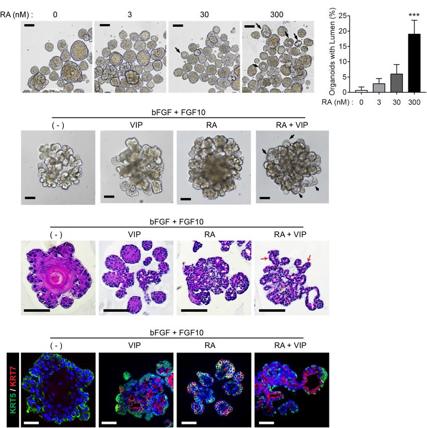
Kim et al.                                                                                                  Lumen-Forming Salivary Gland Organoid

for each other under specific conditions. RA has been reported              R-spondin 1-conditioned media (homemade), 50% Wnt3A-
to be an essential factor in the initial stages of embryonic                CM (homemade) or 5% afamin/Wnt3A-CM (MBL-life science,
development of SMGs in the salivary glands (Wright et al., 2015;            Woburn, MA, United States), and Y-27632 (10µM, Tocris,
Abashev et al., 2017; Metzler et al., 2018). However, due to the            Bristol, United Kingdom) to prepare the SMG organoid growth
limitations of using experimental models, the mechanism of adult            medium. The medium (250 µL/well) was added and changed
mouse SMG tissue morphogenesis, including lumen formation,                  every 2–3 days. The organoids were maintained at 37◦ C in a
has remained elusive.                                                       humidified atmosphere under 5% CO2 .
    Recent advances in 3D organoid culture have enabled the
establishment of long-term stem cell-based organotypic cultures             Immunofluorescence Microscopy
(Clevers, 2016; Kretzschmar and Clevers, 2016), and research                For formalin-fixed paraffin-embedded immunofluorescence
has demonstrated that organoid cultures can recapitulate in vivo            analysis, sections were deparaffinized and rehydrated. The
morphogenesis of tissues, making them of use as model systems               sections were subjected to heat antigen retrieval (10 mM Tris,
for investigating organ development and regeneration. Salivary              1 mM EDTA, 0.05% Tween-20) for 40 min. After cooling
gland organoids can be derived from murine embryonic stem                   the slide in ice water, the sections were blocked for 1 h with
cells (Tanaka et al., 2018), murine adult stem cells (Maimets et al.,       5% normal goat serum (NGS, Jackson ImmunoResearch
2016), or human adult stem cells (Pringle et al., 2016). However,           Laboratories, West Grove, PA, United States) or 5% normal
the application of salivary gland organoids in studies of adult             donkey serum (NDS, Jackson ImmunoResearch Laboratories),
tissue morphogenesis remains unexplored. Therefore, in this                 depending on the secondary antibody. For whole-mount
study, we postulated that a salivary gland organoid culture system          organoid immunofluorescence analysis, the organoids were
could recapitulate lumen formation during SMG morphogenesis,                fixed with either 4% paraformaldehyde for 30–60 min or ice
and investigated whether SMG organoids could be harnessed to                cold acetone/methanol (1:1) for 1 min followed by 0.3% Triton
elucidate the mechanism of lumen formation.                                 X-100 in phosphate-buffered saline (PBS). The organoids
                                                                            were blocked with 5% NGS or NDS in 0.05% PBS-Tween
                                                                            20 for 2 h at 4◦C. The sections or organoids were incubated
MATERIALS AND METHODS                                                       with primary antibodies overnight at 4◦C as follows: chicken
                                                                            anti-KRT5 (905901, BioLegend, 1:1,000); mouse anti-KRT7
Mice                                                                        (ab9021, Abcam, 1:200); rabbit anti-KRT-19 (ab52625, Abcam,
Six to sixteen-week-old female C3H mice were purchased from                 1:500); rabbit anti-CD133 (orb99113, Biorbyt, 1:50); rabbit
Jackson Laboratory (Bar Harbor, ME) and maintained under                    anti-VIPR1 (AVR-001, Alomone, 1:100); mouse anti-MIST1
specific pathogen-free conditions in a facility accredited by               (ab110919, Abcam, 1:50), goat anti-ACTA2 (NB300-978, Novus
AAALAC International. All experiments were approved by                      Bio, 1:1,000), rabbit anti-cleaved Caspase-3 (9661s, CST, 1:500),
the Institutional Animal Care and Use Committee of Yonsei                   and goat anti-TJP1 (ABIN6254231, antibodies-online. 1:1,000) in
University College of Medicine (Approval Number; 2017-0092).                tris-buffered saline (TBS) or phosphate-buffered saline PBS. On
                                                                            the following day, the sections or organoids were rinsed thrice
Cell Isolation and Organoid Culture                                         in TBS or PBS for 5 min (sections) or 30 min (whole-mount).
Mouse salivary gland organoids were generated from the SMGs                 The primary antibodies were detected using 488-, 555-, 647-
of 6–16-week-old female C3H mice. SMG tissues were minced                   conjugated secondary antibodies (Invitrogen), and the nuclei
with a razor blade, and the homogenate was incubated in                     were counter stained using Hoescht 33342 (1:1,000, Invitrogen).
digestion buffer consisting of Hanks’ balanced salt solution                Fluorescence was analyzed using the Carl Zeiss LSM 700 confocal
(HBSS, Biowest, Nuaille, France), 0.63 mg/mL of collagenase                 microscope and Zen software.
type II (Worthinton Biochem, Lakewood, NJ, United States),
0.5 mg/mL of hyaluronidase (Sigma-Aldrich, St. Louis, MO,                   Bright-Field Image Acquisition and
United States), and 6.25 mM CaCl2 (Sigma-Aldrich) for 1 h
at 37◦ C in a shaking incubator. After incubation, debris was
                                                                            Analysis
filtered through a series of 100-, 70-, and 40-µm strainers                 Bright-field images (40× or 100× magnification) of each culture
(SPL Life Sciences Co., Ltd., Seoul, South Korea). The isolated             were acquired using the Nikon Eclipse Ti 2-U microscope
cells were mixed with 20 µL of growth factor reduced (GFR)                  with NIS-Elements BR software (Nikon, Tokyo, Japan). The
Matrigel (Corning, NY, United States) and seeded in 48-well                 number of total organoids and organoids with lumen were
plates (Greiner, Kremsmünster, Austria). The basal medium                   counted manually.
contained advanced DMEM/F12 (Thermo Fisher Scientific,
Waltham, MA, United States) with penicillin/streptomycin                    Quantitative Reverse Transcription-PCR
(Gibco, Grand Island, NY, United States), HEPES (Biowest),                  (qRT-PCR)
and GlutaMAX (Gibco). The basal medium was supplemented                     Total RNA was isolated from the cultured organoids using TRIzol
with Primocin (0.1 mg/mL, InvivoGen, San Diego, CA,                         Reagent (Invitrogen) and processed for reverse transcription
United States), N-acetyl cysteine (1.25 mM, Sigma-Aldrich),                 using the PrimeScript RT Reagent Kit (Takara) according to the
B27 minus vitamin A (Gibco), EGF (5 nM, Gibco), bFGF                        manufacturer’s protocol. Gene expression was assessed by the
(1 nM, PeproTech, Rocky hill, NJ, United States), 10%                       conventional SYBR method using the QuantStudio 5 Real-Time

Frontiers in Cell and Developmental Biology | www.frontiersin.org       2                                   August 2021 | Volume 9 | Article 698292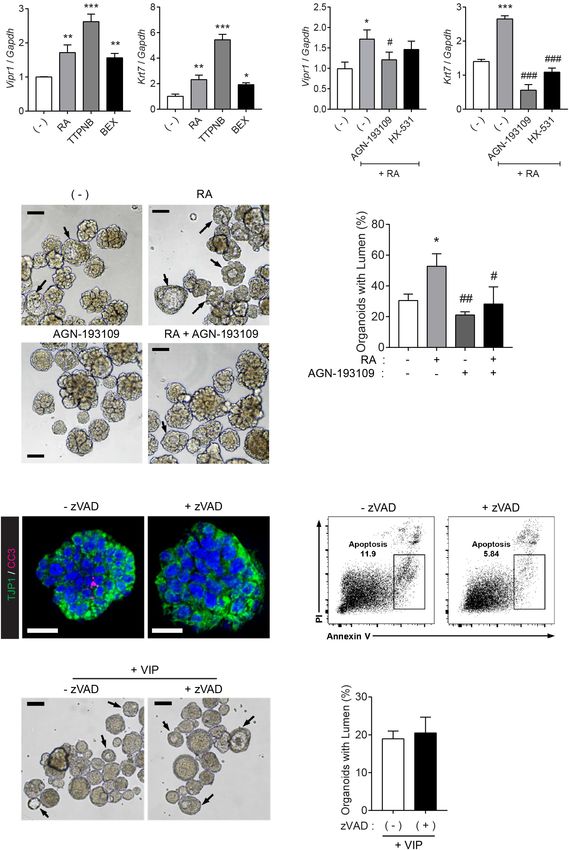
Kim et al. Lumen-Forming Salivary Gland Organoid
PCR Systems (Applied Biosystems). Primer information for monitored the temporal expression kinetics of KRT5, KRT19,
analysis of mouse genes was as follows: Vipr1: GCA GCA AGA KRT7, and CD133, which are known as markers of basal cells,
TGT GGG ACA ACC T (Forward), CAG TTG TGA CCA GCC luminal cells, terminally differentiated luminal cells, and apical
TTC TTC AG (Reverse); Vipr2: TGC CTC TTC AGG AAG membranes of luminal cells, respectively, in developing mouse
CTG CAC T (Forward), TGG AGT AGA GCA CGC TGT CCT SMGs (Walker et al., 2008; Nedvetsky et al., 2014). On day 0,
T (Reverse); Vip: GAT GCC GTT TGA AGG AGC AGG T the organoids consisted of KRT5+ KRT19− KRT7− basal ductal
(Forward), GAA GTC TGC TGT AAT CGC TGG TG (Reverse); cells (Figure 1B). The organoid culture led to the expression
Rarb: GCT TCG TTT GCC AGG ACA AGT C (Forward), TGG of KRT19+ cells on day 3, and also started to express KRT7
CAT CGG TTC CTA GTG ACC T (Reverse); Krt5: TTG GTG on day 4. In addition, from day 5, cells expressing KRT19
TTG GCA GTG GCT TT (Forward), CCC GCT ACC CAA and cells expressing KRT7 began to be distinguished from
ACC AAG AC (Reverse); Krt7: GCT CTC GCT CCA CTG CTT organoids, and microlumen which is not contiguous was clearly
AC (Forward), CGC CAG CAA GCT CTG ATT GA (Reverse); found on days 7 and 9 (Figure 1B, Supplementary Figure 1A,
Aqp5: GCC ACA TCA ATC CGG CCA TT (Forward), GGG and Supplementary Video 1). After 4 days of culture, the
CTG CCA CGT AGA AGA TG (Reverse); Acta2: GCC ATC organoids contained CD133+ luminal cells (Figure 1C), which
ATG CGT CTG GAC TT (Forward), ATC TCA CGC TCG GCA is similar to a presumptive lumen reminiscent of mouse SMG
GTA GT (Reverse); Gapdh: AGG GCA TCT TGG GCT ACA CT development (Nedvetsky et al., 2014). These data indicated that
(Forward), CGG CAT CGA AGG TGG AAG AG (Reverse). Gene luminal cells in this condition were terminally differentiated.
expressions were normalized to the expression of Gapdh. However, other factors were still required for contiguous large
lumen formation. Lumen formation and expansion are known
Flow Cytometry to occur in the presence of vasoactive intestinal protein (VIP) in
To assess cell death in mouse SMG organoids, we used the mouse SMGs (Nedvetsky et al., 2014). Therefore, we determined
EzWay Annexin V-FITC Apoptosis Detection Kit (KOMA whether lumen formation could be achieved with the addition
Biotech, Seoul, South Korea). Briefly, mouse SMG organoids were of VIP into the organoid culture medium. After 2 days of VIP
dissociated with TrypLE Express (Thermo Fisher Scientific) for treatment, the microlumens were enlarged and clearly visible,
20 min at 37◦C, and incubated in 100µL of 1 × annexin V compared to those in the non-treated organoids (Figure 1D).
binding buffer with annexin V-FITC and PI for 15 min at titrated Although the addition of VIP did not affect the number of
concentrations. After incubation, 400µL of binding buffer was organoids (data not shown), we confirmed that VIP promoted
added, and the samples were subsequently subjected to flow the formation of lumens (Figure 1E). Interestingly, VIP not
cytometry using LSRFortessa X-20 (BD Biosciences). Data were only accelerated lumen formation at day 5 but also induced
collected and analyzed using FlowJo software (Tree Star Inc., the formation of contiguous lumen with a defined expression
Ashland, OR, United States). of basal/luminal ductal cell markers on day 9 (Figure 1F).
Basal cells and luminal cells were clearly distinguished in the
Statistical Analysis VIP-treated organoids compared to the non-treated organoids.
All experiments were performed at least three times. Data The expression of the basal marker gene, Krt5, was not
are presented as the mean with standard mean error (SEM). altered; but on day 9, luminal marker genes, Krt19 and Krt7,
Unpaired two-tailed Student’s t-tests (two groups) and one-way were somewhat decreased after VIP treatment, suggesting that
ANOVA (more than two groups) with Tukey’s post hoc test VIP-induced lumen formation occurred after transcriptional
were performed using GraphPad Prism 7 (GraphPad Software regulation (Supplementary Figure 1B). Collectively, these results
Inc., CA). Values of ∗ p < 0.05, ∗∗ p < 0.01, and ∗∗∗ p < 0.001 implied that SMG organoid culture can recapitulate lumen
were considered statistically significant, while p > 0.05 was not formation during SMG morphogenesis in the presence of a niche
considered statistically significant. factor, VIP. In addition, VIP induces clear differentiation between
luminal and basal cells in mSMG organoids.
RESULTS RA Signaling Enhances VIP-Mediated
Lumen Formation in Salivary Gland
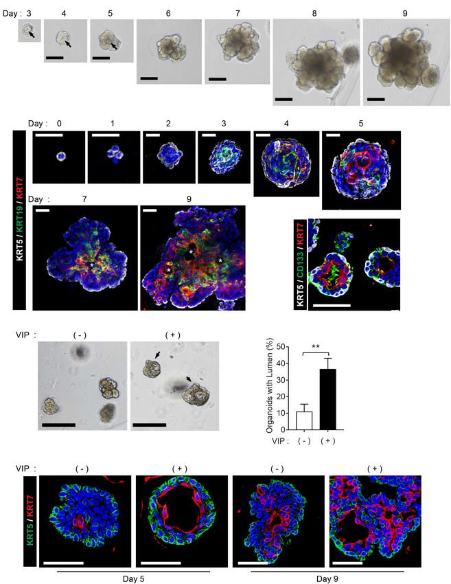
Salivary Gland Organoids Can Organoids
Recapitulate Lumen Formation During Next, we explored whether RA plays a pivotal role in lumen
Salivary Gland Morphogenesis formation in our SMG organoid culture system. Since retinoids
The mouse SMG organoids were generated and maintained are present in serum trace quantities in Wnt3A-conditioned
under specific conditions according to a previously reported medium (Wnt3A-CM) (Kim et al., 1996), further experiments
protocol (Maimets et al., 2016). We could detect microlumen were conducted with serum-free afamin/Wnt3A to exclude the
formation from day 3 of the organoid culture, while lumen in possibility of retinoids in Wnt3A-CM affecting lumen formation
organoids was not clearly seen in brightfield when cultured for (Mihara et al., 2016). In order to examine the effect of RA
longer periods (Figure 1A). Next, we investigated whether the and VIP on lumen formation of SMG organoids, the SMG
cell constitution and organoid characteristics recapitulate the organoids were cultured with different doses of RA and treated
process of lumen formation during morphogenesis. First, we with 200 nM VIP on day 4. RA alone did not induce distinct
Frontiers in Cell and Developmental Biology | www.frontiersin.org 3 August 2021 | Volume 9 | Article 698292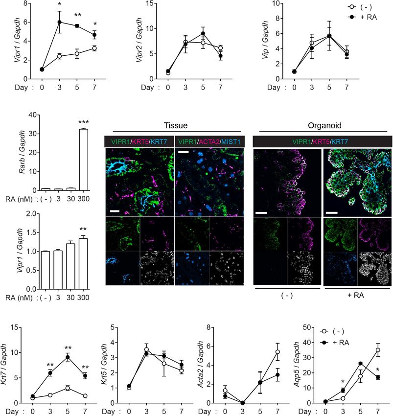
Kim et al.                                                                                                                   Lumen-Forming Salivary Gland Organoid

  FIGURE 1 | Recapitulation of lumen formation during SMG development in a mouse SMG organoid culture. (A) Organoid growth from single cells was monitored at
  each time point. Black arrows indicate internal lumen formation as shown in the bright-field microscopy images. Scale bar indicates 100 µm. (B) SMG organoids
  were harvested at each time point and subjected to whole-mount immunofluorescence microscopy for the evaluation of KRT5 (white), KRT19 (green), and KRT7
  (red) expressions from days 0 to 9. Up to day 5, images were obtained through maximum intensity projection. From days 7 to 9, single images were obtained from
  z-stack. Micro-lumens were denoted with white asterisks. Scale bars indicate 20 µm. Nuclei were counterstained with DAPI (blue). (C) KRT5 (white), CD133 (green),
  and KRT7 (red) expressions on day 4. Scale bars indicate 50 µm. Nuclei were counterstained with DAPI (blue). (D) The organoids were treated with 200 nM of VIP
                                                                                                                                                         (Continued)

Frontiers in Cell and Developmental Biology | www.frontiersin.org                 4                                          August 2021 | Volume 9 | Article 698292
Kim et al. Lumen-Forming Salivary Gland Organoid
FIGURE 1 | Continued
on day 2, and lumen enlargement was observed on day 4. Black arrows indicate internal lumen formation. Scale bars indicate 100 µm. (E) The proportion of
organoids with lumen were determined (n = 3). The results are expressed as the mean ± SD. **p < 0.01. (F) SMG organoids were harvested on days 5 and 9, and
200 nM of VIP was treated on the last 1 day. Organoid were subjected to immunofluorescence staining for KRT5 (green) and KRT7 (red). Scale bars indicate 50 µm.
Nuclei were counterstained with DAPI (blue). All experiments were performed three times independently.
differences in lumen formation in the absence of VIP in the nM of RA significantly induced Rarb, a positive control for
culture medium (data not shown), whereas the addition of VIP RA signaling, and Vipr1 expression (Figure 3B), indicating that
to the organoid culture on day 4 in the RA-containing culture sufficient amounts of RA are required for the Vipr1 expression.
medium increased the proportion of the lumen-containing Immunofluorescence staining was performed to confirm
organoids (Figures 2A,B). Next, we maintained the organoid whether the upregulated mRNA of Vipr1 was translated to the
culture with combination of FGF ligands, which are known expression of VIPR1. Immunofluorescence staining of mouse
to be involved in budding and branching morphogenesis in SMG tissues showed that VIPR1 was present in the basolateral
salivary gland development (Hoffman et al., 2002; Steinberg membrane of KRT7+ luminal cells; however, it was not present
et al., 2005), in order to induce lumen formation in organoids in MIST1+ acinar or ACAT2+ myoepithelial cells in murine
with more complex structure containing several branches and SMG tissues (Figure 3C). In SMG organoids, KRT7+ cells on the
buds. Prior to culture using the combination of FGF, we tested luminal side co-expressed VIPR1 in the presence of RA, although
whether the difference in morphology caused by FGFs in the VIPR1 was also expressed in KRT5+ basal cells of organoid
mouse SMG development stage could be reproduced in organoids culture (Figure 3D). MIST1+ acinar cells were not present in
(Supplementary Figure 2A). In the absence of FGFs, small and the organoids, and ACTA2+ myoepithelial cells were present, but
round-shaped organoids were formed. In the presence of bFGF, VIPR1 was not co-expressed (Supplementary Figure 3). Next, we
short ducts with endbuds were observed, while long ducts were sought to determine whether RA treatment affects the expression
observed in the presence of FGF10. When bFGF and FGF10 of other salivary gland markers in SMG organoids. Consistent
were co-treated, complex types of organoids were observed with protein expressions, RA treatment markedly increased the
(Supplementary Figure 2A). It was also found that basal and expression of Krt7; however, it did not alter the expression of Krt5
luminal cells were present in all conditions (Supplementary or Acta2. The expression of Aqp5 (acinar marker) decreased in
Figure 2B). Therefore, we decided to treat bFGF and FGF10 late time points, compared to non-treated groups (Figure 3E),
and tested whether RA or VIP also affected lumen formation suggesting that RA might regulate acinar cell differentiation.
in organoids with complex morphology. As expected, brightfield Collectively, these results suggested that RA signaling induces
images showed that both RA and VIP treatment induced lumen lumen formation in mouse SMG organoids via the induction of
expansion in branching organoids, while this phenotype was VIPR1 expression and KRT7+ luminal cell differentiation.
severely compromised upon the removal of RA (Figure 2C).
Furthermore, lumen formation in branching ducts was observed
in H&E-stained images when treated with both RA and VIP RAR-Mediated Regulation of Vipr1
(Figure 2D). However, when treated with VIP only, branching Promotes Lumen Formation in Mouse
ducts were scarcely detected, suggesting that RA may promote SMG Organoids
branching of adult SMG (Wang et al., 2006). In addition, RA To investigate which receptor is pivotal to Vipr1 expression and
and VIP promoted the compartmentalization of luminal cells lumen formation, the organoids were treated with RAR agonist
and basal cells, as assessed by immunofluorescence staining for (TTNPB) or RXR-selective agonist (bexarotene) instead of RA,
KRT5 and KRT7 (Figure 2E). Collectively, these results suggested since RA binds with either nuclear RAR or RXR transcription
that RA is necessary for terminal luminal cell differentiation in factors and controls downstream gene expression by binding
salivary gland organoids. to DNA sequences of response elements (Dawson and Xia,
2012; Cunningham and Duester, 2015). The RA-mediated Vipr1
RA Signaling Induces Lumen Formation and Krt7 expressions were higher when treated with RAR-
via VIPR Signaling selective agonist at the same concentration, indicating that these
Next, we assessed whether VIPR signaling is upregulated in genes are regulated in an RAR-dependent manner (Figure 4A).
the organoids when treated with RA. Quantitative real-time To confirm RAR-dependent gene regulation, RAR or RXR-
polymerase chain reaction (qRT-PCR) confirmed that Vipr1 selective antagonists (AGN-193109 and HX-531, respectively)
expression increased in the presence of RA (Figure 3A), whereas were treated along with RA in SMG organoids. We found
Vipr2, another receptor for VIP, and Vip ligand elicited no that the RAR antagonist significantly reduced RA-mediated
significant difference compared to the expression in non-treated gene expression, although partial RXR-mediated gene regulation
groups during organoid culture. Induced Vipr1 expression began was also observed (Figure 4B). Next, we determined whether
from day 3 and continued until day 7, which corresponds RAR-dependent gene induction could affect lumen formation.
with the period of internal lumen formation detected in Consistent with our hypothesis, lumen formation in SMG
immunofluorescence staining, as shown in Figure 1B. To test organoids was promoted by RA treatment and was severely
the dose-dependent induction of Vipr1, mouse SMG organoids compromised when treated with RAR antagonist (Figures 4C,D).
were treated with different doses (0–300 nM) of RA. Only 300 Collectively, we suggest that RA promotes lumen formation
Frontiers in Cell and Developmental Biology | www.frontiersin.org 5 August 2021 | Volume 9 | Article 698292Kim et al. Lumen-Forming Salivary Gland Organoid
FIGURE 2 | Retinoic acid promotes lumen formation in SMG organoids. (A) SMG organoids were cultured with different doses of RA and treated with 200 nM VIP
on day 4. Microscopic images of lumen formation were obtained on day 6. Scale bars indicate 100 µm. (B) The proportion of organoids containing internal lumen
were calculated, followed by statistical analysis (n = 3). Results are expressed as the mean ± SD. ***p < 0.001. (C–E) SMG organoids were cultured with or without
RA in combination with 1 nM bFGF and 5 nM FGF10 throughout the culturing period. At day 9, organoids were either unstimulated or stimulated with VIP for 1 day.
(C) Branching ducts were observed with brightfield microscopy. Black arrows indicate formation of lumens near end buds. Scale bars indicate 100 µm.
(D) Harvested organoid were subjected to H&E staining to observe lumen formation in branching ducts (red arrows). Scale bars indicate 100 µm. (E) Organoids
were subjected to immunofluorescence staining for KRT5 (green) and KRT7 (red). Scale bars indicate 50 µm. Nuclei were counterstained with DAPI (blue). All
experiments were performed three times independently.
through the induction of luminal cell-related gene expression in central epithelial cells in presumptive ducts and endbuds by
a RAR-dependent manner. depleting FGF signals (Jaskoll and Melnick, 1999; Melnick and
Jaskoll, 2000; Teshima et al., 2016). RA has been reported to
antagonize FGFs during development (Cunningham et al., 2013).
RA Signaling Induces Lumen Formation Therefore, we examined whether lumen formation by RA can be
in an Apoptosis-Independent Manner attributed to apoptosis of the inner cells. To do so, the organoid
It has been suggested that the underlying mechanisms of cultures were treated with a pan-caspase inhibitor, z-VAD-
lumen formation in mouse SMGs involve the apoptosis of fmk (zVAD), to inhibit apoptosis. Immunofluorescence results
Frontiers in Cell and Developmental Biology | www.frontiersin.org 6 August 2021 | Volume 9 | Article 698292Kim et al. Lumen-Forming Salivary Gland Organoid FIGURE 3 | Retinoic acid induces Vipr1 gene expression. (A) SMG organoids were treated with or without RA (300 nM) for the indicated time. After harvesting, the organoids were subjected to qRT-PCR to evaluate gene expression associated with VIP signaling (n = 4). (B) SMG organoids were treated with varying doses of RA for 7 days and subjected to qRT-PCR for the evaluation of gene expression of Vipr1 and Rarb, a positive control, for RA-mediated signaling (n = 4). (C) Expression of VIPR1 (green) in mouse SMG tissues (left) was assessed by co-staining with KRT5 (magenta, left), KRT7 (cyan, left), ACTA2 (magenta, right), and MIST1 (cyan, right). Nuclei were counterstained with DAPI (white) and each single channel image was placed below. Scale bars indicate 20 µm. (D) The mouse SMG organoids cultured with (bottom) or without RA (top) were subjected to immunofluorescence for the evaluation of VIPR1 (green), KRT5 (magenta), and KRT7 (cyan) expressions. Nuclei were counterstained with DAPI (white), and each single channel image was placed below. Scale bars indicate 50 µm. (E) The expression of several salivary gland markers in organoids treated with or without RA were analyzed using qRT-PCR at the indicated time points (n = 4). Results are expressed as the mean ± SEM. *p < 0.05, **p < 0.01, and ***p < 0.001. All experiments were performed at least three times independently. showed that zVAD inhibited naturally occurring apoptosis in However, bright-field images of the SMG organoids showed the organoids (Figure 4E). Moreover, zVAD-mediated inhibition that pre-treatment of zVAD had no effect on VIP-induced of apoptosis was confirmed using flow cytometry (Figure 4F). lumen formation (Figure 4G and its quantitative analysis in Frontiers in Cell and Developmental Biology | www.frontiersin.org 7 August 2021 | Volume 9 | Article 698292
Kim et al.                                                                                                                     Lumen-Forming Salivary Gland Organoid

  FIGURE 4 | RAR activation induces lumen formation in SMG organoids. (A) SMG organoids were cultured with 300 nM RA, TTNPB (RAR agonist), or bexarotene
  (RXR agonist) for 7 days and subjected to qRT-PCR for the detection of Vipr1 and Krt7 expression (n = 4). (B) SMG organoids were treated with RA (300 nM),
  RA + 3 µM AGN-193109 (RAR antagonist), or RA + 3 µM HX (HX-531, RXR antagonist) throughout the culturing period and subjected to qRT-PCR for the detection
  of Vipr1 and Krt7 expression (n = 4). (C–D) SMG organoids were treated with RA (300 nM), AGN-193109 (3 µM), or both. The organoids were treated with VIP (200
  nM) on day 2 for lumen formation. After 3 days, lumen formation was observed via microscopic images (C). Black arrows indicate organoids containing visible
  lumen. The proportion of organoids with lumen were calculated and statistically analyzed (D) (n = 3). Scale bars indicate 100 µm in (C). (E) SMG organoids were
  treated with 50µM z-VAD-FMK, a pan-caspase inhibitor, for 2 days, followed by whole-mount immunostaining with antibodies against CC3 (cleaved caspase-3)
  and TJP1 (ZO-1). DAPI (blue) was used for counterstaining the nucleus. Scale bars indicate 20 µm. (F) Apoptotic cell death (Annexin-V+ PI− ) in SMG organoids treated
                                                                                                                                                            (Continued)

Frontiers in Cell and Developmental Biology | www.frontiersin.org                  8                                           August 2021 | Volume 9 | Article 698292
Kim et al. Lumen-Forming Salivary Gland Organoid
FIGURE 4 | Continued
with or without zVAD was examined using flow cytometry. (G) Representative bright-field images of organoids with lumen were obtained on day 5. Black arrows
indicate organoids containing visible lumen. Scale bars indicate 100 µm. (H) The proportion of organoids with lumen were calculated (n = 3). Results are expressed
as the mean ± SEM. *p < 0.05, **p < 0.01, and ***p < 0.001 when compared to the non-treated groups. # p < 0.05, ## p < 0.01, and ### p < 0.001 when
compared to the RA-treated groups. All experiments were performed three times independently.
Figure 4H). Increased zVAD concentration did not inhibit mediated by the cAMP/PKA pathways, respectively (Knox et al.,
VIP-induced lumen formation (data not shown). These results 2010; Nedvetsky et al., 2014). Although RA alone showed limited
indicated that RA induces lumen formation in an apoptosis- effects on lumen formation, we demonstrated that RA promotes
independent manner. lumen formation in the presence of VIP, suggesting that lumen
formation is induced by VIP and accelerated by RA treatment.
Furthermore, we discovered that RA promotes lumen formation
in mouse SMG organoids by the induction of VIPR1 and KRT7
DISCUSSION
expression, and that RA-induced lumen formation is achieved
The salivary glands play important roles in providing lubrication via VIPR1-RAR mediated signaling, not via apoptosis pathways,
for chewing and swallowing, digestion, and vocalization, as as zVAD treatment did not affect lumen formation in SMG
well as protection against microbial infections (Tucker, 2007; organoids. We suggest that the RA-mediated induction of Krt7
Mattingly et al., 2015). Salivary gland hypofunction caused expression can be explained by the direct effects of RA or
by radiation therapy, aging, or autoimmune diseases, severely indirect outcomes of lumen formation, indicating that further
impairs an individual’s oral health and quality of life (Jensen investigation is needed to completely understand the function of
et al., 2010; Rocchi and Emmerson, 2020). Stem cell-based RA in salivary gland development based on our organoid system.
regeneration therapy has been regarded as a potential strategy to In mouse SMG tissues, RA is synthesized and supplied from
overcome salivary gland hypofunction, and translational research the mesenchyme surrounding the developing salivary gland
investigating salivary gland regeneration based on adult stem epithelium. RA signaling begins in the early developmental stages
cell therapies is being intensively performed. The importance and is activated throughout the morphogenetic process of the
of understanding salivary gland morphogenesis has been glands (Wright et al., 2015; Metzler et al., 2018). Intriguingly,
emphasized along with the importance of gaining mechanistic we found that RA promoted lumen formation in our organoid
insights into the regeneration of salivary glands using adult stem culture, but also inhibited differentiation into acinar cells.
cells (Lombaert and Hoffman, 2010; Patel and Hoffman, 2014). Although the regulation of acinar cell differentiation by RA in
In this study, we demonstrated that RA induces lumen formation other exocrine glands, including lacrimal gland and pancreas,
in adult mouse SMGs and this adult organoid culture system is has been studied (Ubels et al., 2002; Tulachan et al., 2003),
capable of recapitulating salivary gland morphogenesis. These the detailed regulatory mechanisms of RA in the differentiation
findings have important implications on our understanding of of acinar cells in the development of salivary glands remain
salivary gland morphogenesis in adult tissues, and provide an unknown. Therefore, further studies will be required to elucidate
opportunity to harness this organoid culture system as a tool for the negative regulation of RA in the differentiation of salivary
studying salivary gland regeneration or modeling. gland acinar cells. As niche factors can be added or withdrawn
Previous studies on salivary glands using adult stem cells have from an organoid culture, our culture system provides a model
exploited floating culture or nanoscaffold microwells to enhance for the investigation of the temporal requirements of niche factors
progenitor cell proliferation and stem cell properties (Shin et al., in tissue development; for example, separate utilization of the
2018; Koslow et al., 2019). However, these salisphere or spheroid growth and differentiation media would enable efficient tissue
cultures are more comparable to cell aggregations, which make recapitulation (Huch et al., 2015). Although our system is based
it difficult to recapitulate complex tissue morphogenesis and on adult stem cells, due to some similarities between embryonic
the surrounding extracellular matrix (ECM). To overcome these development and adult tissue regeneration (Nedvetsky et al.,
limitations, an organoid system cultured in serum-free and 2014; Emmerson et al., 2017), there is a possibility that our
biochemically defined media with ECM (e.g., laminin-111 or findings are applicable to embryonic stages. Moreover, our
Matrigel) has been suggested as an alternative to 3D culture. findings can be extended to other culture systems, including
This 3D organoid culture system facilitates the investigation of embryonic stem cells, iPSC, or tissue explant culture, to
not only personalized medicine and disease models but also explore the role of RA in the development or regeneration of
the developmental processes of organs using adult stem cells salivary glands.
(Fatehullah et al., 2016; Xu et al., 2018). To extend the potential In conclusion, we found that the administration of RA to
of salivary gland organoids as a tool for investigation of tissue organoid culture medium leads to lumen formation via the
morphogenesis, it is important to study the essential niche factors induction of Vipr1 and Krt7 expressions, and that RA promotes
required to develop a 3D organoid culture system for efficient VIP-mediated lumen expansion in a RAR-dependent manner.
tissue recapitulation. Our study suggests that salivary gland organoid culture is a useful
VIP, the ligand for VIPR1, was thought to regulate not only system for investigating the niche factors associated with salivary
luminal cell proliferation but also lumen formation, which is gland morphogenesis.
Frontiers in Cell and Developmental Biology | www.frontiersin.org 9 August 2021 | Volume 9 | Article 698292Kim et al. Lumen-Forming Salivary Gland Organoid
DATA AVAILABILITY STATEMENT and Future Planning (NRF-2018R1A2B3004269 and NRF-
2020M3A9I4039045, Republic of Korea).
The original contributions presented in the study are included
in the article/Supplementary Material, further inquiries can be
directed to the corresponding author/s. SUPPLEMENTARY MATERIAL
The Supplementary Material for this article can be found
ETHICS STATEMENT online at: https://www.frontiersin.org/articles/10.3389/fcell.2021.
698292/full#supplementary-material
The animal study was reviewed and approved by the Institutional
Animal Care and Use Committee of Yonsei University College of Supplementary Figure 1 | Expressions of basal and luminal cell markers in
salivary gland organoids. (A) Maximum intensity projected images of organoid at
Medicine (Approval Number; 2017-0092).
day 7 (up) and day 9 (down). Organoids were subjected to immunofluorescence
for KRT5 (white), KRT19 (green), KRT7 (red), and DAPI (blue). Scale bar indicates
50 µm. (B) SMG organoids were harvested on days 5 and 9, and 200 nM of VIP
AUTHOR CONTRIBUTIONS was treated on the last 1 day. Harvested organoids were subjected to qRT-PCR to
evaluate the expressions of Krt5, Krt19, and Krt7 (n = 3). Results are expressed
DK and Y-JY designed the study, performed the experiments, as the mean ± SEM. ∗ p < 0.05.
analyzed the data, and wrote the manuscript. DC and JK Supplementary Figure 2 | FGF-induced complex structure is observed in
performed H&E staining and immunofluorescence staining. J-YL salivary gland organoids. SMG organoids were cultured in combination with 1 nM
supervised the project and wrote the manuscript. All authors bFGF and 5 nM FGF10 throughout the culture. (A) FGF-induced changes in
morphology were observed via brightfield microscopy in salivary gland organoids.
revised the article and approved the submission of revised article.
Scale bar indicates 500 µm. (B) Organoids were subjected to
immunofluorescence for KRT5 (green) and KRT7 (red). Nuclei were counterstained
with DAPI (blue). Scale bar indicates 50 µm.
FUNDING
Supplementary Figure 3 | VIPR1 expression in salivary gland organoid. The
mouse SMG organoids cultured with RA were subjected to immunofluorescence
This work was supported by the Basic Science Research for the evaluation of VIPR1 (green), ACTA2 (magenta), and MIST1 (cyan)
Program through the National Research Foundation of expressions. Nuclei were counterstained with DAPI (white), and each single
Korea (NRF) funded by the Ministry of Science, ICT, channel image was placed below. Scale bars indicate 50 µm.
REFERENCES Hsu, J. C., and Yamada, K. M. (2010). Salivary gland branching morphogenesis–
recent progress and future opportunities. Int. J. Oral Sci. 2, 117–126. doi:
Abashev, T. M., Metzler, M. A., Wright, D. M., and Sandell, L. L. (2017). Retinoic 10.4248/ijos10042
acid signaling regulates Krt5 and Krt14 independently of stem cell markers Huch, M., Gehart, H., Van Boxtel, R., Hamer, K., Blokzijl, F., Verstegen, M. M.,
in submandibular salivary gland epithelium. Dev. Dyn. 246, 135–147. doi: et al. (2015). Long-term culture of genome-stable bipotent stem cells from adult
10.1002/dvdy.24476 human liver. Cell 160, 299–312. doi: 10.1016/j.cell.2014.11.050
Clevers, H. (2016). Modeling Development and Disease with Organoids. Cell 165, Jaskoll, T., Abichaker, G., Witcher, D., Sala, F. G., Bellusci, S., Hajihosseini, M. K.,
1586–1597. doi: 10.1016/j.cell.2016.05.082 et al. (2005). FGF10/FGFR2b signaling plays essential roles during in vivo
Cunningham, T. J., and Duester, G. (2015). Mechanisms of retinoic acid signalling embryonic submandibular salivary gland morphogenesis. BMC Dev. Biol. 5:11.
and its roles in organ and limb development. Nat. Rev. Mol. Cell Biol. 16, doi: 10.1186/1471-213X-5-11
110–123. doi: 10.1038/nrm3932 Jaskoll, T., and Melnick, M. (1999). Submandibular gland morphogenesis:
Cunningham, T. J., Zhao, X., Sandell, L. L., Evans, S. M., Trainor, P. A., and stage-specific expression of TGF-alpha/EGF, IGF, TGF-beta, TNF,
Duester, G. (2013). Antagonism between retinoic acid and fibroblast growth and IL-6 signal transduction in normal embryonic mice and the
factor signaling during limb development. Cell Rep. 3, 1503–1511. doi: 10.1016/ phenotypic effects of TGF-beta2, TGF-beta3, and EGF-r null mutations.
j.celrep.2013.03.036 Anat. Rec. 256, 252–268. doi: 10.1002/(sici)1097-0185(19991101)25
Dawson, M. I., and Xia, Z. (2012). The retinoid X receptors and their ligands. 6:33.0.co;2-6
Biochim. Biophys. Acta 1821, 21–56. Jensen, S. B., Pedersen, A. M., Vissink, A., Andersen, E., Brown, C. G., Davies,
De Moerlooze, L., Spencer-Dene, B., Revest, J. M., Hajihosseini, M., Rosewell, I., A. N., et al. (2010). A systematic review of salivary gland hypofunction
and Dickson, C. (2000). An important role for the IIIb isoform of fibroblast and xerostomia induced by cancer therapies: prevalence, severity and impact
growth factor receptor 2 (FGFR2) in mesenchymal-epithelial signalling during on quality of life. Support Care Cancer 18, 1039–1060. doi: 10.1007/s0052
mouse organogenesis. Development 127, 483–492. doi: 10.1242/dev.127.3.483 0-010-0827-8
Emmerson, E., May, A. J., Nathan, S., Cruz-Pacheco, N., Lizama, C. O., Maliskova, Kim, N. D., Paik, K. J., and Clifton, K. H. (1996). Inhibitory effects of retinoids
L., et al. (2017). SOX2 regulates acinar cell development in the salivary gland. on development of squamous metaplasia in rat mammary epithelial organoids
Elife 6:e26620. cultured in Matrigel. Cancer Lett. 110, 217–223. doi: 10.1016/s0304-3835(96)
Fatehullah, A., Tan, S. H., and Barker, N. (2016). Organoids as an in vitro model 04514-4
of human development and disease. Nat. Cell Biol. 18, 246–254. doi: 10.1038/ Knox, S. M., Lombaert, I. M., Reed, X., Vitale-Cross, L., Gutkind, J. S., and
ncb3312 Hoffman, M. P. (2010). Parasympathetic innervation maintains epithelial
Hoffman, M. P., Kidder, B. L., Steinberg, Z. L., Lakhani, S., Ho, S., Kleinman, progenitor cells during salivary organogenesis. Science 329, 1645–1647. doi:
H. K., et al. (2002). Gene expression profiles of mouse submandibular gland 10.1126/science.1192046
development: FGFR1 regulates branching morphogenesis in vitro through Koslow, M., O’keefe, K. J., Hosseini, Z. F., Nelson, D. A., and Larsen, M. (2019).
BMP- and FGF-dependent mechanisms. Development 129, 5767–5778. doi: ROCK inhibitor increases proacinar cells in adult salivary gland organoids. Stem
10.1242/dev.00172 Cell Res. 41:101608. doi: 10.1016/j.scr.2019.101608
Frontiers in Cell and Developmental Biology | www.frontiersin.org 10 August 2021 | Volume 9 | Article 698292Kim et al. Lumen-Forming Salivary Gland Organoid Kretzschmar, K., and Clevers, H. (2016). Organoids: modeling Development and cell proliferation and branching morphogenesis. Development 132, 1223–1234. the Stem Cell Niche in a Dish. Dev. Cell 38, 590–600. doi: 10.1016/j.devcel. doi: 10.1242/dev.01690 2016.08.014 Tanaka, J., Ogawa, M., Hojo, H., Kawashima, Y., Mabuchi, Y., Hata, K., et al. (2018). Lombaert, I. M. A., and Hoffman, M. P. (2010). Epithelial stem/progenitor cells Generation of orthotopically functional salivary gland from embryonic stem in the embryonic mouse submandibular gland. Front. Oral Biol. 14, 90–106. cells. Nat Commun. 9:4216. doi: 10.1159/000313709 Tanumihardjo, S. A. (2011). Vitamin A: biomarkers of nutrition for development. Maimets, M., Rocchi, C., Bron, R., Pringle, S., Kuipers, J., Giepmans, B. N., et al. Am. J. Clin. Nutr. 94, 658S–665S. (2016). Long-Term In Vitro Expansion of Salivary Gland Stem Cells Driven by Teshima, T. H., Wells, K. L., Lourenco, S. V., and Tucker, A. S. (2016). Apoptosis in Wnt Signals. Stem Cell Rep. 6, 150–162. doi: 10.1016/j.stemcr.2015.11.009 Early Salivary Gland Duct Morphogenesis and Lumen Formation. J. Dent. Res. Mattingly, A., Finley, J. K., and Knox, S. M. (2015). Salivary gland development and 95, 277–283. doi: 10.1177/0022034515619581 disease. Wiley Interdiscip. Rev. Dev. Biol. 4, 573–590. doi: 10.1002/wdev.194 Tucker, A. S. (2007). Salivary gland development. Semin. Cell Dev. Biol. 18, Melnick, M., and Jaskoll, T. (2000). Mouse submandibular gland morphogenesis: 237–244. a paradigm for embryonic signal processing. Crit. Rev. Oral Biol. Med. 11, Tulachan, S. S., Doi, R., Kawaguchi, Y., Tsuji, S., Nakajima, S., Masui, T., et al. 199–215. doi: 10.1177/10454411000110020401 (2003). All-trans retinoic acid induces differentiation of ducts and endocrine Metzler, M. A., Raja, S., Elliott, K. H., Friedl, R. M., Tran, N. Q. H., Brugmann, S. A., cells by mesenchymal/epithelial interactions in embryonic pancreas. Diabetes et al. (2018). RDH10-mediated retinol metabolism and RARalpha-mediated 52, 76–84. doi: 10.2337/diabetes.52.1.76 retinoic acid signaling are required for submandibular salivary gland initiation. Ubels, J. L., Wertz, J. T., Ingersoll, K. E., Jackson, R. S. II., and Aupperlee, M. D. Development 145:dev164822. (2002). Down-regulation of androgen receptor expression and inhibition of Mihara, E., Hirai, H., Yamamoto, H., Tamura-Kawakami, K., Matano, M., Kikuchi, lacrimal gland cell proliferation by retinoic acid. Exp. Eye Res. 75, 561–571. A., et al. (2016). Active and water-soluble form of lipidated Wnt protein is doi: 10.1006/exer.2002.2054 maintained by a serum glycoprotein afamin/alpha-albumin. Elife 5:e11621. Walker, J. L., Menko, A. S., Khalil, S., Rebustini, I., Hoffman, M. P., Kreidberg, Miyazaki, Y., Nakanishi, Y., and Hieda, Y. (2004). Tissue interaction mediated by J. A., et al. (2008). Diverse roles of E-cadherin in the morphogenesis of neuregulin-1 and ErbB receptors regulates epithelial morphogenesis of mouse the submandibular gland: insights into the formation of acinar and ductal embryonic submandibular gland. Dev. Dyn. 230, 591–596. doi: 10.1002/dvdy. structures. Dev. Dyn. 237, 3128–3141. doi: 10.1002/dvdy.21717 20078 Wang, Z., Dolle, P., Cardoso, W. V., and Niederreither, K. (2006). Retinoic acid Nedvetsky, P. I., Emmerson, E., Finley, J. K., Ettinger, A., Cruz-Pacheco, N., regulates morphogenesis and patterning of posterior foregut derivatives. Dev. Prochazka, J., et al. (2014). Parasympathetic innervation regulates tubulogenesis Biol. 297, 433–445. doi: 10.1016/j.ydbio.2006.05.019 in the developing salivary gland. Dev. Cell 30, 449–462. doi: 10.1016/j.devcel. Wright, D. M., Buenger, D. E., Abashev, T. M., Lindeman, R. P., Ding, J., 2014.06.012 and Sandell, L. L. (2015). Retinoic acid regulates embryonic development of Nitta, M., Kume, T., and Nogawa, H. (2009). FGF alters epithelial competence for mammalian submandibular salivary glands. Dev. Biol. 407, 57–67. doi: 10.1016/ EGF at the initiation of branching morphogenesis of mouse submandibular j.ydbio.2015.08.008 gland. Dev. Dyn. 238, 315–323. doi: 10.1002/dvdy.21780 Xu, H., Lyu, X., Yi, M., Zhao, W., Song, Y., and Wu, K. (2018). Organoid technology Patel, N., Sharpe, P. T., and Miletich, I. (2011). Coordination of epithelial branching and applications in cancer research. J. Hematol. Oncol. 11:116. and salivary gland lumen formation by Wnt and FGF signals. Dev. Biol. 358, 156–167. doi: 10.1016/j.ydbio.2011.07.023 Conflict of Interest: The authors declare that the research was conducted in the Patel, V. N., and Hoffman, M. P. (2014). Salivary gland development: a template absence of any commercial or financial relationships that could be construed as a for regeneration. Semin. Cell Dev. Biol. 2, 52–60. potential conflict of interest. Pringle, S., Maimets, M., Van Der Zwaag, M., Stokman, M. A., Van Gosliga, D., Zwart, E., et al. (2016). Human Salivary Gland Stem Cells Functionally Restore Publisher’s Note: All claims expressed in this article are solely those of the authors Radiation Damaged Salivary Glands. Stem Cells 34, 640–652. doi: 10.1002/stem. and do not necessarily represent those of their affiliated organizations, or those of 2278 the publisher, the editors and the reviewers. Any product that may be evaluated in Rocchi, C., and Emmerson, E. (2020). Mouth-Watering Results: clinical need, this article, or claim that may be made by its manufacturer, is not guaranteed or current approaches, and future directions for salivary gland regeneration. endorsed by the publisher. Trends Mol. Med. 26, 649–669. doi: 10.1016/j.molmed.2020.03.009 Shin, H. S., Lee, S., Hong, H. J., Lim, Y. C., Koh, W. G., and Lim, J. Y. (2018). Stem Copyright © 2021 Kim, Yoon, Choi, Kim and Lim. This is an open-access article cell properties of human clonal salivary gland stem cells are enhanced by three- distributed under the terms of the Creative Commons Attribution License (CC BY). dimensional priming culture in nanofibrous microwells. Stem Cell Res. Ther. The use, distribution or reproduction in other forums is permitted, provided the 9:74. original author(s) and the copyright owner(s) are credited and that the original Steinberg, Z., Myers, C., Heim, V. M., Lathrop, C. A., Rebustini, I. T., Stewart, J. S., publication in this journal is cited, in accordance with accepted academic practice. No et al. (2005). FGFR2b signaling regulates ex vivo submandibular gland epithelial use, distribution or reproduction is permitted which does not comply with these terms. Frontiers in Cell and Developmental Biology | www.frontiersin.org 11 August 2021 | Volume 9 | Article 698292
You can also read


