Events associated with susceptibility to invasive Salmonella enterica serovar Typhi in BALB/c mice previously infected with Plasmodium berghei ...
←
→
Page content transcription
If your browser does not render page correctly, please read the page content below
www.nature.com/scientificreports
OPEN Events associated
with susceptibility to invasive
Salmonella enterica serovar Typhi
in BALB/c mice previously infected
with Plasmodium berghei ANKA
Yasmin Cabral Moreira1,2,9, Maele Jordão1,2,9, Oscar Tadeu Ferreira da Costa3,
Elizangela Farias1, Alysson Guimaraes Costa4,5,6, Viviane de Farias1,
Dorval Antonio Mafra Coimbra1, Tatiana Bacry Cardoza1, Yury Oliveira Chaves1,7,
Patricia Puccinelli Orlandi1, Fabio Trindade Maranhão Costa8 & Paulo Afonso Nogueira
1,2,5,6*
Numerous mechanisms have been proposed to explain why patients with malaria are more susceptible
to bloodstream invasions by Salmonella spp., however there are still several unknown critical factors
regarding the pathogenesis of coinfection. From a coinfection model, in which an S. enterica serovar
Typhi (S_Typhi) was chosen to challenge mice that had been infected 24 h earlier with Plasmodium
berghei ANKA (P.b_ANKA), we evaluated the influence of malaria on cytokine levels, the functional
activity of femoral bone marrow-derived macrophages and neutrophils, and intestinal permeability.
The cytokine profile over eight days of coinfection showed exacerbation in the cytokines MCP-1, IFNγ
and TNFα in relation to the increase seen in animals with malaria. The cytokine profile was associated
with a considerably reduced neutrophil and macrophage count and a prominent dysfunction,
especially in ex vivo neutrophils in coinfected mice, though without bacterial modulation that could
influence the invasion capacity of ex vivo S_Typhi obtained from liver macerate in non-phagocyte
cells. Finally, irregularities in the integrity of intestinal tissue evidenced ruptures in the enterocyte
layer, a presence of mononuclear leukocytes in the enterocyte layer, an increase of goblet cells in the
enterocyte layer and a high volume of leukocyte infiltrate in the sub-mucosa were greatly increased
in coinfected animals. Increases of mononuclear leukocytes in the enterocyte layer and volume of
leukocyte infiltrate in the sub-mucosa were also seen in monoinfected animals with P. berghei ANKA.
Our findings suggest malaria causes a disarrangement of intestinal homeostasis, exacerbation
of proinflammatory cytokines and dysfunction in neutrophils that render the host susceptible to
bacteremia by Salmonella spp.
Bacteremia caused by Salmonella enterica (typhoid or non-typhoid) in malaria patients is a major public health
concern in many Africa c ountries1–8. To date, numerous mechanisms have been proposed to explain suscepti-
bility to malaria-induced Salmonella6,9–14, and hemolysis caused by Plasmodium sp. causes the impairment of
a variety of host defense mechanisms. Pioneering studies have already associated macrophage and neutrophil
1
Fundação Oswaldo Cruz‑Fiocruz, Instituto Leônidas e Maria Deane (ILMD- Fiocruz Amazônia), Manaus, AM,
Brazil. 2Programa de Pós‑Graduação em Biologia da Interação Patógeno Hospedeiro, ILMD- Fiocruz Amazônia,
Manaus, AM, Brazil. 3Instituto de Ciências Biológicas, Universidade Federal do Amazonas, Manaus, AM,
Brasil. 4Diretoria de Ensino e Pesquisa, Fundação Hospitalar de Hematologia e Hemoterapia do Amazonas,
Manaus, AM, Brazil. 5Fundação de Medicina Tropical Dr Heitor Vieira Dourado, Instituto de Pesquisa Clínica Carlos
Borborema, Manaus, AM, Brazil. 6Programa de Pós‑Graduação em Imunologia Básica e Aplicada, Universidade
Federal do Amazonas, Manaus, AM, Brasil. 7Fundação Oswaldo Cruz‑Fiocruz, Programa de Pós-graduação Stricto
sensu em Biologia Parasitária do Instituto Oswaldo Cruz, Rio de Janeiro, RJ, Brazil. 8Laboratório de Doenças
Tropicais, Instituto de Biologia, Universidade Estadual de Campinas, Campinas, SP, Brazil. 9These authors
contributed equally: Yasmin Cabral Moreira and Maele Jordão. *email: paulo.nogueira@fiocruz.br
Scientific Reports | (2021) 11:2730 | https://doi.org/10.1038/s41598-021-82330-0 1
Vol.:(0123456789)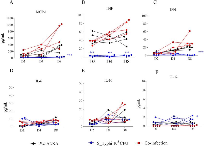
www.nature.com/scientificreports/
Figure 1. Plasmodium berghei ANKA and Salmonella enterica serovar Typhi coinfection model. In the malaria
infection model developed, three groups of animals received an intraperitoneal dose of 1 06 erythrocytes infected
with P. berghei ANKA on D0, and challenged with S_Typhi on D1. Group 1 received 105 S_Typhi CFUs on
D1 (Pb + S_Typhi 105). Group 2 received 1 04 S_Typhi CFUs on D1 (Pb + S_Typhi 1 04), and Group 3 received
103 S_Typhi CFUs (Pb + S_ Typhi 1 03). Three other groups were challenged with only S_Typhi on D1, Group
4 (S_Typhi 105), Group 5 (S_Typhi 104) and Group 6 (S_Typhi 1 03). The animals were euthanized for the
quantification of CFUs of the liver, spleen and intestine in SS-Agar at 37 °C. This experiment was replicated
once.
dysfunctions as having an important role in increasing susceptibility to Salmonella bacteremia, since these cells
are one of the main phagocytes of bacteria2,4,6,8,12,13,15. The hemolysis caused by Plasmodium sp. induces the secre-
tion of the enzyme heme oxygenase 1 (HO-1) that efficiently ensures the iron recycling and, at the same time,
prevents the harmful pro-oxidant effects of the heme group. As a result, malarial hemolysis impairs the ability to
develop neutrophils in order to mount a competent oxidative burst so that they become niches for Salmonella and
other Gram-negative bacterial co-infections6,12,13,16,17. In the case of macrophage, malaria hemolysis also affects
macrophage iron homeostasis. The iron liberated from heme by HO-1 enters macrophages and, since iron is
an essential micronutrient for the replication of Salmonella spp. and other intracellular pathogens, macrophage
iron retention in malaria favors bacterial m ultiplication6,16,17. In addition, macrophage iron contents enhance
cytokine dysregulation and lead to the infiltration of monocytes and neutrophils in the lamina propria, which
are hallmarks of pathology in bacteremia for Salmonella or other invasive bacteria in malaria infection11,16.
Malaria infection also affects intestinal permeability and facilitates invasions of enteric pathogenic
bacteria18–20. One clinical study addressed intestinal permeability in patients with falciparum malaria, and
sucrose, lactulose and mannitol in urine were used as an indication of alterations of this permeability18. There
are still several critical factors that are unknown regarding the pathogenesis of Plasmodium-Salmonella coin-
fection. Several models have proposed plausible epidemiological situations in malaria endemic areas without
basic sanitation. These situations involve simultaneous infections or infections occurring in the days after the
malarial infection10,12–14,21. Thus, a Plasmodium-Salmonella coinfection model on which oral bacterial chal-
lenge happened at the beginning of the rodent malaria simulation can provide insights into how parasitemia
compatible to P. falciparum malaria promotes the susceptibility to bacteremia. Herein, a coinfection model was
established challenging Pb_ANKA-infected BALB/c mice with a serovar S_Typhi. Serum cytokines, functional
phagocytic activity and histopathological-morphometric analyses of the intestinal mucosa were compared with
groups of monoinfected mice with P.b_ANKA or S_Typhi in order to aid the understanding of these factors on
susceptibility to septicemia during malaria.
Results
Susceptibility of mice with malaria to Salmonella enterica sv Typhi infection. Most Salmonella
enterica serovares (sv) can cause intestinal infections. For the selection of the bacterial strain to be used in
the coinfection model, the three following strains were used: Salmonella enterica serovar Typhi (S_Typhi), Sal-
monella enterica serovar Chloreaesius (S_Chloreaesius) and Salmonella enterica serovar Salamae (S_Salamae)
(Figure S1). Only the S. enterica serovar Typhi managed to evolve in the invasion, and increased the number
of colonies established in 48 h. Based on the hypothesis that malaria infection would act as a susceptibility fac-
tor to Salmonella invasion, the experimental design of coinfection was tested in order to define the minimum
bacterial concentration in which bacteremia occurred only in mice previously infected with malaria, and not in
healthy mice. The Plasmodium-Salmonella coinfection model was established by comparing three different doses
of S_Typhi (1 × 10⁵, 1 × 10⁴ and 1 × 10³ CFU). For comparison, another three groups of S_Typhi monoinfected
mice received only 1 × 10⁵, 1 × 10⁴ 1 × 10³ CFU, respectively. The bacterial culture was positive in all coinfection
plates, on all sampled days (D2, D4 and D8), while the group that was monoinfected with S. enterica sv Typhi
infected with 1 × 10³ CFU did not show bacterial growth in any of the macerated organs on any of the analyzed
days. The presence of colony-forming units in the macerate of the liver, spleen and intestine was only found
in the respective Plasmodium-Salmonella-coinfection group (Fig. 1A–C). The results allowed us to determine
that the inoculum of 103 CFU of S. enterica sv Typhi was only able to develop bacteremia in animals previously
Scientific Reports | (2021) 11:2730 | https://doi.org/10.1038/s41598-021-82330-0 2
Vol:.(1234567890)www.nature.com/scientificreports/
Figure 2. Inflammation response on days D2, D4 and D8 in the coinfection model. The plasma of four
animals was sampled for the determination of inflammation response using a cytometric bead array (Mouse
Inflammation CBA Kit; BD-Biosciences/USA). (A) MCP-1; (B) TNFα; (C) IFNγ; (D) IL-6; (E) IL-10 and (F)
IL-12. The data for cytokines and chemokines are grouped by mean and standard deviation in a spreadsheet
using the GraphPad Prism program (version 7). Asterisks: level of significance of *p < 0.05, **p < 0.005 and
***p < 0.0005.
infected with malaria. The parasitemia of P. berghei ANKA in Plasmodium-Salmonella-coinfected mice on days
D2, D4 and D8 did not differ from those of malaria-monoinfected mice (Figure S2).
Plasma cytokine profile during malaria and Salmonella enterica sv Typhi coinfection in rela-
tion to malaria‑ and Salmonella‑ monoinfections. Since production of inflammatory cytokines
and chemokines are hallmarks of pathology in bacteremia for Salmonella or other invasive bacteria in malaria
infection, the inflammation response was evaluated using a cytometric bead array (CBA Mouse Inflammation
Kit; BD-Biosciences/USA). MCP-1, TNFα and IFNγ were the most secreted followed by IL-6, IL-10 and IL-12
(Fig. 2). Both Pb_ANKA-infected BALB/c mice and coinfected mice showed exacerbation in the cytokines
MCP-1, TNFα and IFNγ, which may contribute to the susceptibility of the organism to Salmonella invasion. On
the other hand, no differences were found with IL-6 and IL-10 between groups, while a slight increase in IL-12
was observed with animals with S_Typhi monoinfection. Nonetheless, these data presented inconclusive results.
Evaluation of phagocytic activity in medullary subpopulations. The study of susceptibility to bac-
terial coinfected in malaria patients has focused on dysfunction of phagocytic cells macrophages and neutro-
phils. Our study evaluated the phagocytic activity of ex vivo, PMA-activated, adherent, medullary subpopulation
cells against a suspension of 103 CFU of formalin-inactivated S_Typhi (see the self-explanatory diagram of the
ex vivo phagocytic assay in Figure S3). The total number of adherent cells showed no differences in all groups
Fig. 3). Coinfected mice showed a very low percentage of (neutrophil and macrophage) phagocytes cells based
on their characteristics under Romanowsky’s staining method when compared to other groups. The percentage
of neutrophils with at least one phagocyted bacteria were more greatly affected in coinfected mice in relation to
other groups, including in Pb_ANKA-infected BALB/c mice which was reduced in relation to the control mice.
The percentage of monocytes with at least one phagocyted bacteria was also affected in coinfected mice, but it
was possible though to note that ex vivo neutrophils showed a prominent phagocytic dysfunction.
Scientific Reports | (2021) 11:2730 | https://doi.org/10.1038/s41598-021-82330-0 3
Vol.:(0123456789)www.nature.com/scientificreports/
Figure 3. Evaluation of phagocytic activity of ex vivo medullar neutrophils and macrophages. After 24 h of
ex vivo incubation on glass slides of cells recovered from the femur of anesthetized mice from coinfected,
monoinfected groups with P. berghei ANKA or S_Typhi and control group. After washing, the adherent
cells were incubated with Phorbol 12-myristate 13-acetate (PMA) for activation in the phagocytosis assay. A
suspension of S_Typhi killed by formalization was incubated with the adherent cells. Finally, the slides were
washed, fixed in picric acid and stained using the Romanowsky method. Neutrophils and monocytes were
visualized through staining characteristics and the total number of phagocytic bacteria was calculated. The
phagocytic activity in cells between groups was compared using the percentage of phagocytes between adherent
cells; % of neutrophils and % of macrophage containing phagocytic bacteria, respectively. The phagocytic
activity in cells was compared by multiple comparison by Tukey’s method. The data are shown in a scattered plot
with mean and standard deviations. Asterisks: level of significance of *p < 0.05, **p < 0.005 and ***p < 0.0005.
Evaluation of the presence of bacterial modulation. In malaria monoinfection, an intraperitoneal
dose with 106 erythrocytes infected with P. berghei ANKA on D0 is able to cause BALB/c mice to die around
day 20 due to high levels of parasitemia (> 60–70%). Coinfection did not lead to a worse outcome because para-
sitemia was similar to that of P. berghei ANKA-monoinfected mice, when the animals were euthanized around
twenty days (data not shown). It was also investigated whether the strain of S_Typhi recovered from the livers of
coinfected mice underwent some modulation that could influence the invasion capacity in non-phagocyte cells,
whereas bacterial pathogens tightly regulate the expression of virulence genes in response to very specific condi-
tions. The HeLa cell invasion assay was used to compare the S_Typhi strain used in pre-inoculum conditions
with the ex vivo strain obtained from liver macerate (see the self-explanatory diagram of invasion assay in Fig-
ure S4). Both behaved similarly in relation to the amount of invaded cells (Fig. 4A), however, the pre-inoculum
invaded more HeLa cells after six hours, which indicates that the post-invasion bacterium did not undergo any
modulation that made it more invasive (Fig. 4B).
Histopathological analysis by photomicroscopy. The above results showed that the invasion by S_
Typhi was mainly due to the presence of malaria rather than the invasive capacity of the bacterium. To assess
whether malaria infection affects intestinal tissue integrity or irregularities on mucosa sublayers are a result of
coinfection, histopathological sections were submitted to morphometric analysis. The volume and intestinal
wall did not differ between the groups following the quantification by Cavalieri technique, nor did the submu-
cosal, muscular and serous layers show differences among the groups (data not shown). Morphometric measure-
ments of intestinal volume in the cross sections of histopathological sections increased by magnification 100.
The intestinal tissue integrity and irregularities on mucosa sublayers were evaluated by images of sections using
a stereomicroscope. Blind analyses were evaluated in longitudinal extensions at magnification 400, where count-
ing systems containing points were superimposed on the images in the program Imod version 4.7/stereology
module22 (Figure S5). Four types of changes were evident (Fig. 5): (I) appearance of ruptures in the enterocyte
layer, (II) presence of mononuclear leukocytes in the enterocyte layer, (III) increase in goblet cells in the entero-
cyte layer and (IV) increase in volume of leukocyte infiltrate in the sub-mucosa. Irregularities in the intestinal
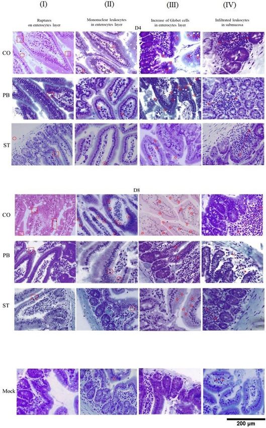
tissues were quantified by the Delesse principle. It was possible to observe ruptures in the enterocyte layer on
days D4 and D8 in the three groups (column I, Fig. 5), but events increased in coinfected animals. Pb_ANKA
monoinfected animals showed a slight increase in ruptures, while the S_Typhi-monoinfected mice (ST) showed
very few ruptures, which indicates that these ruptures may be the gateway for the bacterium (Fig. 6A).
The intestinal mucosa showed the presence of mononuclear leukocytes among the enterocytes (column
II, Fig. 5). In this case, the three groups presented more than the control group on D4, the mononuclear cells
infiltrate was much higher on D8 in the coinfected ones in relation to S_Typhi and control groups, however it
did no differ of P. berghei ANKA infected mice (Fig. 6B). Regarding the presence of mucoid or goblet cells in
the epithelial layer (column III, Fig. 5), only the co-infected animals showed an increase of these cells (Fig. 6C).
The volume of leukocyte infiltrate in the mucosa showed the main differences (column IV, Fig. 5) and the three
groups differed from the control on the 2 days, while the volume in coinfected animals was higher than both
groups which were monoinfected with P. berghei ANKA and S_Typhi (Fig. 6D). Interestingly, the volume of
Scientific Reports | (2021) 11:2730 | https://doi.org/10.1038/s41598-021-82330-0 4
Vol:.(1234567890)www.nature.com/scientificreports/
Figure 4. Comparison of the invasive capacity of pre-inoculum and ex vivo obtained bacteria. HeLa cell
invasion assay to evaluate the possibility of ex vivo Salmonella having its phagocytic activity influenced by
some modulation due to host response. (A) Number of cells invaded and (B) number of bacteria per cell
between aliquots of S_Typhi strain used in pre-inoculum conditions with ex vivo strain obtained from liver
macerate. Pre-inoculum aliquot was obtained after growth of nine hours in LB broth and stored at − 80 °C for
more than 15 days until the moment of the assay. The ex vivo aliquot was obtained from a macerated mouse
liver coinfected on D4 and frozen at − 80 °C. The two ex vivo and pre-inoculum bacteria were thawed and set
for invasion assay in HeLa cells. Pre-inoculum bacteria were diluted in a series of 10–10 and sown in SS-Agar
medium to define the inoculum equivalence of ex vivo bacteria. The invasion time was 1, 3 and 6 h. After these
periods, medium supplemented with gentamicin was added to the culture medium for 1 h in order to eliminate
external bacteria. Slides were then washed, stained using the Romanowsky method and bacteria visualized. The
data are shown in a box plot with mean and standard deviations. Asterisks: level of significance of *p < 0.05,
**p < 0.005 and ***p < 0.0005.
leukocyte infiltrate in S_Typhi-infected mice recovered normal volume on D8 that did not differ of control group.
In relation to enterocyte basal layer, the volume was inconclusive (Fig. 6E). With this, it is possible to suggest that
malaria causes a disruption of intestinal homeostasis and makes it susceptible to Salmonella invasion.
Discussion
Due to the overlap in terms of geographical distribution, children with recent or acute malaria have a higher risk
of bacterial infection and death. This knowledge comes from clinical studies according to which the effects of
malaria on the immune response against Salmonella bacteremia1,2,7. The exact nature of this association is still
unknown, due to heterogeneity of data and lack of adequate controls corresponding to severity (reviewed b y6).
Thus, little is known about whether malaria affects host resistance to intestinal colonization with typhoidal or
non-typhoidal Salmonella enterica after bacterial ingestion. Herein, we managed to develop a model of coinfec-
tion by Salmonella enterica serovar Typhi and P. berghei ANKA in BALB/c mice, in which animals were already
susceptible to Salmonella bacteremia after a day of malarial infection. Our model showed that a level of 1%
parasitemia and 24 h of intraperitoneal infection was able to make the animals susceptible to invasive salmonel-
losis. These parasitemias are commonly observed in human malaria, especially in African children. Therefore,
the leukocyte hemozoin pigment is a stronger predictor of malaria disease than parasitemia itself, which shows
that concomitant infection can mask the diagnosis of salmonellosis infection when antimalarial treatment has
still not improved the child’s symptoms9,23.
Although malaria is an acute systemic infection, it is known that gastrointestinal symptoms, such as nausea,
vomiting, abdominal pain and diarrhea, are common during the acute phase of malaria. Nutrient malabsorp-
tion has been reported in severe falciparum malaria, suggesting that the environment in the intestinal lumen
could be a ltered18,24. In a study on malaria falciparum, damage in the intestinal epithelium was evidenced due
to permeation of compounds, such as monosaccharides and disaccharides, that are normally relatively imper-
meable in a healthy intestinal mucosa but in this case they were observed in the urine of these patients18. In
this context, we performed a thorough analysis of intestinal tissue in the histopathological sections in malaria
infections on D8 in order to evaluate changes in intestinal volume and tissue. The volume of the lumen and
intestinal wall were not altered between the groups probably because on the eighth day the changes are probably
still focal. The intestinal epithelium provides a protective barrier against enteric pathogens and upon disruption
increases susceptibility to infections or impairs immunity25. One of the main findings observed here was that
monoinfection by P. berghei ANKA malaria was able to alter the integrity of the epithelial layer of the intestine.
These findings are in agreement with altered intestinal epithelium permeability in both Plasmodium falciparum
infected patients and murine malaria model10,18,25. The appearance of ruptures in the enterocyte layer seems a
detachment of the intestinal epithelia demonstrated by Taniguchi and cols10. Here, ruptures in the enterocyte
layer were exacerbated in coinfected animals suggesting that ruptures in the epithelial layer caused by malaria
predispose the host to bacteremia23,26.
Scientific Reports | (2021) 11:2730 | https://doi.org/10.1038/s41598-021-82330-0 5
Vol.:(0123456789)www.nature.com/scientificreports/
Figure 5. Irregularities in the small intestines of BALB/c mice caused by coinfection by Plasmodium berghei ▸
ANKA and Salmonella enterica serovar Typhi. Four types of changes were evidenced in photomicrographs and
highlighted in columns I to IV. (I) appearance of ruptures in the enterocyte layer, (II) presence of mononuclear
leukocytes in the enterocyte layer, (III) increase of goblet cells in the enterocyte layer and (IV) volume of
leukocyte infiltrate in the sub-mucosa. Group CO (coinfection): mice infected with 1 × 10⁶ erythrocytes
infected with P. berghei ANKA inoculated intraperitoneally on D0, and on D1 the animals were challenged with
1 × 10³ CFU of Salmonella enterica sp. Typhi by gavage. Group PB: monoinfected mice with 1 × 10⁶ erythrocytes
infected with P. berghei ANKA intraperitoneally on D0. Group ST: monoinfected mice with 1 × 10³ CFU
of Salmonella enterica sp. Typhi inoculated via gavage on D1. Photomicrographs of groups CO, PB and ST
distributed in columns I to IV between D4 and D8. Last sequence of photomicrographs with images of control
animals. Column I: presentation of ruptures at the border of the villus (red square); column II: infiltrate of
mononuclear leukocytes in the enterocyte layer (red circle); column III: highlight of goblet cells (red broad
arrows); column IV: dimension of the volume of leukocyte infiltrate in the mucosa region (red arrow at both
ends). Bar 200 μm.
Changes in the architecture of the intestinal tissue included the increase in the number of goblet cells and
in the cell density, which in the latter is characterized by volume of the leukocyte infiltrate in the sub-mucosa.
The goblet cells are responsible for the layer of mucus that covers the epithelium to facilitate the removal of food
content and are also part of the first line of defense against damage caused by pathogenic bacteria and bacterial
products27,28. Here, coinfected mice showed several of these mucoid cells in the enterocyte layer, which indicates
that the innate response to the infection bacteria was active in these animals. However, the disarrangement of
intestinal homeostasis caused by malaria may compromise the fight against the Salmonella infection, despite
abundance of the goblet cells. In relation to cell density, leukocyte infiltrate was greatly increased in coinfected
mice. Several coinfection models showed that these infiltrates are composed of inflammatory monocytes and
neutrophils6,9,21,25. Despite this increase, the phagocytic activity has been one of the most widely-accepted expla-
nations for susceptibility to bacteremia in m alaria2,4,14,15. Herein, phagocytic dysfunctions of macrophage and
neutrophil progenitors in co-infected mice were greatly reduced, which indicates that these effector cells would
have already been compromised in the primary lymphoid organs. In addition, hematopoiesis impairment may
be associated with factors associated with bacteria, since MCP-1, TNFα and IFNγ levels and parasitemia in co-
infected mice did not differ from those monoinfected by P. berghei ANKA.
In relation to the bacteria, the survival and growth of salmonellae within host cells are important for bacte-
rial virulence, and many genes that are required for invasion are not expressed when bacteria are grown on rich
media29. Herein, we demonstrated that phenotypically the ex vivo S_Typhi recovered from the liver of coinfected
animals did not become more invasive than pre-inoculum bacteria. One study showed that animals previously
infected with P. yoelii exhibited intensified colonization of a noninvasive, commensal, Escherichia coli strain,
which suggests that the disturbance of the microbial community by malaria opens up an ecological niche that
can be harnessed by pathogenic b acteria21. However, we are cautious as to whether our data show that malaria
infection makes mice susceptible to invasion of pathogenic bacteria, despite some modulation in vivo, or if,
phenotypically, the S_Typhi grown in rich media would already be able to invade the host c ells9,14.
One limitation of the study was to use a serovar Typhi from the Salmonella enterica line, since pediatric bacte-
remia caused by P. falciparum infection mainly involves non-typhoid invasive Salmonella. In addition, the models
of coinfection or monoinfection in mice use S. enterica serovar Typhimurium because it spreads systemically in
this host9,12,14,21,30. From three serovars of Salmonella enterica, we defined a dose with S_Typhi that managed to
evolve in the invasion of BALB/c mice only in those previously infected with malaria. Other limitation was that
in our model, the host was challenged with the bacterium the day after malaria infection, while children who
develop malaria-Salmonella coinfection are probably already colonized by these pathogenic b acteria15. Although,
that is assumption that Salmonella infection is subsequent to malaria, i.e. it is reasoned that in endemic areas
individuals would be most likely to become infected with typhoidal or non-typhoidal Salmonella after contracting
malaria9. The number of mice used here seemed another limitation, however they were sufficient to demonstrate
the major differences. Despite these limitations, our model provided us with a new view on intestinal mucosal
alteration that predisposed BALB/c mice to bacteremia due to malaria.
In conclusion, our findings provide descriptive insights into how mucosal responses to a bacterial pathogen
are altered in a simultaneous malaria infection. Although the knowledge of malaria as a susceptibility factor to
Salmonella bacteremia is evident, the pathophysiological mechanisms are still not well understood due to their
complexity and lack of well-established experimental models. This study aimed to establish an experimental
model and evaluate the pathophysiology with better coverage of intestinal histopathology, systemic immune fac-
tors and functional assays of phagocytic dysfunction. The inflammatory exacerbation evidenced by the increase
of cytokines and chemokines with the dysfunction especially of neutrophils is also well known and was well
characterized as an effect of an exacerbated inflammatory response. Finally, our main findings were the altera-
tion in the epithelial integrity of the intestine caused by monoinfection with P. berghei, which was identified as a
possible cause of susceptibility to Salmonella invasion. Knowledge on the influence on intestinal tissue and the
interaction of effector immune mechanisms in Salmonella-malaria coinfection is absolutely essential for creating
strategies to combat the bacterial infections in affected populations.
Scientific Reports | (2021) 11:2730 | https://doi.org/10.1038/s41598-021-82330-0 6
Vol:.(1234567890)www.nature.com/scientificreports/
Scientific Reports | (2021) 11:2730 | https://doi.org/10.1038/s41598-021-82330-0 7
Vol.:(0123456789)www.nature.com/scientificreports/
Figure 6. Quantification of irregularities in the small intestines caused by coinfection by Plasmodium berghei
ANKA and Salmonella enterica serovar Typhi. Multiple comparison by Tukey test among the four types of
changes evidenced in photomicrographs in coinfection Plasmodium berghei ANKA and Salmonella enterica
serovar Typhi. (A) Appearance of ruptures in the enterocyte layer, (B) Presence of mononuclear leukocytes in
the enterocyte layer, (C) Increase in goblet cells in the enterocyte layer and (D) Volume of leukocyte infiltrate in
the mucosa. The data are shown in a box plot with mean and standard deviations. Asterisks: level of significance
of *p < 0.05, **p < 0.005 and ***p < 0.0005.
Material and methods
Strains. Currently, the Salmonella nomenclature system defines the genus as being composed of three spe-
cies: S. subterranea, S. bongori and S. enterica, the latter having more than two thousand serotypes and, as such,
it is classified as a subspecies31. The Salmonella enterica serovar Typhi (S_Typhi), Salmonella enterica serovar
Chloreaesius (S_Chloreaesius) and Salmonella enterica serovar Salamae (S_Salamae) strains were provided by
the Laboratory of Diagnosis and Control of Amazonian Infectious Diseases, Instituto Leonidas e Maria Deane-
ILMD/Fiocruz Amazonia. The Plasmodium berghei ANKA-GFP strain (clone 15cy1) was conceded by Dr.
Claudio Marinho of the Laboratory of Experimental Immunoparasitology, University of São Paulo (USP). The
infected red blood cells (iRBC) used in experimental infections were obtained after in vivo passage in BALB/c
mice. For infections, aliquots containing 5 × 10⁶ infected red blood cells (iRBCs)/100 µl were stored at − 80 °C
were inoculated intraperitoneal (i.p.) on day 0. The controls treated with this simulation were injected with an
equal volume of isotonic and neutral phosphate buffer. The parasitemia was performed on days 2, 4 and 8 after
analysis using flow cytometry.
Animals. Female BALB/c mice aged between 6 and 8 weeks were used. In all experiments, the welfare of the
animals was taken into consideration. The mice were housed in a standard polycarbonate cage with wood shav-
ings bedding, with a maximum of 5 mice per cage. We also attempted to reduce the stress of individual housing
(when necessary) by environmental enrichment with nestlets and small play tunnels. The animal room was kept
at a controlled temperature and humidity, with a light and dark cycle of 12 h. Mice had ad libitum access to food
and water.
Anesthesia and euthanasia. All efforts were made to prevent undue stress or pain to the mice. The mice
were humanely euthanized once they showed the following clinical signs: lethargy; hypothermia and/or dif-
ficulty of breathing. The mice were euthanized with ketamine (300 mg/kg) (Vetbrands, Brazil) and xylazine
(22.5 mg/kg) (Syntec, Brazil), and consciousness was checked by testing the foot reflexes, heartbeats and breath-
ing movements. All experiments were performed in accordance with the ethical guidelines for experiments with
mice, and the protocols were approved by the National Council for the Control of Animal Experimentation,
National Institute of Amazonian Research – INPA (CEUA nº 2018016/2019). The guidelines for animal use and
care were based on the standards established by the Brazilian College of Animal Experimentation (COBEA).
Scientific Reports | (2021) 11:2730 | https://doi.org/10.1038/s41598-021-82330-0 8
Vol:.(1234567890)www.nature.com/scientificreports/
Infection. Initially, one Salmonella enterica serovar was selected to be used in the coinfection model with the
Plasmodium berghei ANKA. The strategy was to select the most invasive of the three bacteria (S_Typhi, S_Chlo-
reaesius and S_Salamae) i.e., the one that presented the most colony forming units (CFU) in the liver, spleen and
intestine at 24 h and 48 h post infection. The animals were divided into three groups containing four animals to
be individually challenged with the three different serovares. The time necessary to reach maximum exponential
growth that was ideal for infection was defined as nine hours of growth in LB broth, and the oral inoculation was
performed by gavage technique with a concentration of 1 × 10⁹ CFU in 100 µl in each individual. After euthanasia
and removal of organs, they were homogenized, diluted and sown in serial dilution in Salmonella—Shigella agar
culture medium (SS-Agar) and incubated at 37 °C. After 24 h, the colony forming units (CFU) were quantified.
Establishment of Plasmodium berghei ANKA and Salmonella enterica serovar Typhi coinfec-
tion model. For establishment of Plasmodium-Salmonella coinfection model, three groups of BALB/c mice
received an intraperitoneal dose of cryopreserved aliquots containing 5 × 10⁶ erythrocytes infected with P. berghei
ANKA on day 0 (D0). One group received a dose of 1 × 10⁵ CFU of S_Typhi, the second received 1 × 10⁴ CFU
and the third 1 × 10³ CFU on D1, by gavage. For the comparison, three other groups containing four mice with
monoinfection by S_Typhi, received, a dose of 1 × 10⁵ CFU by gavage on D1. The second received 1 × 10⁴ CFU
and the third 1 × 10³ CFU, by the same means. The experimental design of malaria-Salmonella coinfection was
evaluated by the presence and/or absence of colony-forming units in the macerate of the liver, spleen and intes-
tine obtained on D2, D4 and D8. After euthanasia, the organs were removed whole, homogenized, diluted and
sown in serial dilution in Salmonella—Shigella agar culture medium (SS-Agar) and incubated at 37 °C. The
group in which S_Typhi monoinfected mice did not show bacterial growth in any of the macerated organs on
any of the analyzed days was chosen. Parasitemia of P. berghei ANKA groups was monitored by flow cytometry.
Plasmodium berghei ANKA and Salmonella enterica serovar Typhi coinfection model. Three
groups containing four mice were classified as S_Typhi monoinfection, P. berghei ANKA monoinfection and
Plasmodium-Salmonella-coinfection. The P. berghei ANKA monoinfection and Plasmodium-Salmonella-coinfec-
tion groups received an intraperitoneal dose of cryopreserved aliquots containing 5 × 10⁶ erythrocytes infected
with P. berghei ANKA on day 0 (D0). The controls and S_Typhi monoinfected mice received an equal volume of
isotonic and neutral phosphate buffer. The determination of parasitemia was performed by flow cytometry. The
controls and S_Typhi monoinfected mice received an equal volume of isotonic and neutral phosphate buffer. The
animals were submitted to the Plasmodium-Salmonella coinfection and S_Typhi monoinfection mice received
100 µL S_Typhi 103 in 24 h LB broth via gavage one day after the malarial infection (D1). Mice of control and
P. berghei ANKA monoinfection groups received an equal volume of isotonic and neutral phosphate buffer.
Parasitemia of P. berghei ANKA monoinfection and Plasmodium-Salmonella-coinfection groups was performed
by flow cytometry. Extraction from the intestine occurred on days four and eight (D4 and D8) after malarial
infection. The animals were euthanized for the quantification of CFUs in the liver, spleen and intestine, and the
homogenized ones were sown in SS-Agar at 37 °C for quantification of CFU.
Evaluation of functional activity. The phagocytic activity of neutrophils and macrophages was evalu-
ated using cell culture after incubation, which was based on the literature and optimized for the methodology
(See the diagram in Figure S3). Briefly, after induction of anesthesia and analgesia, the animals were sacrificed
for the removal of medullary subpopulations from the femur on D8. The recovered cells were plated on glass
slides (Knittel, Brazil) in DMEM 24-well plates at 37 °C, 5% CO2 for panning of adherent cells. After 24 h of
incubation, nonadherent cells were removed after three cycles of washing in DMEM medium. The adherent
cells were incubated with Phorbol 12-myristate 13-acetate (PMA) for 1 h for activation, and then washed three
times in DMEM medium. A suspension of 103 CFU of formalin inactivated S_Typhi that had been treated
previously with 37% formalin for 1-h and washed 4 times with neutral phosphate buffer was applied for 3 h at
37 °C, 5% CO2. Finally, the slides were washed, fixed in picric acid and stained using the Romanowsky method.
Neutrophils were differentiated by the “busy” aspect of nucleus showing several lobes, while cells character-
ized by horseshoe-shaped nucleus with dishwater-gray cytoplasm and a few tiny granules were characterized as
monocyte-macrophage lines. The quantity of phagocytic activity in cells was compared by multiple comparison
by Tukey’s method.
Bacterial invasion assay in HeLa cells. The possibility of ex vivo Salmonella strains having been modu-
lated after infection and influencing phagocytic activity was analyzed. For this, the livers of mice that were
coinfected on D4 were macerated and frozen at − 80 °C. For comparison, an aliquot of S_Typhi was used under
the same conditions as the pre-inoculum (in growth phase of nine hours in LB broth) and stored at − 80 °C for
15 days until the moment of the assay (Figure S4). HeLa cells were grown in 24-well plates containing glass
slides (Knittel, Brazil) in DMEM medium with 10% fetal bovine (Sigma, Brazil) at 5% C O2 at 37 °C until total
confluence. The two ex vivo and pre-inoculum bacteria were thawed and set for the invasion assay in HeLa cells.
Pre-inoculum bacteria were diluted in a series of 10–10 and sown in SS-Agar medium to define the inoculum
equivalence of ex vivo bacteria. The invasion time was 1, 3 and 6 h. After these periods, medium supplemented
with gentamicin was added to the culture medium for 1 h in order to eliminate external bacteria. Slides were
then washed, stained using Romanowsky staining and bacteria were visualized.
Scientific Reports | (2021) 11:2730 | https://doi.org/10.1038/s41598-021-82330-0 9
Vol.:(0123456789)www.nature.com/scientificreports/
Analysis of serum cytokines. Cytokines were measured on D2, D4 and D8 for each of the infection
groups. The plasma from four mice was collected for the determination of cytokines using cytometric bead array
for mouse inflammation factors (CBA Mouse Inflammation Kit; BD-Biosciences/USA).
Histopathological evaluation of the intestine. After euthanasia, the intestines of the animals were
removed and kept in buffered formalin for 48 h at room temperature. The samples were processed in the Quan-
titative Morphology Laboratory (LaMiq/UFAM). For this, the intestines were dehydrated in increasing con-
centrations of 70 and 96% ethanol (2 h in each concentration), pre-infiltrated in 96% ethanol + hydroxyethyl
methacrylate plastic historesin solution (Technovit 7100, Külzer-Heraues, Germany) overnight and infiltrated in
100% resin. The samples were arranged in individual Histobloc teflon molds (Külzer-Heraues, Germany) set in
plastic resin + polymerizing solution. The molds were heated in an oven at 37 °C until complete polymerization.
The analysis of epithelial integrity of the intestine and morphometric measurements were made using stereol-
ogy of the Cavalieri principle of volume and the Delesse principle, both operated using the Imod program (see
Supplemental Material 1).
Statistical analysis. The prism 5.0 statistical program (GraphPad Software, Inc. USA) was used for the
statistical and graphical analysis of this study. The independent variables (treatments) were tested for their nor-
mality by the Kolmogorov–Smirnov test. An ANOVA test using a multiple Tukey comparison was used for the
following assays: (1) cytokines on days D4 and D8, (2) phagocytosis assays on D8, and (3) S_Typhi invasion of
pre-inoculum and ex vivo in HeLa cells. In relation to histopathological analyses, on average 20 serial sections
of the intestine were calculated by appropriate equations developed for stereological analyses22,32,33. An error
coefficient of 5% and deviation of 15% were considered acceptable. The estimate of the volume was determined
according to Cavalieri’s principle, and the results were analyzed using one-way ANOVA (see Supplementary
material).
Received: 28 August 2020; Accepted: 6 January 2021
References
1. Uneke, C. J. Concurrent malaria and typhoid fever in the tropics: the diagnostic challenges and public health implications. J. Vector
Borne Dis. 45, 133–142 (2008).
2. Takem, E. N. N., Roca, A. & Cunnington, A. The association between malaria and non-typhoid Salmonella bacteraemia in children
in sub-Saharan Africa: a literature review. Malar. J. 13, 400 (2014).
3. Nadjm, B. et al. WHO guidelines for antimicrobial treatment in children admitted to hospital in an area of intense Plasmodium
falciparum transmission: prospective study. BMJ 340, 848 (2010).
4. Nyirenda, T. S. et al. Loss of humoral and cellular immunity to invasive nontyphoidal Salmonella during current or convalescent
Plasmodium falciparum infection in Malawian children. Clin. Vaccine Immunol. 24, 1–13 (2017).
5. Mtove, G. et al. Decreasing incidence of severe malaria and community-acquired bacteraemia among hospitalized children in
Muheza, north-eastern Tanzania, 2006–2010. Malar. J. 10, 320 (2011).
6. Mooney, J. P., Galloway, L. J. & Riley, E. M. Malaria, anemia, and invasive bacterial disease: a neutrophil problem?. J. Leukoc. Biol.
105, 645–655 (2019).
7. Mackenzie, G. et al. A decline in the incidence of invasive non-typhoidal salmonella infection in the gambia temporally associated
with a decline in malaria infection. PLoS ONE 5, e10568 (2010).
8. Mabey, D. C. W., Brown, A. & Greenwood, B. M. Plasmodium falciparum malaria and salmonella infections in gambian children.
J. Infect. Dis. 155, 1319–1321 (1987).
9. Mooney, J. P. et al. The mucosal inflammatory response to non-typhoidal Salmonella in the intestine is blunted by IL-10 during
concurrent malaria parasite infection. Mucosal Immunol. 7, 1302–1311 (2014).
10. Taniguchi, T. et al. Plasmodium berghei ANKA causes intestinal malaria associated with dysbiosis. Sci. Rep. 5, 1–12 (2015).
11. Roux, C. M. et al. Both hemolytic anemia and malaria parasite-specific factors increase susceptibility to nontyphoidal Salmonella
enterica serovar typhimurium infection in mice. Infect. Immun. 78, 1520–1527 (2010).
12. Cunnington, A. J., De Souza, J. B., Walther, M. & Riley, E. M. Malaria impairs resistance to Salmonella through heme-and heme
oxygenase-dependent dysfunctional granulocyte mobilization. Nat. Med. 18, 120–127 (2012).
13. Cunnington, A. J. et al. Prolonged neutrophil dysfunction after plasmodium falciparum malaria is related to hemolysis and heme
oxygenase-1 induction. J. Immunol. 189, 5336–5346 (2012).
14. Lokken, K. L. et al. Malaria parasite infection compromises control of concurrent systemic non-typhoidal Salmonella infection
via IL-10-mediated alteration of myeloid cell function. PLoS Pathog. 10, e1004049 (2014).
15. Gómez-Pérez, G. P., Van Bruggen, R., Grobusch, M. P. & Dobaño, C. Plasmodium falciparum malaria and invasive bacterial co-
infection in young African children: the dysfunctional spleen hypothesis. Malar. J. 13, 1–15 (2014).
16. Orf, K. & Cunnington, A. J. Infection-related hemolysis and susceptibility to gram-negative bacterial co-infection. Front. Microbiol.
6, 1–8 (2015).
17. van Santen, S., de Mast, Q., Swinkels, D. W. & van Ven, A. J. A. M. The iron link between malaria and invasive non-typhoid Sal-
monella infections. Trends Parasitol. 29, 220–227 (2013).
18. Wilairatana, P., Meddings, J. B., Ho, M., Vannaphan, S. & Looareesuwan, S. Increased gastrointestinal permeability in patients with
Plasmodium falciparum malaria. Clin. Infect. Dis. 24, 430–435 (1997).
19. Potts, R. A. et al. Mast cells and histamine alter intestinal permeability during malaria parasite infection. Immunobiology 221,
468–474 (2016).
20. Chau, J. Y. et al. Malaria-associated l-arginine deficiency induces mast cell-associated disruption to intestinal barrier defenses
against nontyphoidal salmonella bacteremia. Infect. Immun. 81, 3515–3526 (2013).
21. Mooney, J. P. et al. Inflammation-associated alterations to the intestinal microbiota reduce colonization resistance against non-
typhoidal Salmonella during concurrent malaria parasite infection. Sci. Rep. 5, 1–11 (2015).
22. Kremer, J. R., Mastronarde, D. N. & McIntosh, J. R. Computer visualization of three-dimensional image data using IMOD. J. Struct.
Biol. 116, 71–76 (1996).
Scientific Reports | (2021) 11:2730 | https://doi.org/10.1038/s41598-021-82330-0 10
Vol:.(1234567890)www.nature.com/scientificreports/
23. Scott, J. A. G. et al. Relation between falciparum malaria and bacteraemia in Kenyan children: a population-based, case-control
study and a longitudinal study. Lancet 378, 1316–1323 (2011).
24. Berkley, J. A. et al. HIV, malnutrition and invasive bacterial infection among children with severe malaria. Clin. Infect. Dis. 49,
336–343 (2009).
25. Alamer, E. S. et al. Dissemination of non-typhoidal Salmonella during Plasmodium chabaudi infection affects anti-malarial immu-
nity. Parasitol. Res. 118, 2277–2285 (2019).
26. Park, S. E. et al. The relationship between invasive nontyphoidal Salmonella disease, other bacterial bloodstream infections, and
malaria in Sub-Saharan Africa. Clin. Infect. Dis. 62, s23–s31 (2016).
27. Lv, Y. et al. Injury and mechanism of recombinant E. coli expressing STa on piglets colon. J. Vet. Med. Sci. 80, 205–212 (2018).
28. Kim, Y. S. & Ho, S. B. Intestinal goblet cells and mucins in health and disease: recent insights and progress. Curr. Gastroenterol.
Rep. 12, 319–330 (2010).
29. Pfeifer, C. G., Marcus, S. L., Steele-Mortimer, O., Knodler, L. A. & Finlay, B. B. Salmonella typhimurium virulence genes are induced
upon bacterial invasion into phagocytic and nonphagocytic cells. Infect. Immun. 67, 5690–5698 (1999).
30. Forbester, J. L. et al. Interaction of Salmonella enterica serovar typhimurium with intestinal organoids derived from human induced
pluripotent stem cells. Infect. Immun. 83, 2926–2934 (2015).
31. Su, L.-H. & Chiu, C.-H. Salmonella: clinical importance and evolution of nomenclature. Chang Gung Med. J. 30, 210–219 (2007).
32. Howard, C. V. & Reed, M. G. Unbiased Stereology. Three-Dimensional Measurement in Microscopy (Garland Science, New York,
2005).
33. Gundersen, H. et al. Some new, simple and efficient stereological methods and their use in pathological research and diagnosis.
Apmis 96, 379–394 (1988).
Author contributions
Y.C.M., M.J., V.F., D.A.M.C. performed the experimental model of infection; E.F., T.B.C., and Y.O.C. performed
invasion and phagocytosis assays; A.G.C. and Y.O.C. performed cytokines assay. Y.C.M., M.J. and O.T.F.C. per-
formed histopathology methods, P.P.O., F.T.M.C., and P.A.N. coordinated the study and prepared the manuscript.
Competing interests
The authors declare no competing interests.
Additional information
Supplementary Information The online version contains supplementary material availlable at https://doi.
org/10.1038/s41598-021-82330-0.
Correspondence and requests for materials should be addressed to P.A.N.
Reprints and permissions information is available at www.nature.com/reprints.
Publisher’s note Springer Nature remains neutral with regard to jurisdictional claims in published maps and
institutional affiliations.
Open Access This article is licensed under a Creative Commons Attribution 4.0 International
License, which permits use, sharing, adaptation, distribution and reproduction in any medium or
format, as long as you give appropriate credit to the original author(s) and the source, provide a link to the
Creative Commons licence, and indicate if changes were made. The images or other third party material in this
article are included in the article’s Creative Commons licence, unless indicated otherwise in a credit line to the
material. If material is not included in the article’s Creative Commons licence and your intended use is not
permitted by statutory regulation or exceeds the permitted use, you will need to obtain permission directly from
the copyright holder. To view a copy of this licence, visit http://creativecommons.org/licenses/by/4.0/.
© The Author(s) 2021
Scientific Reports | (2021) 11:2730 | https://doi.org/10.1038/s41598-021-82330-0 11
Vol.:(0123456789)You can also read