Activation of short-chain ketones and isopropanol in sulfate-reducing bacteria
←
→
Page content transcription
If your browser does not render page correctly, please read the page content below
Frey et al. BMC Microbiology     (2021) 21:50
https://doi.org/10.1186/s12866-021-02112-6

 RESEARCH ARTICLE                                                                                                                                  Open Access

Activation of short-chain ketones and
isopropanol in sulfate-reducing bacteria
Jasmin Frey1, Sophie Kaßner1, Dieter Spiteller1, Mario Mergelsberg2, Matthias Boll2, David Schleheck1 and
Bernhard Schink1*

  Abstract
  Background: Degradation of acetone by aerobic and nitrate-reducing bacteria can proceed via carboxylation to
  acetoacetate and subsequent thiolytic cleavage to two acetyl residues. A different strategy was identified in the
  sulfate-reducing bacterium Desulfococcus biacutus that involves formylation of acetone to 2-hydroxyisobutyryl-CoA.
  Results: Utilization of short-chain ketones (acetone, butanone, 2-pentanone and 3-pentanone) and isopropanol by
  the sulfate reducer Desulfosarcina cetonica was investigated by differential proteome analyses and enzyme assays.
  Two-dimensional protein gel electrophoresis indicated that D. cetonica during growth with acetone expresses
  enzymes homologous to those described for Desulfococcus biacutus: a thiamine diphosphate (TDP)-requiring
  enzyme, two subunits of a B12-dependent mutase, and a NAD+-dependent dehydrogenase. Total proteomics of
  cell-free extracts confirmed these results and identified several additional ketone-inducible proteins. Acetone is
  activated, most likely mediated by the TDP-dependent enzyme, to a branched-chain CoA-ester, 2-hydroxyisobutyryl-
  CoA. This compound is linearized to 3-hydroxybutyryl-CoA by a coenzyme B12-dependent mutase followed by
  oxidation to acetoacetyl-CoA by a dehydrogenase. Proteomic analysis of isopropanol- and butanone-grown cells
  revealed the expression of a set of enzymes identical to that expressed during growth with acetone. Enzyme assays
  with cell-free extract of isopropanol- and butanone-grown cells support a B12-dependent isomerization. After
  growth with 2-pentanone or 3-pentanone, similar protein patterns were observed in cell-free extracts as those
  found after growth with acetone.
  Conclusions: According to these results, butanone and isopropanol, as well as the two pentanone isomers, are
  degraded by the same enzymes that are used also in acetone degradation. Our results indicate that the
  degradation of several short-chain ketones appears to be initiated by TDP-dependent formylation in sulfate-
  reducing bacteria.
  Keywords: Anaerobic acetone degradation, Ketone degradation, Pentanone, Sulfate reduction, 2-hydroxyisobutyryl-
  CoA, Thiamine diphosphate, Adenosylcobalamin

* Correspondence: bernhard.schink@uni-konstanz.de
1
 Department of Biology, University of Konstanz, 78457 Constance, Germany
Full list of author information is available at the end of the article

                                       © The Author(s). 2021 Open Access This article is licensed under a Creative Commons Attribution 4.0 International License,
                                       which permits use, sharing, adaptation, distribution and reproduction in any medium or format, as long as you give
                                       appropriate credit to the original author(s) and the source, provide a link to the Creative Commons licence, and indicate if
                                       changes were made. The images or other third party material in this article are included in the article's Creative Commons
                                       licence, unless indicated otherwise in a credit line to the material. If material is not included in the article's Creative Commons
                                       licence and your intended use is not permitted by statutory regulation or exceeds the permitted use, you will need to obtain
                                       permission directly from the copyright holder. To view a copy of this licence, visit http://creativecommons.org/licenses/by/4.0/.
                                       The Creative Commons Public Domain Dedication waiver (http://creativecommons.org/publicdomain/zero/1.0/) applies to the
                                       data made available in this article, unless otherwise stated in a credit line to the data.

                                             Konstanzer Online-Publikations-System (KOPS)
                                    URL: http://nbn-resolving.de/urn:nbn:de:bsz:352-2-1go67kfz2048n7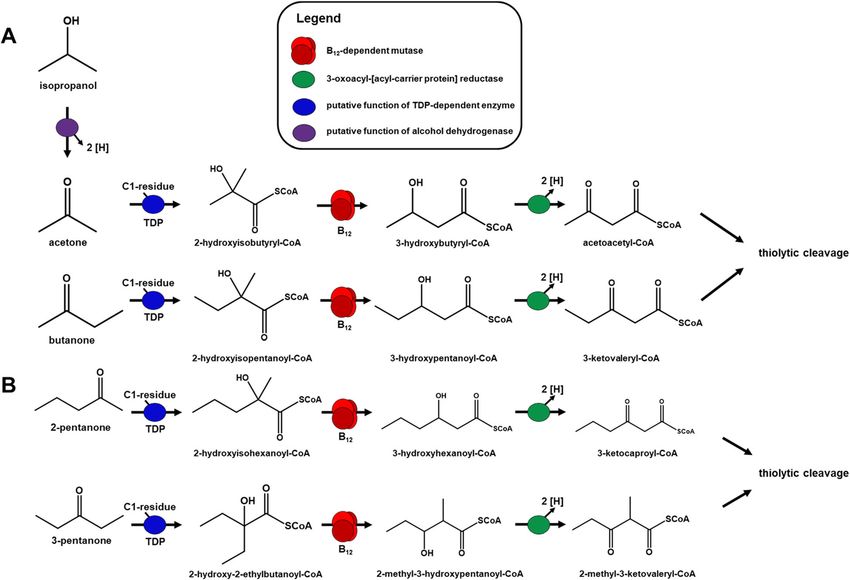
Frey et al. BMC Microbiology   (2021) 21:50                                                                 Page 2 of 11

Background                                                    Thus far, two sulfate-reducing bacteria (SRB) have
Short-chain ketones are used in industry as solvents and   been described that are capable of utilizing acetone and
as precursors for chemical syntheses. Acetone is pro-      butanone as electron donor and carbon source: Desulfo-
duced by a wide range of industrial processes, such as     coccus biacutus strain KMRActS (DSM 5651) and Desul-
the co-production with phenol in the so-called cumene      fosarcina cetonica strain 480 (DSM 7267) [16, 25–27]. In
process. It is also produced by fermenting bacteria such   D. biacutus acetone carboxylase activity was not detect-
as Clostridium spp., or in higher animals and humans       able in cell-free extracts, and the respective candidate
during ketosis [1, 2]. Isopropanol and butanone are com-   genes not found in the genome either. Moreover, no
monly used as solvents in industrial processes and are     acetoacetate-activating enzyme (e.g. acetyl-CoA:acetoa-
also side products of microbial degradation processes in   cetate CoA transferase or acetoacetate CoA ligase) was
soils. Acetone, isopropanol, butanone and 2-pentanone      measurable [16, 26, 28]. Further experiments led to the
belong to the ten most abundant volatile organic com-      assumption that carbon monoxide may be used as co-
pounds (VOCs) in agricultural soils [3]. Next to acetone   substrate leading to a reactive aldehyde species that is
and butanone, 3-pentanone, also known as diethyl ke-       subsequently transformed to acetoacetyl-CoA [29]. Gen-
tone, is a common odor component of landfills [4]. They    omic and proteomic studies revealed a gene cluster in D.
are released into the environment in considerable          biacutus that is strongly induced during acetone
amounts, rendering their microbial degradation a rele-     utilization: the cluster encodes a thiamine diphosphate
vant component of the biogeochemical carbon cycle.         (TDP)-dependent enzyme, a B12-dependent isomerase
Acetone (and other ketones) are present in the human       and a NAD+-dependent dehydrogenase [28]. Results
body in blood, urine and exhaled air (ketosis), but they   from recent studies suggested that the three (potential)
are also present in the human intestinal system as a re-   enzymes catalyze the following reaction sequence [30]:
sult of microbial fermentation: ketones are the second     (i) formylation of acetone to the branched-chain CoA
largest class of volatile organic compounds in human       ester 2-hydroxyisobutyryl-CoA, most likely mediated by
feces [5, 6]. Therefore, degradation of ketones might be   the TDP-dependent enzyme, (ii) conversion to 3-
relevant also in the metabolism of human gut micro-        hydroxybutyryl-CoA by the B12-dependent mutase, and
biota. Other sources of acetone and butanone are the       (iii) oxidation to acetoacetyl-CoA by a dehydrogenase.
emission from plant material during photochemical pro-     While the enzyme identities and intermediates for the
cesses [7, 8].                                             last two reactions were confirmed in cell-free extracts by
  Several metabolic routes are known for acetone           mass spectrometry and by heterologously produced en-
degradation by microorganisms. Aerobic microbes            zymes [30], the (predicted) TDP-dependent enzyme re-
may initiate acetone degradation by activation through     action remained experimentally inaccessible so far. D.
a Baeyer-Villiger monooxygenase to methyl acetate          cetonica harbors genes that are homologous to those
ester, by terminal hydroxylation to acetol (hydroxya-      found in D. biacutus. Therefore, it appears plausible that
cetone), or by carboxylation to acetoacetate [6, 9, 10].   both bacteria, and probably more sulfate-reducing bac-
Nitrate-reducing and phototrophic bacteria activate        teria, use the same metabolic strategy for acetone deg-
acetone by carboxylation which is coupled to the           radation [30].
consumption of two or more ATP equivalents per                In the present paper, we describe acetone degradation
molecule of acetone [11–15]. While such an energy-         in D. cetonica in order to identify similarities and/or dif-
expensive activation reaction is possible for nitrate-     ferences to that studied previously in D. biacutus by dif-
reducing and phototrophic bacteria, it is not feasible     ferential proteomics, enzyme assays, and CoA ester
for sulfate-reducing bacteria due to their much            metabolite analyses. In addition, degradation of buta-
smaller energy budget [11, 16–18].                         none (2-methyl ethyl ketone), 2-pentanone, 3-
  Sulfate reducers play important roles in the envir-      pentanone, and isopropanol (2-propanol) by D. cetonica
onment, especially in marine sediments and in tech-        was examined. In this work, also the first description of
nical settings such as the oil industry or wastewater      pentanone degradation by a sulfate reducer is presented
technology [19]. They are also active in the digestive     on the basis of proteomic data.
system of animals and humans both in healthy indi-
viduals and in persons afflicted with diseases like ul-    Methods
cerative colitis [20, 21]. They also appear to be          Chemicals
involved in or associated with chronic inflammatory        Chemicals were purchased from Sigma-Aldrich (Germany),
bowel diseases (IBD) in humans caused most likely by       Apollo Scientific (UK), AppliChem (Germany) or Carl
release of highly toxic and reactive hydrogen sulfide,     Roth GmbH (Germany). The CoA esters were synthe-
e.g., through H2S-mediated inhibition of butyrate oxi-     sized using the acyl thiophenyl esters as precursors as
dation in colonocytes [22–24].                             described earlier [30].
Frey et al. BMC Microbiology   (2021) 21:50                                                               Page 3 of 11

Bacterial growth conditions                                 Konstanz [28]. For total proteomic analysis of cell-free
Desulfosarcina cetonica strain 480 was cultivated in N2/    extracts (CFE) from cells grown with the different
CO2 (80%/20%)-flushed, butyl rubber-stoppered bottles       substrates (acetone, butyrate, isopropanol or butanone,
containing     sulfide-reduced,    bicarbonate-buffered     respectively), CFEs were analyzed directly with high-
medium [25]. The medium was supplemented with 10            resolution peptide fingerprinting-mass spectrometry
mM (or 20 mM in case of pentanones) Na2SO4 as               (LTQ-Orbitrap, Thermo Fisher) by the Proteomics Facil-
electron acceptor and 5 mM carbon source (acetone,          ity of the University of Konstanz [33].
butyrate, isopropanol, butanone, 2-pentanone, and 3-
pentanone). Cultures were incubated at 30 °C in the         Enzyme assays
dark.                                                       Activities of key enzymes of acetone degradation were
                                                            tested by discontinuous assays analyzed by HPLC. All
Preparation of cell-free extracts (CFE)                     enzyme assays were performed under strictly anoxic
Cells of D. cetonica were harvested by centrifugation       conditions (N2-flushed 4-ml glass vials sealed with butyl
(8200×g, 30 min, 4 °C) and washed twice with Tris–HCl       rubber stoppers) in 25 mM MOPS buffer, pH 7.2, (mu-
buffer (20 mM, pH 7.2). The cell pellet was resuspended     tase) containing 1 g l− 1 NaCl, 0.6 g l− 1 MgCl2 × 6 H2O
in Tris–HCl buffer (20 mM, pH 7.2) supplemented with        and 3 mM DTT. B12-dependent reaction mixes con-
0.5 mg DNase mL− 1 and 10 μL mL− 1 of Halt™ Protease        tained additional 50 μM adenosylcobalamin and were in-
Inhibitor Cocktail (with EDTA; Thermo Scientific). Cells    cubated in the dark at 30 °C. Reactions were started by
were disrupted by three to five passages through a          addition of cell-free extract and were followed discon-
cooled French pressure cell at 140 MPa. Cell debris was     tinuously by HPLC-UV or HPLC-MS measurements.
removed by centrifugation (27,000×g, 30 min, 4 °C) to       Substrate addition and sampling (150–200 μL per sam-
produce cell-free extract (CFE). Membrane fragments         ple) under anoxic conditions was performed with gas-
were separated by ultracentrifugation (50,000×g, 60 min,    tight syringes (Hamilton AG, Switzerland). Samples were
4 °C); the supernatant was termed soluble protein frac-     mixed thoroughly with dichloromethane to stop the re-
tion. Membrane fragments were washed once with buf-         action and remove protein. After centrifugation (16,000
fer (the remaining supernatant was termed the wash          x g, 5 min, RT (20–23 °C)), the aqueous phase was used
fraction) and resuspended in the same buffer.               for analysis by HPLC or LC-MS.
                                                              All photometric assays were carried out in MOPS
Two-dimensional polyacrylamide gel electrophoresis (2D-     buffer under strictly anoxic conditions as described
PAGE) and proteome analysis                                 above. 3-hydroxybutyryl-CoA dehydrogenase was
2D-PAGE of soluble proteins was performed using a           measured photometrically as 3-hydroxybutyryl-CoA-
BioRad Ready Strip IPG/Protean II system. The soluble       dependent NAD+ reduction at 340 nm. A coupled
protein fraction was desalted by Illustra NAP-25 col-       photometric assay for measurement of the B12-
umns (GE Healthcare, Germany) and each sample of 4          dependent isomerization of 2-hydroxyisobutyryl-CoA to
mg total protein was precipitated overnight at − 20 °C by   3-hydroxybutyryl-CoA was performed in a similar setup
addition of 4 volumes of ice-cold acetone. Precipitated     containing 2-hydroxyisobutyryl-CoA instead of 3-
protein was collected by centrifugation (10,000×g, 10       hydroxybutyryl-CoA. The subsequent oxidation of the
min, 4 °C) and air-dried at room temperature. The dried     isomerization product 3-hydroxybutyryl-CoA was mea-
protein pellet was solubilized in rehydration buffer        sured photometrically as described above.
(350 μL) and loaded onto an isoelectric focusing (IEF)
strip (BioRad IPG strips, 17 cm, pH 4–7) [31]. The iso-     High pressure liquid chromatography (HPLC) and HPLC-
electric focusing program involved a voltage ramp           mass spectrometry (MS) measurements
(rapid) to a maximal voltage of 10,000 V for at least 3 h   CoA esters were analyzed by HPLC using a Kinetex PFP
and a total focusing of 60,000 Volt-hours (Vh). Strips      column (5 mm, 100 A°, 250 3 4.6 mm; Phenomenex,
were equilibrated in SDS equilibration buffers I and II     USA) on a Shimadzu Prominence system with PDA de-
(with DTT and iodoacetamide, respectively) as de-           tector (SPD-M20A). The temperature was set to 40 °C
scribed earlier [28] and placed onto a 12% SDS-PAGE         and a flow rate of 0.75 mL min− 1 was used. The injection
gel. Gels were stained by colloidal Coomassie staining      volume was 5 μL. 100 mM ammonium acetate (eluent B)
with (final concentrations) 2% H3PO4, 10%                   and acetonitrile (eluent A) were used as eluents. The
(NH4)2SO4, 20% methanol, and 0.08% (w/v) Coomas-            separation started with 5% eluent A for 15 min followed
sie Brilliant Blue R-250 [32].                              by a gradient step (1 min) up to 80% eluent A, holding
  Protein spots of interest were excised from the gels      80% eluent A for 1 min, an additional gradient (1 min)
and analyzed by peptide fingerprinting-mass spectrom-       back to 5% eluent A and a re-equilibration with 5%
etry by the Proteomics Facility of the University of        eluent A for 8 min. For HPLC-electrospray ionization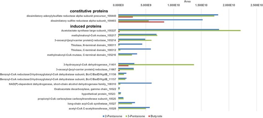
Frey et al. BMC Microbiology    (2021) 21:50                                                                                    Page 4 of 11

(ESI)-MS/MS, an Agilent 1100 HPLC system and the                        conducted using a Waters Acquity UPLC HSS T3 col-
Kinetex PFP column (see above) connected to an LCQ                      umn (2.1 × 100 mm) with a 20 min linear gradient of 2%
ion trap mass spectrometer (Thermo Fisher Scientific)                   acetonitrile to 40% acetonitrile in 10 mM ammonium
was used [30]. The injection volume was 50 μL. The col-                 acetate buffer at 0.35 ml/min. The mass spectrometer
umn was run isocratically with 95% 100 mM ammonium                      was operated in positive ion mode with a capillary volt-
acetate and 5% acetonitrile for 10 min at a flow rate of                age of 3.0 kV, a source temperature of 150 °C, a desolva-
0.75 ml/min and a temperature of 40 °C.                                 tion temperature of 450 °C and desolvation gas flow of
                                                                        1000 L/h. Theoretical m/z values were calculated and
CoA ester extraction from intact cells and UPLC-MS                      data were evaluated with Waters MassLynx V4.1
analysis                                                                SCN916.
For analysis of the CoA ester pool in growing cells, D.
cetonica was grown with acetone or butyrate as sole                     Results and discussion
carbon source. Cells were harvested in the mid-                         General properties of D. cetonica
exponential growth phase, lysed on ice with an ice-cold                 Desulfosarcina cetonica is next to Desulfococcus biacutus
solution containing 0.1 M formic acid and 80% aceto-                    the only described acetone- and butanone-degrading
nitrile, and subsequently lyophilized [34]. Lyophilized                 sulfate-reducing bacterium described so far. It was iso-
samples were dissolved in 200 μL 10 mM ammonium                         lated from stratal waters of Apsheron peninsula, Lokba-
acetate and centrifuged two times. The supernatant was                  tanskii deposit in Azerbaijan. D. biacutus was isolated
used for UPLC-MS measurements.                                          from anoxic sludge of a sewage plant in Marburg,
  Samples were analyzed with a Waters Acquity I-Class                   Germany [16, 27].
UPLC coupled to a Waters Synapt G2-Si HDMS ESI-                           In this study, proteome data were analyzed for all
QTOF mass spectrometer. UPLC separation was                             major metabolic pathways of D. cetonica. A full set of

 Fig. 1 Representative HPLC-MS chromatograms of an in vitro enzyme assay of the B12-dependent 2-hydroxyisobutyryl-CoA mutase reaction. Time
 course measurement of the reaction showing formation of 3-hydroxybutyryl-CoA (peak eluting between 5.87 and 6.21 min), concomitant with
 disappearance of 2-hydroxyisobutyryl-CoA (peak eluting between 6.24 and 6.62 min). The product 3-hydroxybutyryl-CoA eluted before the
 substrate 2-hydroxyisobutyryl-CoA
Frey et al. BMC Microbiology (2021) 21:50 Page 5 of 11
enzymes was identified that allows dissimilatory sulfate that was also reported to isomerize 2-hydroxyisobutyryl-
reduction and complete oxidation of acetyl residues via CoA to 3-hydroxybutyryl-CoA using a B12-dependent
the reversed Wood-Ljungdahl pathway. Furthermore, mutase followed by dehydrogenation to acetoacetyl-CoA
most enzymes of the citric acid cycle (except for malate (initially annotated as a 3-oxoacyl-[acyl carrier protein
dehydrogenase and aconitase) were detected by total (ACP)] reductase) [28, 30].
proteomics. Attempts to demonstrate the predicted TDP-
dependent enzymatic conversion of acetone to 2-
Inducible 2-hydroxyisobutyryl-CoA mutase and NAD+- hydroxyisobutyryl-CoA (Fig. 2A) were unsuccessful with
dependent 3-hydroxybutyryl-CoA dehydrogenase activity CFE of D. cetonica, as previously reported with CFE of
in cell-free extracts of acetone-grown cells of D. cetonica D. biacutus: reaction of CFE/acetone with the potential
Cell-free extracts (CFE) of acetone-grown cells of D. C1-cosubstrates CO, CO2, formate, formaldehyde,
cetonica were analyzed for two key enzyme activities of formyl-CoA or oxalyl-CoA as formyl donor with or
acetone metabolism observed previously in D. biacutus. without addition of TDP, ATP, FAD, FMN, tetrahydro-
2-hydroxyisobutyryl-CoA mutase activity was tested in folate, NAD+ and/or NADH as cofactors (see also
reactions containing CFE, 2-hydroxyisobutyryl-CoA and Material and methods) did not show any formation of 2-
B12 (adenosylcobalamin) using HPLC fragmentation- hydroxybutyryl-CoA. Therefore, the nature of the
mass spectrometry. The B12- dependent isomerization of formylating C1 unit remains elusive. It is likely that a
2-hydroxyisobutyryl-CoA to 3-hydroxybutyryl-CoA was C1-intermediate of the Wood-Ljungdahl pathway may
detectable in extracts of cells grown with acetone, but be used as co-substrates for acetone activation. In
not with butyrate (Fig. 1), suggesting that the synthesis former studies with D. biacutus, a co-localization of the
of the enzyme is induced during growth with acetone. TDP-dependent enzyme with membrane proteins was
Further, the activity of a NAD+-dependent 3- proposed [28] but there is no indication for a functional
hydroxybutyryl-CoA dehydrogenase was detected in CFE association with the membrane so far.
of acetone-grown cells (449.6 ± 44.2 mU mg− 1); the ac-
tivity in butyrate-grown cells was lower (222.9 ± 66.4 Detection of 2-hydroxyisobutyryl-CoA as a metabolite
mU mg− 1 protein). When the B12-dependent isomeriza- Freshly harvested, freeze-dried cells of D. cetonica grown
tion of 2-hydroxyisobutyryl-CoA was coupled to the with acetone were examined for CoA metabolites by
NAD+-dependent oxidation of 3-hydroxybutyryl-CoA to HPLC-MS (see Material and methods). Comparison of
acetoacetyl-CoA by addition of 2-hydroxyisobutyryl- acetone- and butyrate-grown cells indicated the presence
CoA, B12 and NAD+, the activity was 1.0 ± 0.2 mU of 2-hydroxyisobutyryl-CoA in acetone-grown cells at
mg−1protein in CFE of acetone-grown cells, but no such about ten-fold higher abundance than in butyrate-grown
activity was found in CFE of butyrate-grown cells. This cells (see Supporting information Fig. S1), thus support-
finding agrees with previous studies with D. biacutus ing the proposed pathway (Fig. 2A). However, formyl-
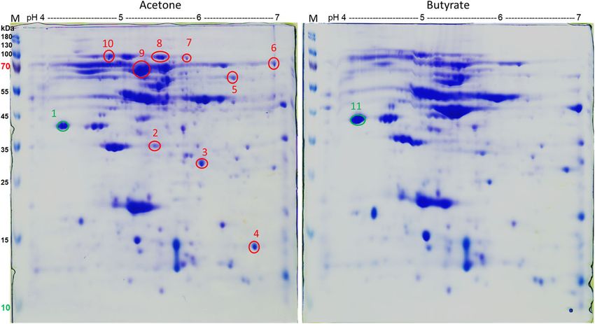
Fig. 2 Proposed pathway for acetone degradation in Desulfosarcina cetonica strain JCM 12296. a Activation of acetone by a yet unknown C1-
residue, followed by 2-hydroxyisobutyryl-CoA mutase and 3-oxoacyl-[acyl-carrier protein] reductase. b Structure of the corresponding gene cluster
in D. cetonica. Corresponding genes/enzymes (IMG locus tags prefix, Ga0122881_) are color-codedFrey et al. BMC Microbiology (2021) 21:50 Page 6 of 11
Fig. 3 2D PAGE analysis of soluble proteins obtained from acetone- and butyrate-grown cells of D. cetonica. Red-labeled spots are acetone-
induced, green-labeled spots were of similar size in both samples. Spots were excised and analyzed by peptide mass fingerprinting (for results
see Table 1)
CoA as a potential formyl donor for the predicted TDP- the tryptic peptides obtained were analyzed by MS. The
dependent conversion of acetone to 2- proteins identified are listed in Table 1. Nine protein
hydroxyisobutyryl-CoA was not detectable by HPLC-MS spots (spots 2–10, Fig. 3) were either exclusively present
in the cells under these conditions. No further promin- or were more abundant in the soluble protein fraction of
ent CoA-intermediate was found exclusively in acetone- acetone-grown cells. Two further protein spots produced
grown cells during analyses of ion chromatograms with at almost equal abundance under both growth condi-
CoA ester-specific fragmentation patterns. tions were excised as controls to ensure correct
localization in the gel and exclusion of carry-over of
Proteomic identification of acetone-inducible enzymes in other proteins (spots 1 and 11, Fig. 3).
D. cetonica Spots 3, 4, 6 and 9 were clearly higher abundant in
Soluble protein fraction of acetone- and butyrate-grown cells grown with acetone (Fig. 3) and are annotated as 3-
cells were compared by two-dimensional protein electro- oxoacyl-[acyl-carrier protein] reductase (IMG locus tag:
phoresis (2D PAGE). All protein spots with higher abun- Ga0122881_105,214; in the following the tag prefix
dance in acetone-grown cells (Fig. 3) were excised, and Ga0122881_ is omitted), methylmalonyl-CoA mutase
Table 1 Identification of proteins after 2D-PAGE analysis of D. cetonica grown with acetone/sulfate vs. butyrate/sulfate. Spots 2–10
were specifically induced after growth with acetone. Proteins of the proposed acetone degradation cluster are marked in bold.
Spots 1 and 11 served as control spot and were found to be identical in both conditions
Spot ID IMG locus tag Annotation Score MW calc. pI
1 Ga0122881_14251 electron transfer flavoprotein beta subunit 2029 26.6 4.35
2 Ga0122881_108331 hypothetical protein 3373 19.3 5.73
3 Ga0122881_105,214 3-oxoacyl-[acyl-carrier protein] reductase 23,994 28.0 5.72
4 Ga0122881_105,216 methylmalonyl-CoA mutase, C-terminal domain 15,935 14.5 6.54
5 Ga0122881_103346 Formate-tetrahydrofolate ligase 10,064 53.4 6.55
6 Ga0122881_105,227 Acetolactate synthase large subunit 1857 77.6 6.62
7 Ga0122881_10451 pyruvate carboxylase subunit B 11,267 63.7 5.73
8 Ga0122881_10843 acetyl-CoA decarbonylase/synthase beta subunit 20,590 81.0 5.35
9 Ga0122881_105,217 methylmalonyl-CoA mutase 14,980 64.7 5.11
10 Ga0122881_103051 iron complex outermembrane receptor protein 9818 73.2 4.96
Ga0122881_11832 outer membrane receptor for ferrienterochelin and colicins 7161 74.0 5.00
11 Ga0122881_14251 electron transfer flavoprotein beta subunit 13,308 26.6 4.35Frey et al. BMC Microbiology (2021) 21:50 Page 7 of 11 (C-terminal domain) (105216), TDP-dependent acetolac- Spots 2, 5, 7, 8, and 10 were identified as a hypothet- tate synthase large subunit (105227), and ical protein, formate-tetrahydrofolate ligase, pyruvate methylmalonyl-CoA mutase (105217) (Table 1), respect- carboxylase subunit B, acetyl-CoA decarbonylase/syn- ively. The genes for these proteins are located in the thase beta subunit and iron complex outer membrane same gene cluster (Fig. 2B) and most likely the two pro- receptor protein / outer membrane receptor for ferrien- teins annotated as methylmalonyl-CoA mutase (105,216, terochelin and colicins. However, these five proteins ap- 105,217) are two different assingments of one enzyme, peared to be constitutively expressed under both growth as described for the homologous proteins in D. biacutus conditions. [30]. In D. biacutus, homologous proteins were observed In summary, these findings, as well as the identifica- to be acetone-induced, and the encoding genes are also tion of 2-hydroxyisobutyryl-CoA in extracts of acetone- located in one gene cluster [28]. The acetolactate syn- grown cells in the metabolomics assay strongly suggest thase (105227) has high similarities (87.7% identity at that acetone is degraded in D. cetonica through the same the amino acid level using IMG Genome BLASTP) to an pathway as proposed for D. biacutus [30]. acetone-induced TDP-dependent enzyme of D. biacutus. Also the two subunits of B12-dependent methylmalonyl- Proteomic identification of inducible proteins in D. CoA mutase (105,216, 105,217) showed high similarities cetonica during growth with isopropanol and butanone (85.6 and 85.5%, respectively amino acid sequence iden- We examined the expression patterns of proteins in CFE tity) to the acetone-induced homologs of D. biacutus. of D. cetonica after growth with four different electron The 3-oxoacyl-[acyl-carrier protein] reductase of D. ceto- donors, acetone, butanone, isopropanol, and butyrate nica exhibited 82.2% sequence identity at the amino acid (control), by total proteomic analysis without preceding level to an acetone-induced enzyme in D. biacutus. 2D-PAGE separation. For these analyses, cultures had Furthermore, it was demonstrated that the acetone-spe- been transferred at least 10 times with the respective cific enzyme initially annotated as 3-oxoacyl-[acyl-carrier substrate prior to analysis to ensure adaptation. The data protein] reductase of D. biacutus oxidizes 3-hydroxybutyryl- obtained confirmed the proteins that were identified by CoA to acetoacetyl-CoA [30]. 2D-PAGE. Several proteins with higher abundance in Fig. 4 Total proteome analysis of cell-free extracts of D. cetonica after growth with different substrates (acetone [blue], butyrate [red], isopropanol [purple] and butanone [green]). Induced and constitutively expressed proteins are shown. IMG locus tag numbers (Ga0122881_number) are given for each protein. Higher area values represent higher protein abundance
Frey et al. BMC Microbiology      (2021) 21:50                                                                                            Page 8 of 11

cells grown with acetone vs butyrate were identified                         the genes of these six induced proteins (10522–10,528)
(Fig. 4). A TDP-dependent protein annotated as aceto-                        are clustered together in one gene cluster in which also
lactate synthase (105227) was highly induced. In                             genes coding for the TDP-dependent enzyme, the two
addition, the small and the large subunit (105,216, 105,                     subunits of the B12-dependent mutase and the 3-
217) of a B12-dependent isomerase acting on CoA esters                       oxoacyl-(ACP) reductase described above are located.
(annotated as methylmalonyl-CoA mutase) and a 3-                             This strongly implies that these enzymes are involved in
oxoacyl-[acyl-carrier protein] reductase (105214) were                       the degradation of acetone, isopropanol, and butanone.
more abundant in acetone-grown cells.                                        One might speculate that these enzymes are needed for
  In addition, several proteins abundant in acetone-grown                    production of another precursor molecule (e.g. formyl-
cells were identified that escaped detection during 2D-                      CoA) which is used for acetone activation. Moreover, an
PAGE and were annotated as: hydroxymethylglutaryl-CoA                        alcohol dehydrogenase (118516) was nearly 20-fold
lyase (105210), acetyl-CoA C-acetyltransferase (10528),                      higher abundant during growth with isopropanol than in
long-chain acyl-CoA synthetase (10527), propionyl-CoA                        acetone- or butanone-grown cells, and was not detected
carboxylase carboxyltransferase subunit (10525), oxaloace-                   in extracts of butyrate-grown cells. Thus, this enzyme
tate decarboxylase beta subunit (10524), a hypothetical                      most likely catalyzes the dehydrogenation of isopropanol
protein (10523), oxaloacetate decarboxylase gamma chain                      to acetone, but is encoded in a gene cluster different
(10522), and two subunits of a benzoyl-CoA reductase/2-                      from that of the genes involved in acetone degradation.
hydroxyglutaryl-CoA dehydratase (11,156, 11,157). Many                         The main difference between D. biacutus and D. ceto-
of these proteins were found to be acetone-inducible also                    nica is that the latter does not contain a homologue of
in D. biacutus [28].                                                         the acetone-inducible threonine dehydrogenase (Debia-
  All these acetone-specific (compared to butyrate) pro-                     DRAFT_04514), and no homologous gene of the re-
teins were found also in CFE of butanone- and of                             spective protein was found in the genome. In an earlier
isopropanol-grown cells. With the exception of the two                       study, this dehydrogenase was proposed to have a de-
benzoyl-CoA reductase/2-hydroxyglutaryl-CoA dehydra-                         toxifying function, as it utilizes a broad variety of short-
tase subunits, all other genes of the respective acetone-                    chain alcohols, aldehydes, and ketones [36]. One may
induced proteins are located in one gene cluster (see Fig.                   speculate that lack of this protein could explain the
S2 in supporting information). Additionally, a LAO/AO                        measured difference in growth yields (for D. cetonica
transport system kinase (105215) related to ArgK                             only around 1/3 compared to D. biacutus) between D.
(MeaB) was found which may serve as a stabilizing G                          biacutus and D. cetonica growing with the same sub-
protein for the B12-dependent enzyme [35]. Interestingly,                    strate [16, 25, 26].

 Fig. 5 Total proteome analysis of cell-free extracts of D. cetonica after growth with different substrates (2-pentanone [blue], 3-pentanone [red]
 and butyrate [green]). IMG locus tag numbers (Ga0122881_number) are given for each protein. Higher area values represent higher
 protein abundance
Frey et al. BMC Microbiology     (2021) 21:50                                                                                        Page 9 of 11

  The finding that the same enzyme proteins are highly                       NAD+-dependent oxidation of 3-hydroxybutyryl-CoA
abundant during growth with acetone, butanone, and                         was detected in isopropanol- and butanone-grown CFEs
isopropanol strongly suggests that degradation of these                    with specific activities of 250.1 ± 37.1 and 391.0 ± 76.3
three substrates involves common proteins.                                 mU mg− 1 protein, respectively. D. cetonica grows with
                                                                           isopropanol slower and to lower cell densities than with
                                                                           acetone or butanone [27].
Differential in vitro activities in cell-free extracts of                    D. biacutus has been described as well to grow with
isopropanol- and butanone-grown D. cetonica                                butanone and isopropanol [16]. Under both growth
Enzyme assays with CFE of isopropanol-grown cells ex-                      conditions, also a B12-dependent isomerization of 2-
hibited a specific activity of 35.8 ± 5.1 mU mg− 1 protein                 hydroxyisobutyryl-CoA to 3-hydroxybutyryl-CoA was
for reduction of 5 mM acetone with 0.5 mM NADH, and                        detected using a coupled assay with NAD+. Here,
14.0 ± 1.0 mU mg− 1 protein for oxidation of 5 mM                          CFE of isopropanol-grown cells of D. cetonica exhib-
isopropanol with 5 mM NAD+, whereas no activity (de-                       ited an activity of 1.8 ± 0.5 mU mg− 1 protein and
tection limit: < 0.5 mU mg− 1 protein) was detected in ex-                 1.3 ± 0.4 mU mg− 1 protein for CFE of butanone-grown
tracts of butyrate-grown cells. These activities were                      cells.
around 10-fold higher than in acetone- or butanone-                          Degradation of butanone via initial formylation would
grown cells. Activities of NADH-dependent acetone                          lead to a C5-CoA ester, which may be cleaved thiolyti-
reduction were lower in CFE of acetone- or butanone-                       cally to acetyl-CoA and propionyl-CoA. Propionate is
grown cells: 3.1 ± 0.3 and 4.9 ± 2.4 mU mg− 1 protein,                     not excreted to the growth medium during growth with
respectively. Enzyme activities of NAD+-dependent iso-                     butanone, obviously because D. cetonica can grow also
propanol oxidation were below the detection limit in                       with propionate as sole carbon source [27]. Degradation
CFE of acetone- or butanone-grown cells.                                   pathways in the sulfate-reducing bacteria D. biacutus

 Fig. 6 Proposed degradation pathways for acetone, isopropanol and butanone in the sulfate-reducing bacteria D. biacutus and D. cetonica (Fig.
 6a) and for 2-pantanone and 3-pentanone in D. cetonica (Fig. 6b)
Frey et al. BMC Microbiology (2021) 21:50 Page 10 of 11
and D. cetonica for acetone, isopropanol and butanone Supplementary Information
are depicted in Fig. 6A. This strategy of activation may The online version contains supplementary material available at https://doi.
org/10.1186/s12866-021-02112-6.
be used also for longer ketones and might reflect a com-
mon concept for activation and utilization of these Additional file 1: Figure S1. HPLC-MS chromatograms from lysates of
ketones. acetone (Sample Acetone 1–3)- and butyrate (Sample Butyrate 1–3)-
grown cells compared to pure standards of 2-hydroxyisobutyryl-CoA (2-
HiB-CoA) and 3-hydroxybutyryl-CoA (3-HB-CoA). Acetone-grown cells
show substantially more 2-hydroxyisobutyryl-CoA (elutes between 10.30–
Proteomic identification of inducible proteins in D. 10.45 min) whereas in butyrate-grown cells mainly 3-hydroxybutyryl-CoA
cetonica during growth with 2-pentanone and 3- (elutes between 10.48–10.58 min) was identified. Figure S2. Gene cluster
pentanone showing specifically acetone-induced genes in Desulfosarcina cetonica.
Most prominent genes are symbolized as red arrows and are annotated
Protein expression patterns after growth with C5- as acetolactate synthase (105227), a small and a large subunit of a
compounds like 2-pentanone and 3-pentanone were methylmalonyl-CoA mutase (105,216, 105,217) and a 3-oxoacyl-[acyl-car-
studied in comparison to butyrate as control substrate. rier protein] reductase (105214). Green genes are found to be expressed
acetone-specifically (also in butanone- and isopropanol-grown, not in
Proteomic analysis was performed with CFEs of 2- butyrate-grown CFE) by proteome analysis. These genes are annotated as
pentanone- and 3-pentanone-grown cells which had an oxaloacetate decarboxylase, gamma chain (10522), a hypothetical pro-
been transferred at least 10 times on the respective sub- tein (10523), an oxaloacetate decarboxylase beta subunit (10524), a
propionyl-CoA carboxylase carboxyltransferase subunit (10525), a long-
strate. Several induced proteins were identified that ex- chain acyl-CoA synthetase (10527), acetyl-CoA C-acetyltransferase (10528),
hibited enhanced abundance compared to growth with as well as a hydroxymethylglutaryl-CoA lyase (105210), a hypothetical pro-
butyrate (Fig. 5). tein (105211), a glyoxylase, beta-lactamase superfamily II (105213), LAO/
AO transport system kinase (105215), an acyl-CoA hydrolase (105218), an
One of these most abundant proteins is a TDP- anaerobic selenocysteine-containing dehydrogenase (105220), a two
dependent acetolactate synthase (105227). Also specific- component transcriptional regulator, LuxR family (105224) and a histidine
ally induced during growth with both pentanones were kinase-, DNA gyrase B-, and HSP90-like ATPase (105225).
the small and large subunit (105,216, 105,217) of a B12-
dependent mutase (annotated as methylmalonyl-CoA Acknowledgements
We are grateful to Fabian Schneider and Thomas Huhn for the synthesis of
mutase) and a 3-oxoacyl-[acyl-carrier protein] reductase reaction compounds. We thank Benjamin Frommeyer and Julia Schmidt for
(105214). help and technical support, as well as Andreas Marquardt for the proteomic
Further proteins with abundance in 2−/3-pentanone- analyses.
grown cells were identified such as two subunits of a
Authors’ contributions
benzoyl-CoA reductase/2-hydroxyglutaryl-CoA dehydra-
J.F. and S.K. did most of the experiments, D.S. helped with the synthesis and
tase (11,156, 11,157), as well as four proteins whose analysis of reaction intermediates, M.M. and M.B. ran the experiment for
genes are directly adjacent to each other: a propionyl- identification of the first reaction intermediate, J.F., D.S. and B.S. wrote the
manuscript. The authors read and approved the final manuscript.
CoA carboxylase carboxyltransferase subunit (10525), an
oxaloacetate decarboxylase beta subunit (10524), a hypo-
Funding
thetical protein (10523) and an oxaloacetate decarboxyl- Research in the Konstanz lab was funded by the Deutsche
ase gamma chain (10522). Also an acetyl-CoA C- Forschungsgemeinschaft (DFG), Bonn, Germany, under grant number Schi
acetyltransferase (10528) and a long-chain acyl-CoA syn- 180/16–1. Further financial support came from the universities Freiburg and
Konstanz.
thetase (10527) were identified to be induced during
growth with the two pentanones. All of the above- Availability of data and materials
mentioned proteins were discovered to be highly abun- All supporting data are presented in the main paper and the supplementary
dant also after growth with acetone, butanone and files. The genome annotation of Desulfococcus biacutus strain KMRActS and
the nucleotide and amino-acid sequences of locus tag DebiaDRAFT_04514
isopropanol. are publicly available within the Joint Genome Institute (JGI) Integrated Mi-
The results, although still preliminary, indicate that crobial Genomes (IMG) system under IMG genome ID 2512047085; the gen-
the two pentanones are degraded analogous to ome sequencing and annotation has been described in ref. [20].
acetone and butanone via different intermediates:
Ethics approval and consent to participate
Pentanone-2 degradation after formylation and Not applicable.
linearization would lead to an acetyl and a butyryl
residue, whereas degradation of 3-pentanone would Consent for publication
form two propionyl-CoA as intermediates (Fig. 6B). It Not applicable.
appears that 2-pentanone is easier to degrade than 3-
Competing interests
pentanone, as 3-pentanone cultures need more than 3 The authors declare that they have no competing interests.
times longer to reach the plateau phase (data not
shown). One might speculate that the respective en- Author details
1
Department of Biology, University of Konstanz, 78457 Constance, Germany.
zymes involved may be sterically hindered by the two 2
Institute of Biology, Albert-Ludwigs-Universität, Freiburg, 79104 Freiburg,
ethyl residues of 3-pentanone. Germany.Frey et al. BMC Microbiology (2021) 21:50 Page 11 of 11
Received: 22 October 2020 Accepted: 29 January 2021 25. Janssen PH, Schink B. Metabolic pathways and energetics of the acetone-
oxidizing, sulfate-reducing bacterium. Desulfobacterium cetonicum Archives
of Microbiology. 1995;163(3):188–94.
26. Janssen PH, Schink B. Catabolic and anabolic enzyme activities and
References energetics of acetone metabolism of the sulfate-reducing bacterium
1. Sifniades S, Levy AB, Bahl H: Acetone. In: Ullmann's Encyclopedia of Industrial Desulfococcus biacutus. J Bacteriol. 1995;177(2):277–82.
Chemistry. Wiley-VCH Verlag GmbH & Co. KGaA; 2011. 27. Galushko A, Rozanova E: Desulfobacterium cetonicum spec. nov., a sulfate-
2. Han B, Gopalan V, Ezeji TC. Acetone production in solventogenic Clostridium reducing bacterium oxidizing fatty acids and ketones. Microbiology (Moscow,
species: new insights from non-enzymatic decarboxylation of acetoacetate. Russ Fed) 1991, 60:102–107.
Appl Microbiol Biotechnol. 2011;91(3):565–76. 28. Gutiérrez Acosta OB, Schleheck D, Schink B: Acetone utilization by sulfate-
3. Zhao J, Wang Z, Wu T, Wang X, Dai W, Zhang Y, Wang R, Zhang Y, Shi C. reducing bacteria: draft genome sequence of Desulfococcus biacutus and a
Volatile organic compound emissions from straw-amended agricultural soils proteomic survey of acetone-inducible proteins. BMC genomics 2014, 15
and their relations to bacterial communities: a laboratory study. J Environ (1)(584).
Sci. 2016;45:257–69. 29. Gutiérrez Acosta OB, Hardt N, Schink B. Carbonylation as a key reaction in
4. Fang J-J, Yang N, Cen D-Y, Shao L-M, He P-J. Odor compounds from anaerobic acetone activation by Desulfococcus biacutus. Appl Environ
different sources of landfill: characterization and source identification. Waste Microbiol. 2013;79(20):6228–35.
Manag. 2012;32(7):1401–10. 30. Frey J, Schneider F, Huhn T, Spiteller D, Schink B, Schleheck D. Two
5. Garner CE, Smith S, de Lacy CB, White P, Spencer R, Probert CS, Ratcliffe NM. enzymes of the acetone degradation pathway of Desulfococcus biacutus:
Volatile organic compounds from feces and their potential for diagnosis of coenzyme B12-dependent 2-hydroxyisobutyryl-CoA mutase and 3-
gastrointestinal disease. FASEB J. 2007;21(8):1675–88. hydroxybutyryl-CoA dehydrogenase. Environ Microbiol Rep. 2018;10(3):
6. Kalapos MP: On the mammalian acetone metabolism: from chemistry to 283–92.
clinical implications. Biochimica et Biophysica Acta (BBA)-General Subjects 31. Schmidt A, Müller N, Schink B, Schleheck D. A proteomic view at the
2003, 1621(2):122–139. biochemistry of syntrophic butyrate oxidation in Syntrophomonas wolfei.
7. de Gouw JA, Howard CJ, Custer TG, Fall R. Emissions of volatile organic PLoS One. 2013;8(2):e56905.
compounds from cut grass and clover are enhanced during the drying 32. Neuhoff V, Arold N, Taube D, Ehrhardt W. Improved staining of proteins in
process. Geophys Res Lett. 1999;26(7):811–4. polyacrylamide gels including isoelectric focusing gels with clear
8. Mopper K, Stahovec WL. Sources and sinks of low molecular weight organic background at nanogram sensitivity using Coomassie brilliant blue G-250
carbonyl compounds in seawater. Mar Chem. 1986;19(4):305–21. and R-250. Electrophoresis. 1988;9(6):255–62.
9. Kotani T, Yurimoto H, Kato N, Sakai Y. Novel acetone metabolism in a 33. Keller A, Schink B, Müller N. Alternative pathways of Acetogenic ethanol and
propane-utilizing bacterium, Gordonia sp. strain TY-5. J Bacteriol. 2007;189(3): methanol degradation in the Thermophilic anaerobe Thermacetogenium
886–93. phaeum. Front Microbiol. 2019;10:423.
10. Hausinger RP. New insights into acetone metabolism. J Bacteriol. 2007; 34. Rabinowitz JD, Kimball E. Acidic acetonitrile for cellular metabolome
189(3):671–3. extraction from Escherichia coli. Anal Chem. 2007;79(16):6167–73.
35. Korotkova N, Lidstrom ME. MeaB is a component of the methylmalonyl-CoA
11. Heider J, Schühle K, Frey J, Schink B. Activation of acetone and other
mutase complex required for protection of the enzyme from inactivation. J
simple ketones in anaerobic bacteria. J Mol Microbiol Biotechnol. 2016;
Biol Chem. 2004;279(14):13652–8.
26(1–3):152–64.
36. Frey J, Rusche H, Schink B, Schleheck D. Cloning, functional expression and
12. Schühle K, Heider J. Acetone and butanone metabolism of the denitrifying
characterization of a bifunctional 3-hydroxybutanal dehydrogenase
bacterium “Aromatoleum aromaticum” demonstrates novel biochemical
/reductase involved in acetone metabolism by Desulfococcus biacutus. BMC
properties of an ATP-dependent aliphatic ketone carboxylase. J Bacteriol.
Microbiol. 2016;16(1):280.
2012;194(1):131–41.
13. Platen H, Schink B. Anaerobic degradation of acetone and higher ketones
via carboxylation by newly isolated denitrifying bacteria. J Gen Microbiol. Publisher’s Note
1989;135(4):883–91. Springer Nature remains neutral with regard to jurisdictional claims in
14. Platen H, Schink B. Enzymes involved in anaerobic degradation of acetone published maps and institutional affiliations.
by a denitrifying bacterium. Biodegradation. 1990;1(4):243–51.
15. Madigan MT. Photocatabolism of acetone by nonsulfur purple bacteria.
FEMS Microbiol Lett. 1990;71(3):281–5.
16. Platen H, Temmes A, Schink B. Anaerobic degradation of acetone by
Desulfococcus biacutus spec. Nov. Arch Microbiol. 1990;154(4):355–61.
17. Oosterkamp MJ, Boeren S, Atashgahi S, Plugge CM, Schaap PJ, Stams AJ:
Proteomic analysis of nitrate-dependent acetone degradation by
Alicycliphilus denitrificans strain BC. FEMS microbiology letters 2015, 362(11):
fnv080.
18. Platen H, Schink B. Methanogenic degradation of acetone by an
enrichment culture. Arch Microbiol. 1987;149(2):136–41.
19. Muyzer G, Stams AJ. The ecology and biotechnology of sulphate-reducing
bacteria. Nat Rev Microbiol. 2008;6(6):441–54.
20. Kováč J, Vítězová M, Kushkevych I. Metabolic activity of sulfate-reducing
bacteria from rodents with colitis. Open Medicine. 2018;13(1):344–9.
21. Kushkevych I, Castro Sangrador J, Dordević D, Rozehnalová M, Černý M,
Fafula R, Vítězová M, Rittmann SK-M. Evaluation of physiological parameters
of intestinal sulfate-reducing bacteria isolated from patients suffering from
IBD and healthy people. J Clin Med. 2020;9(6):1920.
22. Kushkevych I, Dordević D, Vítězová M. Toxicity of hydrogen sulfide toward
sulfate-reducing bacteria Desulfovibrio piger Vib-7. Arch Microbiol. 2019;
201(3):389–97.
23. Cummings J, Macfarlane G, Macfarlane S. Intestinal bacteria and ulcerative
colitis. Current issues in intestinal microbiology. 2003;4(1):9–20.
24. Kushkevych I, Cejnar J, Treml J, Dordević D, Kollar P, Vítězová M. Recent
advances in metabolic pathways of sulfate reduction in intestinal Bacteria.
Cells. 2020;9(3):698.You can also read