Oncolytic peptides DTT 205 and DTT 304 induce complete regression and protective immune response in experimental murine colorectal cancer - Nature
←
→
Page content transcription
If your browser does not render page correctly, please read the page content below
www.nature.com/scientificreports
OPEN Oncolytic peptides DTT‑205
and DTT‑304 induce complete
regression and protective immune
response in experimental murine
colorectal cancer
Karianne Giller Fleten1,2, J. Johannes Eksteen3, Brynjar Mauseth4, Ketil André Camilio4,
Terje Vasskog5, Baldur Sveinbjørnsson4,6, Øystein Rekdal4, Gunhild M. Mælandsmo1,6,8 &
Kjersti Flatmark1,2,7,8*
Oncolytic peptides represent a novel, promising cancer treatment strategy with activity in a broad
spectrum of cancer entities, including colorectal cancer (CRC). Cancer cells are killed by immunogenic
cell death, causing long-lasting anticancer immune responses, a feature of particular interest in
non-immunogenic CRC. Oncolytic peptides DTT-205 and DTT-304 were administered by intratumoral
injection in subcutaneous tumors established from murine CRC cell lines CT26 and MC38, and
complete regression was obtained in the majority of animals. When cured animals were rechallenged
by splenic injection of tumor cells, 1/23 animals developed liver metastases, compared to 19/22
naïve animals. Treatment with both peptides was well tolerated, but monitoring post-injection
hemodynamic parameters in rats, less extensive changes were observed with DTT-205 than DTT-
304, favoring DTT-205 for future drug development. DTT-205 was subsequently shown to have
strong in vitro activity in a panel of 33 cancer cell lines. In conclusion, both peptides exerted a strong
inhibitory effect in two immunocompetent CRC models and induced a systemic effect preventing
development of liver metastases upon splenic rechallenge. If a similar effect could be obtained
in humans, these drugs would be of particular interest for combinatory treatment with immune
checkpoint inhibitors in metastatic CRC.
Metastatic colorectal cancer (mCRC) is a leading cause of cancer related mortality worldwide, with the liver
as the most common metastatic s ite1. Surgery is a curative treatment option in a subgroup of mCRC patients
with limited disease burden, while the majority of cases will receive palliative systemic chemotherapy only2,3.
Recently, immunotherapy, including inhibitors of programmed death protein 1 (PD-1) and its ligand PD-L1,
has been shown to increase the survival in patients with different types of cancer, including melanoma, lung
cancer, and renal cell c ancer4,5. However, with the exception of the small subgroup of microsatellite instable (MSI)
tumors, CRC remains a non-immunogenic cancer that responds poorly to immunotherapy4,6. Extensive research
efforts are therefore being mounted to develop strategies for inducing immune responses in non-immunogenic
cancers. Clinical development of oncolytic therapies is gaining momentum, with one drug i.e. T-VEC already
approved for treating patients. A promising new approach to oncolytic therapy is the use of oncolytic peptides.
These peptides are based on host defense peptides (cationic antimicrobial peptides; CAPs) which are part of the
innate immune system of virtually all living organisms, protecting the organisms against foreign pathogens7.
1
Department of Tumor Biology, Institute for Cancer Research, The Norwegian Radium Hospital, Oslo University
Hospital, Montebello, 0379 Oslo, Norway. 2Faculty of Medicine, Institute of Clinical Medicine, University of
Oslo, Oslo, Norway. 3NORCE Norwegian Research Centre AS, SIVA Innovation Centre, Tromsø, Norway. 4Lytix
Biopharma, Oslo, Norway. 5Department of Pharmacy, The Arctic University of Norway – University of Tromsø,
Tromsø, Norway. 6Institute of Medical Biology, Faculty of Health Sciences, The Arctic University of Norway –
University of Tromsø, Tromsø, Norway. 7Department of Gastroenterological Surgery, The Norwegian Radium
Hospital, Oslo University Hospital, Oslo, Norway. 8These authors contributed equally: Gunhild M. Mælandsmo and
Kjersti Flatmark. *email: kjersti.flatmark@rr-research.no
Scientific Reports | (2021) 11:6731 | https://doi.org/10.1038/s41598-021-86239-6 1
Vol.:(0123456789)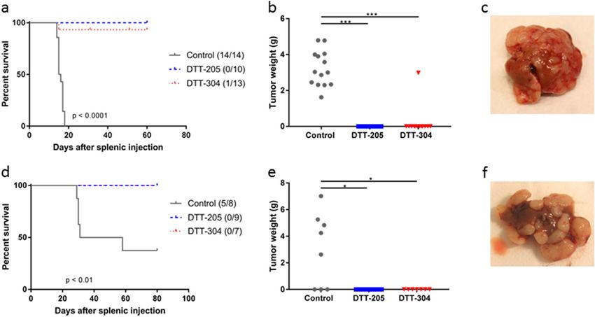
www.nature.com/scientificreports/
Figure 1. Response to DTT-205 or DTT-304 treatment in CT26 tumors. (a) Tumor growth curves of CT26
tumors treated with vehicle, 1 mg DTT-205 or 1 mg DTT-304. Tumors were treated for three consecutive days.
(b) Tumor growth curves of CT26 tumors treated with vehicle, 1.5 mg DTT-205 or 1.5 mg DTT-304 for four
consecutive days.
CAPs preferentially kill cancer cells over non-cancerous cells, due to higher expression of anionic macromol-
ecules on the cell membranes, and kill dividing and non-dividing cells as well as chemotherapy resistant cells
by their membranolytic a ction7–9. Following intralesional injection, tumor cells are rapidly killed by immuno-
genic cell death with release of damage associated molecular patterns (DAMPs) and tumor antigens. This will
result in recruitment and activation of dendritic cells with subsequent T-cell activation and long-term tumor-
specific immune r esponse10. Several CAPs have shown to cure cancer in vivo and also to induce a long-lasting
immune response11–14. Some, such as LTX-315 has even progressed to early phase clinical trials (NCT01058616,
NCT01223209, NCT01986426, NCT03725605).
Oncolytic peptides offer several potential advantages over oncolytic viruses in terms of efficacy, safety regula-
tions, manufacturing and handling. But more importantly, oncolytic peptides are less likely to have unwanted
immune effects. For example, unlike viruses, peptides cannot self-replicate and they are generally too small
to induce an adverse immune response against itself. Furthermore, proteolytic degradation into harmless
metabolites will usually prevent systemic accumulation and toxicity as is often observed during conventional
chemotherapy15,16. Intralesional oncolytic peptide therapy therefore represents a novel local treatment modality
with systemic effect and, potentially, an improved therapeutic window.
In the present study, the therapeutic efficacy of administering local injections of DTT-205 and DTT-304
was investigated in subcutaneous (s.c.) tumors in two syngeneic models of murine CRC. Cured animals were
rechallenged by splenic injection of tumor cells for establishment of liver metastases. In vitro pharmacologi-
cal and in vivo cardiovascular safety studies were subsequently performed as a preliminary assessment of the
peptides’ therapeutic potential. Finally, an extended cytotoxicity profile for DTT-205 was determined in a large
panel screen with additional cancer cell lines in order to pave the way for in vivo studies in other cancer models.
Results
Peptide injections induced complete regression of CT26 and MC38 tumors. CT26 tumors were
established in Balb/c mice, and two intratumoral injection regimens were investigated. In the first experiment,
injections of vehicle, DTT-205 (1 mg) or DTT-304 (1 mg) were administered for three consecutive days (8
tumors in each group). Tumors treated with vehicle grew rapidly, and the mice were sacrificed because of large
tumor size (> 15 mm) median 14 days after treatment initiation. All tumors treated with peptide initially disap-
peared, and complete regression was observed in 2 and 5 animals treated with DTT-205 and DTT-304, respec-
tively, while regrowth occurred in 6 of 8 tumors treated with DTT-205 and 3 of 8 tumors treated with DTT-304
(Fig. 1a). In a second experiment, to investigate if administration of a higher peptide dose would increase the
cure rate, injections of 1.5 mg were given for four consecutive days. Again, vehicle-treated tumors grew rap-
idly, and the mice were sacrificed median 19 days after treatment initiation, while all peptide-treated tumors
regressed completely and no regrowth was observed (Fig. 1b).
Scientific Reports | (2021) 11:6731 | https://doi.org/10.1038/s41598-021-86239-6 2
Vol:.(1234567890)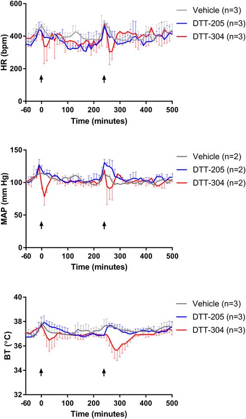
www.nature.com/scientificreports/
Figure 2. Response to DTT-205 or DTT-304 treatment in MC38 tumors. Tumor growth curves of MC38
tumors treated with saline, 0.5 mg DTT-205, 1 mg DTT-205 or 1 mg DTT-304. Mice were treated for three
consecutive days.
In the MC38 model, s.c. tumors were established in C57Bl/6 mice and treated with injections of vehicle (n = 7),
DTT-205 (0.5 mg; n = 6), DTT-205 (1 mg; n = 9), or DTT304 (1 mg; n = 8) for three consecutive days. Tumors
treated with vehicle grew rapidly and the mice were sacrificed within 28 days of treatment initiation. Complete
regression was observed in 21 of 23 peptide-treated animals, while two tumors treated with 1 mg DTT-205
regrew after 14 and 28 days (Fig. 2).
Curative peptide treatment prevented development of liver metastases. To investigate if cura-
tive treatment with DTT-205 or DTT-304 would protect animals from developing liver metastases, mice cured
for s.c. tumors by peptide treatment were rechallenged by splenic injection of CT26 or MC38 cells in Balb/c or
C57Bl/6 mice, respectively. Naïve mice injected with the same number of cells served as experimental controls.
All Balb/c control animals had to be sacrificed within 20 days of splenic injection due to tumor growth, and
liver tumors (3.26 ± 1.02 g) and bloody ascites (1.21 ± 0.52 g) were detected at autopsy. None of the 10 mice
previously cured by DTT-205 and only 1 of 13 mice previously cured by DTT-304 developed liver metastases.
The previously cured mouse that developed liver metastases had similar survival (15 days) and tumor burden
(2.98 g tumor and 1.4 mL ascites) as the control mice. Two of the mice cured by DTT-304 had to be sacrificed
after 31 and 51 days because of ulcerations at the previous s.c. treatment site (Fig. 3a,b).
None of the 16 C57Bl/6 mice previously cured by peptide injections developed liver tumors, while 5 of 8
naïve mice developed tumor (Fig. 3d). C57Bl/6 mice that developed tumor had 4.97 ± 1.77 g tumor in the liver
at time of sacrifice (Fig. 3e). Ascites was present in one mouse (0.4 g). Representative images of tumor-bearing
livers are shown in Fig. 3c,f.
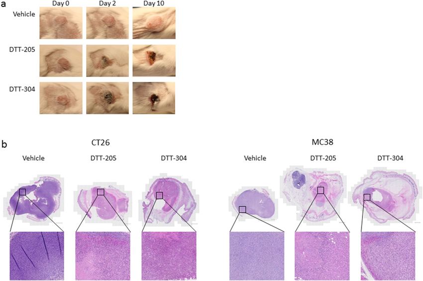
Induction of necrosis by peptide treatment. Intratumoral injection with peptide induced rapid swell-
ing of the tissue surrounding the s.c. tumors, interpreted as local inflammation. Images taken before, and at
various time points after treatment showed growth of the tumor and development of necrosis in Balb/c mice
(Fig. 4a). No macroscopic differences were observed between tumors treated with DTT-205 or DTT-304. In a
separate experiment, tumors were treated with two daily injections (1 mg) of DTT-205 or DTT-304 and har-
Scientific Reports | (2021) 11:6731 | https://doi.org/10.1038/s41598-021-86239-6 3
Vol.:(0123456789)www.nature.com/scientificreports/
Figure 3. Survival after rechallenge with splenic injection of tumor cells. (a) Kaplan–Meier curves comparing
naïve and previously cured Balb/c mice after splenic injection of CT26 cells. In parentheses, the number of
animals that developed liver metastases/total number of mice. (b) Tumor weight in the liver in Balb/c mice
after splenic injection (tumor was dissected from liver tissue). (c) Representative image of CT26 tumor in the
liver in Balb/c mice. (d) Kaplan–Meier curves comparing naïve and previously cured C57Bl/6 mice after splenic
injection of MC38 cells. In parentheses, the number of animals that developed liver metastases/total number of
mice. (e) Tumor weight in the liver in C57Bl/6 mice after splenic injection. (f) Representative image of MC38
tumor in the liver in C57Bl/6 mice. *p < 0.05, **p < 0.01, ***p < 0.001.
vested for histological analyses 48 and 120 h after the first injection (n = 4). Treatment with DTT-205 and DTT-
304 induced necrosis and significant reduction of tumor size resulting in no to little viable tumor tissue in CT26
tumors harvested after 48 and 120 h and in MC38 tumors harvested after 48 h (Fig. 4b). In MC38 tumors har-
vested after 120 h, there was little viable tumor tissue after treatment with DTT-304 while a considerable amount
of viable tumor tissue was left in 3 of 4 tumors treated with DTT-205.
Rapid proteolytic degradation. It is expected that oncolytic peptides, after exerting its desired therapeu-
tic effect, will be degraded and its metabolites rapidly removed from circulation. As a result, oncolytic peptides,
unlike chemotherapeutics, are not expected to accumulate in the patient’s body, and this represents a major
therapeutic advantage. The degradation assay performed in this study, illustrated that as expected, both DTT-
205 and DTT-304 were rapidly degraded in vitro by enzymes having cleavage sites in the peptide sequences
(Fig. 5). DTT-304 does not have a cleavage site for chymotrypsin and consequently no degradation was observed
at the end of the experiment. MS analysis indicated that DTT-205 were broken down into several species of short
peptide fragments of which the majority were 2–8 amino acids in length.
Assessment of adverse effects of DTT‑205 and DTT‑304. It is anticipated that following intratu-
moral injection, some of the peptide will escape into the blood stream, which could cause systemic adverse
effects, typically hypotension. A possible mechanism for this could be binding of DTT-205 to the MRGX2 recep-
tor. A dose dependent increase in calcium flux indicating binding to the receptor was observed in recombinant
cells when treated with increasing concentration of DTT-205, indicating that DTT-205 act as a MRGX2 receptor
agonist (Fig. 6). No effect was observed in the host cells without MRGX2 expressed (null cells).
To further explore the cardiovascular activity of the peptides, DTT-205 and DTT-304 were injected intrave-
nously (i.v) in rats, and body temperature, heart rate, mean arterial pressure (Fig. 7), systolic arterial pressure,
diastolic arterial pressure, and pulse pressure (Supplementary Fig. S1) were measured. DTT-205 had no effect
on cardiovascular parameters or body temperature, while DTT-304 induced a significant decrease in mean arte-
rial pressure after the first dose (p = 0.01) and in body temperature after the second dose (p = 0.02) compared
to vehicle treated rats. For the other parameters, a trend towards reduction was observed for DTT-304, but the
differences were not significant. Only measurements from 60 min before to 500 min after the first injection are
shown in Fig. 7. The full graph showing measurement from 60 min before to 1440 min after the first injection is
shown in Supplementary Fig. S2. Based on the results from the preliminary safety studies, DTT-205 was judged
to be superior to DTT-304 in terms of therapeutic index and was therefore selected as lead compound.
Scientific Reports | (2021) 11:6731 | https://doi.org/10.1038/s41598-021-86239-6 4
Vol:.(1234567890)www.nature.com/scientificreports/
Figure 4. Macroscopic and histological analyses of CT26 and MC38 tumors after treatment with DTT-205 and
DTT-304. (a) Images of CT26 tumors before treatment, and at day 2 and 10 after three injections with vehicle,
1 mg DTT-205 or 1 mg DTT-304. (b) HE staining of whole tumor (scale bar 1000 µm) and enlargement of
selected areas of CT26 and MC38 treated with 2 injections of 1 mg DTT-205, DTT-304 or saline. Tumors were
harvested 48 h after the first peptide injection.
Figure 5. Degradation of peptides by trypsin or chymotrypsin. Degradation of DTT-205 and DTT-304 after
administration of trypsin or chymotrypsin every 13 min, shown as % of remaining peptide. Error bars indicate
standard deviation.
DTT‑205 is cytotoxic against a variety of cancer cell lines. The cell viability assay revealed that DTT-
205 is highly cytotoxic against most types of cancer cells, with I C50 values in the range of 1.5–6 µM for the major-
ity of the cell lines. The only cancer type that was not as responsive to the peptide was kidney cancer with I C50
values of 8.3–20.9 µM. DTT-205 also had some toxicity towards the normal cell types that were tested with IC50
values varying from 1.8 to 12.9 µM (Table 1).
Discussion
The main finding of this experimental study was that in two syngeneic CRC models, local injections of onco-
lytic peptides DTT-205 and DTT-304 resulted in complete s.c. tumor regression and cure in 44 of 55 treated
animals. Furthermore, when the cured mice were rechallenged with splenic injection of tumor cells, only one of
39 animals developed liver metastases, suggesting that the initial treatment response prevented establishment
of liver metastases.
The first generation of DTT peptides was originally designed for treating lymphoma, and to generate peptides
with a broader anti-cancer spectrum, DTT-205 and DTT-304 were developed17. The two peptides have similar,
Scientific Reports | (2021) 11:6731 | https://doi.org/10.1038/s41598-021-86239-6 5
Vol.:(0123456789)www.nature.com/scientificreports/
Figure 6. Calcium flux after treatment with DTT-205. Release of calcium flux, shown as % of calcium flux
induced by PAMP-20 agonist in recombinant and null cells after treatment with increasing doses of DTT-205.
Error bars indicate standard deviation. **p < 0.01, ***p < 0.001.
Figure 7. Cardiovascular and temperature assessment in rats after treatment with DTT-205 or DTT-304. Heart
rate (HR), mean arterial pressure (MAP) and body temperature (BT) in rats treated with DTT-205 or DTT-304
in the period − 60 to 500 min after injection. Arrows indicate time of injection. Error bars indicate standard
deviation.
Scientific Reports | (2021) 11:6731 | https://doi.org/10.1038/s41598-021-86239-6 6
Vol:.(1234567890)www.nature.com/scientificreports/
# Cell line Disease/histological subtype Origin IC50 (µM)
1 CCD-18Co NORMAL Colon 4.4
2 MRC-5 NORMAL, fœtal Fibroblast 8.8
3 BEAS-2B NORMAL, broncheal, virus tranformed Lung 1.8
4 HMVEC NORMAL, microvascular endothelial Lung 12.9
5 UM-UC-3 Transitional papilloma Bladder 4.2
6 RT-4 Transitional cell carcinoma Bladder 6.1
7 LN-229 Glioma Brain 5.1
8 U-87 MG Glioblastoma Brain 4.5
9 BT-474 Invasive ductal carcinoma, primary Breast 3.0
10 MCF-7 Invasive ductal carcinoma, pleural effusion metastasis Breast 3.7
11 MDA-MB-231 Invasive ductal carcinoma, pleural effusion metastasis Breast 3.7
12 HT-29 Colorectal adenocarcinoma Colon 5.5
13 NCI-H747 Adenocarcinoma Colon 3.8
14 SW-480 Colorectal adenocarcinoma Colon 3.2
15 FaDu Pharynx carcinoma, squamous Head & neck 3.9
16 KB Mouth epidermoid carcinoma Head & neck 3.2
17 786-O Clear cell adenocarcinoma Kidney 8.3
18 A-498 Carcinoma Kidney 20.9
19 Caki-1 Clear cell carcinoma, skin metastasis Kidney 10.9
20 Hep G2 Hepatocellular carcinoma Liver 3.8
21 SK-HEP-1 Adenocarcinoma, ascites metastasis Liver 5.1
22 Calu-3 Adenocarcinoma; non-small cell lung cancer Lung 4.1
23 NCI-H1975 Adenocarcinoma; non-small cell lung cancer Lung 5.9
24 NCI-H146 Carcinoma; small cell lung cancer carcinoma Lung 3.4
25 NCI-H209 Carcinoma; small cell lung cancer carcinoma Lung 1.5
26 A2780 Adenocarcinoma Ovary 3.1
27 NIH:OVCAR-3 Adenocarcinoma, ascites metastasis Ovary 8.9
28 SW-626 Adenocarcinoma Ovary 4.2
29 BxPC-3 Adenocarcinoma Pancreas 6.3
30 Capan-1 Adenocarcinoma, liver metastasis Pancreas 5.5
31 Capan-2 Adenocarcinoma Pancreas 3.4
32 DU 145 Carcinoma, brain metastasis Prostate 2.4
33 MDA PCa 2b Adenocarcinoma, bone metastasis Prostate 3.1
34 PC-3 Adenocarcinoma, bone metastasis Prostate 5.1
35 A-375 Malignant melanoma Skin 1.7
36 A-431 Epidermoid carcinoma Skin 7.2
37 SK-MEL-28 Malignant melanoma Skin 1.9
Table 1. IC50 values of DTT-205 against various cell lines.
but not identical, mode-of-actions involving lysosomal localization. In addition, DTT-205 and DTT-304 may
influence pro-oxidant and BAX/BAK dependent processes, as inhibition of these also led to reduced efficacy18.
Intratumoral injections of DTT-205 and DTT-304 have previously been investigated in s.c. models in C57Bl/6
mice using the mouse fibrosarcoma cell line MCA205 and the lung carcinoma cell line TC-1. Both peptides
induced regression, indicating that the effect was independent of cancer type. Prior depletion of T-cells using
antibodies targeting CD4 and CD8 abolished the sustained growth inhibitory effect. In cured animals, tumors
could not be reestablished when rechallenged by s.c. i njection18. To evaluate the presence of a sustained growth
inhibitory effect in our models, mice cured of s.c. tumors were rechallenged using a liver colonization m odel19.
The majority of the mice previously cured by peptide treatment (38/39 animals) did not develop liver metastases,
which is in accordance with previous findings in other cancer types and tumor locations13. Prevention of tumor
growth in a secondary site in these experiments further strengthens the hypothesis that oncolytic peptide treat-
ment can induce a systemic long-term immune protective effect. A possible treatment strategy for patients could
be to inject the peptides in the primary tumor before removal to possibly prevent establishment of metastases
at a later time point.
Previous results have shown that DTT-205 and DTT-304 can trigger immunogenic cell death (ICD) with the
release of damage associated molecular patterns such as ATP, HMGB1 and C ALR118. Induction of ICD can lead
20,21
to increased T-cell infiltration in t umors , which has been shown to be associated with improved efficacy of
immune checkpoint inhibitors22 and strategies to increase the infiltration of immune cells in non-immunogenic
or cold tumors are of great interest. In mCRC, checkpoint inhibitors have exhibited poor activity in patients
Scientific Reports | (2021) 11:6731 | https://doi.org/10.1038/s41598-021-86239-6 7
Vol.:(0123456789)www.nature.com/scientificreports/
with microsatellite stable (MSS) tumors, and this group is therefore a particular focus in ongoing research in
the field23. It has previously been shown that oncolytic peptide treatment lead to increased tumor infiltration of
T-cells14,24. In our studies, no increase of T-cell infiltration was observed after treatment, but a possible explana-
tion may be that treated tumors largely consisted of necrotic tissue, making comparisons with vehicle-treated
tumors difficult (Supplementary Fig. S3). With the CT26 and MC38 being murine equivalents of MSS and MSI
CRC, respectively25,26, the subsequent prevention of liver metastasis establishment still suggests that results have
potential relevance both in MSS and MSI CRC.
In our experiments, the toxicity of DTT-205 and DTT-304 was low and comparable, with survival rates
close to 100%, while a higher toxicity was previously observed in vivo with DTT-304 compared to DTT-205
with survival rates of 72% and 90%, respectively27. The underlying cause of toxicity has not been established,
but hypotension has been the prime suspect. Since oncolytic peptide therapy involves transdermal injection of
peptide directly into the tumor, there is a possibility that some of the injected peptide may escape the injection
site to cause systemic side effects. Although DTT-205 was shown to interact with the promiscuous MRGX2
receptor, known to induce histamine release from mast cells and cause hypotension28, no major toxicity and
no hypotension were observed upon DTT-205 administration in vivo. A possible explanation could be that the
peptides are rapidly degraded by proteolytic enzymes (i.e. trypsin and chymotrypsin) present in blood. On the
other hand, it might be necessary to investigate potential systemic effects of higher DTT-205 doses, since the
DTT-205 dose administered i.v. to rats in the cardiovascular studies (0.3–1 mg/kg) was lower than doses used
for tumor injection in mice (25–75 mg/kg).
The extensive cell line screening showed that DTT-205 exhibited high in vitro activity against a broad range
of cancer cell types, but also that some normal cells may be affected. These results suggest that DTT-205 has the
potential to treat most kinds of tumors, provided that some degree of damage to normal tissue surrounding the
tumor is clinically acceptable. Therefore, while DTT-205 has a much broader anti-tumor activity than the more
tumor specific peptide predecessors, it also seems to be less selective. Since oncolytic peptides are best suited for
tumors that are accessible to local injections, this aspect should be taken into account in the clinical development
of DTT-205 as an anti-cancer drug.
In conclusion, oncolytic peptides DTT-205 and DTT-304 both displayed strong growth inhibitory effects in
two immunocompetent murine CRC models and induced a long-lasting systemic effect preventing tumor growth
in the liver. The observed results are consistent with the peptides inducing an immunogenic response, which
could be of particular interest in MSS mCRC for a potential clinical application in combination with immune
check point inhibitors or adoptive cell therapies.
Materials and methods
Peptide synthesis. DTT-205 and DTT-304 were synthesized with a Prelude instrument (Protein Tech-
nologies Inc. Tucson, AZ, USA) using a solid-phase Fmoc-approach. All synthesized peptides were prepared as
C-terminal amides. The resin (Rink amide) and Fmoc-amino acids used were standard derivatives purchased
from Novabiochem (Merck Millipore, Billerica, MA, USA). Fmoc-derivative activations were done in dimethyl-
formamide (DMF) using HCTU (2-(6-chloro-1H-benzotriazole-1-yl) − 1,1,3,3-tetramethylaminium hexafluo-
rophosphate) and diisopropylethylamine (5 eq and 10 eq relative to the resin, respectively). To ensure complete
reactions, double couplings (2 × 30 min) were performed. Coupling reactions were follow-up with a washing
(DMF, 3 × 30 s) and Fmoc-deprotection step (20% piperidine in DMF, 5 + 10 min). Following the final Fmoc-
deprotection, the completed peptides were cleaved from the resin using a cocktail containing 95% trifluoroacetic
acid (TFA), 2.5% MilliQ water (MQ) and 2.5% triisopropylsilane for 3 h. The spent resin particles were removed
by passing the peptide-containing cleavage cocktail through a sintered glass funnel. The TFA was removed
under reduced pressure (Hei-VAP Advantage rotavapor, Heidolph Instruments, Schwabach, Germany). The
fully deprotected peptides were precipitated using cold diethyl ether, afterwards the ether was decanted and the
precipitated crude peptide air dried.
Peptide purification and characterization. MQ water (solvent A) and acetonitrile (MeCN) (solvent B),
both modified with 0.1% TFA, were used for analytical and preparative liquid chromatography. Purification was
done using an AutoPurification System (Waters, Milford, MA, USA) fitted with a XSelect CSH C18 OBD column
(19 × 250 mm, 5 µm). A default gradient of 10–40% solvent B over 30 min with a flow rate of 20 mL/min was
used, but was adjusted when necessary. Fractions containing peptides were automatically collected and further
analyzed for desired material. Purity analyses were done using an ACQUITY UPLC H-class system fitted with
an ACQUITY BEH C18 UPLC column (Waters, 2.1 × 50 mm, 1.7 µm). Detection was done at 200–500 nm using
a Waters ACQUITY photodiode array detector. A gradient of 0–50% solvent B over 30 min with a flow rate of
1 mL/min was used. Fractions with a purity of > 95% were pooled and lyophilized (Labconco FreeZone 4.5 Plus,
Kansas City, MO, USA) as TFA-salts. The correct molecular weight of the peptides was confirmed on a Waters
Xevo G2 Q-TOF with ACQUITY UPLC I-Class system.
Converting peptides into acetate salts. In order to convert the peptides into acetate salts, the TFA
counterions first had to be removed. This was done by dissolving the TFA-containing peptides in MQ and pass-
ing it through an Agilent StratoSphere ML-HCO3 MP cartridge (Agilent Technologies, Santa Clara, CA, USA).
The free base peptides eluting from the cartridge were collected, a small amount (0.1% v/v) of acetic acid added,
and the solution lyophilized. The resulting peptide acetate salt was stored at − 20 °C until further use.
Cell lines. Two murine CRC cell lines were used for treatment experiments. The CT26 cell line (ATCC,
Manassas, VA, USA) was cultured in RPMI-1640 supplemented with 10% fetal bovine serum (Sigma-Aldrich,
Scientific Reports | (2021) 11:6731 | https://doi.org/10.1038/s41598-021-86239-6 8
Vol:.(1234567890)www.nature.com/scientificreports/
St. Louis, MO, USA), 2 mM glutamine (Life Technologies, Carlsbad, CA, USA) and 10 mM HEPES (Life Tech-
nologies). The MC38 cell line (MC38-CEA; Kerafast, Boston, MA, USA) was cultured in high glucose DMEM
supplemented with 10% fetal bovine serum, 2 mM glutamine, 5 mM HEPES and 0.1 mM non-essential amino
acids (Life Technologies). Both cell lines were cultured at 37 °C with 5% CO2, and were routinely tested for
mycoplasma.
Treatment experiments in mice. All procedures and experiments including mice were approved by
the National Animal Research Authority in Norway (Application number 11654), and conducted according to
the regulations of European Laboratory Animals Science Association29 and the ARRIVE guidelines30. Female
Balb/c and C57Bl/6 mice (age 5–12 weeks, weight 18–24 g) were purchased from Janvier (Le Genest-Saint-
Isles, France), and kept in specific pathogen free environment, at constant temperature (22 ± 1 °C) and humidity
(62 ± 5%), 15 air changes/hour and a 12 h light/dark cycle. Food and water were supplied ad libitum, and the
mice were given paper and cardboard houses for environmental stimulation. MAK III cages were used to house
the mice, and maximum 10 mice were kept in each cage and the different treatment groups were mixed in the
cages. For treatment experiments, 6–9 mice were included in each group. For histology experiments, 4 mice were
included in each group. Animals were sacrificed using cervical dislocation when the s.c. tumors reached 15 mm,
or if the animals showed signs of illness or discomfort according to standardized criteria.
Tumors were established by s.c. injection of 2.5 × 10⁶ CT26 cells or 1 × 10⁶ MC38 cells using an injection
volume of 100 µL. Peptide treatment was initiated when the tumors reached a mean of ~ 80 m m3 (± 20 mm3).
2
Tumor size was measured by caliper, and the volume was calculated using the formula: width × length × 0.5. The
peptides DTT-205 and DTT-304 were dissolved in 0.9% NaCl to a final concentration of 10–30 mg/mL and 50
µL solution (0.5–1.5 mg) or vehicle (NaCl) was injected intratumorally for 2–4 consecutive days. The treatment
was generally well tolerated, but three out of 91 animals treated died shortly after the first or second injection
(two C57Bl/6 mice treated with DTT-304 (1 mg) and one Balb/c mouse treated with DTT-205 (1 mg)). Animals
were considered cured if s.c. tumors disappeared and did not regrow within 35 days.
Rechallenge. For splenic injection of tumor cells, the mice were anesthetized with sevoflurane (Baxter,
Deerfield, IL, USA). Buprenorphine (0.15 mg/kg) and carprofen (5 mg/kg) were given s.c. prior to the surgi-
cal procedure for postoperative pain relief. The spleen was exteriorized through a transverse laparotomy in the
left hypochondrium, and 50 µL of cell suspension containing 0.5 × 10⁶ CT26 cells or 1.0 × 10⁶ MC38 cells, was
injected 2–3 mm into the splenic parenchyma. Splenectomy was performed seven minutes after injection of cells
after ligating the splenic vessels using titanium clips (Ligaclip; Ethicon, Cincinnati, OH, USA) to avoid tumor
growth in the spleen that would affect survival, preventing assessment of growth in the liver. The wound was
closed using absorbable sutures (Polysorb 5-0, Covidien, Dublin, Ireland). Five of the C57Bl/6 mice (four previ-
ously treated with DTT-205 and one treated with DTT-304) died shortly after splenic injection. At autopsy, each
liver containing tumor was excised and weighed before the tumor tissue was dissected and weighed separately.
If the mice did not develop tumor within 60 or 80 days, for experiments with CT26 and MC38 respectively, the
mice were sacrificed as it was then assumed they would not develop tumor.
Histology. Tumors were fixed in 4% formaldehyde and paraffin embedded, and sections were stained with
hematoxylin and eosin (HE).
Assessment of adverse effects. Male Sprague–Dawley (Charles River, Portage, MI, USA) rats (320–
400 g) were instrumented with DSI (St. Paul, MN, USA) telemetry transmitters. Solutions of DTT-205 and
DTT-304 were prepared for i.v. injection at dose volumes of 0.5 mL/kg/dose in 0.9% NaCl. Doses were filtered
through a 0.2 μM polypropylene (PP) filter prior to injection. Each rat was manually restrained and vehicle or
DTT solution was dosed twice intravenously over approximately 1–2 min. The injections were separated by
approximately 240 min. The first dose was 0.3 mg/kg, while for the second injection the dose was increased
to 1.0 mg/kg. Thus, from 0 to 240 min the test line represents 0.3 mg/kg, while > 240 min it represents 1.0 mg/
kg. Heart rate (HR), mean arterial pressure (MAP), diastolic arterial pressure (DAP), systolic arterial pressure
(SAP), pulse pressure (PP) and body temperature (BT) were recorded for − 1 to 24 h following the first dose via
telemetry and reported in 10 min averages from − 1 to 24 h following the first dose. This study was performed by
CorDynamics Inc. (Chicago, IL, USA).
Degradation time study. For degradation studies with trypsin and chymotrypsin, the peptides were dis-
solved in a concentration of approximately 1 mg/mL in a buffer consisting of 50 mM N H4HCO3 for trypsination
and 50 mM Tris–HCl (pH 7.8), 1 mM CaCl2 for chymotrypsination. Trypsin or chymotrypsin was added in a
1:100 enzyme/peptide weight ratio and incubated at 37 °C in the UPLC sample manager with injections (5 µL)
immediately after addition of enzyme and thereafter every 13 min until all peptide was degraded. If the peptides
did not show sign of degradation, the experiments were ended after 52 min. The samples were analyzed on a
Waters UPLC H-class with a PDA detector equipped with a Waters Acquity UPLC BEH C18 1.7 µm 2.1 × 50 mm
column. Separation was obtained with a gradient starting with 5% solvent B with a linear increase to 50% B over
10 min. The column compartment was set to 60 °C and the sample compartment to 37 °C to mimic body tem-
perature on the sample. One injection was made every 13 min to determine how fast DTT-205 and DTT-304 are
degraded by trypsin and chymotrypsin. The trypsination and chymotrypsination experiments were ended after
13 min and 52 min, respectively. The reduction of peak areas of the intact peptide were determined over time
using integration software and was expressed as a percentage of the original peak area (i.e. before enzyme was
added) and was plotted as a function of time. Each experiment was done in triplicate.
Scientific Reports | (2021) 11:6731 | https://doi.org/10.1038/s41598-021-86239-6 9
Vol.:(0123456789)www.nature.com/scientificreports/
MRGX2 agonist fluorimetric assay. Evaluation of the agonist activity of DTT-205 at the human MRGX2
receptor (UniProt nr Q96LB1) expressed in rat basophil leukemia cells, determined by measuring their effect
on cytosolic Ca2+ ion mobilization using a fluorimetric detection method based on Kamohara et al.31 was per-
formed by Eurofins-Cerep SA (Celle-Lévescault, France). Chem-1 cells without (null cells) and with (recombi-
nant cells) induced expression of human MRGX2 receptor were suspended in HBSS buffer (Thermo Fisher Sci-
entific, Waltham, MA, USA) complemented with 20 mM HEPES and seeded at a density of 6.5 × 10⁴ cells/well in
96-well plates. The following day, medium was removed and cells were washed with assay buffer (20 mM HEPES,
2.5 mM Probenicid). Loading buffer (100 µL, assay buffer with 5 mM Fluo8 Dye (AAT Bioquest)) was added
and incubated for 1.5 h at room temperature. The plate was placed in the microplate reader FLIPR Tetra with a
run time of 180 s. The ligand DTT-205 was dissolved in dimethyl sulfoxide (DMSO) and concentrations of 0.1,
1, 10, and 100 µM, 10 µM PAMP-20 (CAS nr. 150238-87-2) or HBSS buffer (basal control) were added to the
cells after 10 s. Cells were excited at 470–495 nm and emission was detected at 515–575 nm. The cellular agonist
effect, i.e. calcium flux, was calculated as a percentage of the response to the known reference agonist PAMP-20.
Cancer cell panel screening. The MTS cell viability assay was used to assess the efficacy of DTT-205 in
several cancer cell lines. Adherent tumor cells were grown as monolayer and non-adherent tumor cells were
grown in suspension at 37 °C in a humidified atmosphere (5% CO2). Cells were grown in their respective cul-
ture media. For treatment experiments, cells were seeded in 96-well plate and incubated at 37 °C overnight in
medium supplemented with FBS before treatment. Before DTT-205 treatment, the cells were washed in serum-
free RPMI-1640 and fresh medium (90 μL) without serum was added. DTT-205 was dissolved in RPMI-1640
(without FBS) and a concentration range from 0.02 to 100 µg/mL was prepared (9 points). Cells were incubated
in triplicate for 4 h in a 100 µL final volume of culture medium containing DTT-205 at 37 °C with 5% C
O2. Triton
X100 (final concentration of 2% v/v) was used as a positive control. At the end of incubation, 20 μL of a 0.22 μm
freshly filtered combined solution of MTS (2 mg/mL) and PMS (0.92 mg/mL) in Dulbecco’s Phosphate Buffered
Saline (DPBS) were added in each well. Culture plates were further incubated for 1 to 4 h depending on the
cell line at 37 °C. Optical Density (OD) was measured at 490 nm in each well using Envision microplate reader
(PerkinElmer, Courtaboeuf, France). This study was performed by OncoDesign (Dijon, France).
Statistical analyses. Statistical analyses were conducted using GraphPad Prism v7 (GraphPad Software,
LaJolla, California, USA) or SPSS 21 (IBM, Armonk, NY, USA). Survival curves (Kaplan–Meier plot) were com-
pared using log-rank test. Differences in tumor weight and calcium flux were calculated using unpaired t-test.
Analyses of changes in cardiovascular parameters and body temperature were performed by calculating area
under curve (AUC) from the time of injection and in the 60 following minutes and comparing the values using
unpaired t-test. p-values < 0.05 were considered statistically significant.
Received: 27 November 2020; Accepted: 23 February 2021
References
1. Ferlay, J. et al. Cancer incidence and mortality worldwide: Sources, methods and major patterns in GLOBOCAN 2012. Int. J.
Cancer 136(5), E359–E386 (2015).
2. Van Cutsem, E., Cervantes, A., Nordlinger, B. & Arnold, D. Metastatic colorectal cancer: ESMO Clinical Practice Guidelines for
diagnosis, treatment and follow-up. Ann. Oncol. 25(Suppl 3), 1–9 (2014).
3. Kopetz, S. et al. Improved survival in metastatic colorectal cancer is associated with adoption of hepatic resection and improved
chemotherapy. J. Clin. Oncol. 27(22), 3677–3683 (2009).
4. Overman, M. J. et al. Nivolumab in patients with metastatic DNA mismatch repair-deficient or microsatellite instability-high
colorectal cancer (CheckMate 142): An open-label, multicentre, phase 2 study. Lancet Oncol. 18(9), 1182–1191 (2017).
5. Topalian, S. L. et al. Safety, activity, and immune correlates of anti–PD-1 antibody in cancer. N. Engl. J. Med. 366(26), 2443–2454
(2012).
6. Guinney, J. et al. The consensus molecular subtypes of colorectal cancer. Nat. Med. 21(11), 1350–1356 (2015).
7. Riedl, S., Zweytick, D. & Lohner, K. Membrane-active host defense peptides—Challenges and perspectives for the development
of novel anticancer drugs. Chem. Phys. Lipid. 164(8), 766–781 (2011).
8. Mader, J. S., Salsman, J., Conrad, D. M. & Hoskin, D. W. Bovine lactoferricin selectively induces apoptosis in human leukemia and
carcinoma cell lines. Mol. Cancer Ther. 4(4), 612–624 (2005).
9. Haug, B. E. et al. Discovery of a 9-mer cationic peptide (LTX-315) as a potential first in class oncolytic peptide. J. Med. Chem.
59(7), 2918–2927 (2016).
10. Eike, L. M., Yang, N., Rekdal, O. & Sveinbjornsson, B. The oncolytic peptide LTX-315 induces cell death and DAMP release by
mitochondria distortion in human melanoma cells. Oncotarget 6(33), 34910–34923 (2015).
11. Riedl, S. et al. In vitro and in vivo cytotoxic activity of human lactoferricin derived antitumor peptide R-DIM-P-LF11-334 on
human malignant melanoma. Oncotarget 8(42), 71817–71832 (2017).
12. Maletzki, C. et al. Host defense peptides for treatment of colorectal carcinoma—A comparative in vitro and in vivo analysis.
Oncotarget 5(12), 4467–4479 (2014).
13. Camilio, K. A., Berge, G., Ravuri, C. S., Rekdal, O. & Sveinbjornsson, B. Complete regression and systemic protective immune
responses obtained in B16 melanomas after treatment with LTX-315. Cancer Immunol. Immunother. 63(6), 601–613 (2014).
14. Nestvold, J. et al. Oncolytic peptide LTX-315 induces an immune-mediated abscopal effect in a rat sarcoma model. Oncoimmunol-
ogy 6(8), e1338236 (2017).
15. Marabelle, A. et al. Starting the fight in the tumor: Expert recommendations for the development of human intratumoral immu-
notherapy (HIT-IT). Ann. Oncol. 29(11), 2163–2174 (2018).
16. Sveinbjørnsson, B., Camilio, K. A., Haug, B. E. & Rekdal, Ø. LTX-315: A first-in-class oncolytic peptide that reprograms the tumor
microenvironment. Future Med. Chem. 9(12), 1339–1344 (2017).
Scientific Reports | (2021) 11:6731 | https://doi.org/10.1038/s41598-021-86239-6 10
Vol:.(1234567890)www.nature.com/scientificreports/
17. Eksteen, J. J. et al. Iterative design and in vivo evaluation of an oncolytic antilymphoma peptide. J. Med. Chem. 60(1), 146–156
(2017).
18. Zhou, H. et al. Oncolysis with DTT-205 and DTT-304 generates immunological memory in cured animals. Cell Death Dis. 9(11),
1086 (2018).
19. Fleten, K. G. et al. Use of non-invasive imaging to monitor response to aflibercept treatment in murine models of colorectal cancer
liver metastases. Clin. Exp. Metastasis 34(1), 51–62 (2017).
20. Dagenborg, V. J. et al. Neoadjuvant chemotherapy is associated with a transient increase of intratumoral T-cell density in micro-
satellite stable colorectal liver metastases. Cancer Biol. Ther. 21(5), 432–440 (2020).
21. Galluzzi, L., Senovilla, L., Zitvogel, L. & Kroemer, G. The secret ally: Immunostimulation by anticancer drugs. Nat. Rev. Drug
Discov. 11(3), 215–233 (2012).
22. Ji, R. R. et al. An immune-active tumor microenvironment favors clinical response to ipilimumab. Cancer Immunol. Immunother.
61(7), 1019–1031 (2012).
23. Ganesh, K. et al. Immunotherapy in colorectal cancer: Rationale, challenges and potential. Nat. Rev. Gastroenterol. Hepatol. 16(6),
361–375 (2019).
24. Liao, H.-W. et al. LTX-315 sequentially promotes lymphocyte-independent and lymphocyte-dependent antitumor effects. Cell
Stress 3(11), 348–360 (2019).
25. Castle, J. C. et al. Immunomic, genomic and transcriptomic characterization of CT26 colorectal carcinoma. BMC Genomics 15,
190 (2014).
26. Efremova, M. et al. Targeting immune checkpoints potentiates immunoediting and changes the dynamics of tumor evolution. Nat.
Commun. 9(1), 32 (2018).
27. Xie, W. et al. Tumor lysis with LTX-401 creates anticancer immunity. Oncoimmunology 8(7), 1594555 (2019).
28. McNeil, B. D. et al. Identification of a mast-cell-specific receptor crucial for pseudo-allergic drug reactions. Nature 519(7542),
237–241 (2015).
29. Guillen, J. FELASA guidelines and recommendations. J. Am. Assoc. Lab. Anim. Sci. 51(3), 311–321 (2012).
30. Percie-du-Sert, N. et al. The ARRIVE guidelines 2.0: Updated guidelines for reporting animal research. PLoS Biol. 18(7), e3000410
(2020).
31. Kamohara, M. et al. Identification of MrgX2 as a human G-protein-coupled receptor for proadrenomedullin N-terminal peptides.
Biochem. Biophys. Res. Commun. 330(4), 1146–1152 (2005).
Acknowledgements
This study received funding from the Norwegian Research Council (Grant number 254800/O30) and Norwegian
Cancer Society (Grant number 197837).
Author contributions
K.G.F., J.J.E. and K.F. drafted the manuscript. K.G.F., J.J.E., B.S., G.M.M. and K.F. designed the experiments.
K.G.F., J.J.E. and T.V. contributed to data acquisition. K.G.F. has taken the photographs. K.G.F., J.J.E., B.M.,
K.A.C., B.S., Ø.R., G.M.M. and K.F. contributed to data interpretation. All authors approved the final version
of the manuscript.
Competing interests
BM is an employee of Lytix Biopharma AS. ØR, BS, and KAC are employees and shareholders in Lytix Biopharma
AS. The other authors declare no competing interests.
Additional information
Supplementary Information The online version contains supplementary material available at https://doi.org/
10.1038/s41598-021-86239-6.
Correspondence and requests for materials should be addressed to K.F.
Reprints and permissions information is available at www.nature.com/reprints.
Publisher’s note Springer Nature remains neutral with regard to jurisdictional claims in published maps and
institutional affiliations.
Open Access This article is licensed under a Creative Commons Attribution 4.0 International
License, which permits use, sharing, adaptation, distribution and reproduction in any medium or
format, as long as you give appropriate credit to the original author(s) and the source, provide a link to the
Creative Commons licence, and indicate if changes were made. The images or other third party material in this
article are included in the article’s Creative Commons licence, unless indicated otherwise in a credit line to the
material. If material is not included in the article’s Creative Commons licence and your intended use is not
permitted by statutory regulation or exceeds the permitted use, you will need to obtain permission directly from
the copyright holder. To view a copy of this licence, visit http://creativecommons.org/licenses/by/4.0/.
© The Author(s) 2021
Scientific Reports | (2021) 11:6731 | https://doi.org/10.1038/s41598-021-86239-6 11
Vol.:(0123456789)You can also read