Keratinocyte growth factor in the rat ventral prostate: androgen-independent expression
←
→
Page content transcription
If your browser does not render page correctly, please read the page content below
115

Keratinocyte growth factor in the rat ventral prostate:
androgen-independent expression
J A Nemeth, D J Zelner, S Lang and C Lee
Department of Urology, Northwestern University Medical School, Chicago, Illinois 60611, USA
(Requests for offprints should be addressed to C Lee, Department of Urology, Tarry 11–715, Northwestern University Medical School, 303 E Chicago Avenue,
   Chicago, Illinois 60611, USA)

Abstract
Keratinocyte growth factor (KGF/FGF-7) is a stromally                          reverse transcriptase-PCR analysis of prostate stromal cells
derived factor which exerts proliferative and differentiat-                    isolated from 4- and 21-day castrated animals, and no gross
ing effects on a variety of epithelial cells. Results of recent                change in message level was observed following castration.
studies utilizing in vitro methods such as tissue culture and                  Furthermore, no significant change in either stromal
organ culture have suggested that KGF may act as a                             staining or message for KGF was observed in newborn
paracrine mediator of androgen-induced growth and de-                          rat prostates 10 days after castration, suggesting a similar
velopment of the prostate and seminal vesicle. We under-                       regulatory mechanism for KGF in the adult and immature
took the present study to determine the distribution of                        prostate. Epithelial staining for KGF decreased following
KGF in relation to the functional regions of the rat                           castration, and greatly increased upon androgen replace-
prostatic ductal system, and whether KGF expression is                         ment, possibly indicating a change in KGF internalization.
influenced by androgen in vivo. Immunohistochemical                            These observations suggest that the presence of KGF
staining revealed KGF to be present in the stroma                              protein is not related to functional differences in the
throughout the prostate, regardless of the functional re-                      prostate epithelium, and that expression of KGF in vivo is
gion, and staining for KGF remained high through 21 days                       not greatly influenced by androgen.
post-castration. Message for KGF could also be detected by                     Journal of Endocrinology (1998) 156, 115–125

Introduction                                                                   study the mechanisms that govern androgen action in the
                                                                               prostate.
The prostate is absolutely dependent on an adequate                               Normal prostatic growth, development and functional
supply of circulating androgen for normal growth and                           activity are clearly dependent upon interactions between
maintenance of differentiated function. In the adult                           the stroma and epithelium (Tenniswood 1986, Chang
prostate, the presence of androgen also serves to maintain                     & Chung 1989), and numerous studies have suggested
a homeostatic balance between cell proliferation and cell                      that the epithelial response to androgen is mediated
death, resulting in no net change in prostate size (Isaacs                     indirectly through soluble factors produced by the stroma
1984). Results of our previous studies have shown that                         (McKeehan et al. 1984). The work of Cunha & Chung
epithelial cell proliferation, differentiation and cell death                  (1981) and Cunha et al. (1981, 1983) has provided further
take place in specific regions of the prostatic duct in                        insights into the indirect nature of androgen action in the
normal rats (Lee et al. 1990). Cells at the distal tips of the                 prostate. Through the use of elegant tissue recombination
ducts are capable of proliferation, as evidenced by DNA                        techniques, it was demonstrated that the prostatic stroma
synthesis (Sugimura et al. 1986), the presence of apical                       or mesenchyme is the target and mediator of androgen-
nuclei (Lee et al. 1994) and occasional mitotic figures                        induced glandular morphogenesis and epithelial cell
(Lee et al. 1990). Cells lining the intermediate ductal                        growth. Further study into the relationship between
region are in a differentiated state and actively secrete                      androgen and stroma has provided clues as to possible
prostate-specific proteins. Many epithelial cells in the                       mechanisms behind the regional variation in epithelial
proximal region, near the urethra, are undergoing active                       function along the prostatic duct. Prins et al. (1991) have
cell death as determined by high levels of cathepsin D                         demonstrated differences in androgen receptor expression
(Lee et al. 1990) and the presence of apoptotic bodies                         between various cell types within the stroma, and hypoth-
(Kerr & Searle 1973). This observed regional variation                         esized that variations in the proportion of stromal cell
in epithelial response to a homogeneous level of circu-                        types along the duct could be involved in mediating epi-
lating androgen has provided us with an opportunity to                         thelial activity (Prins et al. 1992). Recently we described

Journal of Endocrinology (1998) 156, 115–125   ? 1998 Journal of Endocrinology Ltd   Printed in Great Britain
0022–0795/98/0156–0115 $08.00/0

                                                                                                            Downloaded from Bioscientifica.com at 03/14/2020 11:40:04AM
                                                                                                                                                          via free access
116 J A NEMETH and others · Androgen-independent KGF expression
region-specific variations in the distribution of smooth members of the FGF family (FGF-1, -2, -4, -5, -6, -7;
muscle and fibrous tissue along the prostatic duct (Nemeth R&D Systems, Minneapolis, MN, USA). Aliquots of
& Lee 1996). The distal tips of the ducts were surrounded 100 ng of each protein were reduced by heating in the
mainly by fibrous tissue. Smooth muscle became more presence of 5% â-mercaptoethanol, separated by SDS/
abundant in the intermediate region, and multiple layers of PAGE (Laemmli 1970), and blotted to nitrocellulose.
smooth muscle cells were found surrounding the proximal Alternatively, samples of rat prostate tissue were homogen-
ducts. Thus it is possible that the regional variation in ized in PBS containing 0·1% SDS, 10 µ aprotinin and
epithelial response to androgen may result from differences 1 m phenylmethylsulfonyl fluoride, and aliquots of 100 µg
in the ratio of smooth muscle and fibrous tissue, and their were reduced by heating with 5% â-mercaptoethanol
respective secretory products, along the prostatic duct. and separated as described above. The membrane was
Keratinocyte growth factor (KGF/FGF-7) is a member blocked overnight in PBS containing 3% BSA and
of the fibroblast growth factor (FGF) family which has 0·05% Tween 20. After blocking, the membrane was
begun to receive attention for its possible role in prostate incubated at 4 )C overnight with a 1:200 dilution of either
biology. Observations that KGF is only produced by cells whole rabbit anti-KGF or preimmune rabbit serum in
of mesenchymal origin and acts specifically on epithelial- 0·05% Tween/PBS, followed by a 2 h incubation with
type cells have established a unique role for it as a mediator alkaline phosphatase-conjugated goat anti-rabbit antiserum
of stromal to epithelial communication (see Rubin et al. (1:6000; Bio-Rad, Hercules, CA, USA). Immunoreactive
(1995) for a review). Prostate stromal cells in culture have bands were visualized using NBT/BCIP as a substrate
been shown to secrete KGF (Yan et al. 1992). Likewise, (Boehringer-Mannheim Biochemicals, Indianapolis, IN,
the FGF receptor 2 containing the IIIb exon, which USA).
imparts specificity for KGF (Miki et al. 1992), has been
demonstrated in prostate epithelial cells (Story et al. 1994).
Animals
The recent finding that androgen can enhance expression
of KGF by prostatic fibroblasts in culture (Yan et al. 1992) Male Sprague–Dawley rats (275–300 g) were purchased
suggested that KGF may be a mediator of androgen action from Harlan Industries (Cumberland, IN, USA) and
in the prostate. Further support for the importance of KGF maintained according to the NIH standards established in
in the growth and development of the urogenital system the Guidelines for the Care and Use of Experimental
has come from organ culture studies in which the Animals. Castrations were performed via scrotal incision
androgen-induced growth and branching of newborn while under anesthesia with methoxyflurane vapor. For
prostate (Sugimura et al. 1996) or seminal vesicle (Alarid androgen replacement experiments, 21-day castrated
et al. 1994) could be blocked by neutralization of KGF rats were anesthetized, and 2 cm silastic tubes (internal
activity. diameter 1·575 mm; outer diameter 3·175 mm; Dow-
To define more precisely the role of KGF in the Corning, Midland, MI, USA) containing crystalline di-
regulation of prostate growth, we have examined its hydrotestosterone (DHT; Sigma, St Louis, MO, USA)
localization with respect to the functional regions of the rat were implanted subcutaneously. Before prostate harvest-
prostatic ductal system, and tested whether its expression ing, animals were decapitated while under anesthesia with
is under androgen control in the prostate in situ. By methoxyflurane vapor. The ventral prostate lobes were
immunohistochemical analysis, KGF protein was found to carefully removed, weighed and fixed in Bouin’s fluid for
be present in the stroma throughout the ductal system, 2 h. After embedment in paraffin, 4 µm sections were cut
independent of the functional region. Using a combination and subjected to immunohistochemical staining for KGF.
of immunohistochemistry and semiquantitative reverse For consistency, all tissues were harvested and processed at
transcriptase (RT)-PCR, we also found that KGF was the same time. For studies on neonatal prostate, pregnant
expressed constitutively, independent of androgen status, female rats were purchased and maintained as above. On
in both the adult and newborn rat prostate. day 4 after birth, male pups were castrated via a small
perineal incision while under anesthesia with methoxy-
flurane vapor. On day 14 after birth, prostates were
Materials and Methods harvested from the pups as above, weighed and fixed over-
night in Optiprobe (Oncor, Gaithersburg, MD, USA).
Antiserum preparation and Western blot analysis After embedment in paraffin, 4 µm sections were cut and
Rabbit antiserum to KGF was produced by Research subjected to immunohistochemical staining for KGF.
Genetics, Inc. (Huntsville, AL, USA) against a synthetic
peptide corresponding to the 16 C-terminal amino acids of
Immunohistochemistry
rat KGF (TKKEQKTAHFLPMAIT). Serum from the
10-week bleed was used for all experiments. To control for Colorimetric staining for KGF was carried out using a
antibody cross-reactivity, Western blot analysis was per- commercial kit (ABC Elite kit; Vector Laboratories,
formed on purified samples of commercially prepared Burlingame, CA, USA). Briefly, histological sections were
Journal of Endocrinology (1998) 156, 115–125
Downloaded from Bioscientifica.com at 03/14/2020 11:40:04AM
via free accessAndrogen-independent KGF expression · J A NEMETH and others 117
deparaffinized, rehydrated and treated with 0·3% hydro- diluted with 1#PBS to make 10% (1 volume stock to 1
gen peroxide for 30 min to inactivate endogenous volume PBS) and 5% (1 volume stock to 3 volumes PBS)
peroxidase. Normal goat serum was used to block non- Percoll. The Percoll solutions were layered in 50 ml
specific binding to sections. In a humidified chamber, polypropylene tubes, starting with 10 ml of 10%, then
Bouin’s-fixed sections were incubated with whole anti- 10 ml of 5% Percoll. Aliquots (5 ml) of the dissociated cell
serum to KGF at a dilution of 1:2000 for 18 h at 4 )C, suspension were layered on top of the 5% Percoll, and the
while KGF-control sections were incubated with pre- gradient tubes were centrifuged at room temperature for
immune rabbit serum at the same dilution. An additional 30 min at 750 g (Beckman TJ-6 centrifuge; 2000 r.p.m.).
control was included to ensure the specificity of KGF The cells were collected by careful removal of each Percoll
staining. Aliquots of diluted KGF antiserum were pre- layer and collection of the density interfaces. The cells
incubated with 100 µg/ml recombinant human KGF were washed 3 times with PBS at 4 )C, and cell viability
(R&D Systems) for 1 h at room temperature, then applied was determined by trypan blue exclusion. Washed
to tissue sections. Positive reaction sites were detected by cells were pelleted and frozen immediately in liquid
subsequent incubation with biotinylated secondary anti- nitrogen, and stored at "80 )C until use for RNA sample
serum, avidin–biotin–horseradish peroxidase complex preparation.
and diaminobenzidine tetrahydrochloride. Nuclei were To assess the purity and identity of each cell fraction,
counterstained with Gill’s hematoxylin. aliquots of the stromal and epithelial fractions were
Photomicrographs were taken through an Olympus adhered to slides by cytocentrifugation, fixed for 5 min in
BH2 microscope (Olympus Camera Co., Woodbury, NY, methanol and stained as described above using antisera to
USA) equipped with a camera. Color prints of colori- basal cell cytokeratin 5 (ab 903; Enzo Diagnostics, New
metric staining were developed from Ektachrome 64T York, NY, USA; 1:5 dilution) and luminal cell cytokeratin
films followed by photoprocessing (Eastman Kodak, 18 (á-CKSE, number RPN.1160; Amersham, Arlington
Rochester, NY, USA). Heights, IL, USA). Percentages of cytokeratin-positive
and -negative cells for each fraction were determined
visually.
Tissue dissociation and Percoll step-gradient centrifugation
In preparation for RT-PCR experiments, adult rat ventral
RNA isolation and RT-PCR
prostates were harvested, placed in a sterile Petri dish and
minced into small pieces (approximately 1 mm3) with Whole RNA was prepared from either isolated epithelial
curved scissors in a laminar flow hood. The minced tissues and stromal cell fractions from adult prostates, or whole
were transferred to a 50 ml polypropylene tube containing neonatal prostates, using Trizol reagent following the
40 ml 1·0 m dithiothreitol (Bio-Rad Laboratories, manufacturer’s recommended protocol (Gibco). RT-PCR
Richmond, CA, USA) in PBS (pH 7·4). The tube was was performed on 750 ng samples of RNA using a Gene
placed on a rotating agitator for 30 min at 37 )C. The Amp RNA PCR kit (Perkin-Elmer, Norwalk, CT, USA)
tissues were allowed to settle briefly, the supernatant was following the manufacturer’s suggested protocol for single-
discarded, and 10 ml of a dissociation solution containing tube RT-PCR. The respective 5* and 3* primers for rat
1% DNase type I (Sigma), 0·28% collagenase type I KGF spanned nucleotides 200–223 (CTTGCAATGACA
(Sigma) and 10% fetal bovine serum (Gibco, Gaithersburg, TGAGTCCAGAGC) and 592–616 (CCCCTCCGCTG
MD, USA) in RPMI 1640 (Gibco) were added. The tube TGTGTCCATTTAGC) of the KGF cDNA sequence
was then rotated for 30 min at 37 )C. After a 5 min settling reported by Yan et al. (1992). Separate reactions were also
period, the supernatant was collected and reserved, and performed in parallel using primers for the rat â-actin
the remaining tissue pieces were subjected to a second cDNA (Clontech, Palo Alto, CA, USA) for use in
digestion as described above. The first and second dis- comparative measurements. The conditions for reverse
sociation supernatants were pooled and passed through a transcription using the 3* primer were 45 )C for 15 min,
series of three tissue sieves with pore sizes ranging from 99 )C for 5 min and 5 )C for 5 min. After reverse
230 to 46 µm (Bellco Glass Co., Vineland, NJ, USA). The transcription, PCR was performed using the following
cells were then rinsed twice in RPMI 1640 containing conditions: 94 )C for 1 min, 60 )C for 1 min, 72 )C for
10% fetal bovine serum, passed through a 25 gauge needle, 2 min, for 26 cycles, followed by 10 min at 72 )C. The
and counted in a Coulter Counter (Coulter Electronics, authenticity of the KGF product was confirmed by
Hialeah, FL, USA). diagnostic restriction digestion with BamHI (Promega,
Separation of epithelial and stromal cell fractions was Madison, WI, USA). As an additional control to detect
performed by a method similar to that of Montpetit & possible contamination of RNA samples by genomic
Tenniswood (1989). A 20% stock of isoionic Percoll DNA, duplicate reactions were performed omitting the
(Sigma) was prepared by the addition of 1 volume reverse transcriptase. PCR products were separated by
10#PBS to 9 volumes Percoll. This stock was then electrophoresis and visualized with ethidium bromide.
Journal of Endocrinology (1998) 156, 115–125
Downloaded from Bioscientifica.com at 03/14/2020 11:40:04AM
via free access118 J A NEMETH and others · Androgen-independent KGF expression
Figure 1 Specificity of antiserum to KGF synthetic peptide. A, Sections of rat ventral prostate were incubated with anti-KGF serum alone
(left panel) or with antiserum that had been preincubated with 100 ìg/ml recombinant human KGF (right panel). Immunoreactivity is
indicated by brown staining. B, Samples (100 ng) of recombinant human FGF-1, -2, -4, -5, -6, and -7 were separated by SDS/PAGE, blotted
and detected using anti-KGF serum. C, Samples (100 ìg) of prostate homogenate were separated by SDS/PAGE, blotted and detected
with anti-KGF serum (lane 1), preimmune rabbit serum (lane 2) or amido black for total protein detection.
Densitometric measurements of band intensity were Results
performed on black and white photographs (PDI
Discovery Series, Huntington Station, NY, USA). Specificity of KGF antiserum
For the stromal-cell samples, comparative measurement Before immunohistochemical staining, the specificity of
of KGF expression by RT-PCR was performed essentially our rabbit antiserum to KGF was assessed in three ways.
as described by Camp et al. (1991). Briefly, the linear First, to assess specificity of the antiserum to KGF in tissue,
range of the PCR was determined by performing sections of rat ventral prostate were incubated with either
RT-PCR as described above and stopping the amplifica- anti-KGF serum alone or antiserum that had been pre-
tion of replicate samples at regular intervals. We chose to incubated with 100 µg/ml recombinant human KGF. Pre-
use 26 cycles for our measurements, as this was within the incubation of the antiserum with KGF was able to block
linear range of amplification for KGF and â-actin (data not nearly all staining in tissue sections (Fig. 1A), suggesting a
shown). Band intensities for each sample were then minimal level of non-specific immunoreactivity.
normalized to the corresponding â-actin band intensity, Western blot analysis was also performed using samples
and values were expressed as percentage of uncastrated of commercially prepared recombinant human FGF-1, -2,
(day 0) controls. Statistical analysis was performed using -4, -5, -6 and -7. As seen in Fig. 1B, only purified FGF-7
the two-tailed Student’s t-test. Analysis of epithelial RNA was recognized by the antiserum.
samples was also performed to confirm a lack of KGF As a third test of specificity, Western blot analysis was
expression (data not shown). performed on samples of whole prostate homogenate. As
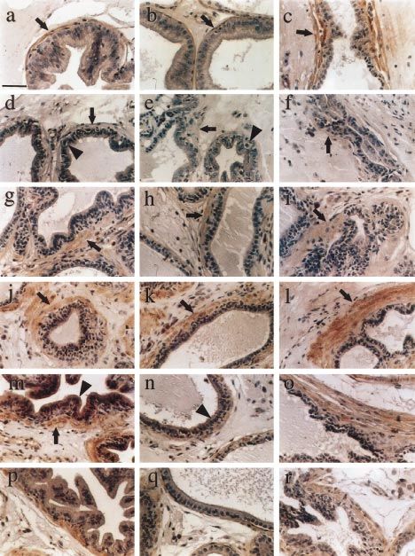
Figure 2 Immunohistochemical analysis of KGF in the normal and androgen-manipulated rat ventral prostate. Sections of ventral prostate
from normal or androgen-manipulated rats were stained for KGF protein as described in Materials and Methods. Representative fields from
the three functional regions of the ductal system are presented (distal region, a, d, g, j, m, p; intermediate region, b, e, h, k, n, q; proximal
region, c, f, i, l, o, r). In normal prostate (a, b, c), brown KGF staining was present mainly in the stromal cells (arrows). Day 4 post-
castration (d, e, f), staining was reduced (arrows) and apoptotic epithelial cells were visible (arrowheads). Day 7 (g, h, i) and day 21
(j, k, l) post-castration, stromal immunoreactivity returned (arrows). Day 21 castrate with 24 h DHT replacement therapy (m, n, o), strong
epithelial immunoreactivity appeared in regions of epithelial regrowth (arrowheads) and persisted through 48 h of DHT replacement
therapy (p, q, r). Scale bar represents 50 ìm.
Journal of Endocrinology (1998) 156, 115–125
Downloaded from Bioscientifica.com at 03/14/2020 11:40:04AM
via free accessAndrogen-independent KGF expression · J A NEMETH and others 119
Journal of Endocrinology (1998) 156, 115–125
Downloaded from Bioscientifica.com at 03/14/2020 11:40:04AM
via free access120 J A NEMETH and others · Androgen-independent KGF expression
seen in Fig. 1C lane 1, the KGF antiserum recognized a staining was also seen in the intermediate epithelium
single protein band with a molecular mass of 85% for all cell isolates). The identity and
most pronounced in the distal region of the duct.
purity of the cell fractions was confirmed by immuno-
cytochemical staining for cytokeratins 5 and 18 (Peehl et al.
Persistence of KGF protein after androgen withdrawal and 1994). The epithelial cell fraction contained a mixture of
replacement luminal and basal cells, and contamination by non-
epithelial cells was less than 1%. Contamination of the
To determine the effect of androgen removal on the
stromal cell fraction by epithelial cells was also minimal
pattern of KGF expression, sections of ventral prostates
(5–7%). The separated epithelial and stromal cell fractions
harvested at 0, 4, 7 and 21 days post-castration were
were used directly for RNA preparation and RT-PCR
assessed by immunohistochemical examination for the
analysis to eliminate possible changes in protein expression
presence of KGF protein. The effects of castration on
due to culture conditions.
prostate weight have been described previously, and
supplementation of castrated animals with 2 cm lengths of
DHT tubing has been shown to restore ventral prostates
to control proportions (Lee et al. 1981). Prostate weights KGF message levels during androgenic manipulation
increased and decreased in response to androgen manipu- To assess further the possible effects of androgen on KGF
lation as expected (data not shown). By day 4 post- expression, steady-state levels of KGF message were
castration, immunoreactivity for KGF was greatly reduced examined after androgen removal and replacement.
in the stroma throughout the ductal system, and was barely RT-PCR analysis was performed on RNA samples pre-
detectable in the intermediate and proximal regions pared from isolated stromal cells, using primer pairs
(Fig. 2d, e and f, arrows). The epithelium at this time was specific to the rat KGF or â-actin gene. As seen in Fig. 3A,
rapidly regressing, as demonstrated by the presence of the expected product of 392 bp was amplified from all
numerous apoptotic cells in the distal and intermediate RNA samples. In the absence of reverse transcriptase, no
regions (Fig. 2d and e, arrowheads). By day 7, the product was observed, suggesting that the observed PCR
involution process had slowed, and epithelial cell death product was not related to DNA contamination of RNA
had nearly stopped in the entire prostate (Fig. 2g, h and i). samples. The authenticity of the product was further
KGF immunoreactivity had returned in the stroma at all confirmed by restriction digestion with BamHI (data not
levels of the ductal system (arrows) to a level similar to that shown). As an internal control, similar reactions were
seen at day 0. Likewise, the periductal stromal cells at carried out using primers specific to the rat â-actin gene.
day 21 were highly positive for KGF protein in all regions Again, a product of the expected size (764 bp) was
(Fig. 2j, k and l). observed in all samples (Fig. 3A). RT-PCR analysis was
After androgen replacement for 24 h, no change in also performed on RNA samples from the epithelial cell
stromal KGF staining could be seen (Fig. 2m, n and o); fractions. No message for KGF was detected in any of
however, the epithelium in the distal region showed the samples, confirming the lack of KGF expression by
intense immunoreactivity (Fig. 2m, arrowhead), and some prostatic epithelial cells (data not shown).
Journal of Endocrinology (1998) 156, 115–125
Downloaded from Bioscientifica.com at 03/14/2020 11:40:04AM
via free accessAndrogen-independent KGF expression · J A NEMETH and others 121
castration also suggests that the diminished immuno-
reactivity for KGF seen in Fig. 2d, e and f was not related
to loss of the KGF transcript.
KGF expression in immature prostate
Previous studies have demonstrated the importance of
KGF in the androgen-induced growth of seminal vesicle
and prostate in organ culture, using fetal or newborn
tissues (Alarid et al. 1994, Sugimura et al. 1996). To
address the possibility that KGF expression may be regu-
lated differently in immature versus adult rat prostate,
immunohistochemical and RT-PCR analyses described
above were performed on prostate tissue from 14-day-old
newborn rats or 14-day-old rats that had been castrated on
day 5 after birth. As seen in Fig. 4a, KGF protein could be
detected in stromal cells surrounding the developing ducts
(arrow). Strong stromal immunoreactivity could still be
observed in neonatally castrated rat prostate tissue (Fig. 4b,
arrow), suggesting that KGF was still being expressed in
the absence of androgen. In contrast with the stromal
staining, a striking difference was seen in the epithelial
KGF immunoreactivity. KGF protein was high in the
rapidly growing ductal epithelium of the normal neonatal
prostate (Fig. 4a, arrowhead); however, the epithelium in
the prostate of the castrated neonate was nearly devoid of
Figure 3 Effect of androgen withdrawal and replacement on KGF staining for KGF (Fig. 4b, arrowhead). Therefore, as seen
message levels in the rat ventral prostate. A, Prostate stromal cells in the adult, only epithelial staining for KGF was affected
were isolated from normal or androgen-manipulated rats as by androgen status in the neonatal prostate.
described in Materials and Methods. RT-PCR analysis was
performed, in the presence (+) or absence (") of reverse Steady-state levels of KGF message were also compared
transcriptase, on RNA samples prepared from isolated prostate in prostates from normal and castrated neonates. Using the
stromal cells using primers specific to rat KGF or â-actin. Products comparative RT-PCR method as described above, KGF
were separated by agarose gel electrophoresis, visualized by message could be detected in samples from both normal
ethidium bromide staining, and measured by scanning
densitometry. B, Values for KGF message were normalized to
and castrated neonatal rats (Fig. 5A), and no significant
â-actin control, and plotted as percentage of day 0 control (n=3, difference was found in KGF message levels between
mean& S.D.). KGF message levels did not vary significantly from the two samples in comparison with the â-actin control
control after any treatment (P>0·05). (Fig. 5B). Taken together, these data support the
hypothesis that KGF expression is not positively regulated
by androgen in the rat prostate in vivo.
For purposes of comparison, we then determined the
range of linearity with regard to the number of PCR
amplification cycles and amount of input RNA (data not Discussion
shown). We chose 26 cycles and 750 ng of RNA sample
for all subsequent experiments, since these conditions were The results of the present study reveal that production of
within the linear range of amplification for both the KGF KGF by prostatic stromal cells was not greatly influenced
and â-actin primer sets. After scanning photographs of the by androgen status in vivo. According to the hypothesis
ethidium bromide-stained gels, band intensities for KGF that KGF is an andromedin (Yan et al. 1992), a decrease
products were normalized to their â-actin counterparts, in KGF expression would be expected after androgen
and the mean values of three experiments were plotted as removal; however, we observed no significant decrease in
percentages of day 0 control (Fig. 3B). Although this KGF message or protein after androgen manipulation.
method was not strictly quantitative, we observed that This result is consistent with previous reports by Sugimura
KGF message persisted at a high level after castration, and et al. (1996), who saw continued presence of the KGF
was abundant even at 21 days post-castration (Fig. 3B). message in the prostate after castration, and by Nishi et al.
Androgen replacement for 24 or 48 h also had no signifi- (1996), who also did not see a decrease in KGF message
cant effect on the steady-state level of KGF message. The after castration, but actually observed a large induction of
high level of KGF message observed on day 4 post- KGF message. This induction of KGF message may have
Journal of Endocrinology (1998) 156, 115–125
Downloaded from Bioscientifica.com at 03/14/2020 11:40:04AM
via free access122 J A NEMETH and others · Androgen-independent KGF expression
Figure 4 Immunohistochemical analysis of prostatic KGF in the
normal and castrated neonatal rat. Sections of ventral prostate
from normal or 10-day castrated neonatal rats were stained for
KGF protein as described in Materials and Methods. In the normal
day 14 rat prostate (a), KGF immunoreactivity was seen in the
stromal cells (arrow), as well as in the rapidly growing epithelium
(arrowhead). In the prostate of the castrated neonate (b), stromal
immunoreactivity was present (arrow), but epithelial staining was
absent (arrowhead). Scale bar represents 50 ìm.
been related to a relative increase in stromally derived
mRNAs after castration, since most of the prostatic epi-
thelium is lost as the result of active cell death after
androgen removal (Coffey et al. 1968), thus greatly in-
creasing the relative proportion of stromal cells in the
prostate. Problems resulting from changing cell ratios were
minimized in the present study by separating the stromal
and epithelial cell fractions before measurement of KGF
message.
Our present observations are not surprising in view of
mounting evidence suggesting that regulation of KGF
expression may be highly complex, involving a number
of inductive and inhibitory factors. Yan et al. (1992)
have demonstrated increased KGF expression by prostate
stromal cultures in response to androgen, and Fasciana
et al. (1996) have found an androgen-inducible element in
the KGF promoter. In addition, interleukin-á1, platelet-
derived growth factor-BB and transforming growth
factor-á have all been shown to stimulate KGF production Figure 5 Effect of androgen withdrawal on KGF message levels in
the immature rat ventral prostate. A, Ventral prostates were
by human fibroblasts, while neither basic FGF nor tumor isolated from normal (D 14) or castrated (C 10) neonatal rats as
necrosis factor-á had any significant effect (Chedid et al. described in Materials and Methods. RT-PCR analysis was
1994). On the other hand, KGF synthesis could be performed, in the presence (+) or absence (") of reverse
inhibited in human fibroblasts by glucocorticoids such as transcriptase, on RNA samples prepared from the prostates using
dexamethasone or hydrocortisone (Chedid et al. 1996). primers specific to rat KGF or â-actin. Products were separated by
agarose gel electrophoresis, visualized by ethidium bromide
Koji et al. (1994) have also found that KGF expression was staining, and measured by scanning densitometry. B, Values for
sensitive to progesterone in the primate endometrium; KGF message were normalized to â-actin control, and plotted as
however, this response was limited to stromal smooth percentage of day 0 control (n=3, mean& S.D.). KGF message
muscle cells in the basalis region, and KGF expression was levels did not vary significantly from control after castration
(P>0·05).
independent of progesterone in smooth muscle cells of the
myometrium. Although KGF expression may be respon-
sive to a number of factors individually in a culture, the
interaction of these various factors within an animal may due to proteolytic degradation of extracellular matrix
have a very different effect, and the reaction to a specific (ECM)-associated pools of the protein. After castration,
factor may vary between cell types in the same tissue. there is a massive induction of ECM-modifying enzyme
Further studies on intact prostates may provide insights activity in the prostate, including cathepsins (Lee et al.
into KGF regulatory mechanisms in vivo. 1990), urokinase (Andreasen et al. 1990, Wilson et al.
The results of our immunohistochemical experiments 1996), gelatinases (Wilson et al. 1991) and collagenases (see
show a transient loss of KGF immunostaining on day 4 Guenette & Tenniswood (1994) for a review). A number
after castration (Fig. 2d, e and f ), which was most likely of these enzymes have also been shown to be involved in
Journal of Endocrinology (1998) 156, 115–125
Downloaded from Bioscientifica.com at 03/14/2020 11:40:04AM
via free accessAndrogen-independent KGF expression · J A NEMETH and others 123
the release of matrix-bound members of the FGF family, Although our results indicate that KGF production
such as plasmin (Saksela & Rifkin 1990) and heparitinase is not androgen-responsive in vivo in the prostate, its
(Bashkin et al. 1989). So it is possible that the KGF protein mitogenic activity clearly is, as evidenced by the organ
is lost or destroyed as a result of rapid matrix turnover culture experiments of Sugimura et al. (1996). FGFs,
during involution, and reappears once matrix degradation including KGF, are stored in complex with heparin-
has slowed by day 7. Post-transcriptional changes could containing matrix and cell-surface molecules, and it is
also explain the loss of KGF protein despite the presence of thought that the matrix association of the FGF molecule
abundant message. Such a mechanism is unlikely though, must be modified to allow the factor to interact with its
since immunoreactive KGF protein was fully restored after cellular receptor and produce a signal (Bashkin et al. 1989).
regression had ceased, without any change in androgen It is possible that this modification process is under
status. androgenic control in the prostate, perhaps through
Surprisingly, we observed the most dramatic change in local elaboration of specific matrix-modifying enzymes. It
KGF immunostaining in the prostate epithelium, where has already been noted that numerous ECM-modifying
high levels of the protein were seen in the rapidly growing enzymes are induced during prostate regression (Wilson
epithelium of the neonate (Fig. 4a) and in the castrated et al. 1991, 1996), and modification of the ECM is
adult in response to androgen replacement (Fig. 2m). This intimately involved in the processes of growth and
staining is probably not due to expression by the epi- morphogenesis (Fukada et al. 1988, Brenner et al. 1989,
thelium, as KGF has been demonstrated to be produced Behrendsten et al. 1992). This mechanism of KGF
solely by stromal cells in normal prostate (Yan et al. 1992, regulation by androgen could be consistent with the
Story et al. 1997), and we were unable to detect KGF observations of Sugimura et al. (1996), and may be at least
message in our isolated epithelial cell fractions. It is possible partially responsible for those of Yan et al. (1992), who
that the epithelial localization of KGF is related to detec- observed increased KGF activity in response to androgen
tion of internalized ligand after receptor activation, which in prostatic fibroblast cultures. This release mechanism
has been clearly demonstrated for other members of the could also help clarify the apparent paradox regarding the
FGF family. For example, FGF-2 has been shown to distribution of KGF protein in the ductal system. In the
accumulate intracellularly in bovine endothelial cells, present study we observed that KGF protein was present at
both within the nucleolus (Bouche et al. 1987) and in high levels in the stroma throughout the ductal system.
lysosomal compartments (Moscatelli 1988), where it is Epithelial staining for KGF, presumably resulting from
then degraded slowly with an apparent half-life of 8 h. internalization of the activated receptor–ligand complex
Likewise, exogenous FGF-1 has been found to accumu- (Moscatelli 1988, Prudovsky et al. 1994), was consistently
late in the cytoplasm and associate with the nucleus associated with regions of epithelial proliferation, such as
by a receptor-dependent mechanism (Zhan et al. 1993, the distal segment in the adult and the normal newborn
Prudovsky et al. 1994). So it is plausible that receptor- rat prostate. Further investigation into the mechanisms
mediated internalization of stromally derived KGF could governing the release and presentation of KGF to its
result in the intracellular accumulation of KGF protein in receptor is clearly warranted.
regions of rapid epithelial cell growth. Further investi-
gation into the intracellular fate of KGF is needed to Acknowledgement
understand this mechanism fully.
It is clear that KGF action is important to the androgen- This work was supported in part by NIH grants DK47561,
induced growth and development of the prostate. In HD28048 and DK07706, and the American Foundation
organ culture studies, it has been found that antibody for Urologic Disease.
neutralization of KGF activity could inhibit growth and
branching morphogenesis of prostatic buds in response to
androgen, and that KGF could actually stimulate ductal References
growth in the absence of androgen (Sugimura et al. 1996). Alarid ET, Rubin JS, Young P, Chedid M, Ron D, Aaronson SA &
Recently a KGF knockout mouse was described (Guo Cunha GR 1994 Keratinocyte growth factor functions in epithelial
et al. 1996). Upon examination of the ventral prostate, no induction during seminal vesicle development. Proceedings of the
gross histologic abnormalities were detected; however, National Academy of Sciences of the USA 91 1074–1078.
closer examination may reveal subtle anomalies in epi- Andreasen PA, Kristensen P, Lund LR & Dano K 1990 Urokinase-
type plasminogen activator is increased in the involuting ventral
thelial function or ductal structure. Certainly it is possible prostate of castrated rats. Endocrinology 126 2567–2576.
that other members of the FGF family may have compen- Bashkin P, Doctrow S, Klagsbrun M, Svahn CM, Folkman J &
sated somewhat for the lack of KGF, as FGF-1 can bind to Vlodavsky I 1989 Basic fibroblast growth factor binds to
the KGF receptor with high affinity (Miki et al. 1992, subendothelial extracellular matrix and is released by heparitinase
and heparin-like molecules. Biochemistry 28 1737–1743.
Wang et al. 1995), but certain specific effects unique to Behrendsten O, Alexander CM & Werb Z 1992 Metalloproteinases
KGF may also exist which are important for normal mediated extracellar degradation by cells from mouse blastocyst
morphogenesis or differentiated function. outgrowths. Development 114 447–456.
Journal of Endocrinology (1998) 156, 115–125
Downloaded from Bioscientifica.com at 03/14/2020 11:40:04AM
via free access124 J A NEMETH and others · Androgen-independent KGF expression
Bouche G, Gas N, Prats H, Baldin V, Tauber J, Teissie J & Amalric F Lee C, Goolsby CL & Sensibar JA 1994 Cell cycle kinetics in rat
1987 Basic fibroblast growth factor enters the nucleolus and prostatic epithelia: nuclear migration during G2 phase. Journal of
stimulates the transcription of ribosomal genes in ABAE cells Urology 152 2294–2299.
undergoing G0–G1 transition. Proceedings of the National Academy of McKeehan WL, Adams PS & Rosser MP 1984 Direct mitogenic
Sciences of the USA 84 6770–6774. effects of insulin, epidermal growth factor, glucocorticoid, cholera
Brenner CA, Adler RR, Rappolee DA, Pederson RA & Werb Z toxin, unknown pituitary factors, and possibly prolactin, but not
1989 Genes for extracellular matrix-degrading metalloproteinases androgen on normal rat prostatic epithelial cells in serum-free,
and their inhibitor, TIMP, are expressed during early mammalian primary culture. Cancer Research 44 1998–2010.
development. Genes and Development 3 848–859. Miki T, Bottaro DP, Fleming TP, Smith CL, Burgess WH, Chan
Camp TA, Rahal JO & Mayo KE 1991 Cellular localization and AM-L & Aaronson SA 1992 Determination of ligand-binding
hormonal regulation of follicle-stimulating hormone and luteinizing specificity by alternative splicing: two distinct growth factor
hormone receptor messenger RNAs in the rat ovary. Molecular receptors encoded by a single gene. Proceedings of the National
Endocrinology 5 1405–1417. Academy of Sciences of the USA 89 246–250.
Chang S-M & Chung LWK 1989 Interaction between prostatic Montpetit ML & Tenniswood MP 1989 Separation of mature rat
fibroblasts and epithelial cells in culture: role of androgen. ventral prostate epithelial and fibroblast cells. Prostate 15 315–325.
Endocrinology 125 2719–2727. Moscatelli D 1988 Metabolism of receptor-bound and matrix-bound
Chedid M, Rubin JS, Csaky KG & Aaronson SA 1994 Regulation of basic fibroblast growth factor by bovine capillary endothelial cells.
keratinocyte growth factor gene expression by interleukin-1. Journal Journal of Cellular Biology 107 753–759.
of Biological Chemistry 269 10753–10757. Nemeth JA & Lee C 1996 Prostatic ductal system in rats: regional
Chedid M, Hoyle JR, Csaky KG & Rubin JS 1996 Glucocorticoids variation in stromal organization. Prostate 28 124–128.
inhibit keratinocyte growth factor production in primary dermal Nishi N, Oya H, Matsumoto K, Nakamura T, Miyanaka H & Wada
fibroblasts. Endocrinology 137 2232–2237. F 1996 Changes in gene expression of growth factors and their
Coffey DS, Shimazaki J & Williams-Ashman HG 1968 Polymerization receptors during castration-induced involution and androgen-
of deoxyribonucleotides in relation to androgen-induced prostatic induced regrowth of rat prostates. Prostate 28 139–152.
growth. Archives of Biochemistry and Biophysics 124 184–198. Peehl DM, Leung GK & Wong ST 1994 Keratin expression: a
Cunha GR & Chung LWK 1981 Stromal-epithelial interactions. I. measure of phenotypic modulation of human prostatic epithelial
Induction of prostatic phenotype in urothelium of testicular cells by growth inhibitory factors. Cell and Tissue Research 277
feminized (Tfm/y) mice. Journal of Steroid Biochemistry and Molecular 11–18.
Biology 14 1317–1321.
Prins GS, Birch L & Greene GL 1991 Androgen receptor localization
Cunha GR, Shannon JM, Newbauer BL, Sawyer LM, Fuji H,
in different cell types of the adult rat prostate. Endocrinology 129
Taguchi O & Chung LWK 1981 Mesenchymal–epithelial
3187–3199.
interactions in sex differentiation. Human Genetics 56 68–77.
Prins GS, Cooke PS, Birch L, Donjacour AA, Yalcinkaya TM, Siiteri
Cunha GR, Chung LWK, Shannon JM, Taguchi O & Fujii H 1983
PK & Cunha GR 1992 Androgen receptor expression and
Hormone-induced morphogenesis and growth: role of
5á-reductase activity along the proximal–distal axis of the rat
mesenchymal–epithelial interactions. Recent Progress in Hormone
prostatic duct. Endocrinology 130 3066–3073.
Research 39 559–598.
Fasciana C, van der Made AC, Faber PW & Trapman J 1996 Prudovsky I, Savion N, Zhan X, Friesel R, Xu J, Hou J, McKeehan
Androgen regulation of the rat keratinocyte growth factor WL & Maciag T 1994 Intact and functional fibroblast growth factor
(KGF/FGF7) promoter. Biochemical and Biophysical Research (FGF) receptor-1 trafficks near the nucleus in response to FGF-1.
Communications 220 858–863. Journal of Biological Chemistry 269 31720–31724.
Fukada Y, Masuda Y, Kishi J, Hashimoto Y, Hayakawa T & Nogawa Rubin JS, Bottaro DP, Chedid M, Miki T, Ron D, Cheon H-G,
H 1988 The role of interstitial collagens in cleft formation of mouse Taylor WG, Fortney E, Sakata H, Finch PW & LaRochelle WJ
embryonic submandibular gland formation during initial branching. 1995 Keratinocyte growth factor. Cell Biology International 19
Development 103 259–267. 399–411.
Guenette RS & Tenniswood M 1994 The role of growth factors in Saksela O & Rifkin DB 1990 Release of basic fibroblast growth
the suppression of active cell death in the prostate: an hypothesis. factor–heparan sulfate complexes from endothelial cells by
Biochemistry and Cell Biology 72 553–559. plasminogen activator-mediated proteolytic activity. Journal of
Guo L, Degenstein L & Fuchs E 1996 Keratinocyte growth factor is Cellular Biology 110 767–775.
required for hair development but not for wound healing. Genes Story MT, Hopp KA, Molter M & Meier DA 1994 Characteristics of
and Development 10 165–175. FGF-receptors expressed by stromal and epithelial cells cultured
Isaacs J 1984 Antagonistic effect of androgen on prostatic cell death. from normal and hyperplastic prostates. Growth Factors 10 269–280.
Prostate 5 545–557. Story MT, Hopp KA, Meier DA, Haq R-U & Rosen MA 1997
Kerr JFR & Searle J 1973 Deletion of cells by apoptosis during Expression of keratinocyte growth factor (FGF7) mRNA and
castration-induced involution of the rat prostate. Virchows Archiv. B. secretion of the protein by human prostate stromal cells. Prostate (In
Cell Pathology 13 87–102. Press).
Koji T, Chedid M, Rubin JS, Slayden OD, Csaky KG, Aaronson SA Sugimura Y, Cunha GR, Donjacour AA, Bigsby RM & Brody JR
& Brenner RM 1994 Progesterone-dependent expression of 1986 Whole-mount autoradiography study of DNA synthetic
keratinocyte growth factor mRNA in stromal cells of the primate activity during postnatal development and androgen-induced
endometrium: keratinocyte growth factor as a progestomedin. regeneration in the mouse prostate. Biology of Reproduction 34
Journal of Cellular Biology 125 393–401. 985–995.
Laemmli UK 1970 Cleavage of structural proteins during the assembly Sugimura Y, Foster BA, Hom YK, Lipschutz JH, Rubin JS, Finch
of the head of bacteriophage T4. Nature 227 680–685. PW, Aaronson SA, Hayashi N, Kawamura J & Cunha GR 1996
Lee C, Prins GS, Henneberry MO & Grayhack JT 1981 Effect of Keratinocyte growth factor (KGF) can replace testosterone in the
estradiol on the rat prostate in the presence and absence of ductal branching morphogenesis of the rat ventral prostate.
testosterone and pituitary. Journal of Andrology 2 293–299. International Journal of Developmental Biology 40 941–951.
Lee C, Sensibar JA, Dudek SM, Hiipakka RA & Liao S 1990 Tenniswood M 1986 Role of epithelial–stromal interactions in the
Prostatic ductal system in rats: regional variation in morphological control of gene expression in the prostate: an hypothesis. Prostate 9
and functional activities. Biology of Reproduction 43 1079–1086. 375–385.
Journal of Endocrinology (1998) 156, 115–125
Downloaded from Bioscientifica.com at 03/14/2020 11:40:04AM
via free accessAndrogen-independent KGF expression · J A NEMETH and others 125
Wang F, Kan M, Xu J, Yan G & McKeehan WL 1995 Yan G, Fukabori Y, Nikolaropoulos S, Wang S & McKeehan WL
Ligand-specific structural domains in the fibroblast growth factor 1992 Heparin-binding keratinocyte growth factor is a candidate
receptor. Journal of Biological Chemistry 270 10222–10230. stromal to epithelial cell andromedin. Molecular Endocrinology 6
Wilson MJ, Strasser M, Vogel MM & Sinha AA 1991 2123–2128.
Calcium-dependent and calcium-independent gelatinolytic Zhan X, Hu X, Friesel R & Maciag T 1993 Long term growth factor
proteinase activities of the rat ventral prostate and its secretion: exposure and differential tyrosine phosphorylation are required for
characterization and effect of castration and testosterone treatment. DNA synthesis in BALB/c 3T3 cells. Journal of Biological Chemistry
Biology of Reproduction 44 776–785. 268 9611–9620.
Wilson MJ, Ludowese C, Sinha AA & Estensen RD 1996 Effects of
castration on plasminogen activator activities and plasminogen
Received 4 March 1997
activator inhibitor type I in the rat ventral prostate. Prostate 28 Revised manuscript received 30 June 1997
239–250. Accepted 26 August 1997
Journal of Endocrinology (1998) 156, 115–125
Downloaded from Bioscientifica.com at 03/14/2020 11:40:04AM
via free accessYou can also read