Intravenous allogeneic umbilical cord blood-derived mesenchymal stem cell therapy in recessive dystrophic epidermolysis bullosa patients - JCI Insight
←
→
Page content transcription
If your browser does not render page correctly, please read the page content below
Intravenous allogeneic umbilical cord blood–derived
mesenchymal stem cell therapy in recessive dystrophic
epidermolysis bullosa patients
Sang Eun Lee, … , Kyounghwan Roh, Soo-Chan Kim
JCI Insight. 2021;6(2):e143606. https://doi.org/10.1172/jci.insight.143606.

 Clinical Medicine   Clinical trials   Dermatology

Recessive dystrophic epidermolysis bullosa (RDEB) is an incurable disease that causes severe mucocutaneous fragility
due to mutations in COL7A1 (encoding type VII collagen [C7]). In this phase I/IIa trial, we evaluated the safety and
possible clinical efficacy of intravenous infusion of allogeneic human umbilical cord blood–derived mesenchymal stem
cells (hUCB-MSCs) in patients with RDEB.

Four adult and two pediatric patients with RDEB were treated with 3 intravenous injections of hUCB-MSCs (1 × 10⁶ to 3 ×
106 cells/kg) every 2 weeks and followed up for 8–24 months after treatment. The primary endpoint was safety.
Secondary endpoints related to efficacy included clinical parameters, such as disease severity score, wound assessment,
itch and pain score, and quality of life. C7 expression levels and inflammatory infiltrates in the skin, as well as serum
levels of inflammatory markers and neuropeptides, were also assessed.

Intravenous hUCB-MSC infusions were well tolerated, without serious adverse events. Improvements in the Birmingham
Epidermolysis Bullosa Severity Score, body surface area involvement, blister counts, pain, pruritus, and quality of life
were observed with maximal effects at 56–112 days after treatment. hUCB-MSC administration induced M2 macrophage
polarization and reduced mast cell […]

Find the latest version:
https://jci.me/143606/pdf
CLINICAL MEDICINE

                                         Intravenous allogeneic umbilical cord
                                         blood–derived mesenchymal stem
                                         cell therapy in recessive dystrophic
                                         epidermolysis bullosa patients
                                         Sang Eun Lee,1 Seung-Ju Lee,1 Song-Ee Kim,1 Kinam Kim,2 Boyoung Cho,2 Kyounghwan Roh,3
                                         and Soo-Chan Kim4
                                         Department of Dermatology and Cutaneous Biology Research Institute, Gangnam Severance Hospital, Yonsei University
                                         1

                                         College of Medicine, Seoul, South Korea. 2Cellular Therapeutics Team, Daewoong Pharmaceutical Co. Ltd., Seoul,
                                         South Korea. 3Department of Clinical Development, Kangstem Biotech Co. Ltd., Seoul, South Korea. 4Department of
                                         Dermatology, Yongin Severance Hospital, Yonsei University College of Medicine, Yongin, South Korea.

                                                 BACKGROUND. Recessive dystrophic epidermolysis bullosa (RDEB) is an incurable disease that
                                                 causes severe mucocutaneous fragility due to mutations in COL7A1 (encoding type VII collagen
                                                 [C7]). In this phase I/IIa trial, we evaluated the safety and possible clinical efficacy of intravenous
                                                 infusion of allogeneic human umbilical cord blood–derived mesenchymal stem cells (hUCB-MSCs)
                                                 in patients with RDEB.

                                                 METHODS. Four adult and two pediatric patients with RDEB were treated with 3 intravenous
                                                 injections of hUCB-MSCs (1 × 10⁶ to 3 × 10⁶ cells/kg) every 2 weeks and followed up for 8–24 months
                                                 after treatment. The primary endpoint was safety. Secondary endpoints related to efficacy included
                                                 clinical parameters, such as disease severity score, wound assessment, itch and pain score, and
                                                 quality of life. C7 expression levels and inflammatory infiltrates in the skin, as well as serum levels
                                                 of inflammatory markers and neuropeptides, were also assessed.

                                                 RESULTS. Intravenous hUCB-MSC infusions were well tolerated, without serious adverse
                                                 events. Improvements in the Birmingham Epidermolysis Bullosa Severity Score, body surface
                                                 area involvement, blister counts, pain, pruritus, and quality of life were observed with maximal
                                                 effects at 56–112 days after treatment. hUCB-MSC administration induced M2 macrophage
                                                 polarization and reduced mast cell infiltration in RDEB skin. Serum levels of substance P were
                                                 decreased after therapy. Increased C7 expression was observed at the dermoepidermal junction in
                                                 1 of 6 patients at day 56.

                                                 CONCLUSION. To the best of our knowledge, this is the first clinical trial of systemic administration
                                                 of allogeneic hUCB-MSCs in patients with RDEB, demonstrating safety and transient clinical
Conflict of interest: KR works in the
Department of Clinical Development               benefits.
of Kangstem Biotech Co. Ltd., a
                                                 TRIAL REGISTRATION. ClinicalTrials.gov NCT04520022.
biotechnology company focused on
stem cell therapeutics. KK and BC                FUNDING. This work was supported by Daewoong Pharmaceutical Co. Ltd. and Kangstem
work in the Cellular Therapeutics Team           Biotech Co. Ltd.
at Daewoong Pharmaceutical Co. Ltd.

Copyright: © 2021, Lee et al. This is
an open access article published under
the terms of the Creative Commons
Attribution 4.0 International License.   Introduction
Submitted: August 25, 2020               Epidermolysis bullosa (EB) is a group of genetic diseases characterized by mechanical fragility of skin and
Accepted: December 9, 2020               mucosa (1). Recessive dystrophic EB (RDEB) is caused by mutations in COL7A1, which encodes type VII
Published: January 25, 2021              collagen (C7), the main constituent of anchoring fibrils at the dermoepidermal junction (DEJ). RDEB is
                                         one of the most severe forms of EB; it is characterized by recurrent blistering, chronic wounds, disabling
Reference information: JCI Insight.
2021;6(2):e143606.                       scarring in the skin, and mucosa and internal organ dysfunctions, leading to substantial morbidity and mor-
https://doi.org/10.1172/jci.             tality (2–4). Currently, there is no cure for this severe subtype of EB; however, novel therapeutic strategies
insight.143606.                          have been developed in the fields of gene and cell therapies (5–15).

                                                                                                                                                             1
CLINICAL MEDICINE

                                      Mesenchymal stem cells (MSCs) have been identified as an attractive option for allogeneic cell therapy
                                 for RDEB based on their potential mechanisms of action, including immunomodulation, migration to
                                 damaged tissue, stimulation of tissue regeneration, and reduction of fibrosis, mainly through paracrine
                                 activities (8–11, 14–16). Locally injected allogeneic bone marrow–derived MSCs (BM-MSCs) have shown
                                 to accelerate wound healing, with transient C7 restoration in patients with RDEB and a mouse model of
                                 dystrophic EB (10). Two early-phase clinical trials of systemic administration of allogeneic BM-MSCs in
                                 23 pediatric patients with RDEB reported variable clinical benefits that lasted for several months with sat-
                                 isfactory safety (8, 9). An additional recently published phase I/II trial of intravenous BM-MSC injection
                                 in 10 adult patients with RDEB also showed transient, but clinically meaningful, improvements in disease
                                 severity, skin inflammation, and pruritus, with no serious adverse events (AEs) (14).
                                      To our knowledge, previous clinical trials for RDEB have examined the potential of BM-MSCs (8, 9,
                                 14). However, umbilical cord blood (UCB) has become an attractive source of stem cells, because of its
                                 noninvasive collection procedure and rapid availability from cord blood banking (17, 18). Human UCB-de-
                                 rived MSCs (hUCB-MSCs) exhibit higher proliferation capacity and lower immunogenicity compared with
                                 BM-MSCs (17, 19). Data from a few reports support that UCB-MSCs may have greater immunosuppres-
                                 sive potential than other sources of MSCs (17–22). In addition, hUCB-MSCs have shown greater immuno-
                                 suppressive and regenerative potential than BM- or peripheral blood–derived MSCs in murine wounding
                                 model (23). A preclinical study has demonstrated that repeated systemic infusions of human UCB-de-
                                 rived unrestricted somatic stem cells, a subpopulation of nonhematopoietic stromal stem cells, significantly
                                 extended the life span and reduced blistering in a RDEB mouse model (16). Given the promising results
                                 of the preclinical study, we conducted a first-in-human, phase I/IIa clinical trial of intravenous adminis-
                                 trations of allogeneic hUCB-MSCs in patients with RDEB to determine safety, tolerability, and potential
                                 efficacy. We also analyzed changes in serum inflammatory markers, neuropeptides, and skin inflammatory
                                 infiltrates as well as C7 expression following hUCB-MSC treatment.

                                 Results
                                 Patient characteristics. Between October 2016 and May 2019, 6 patients with RDEB were assessed for eligi-
                                 bility. Three adult and two pediatric patients were sequentially enrolled in the trial and received 3 repeated
                                 intravenous hUCB-MSC injections. One additional adult patient was treated with the same investigational
                                 product under the treatment use approval from the Korea Food & Drug Administration (KFDA), because
                                 they were too late for trial enrollment (Figure 1). All patients had moderate-to-severe or severe phenotypes,
                                 with various extracutaneous symptoms. Negative or markedly decreased expression of C7 noncollage-
                                 nous-1 domain was found in baseline skin biopsies. Analysis of circulating autoantibodies against C7 using
                                 indirect immunofluorescence (IIF) was negative for all patients (Table 1). All adult patients received 3 × 10⁶
                                 hUCB-MSCs/kg every 2 weeks, whereas the 2 pediatric patients received 1 × 10⁶ to 2 × 10⁶ hUCB-MSCs/
                                 kg every 2 weeks. All patients were carefully observed for clinical signs and laboratory test results related to
                                 potential thromboembolic events were monitored, even though a recent meta-analysis of randomized con-
                                 trolled trials reported no significant increase in the risk of thromboembolic events for patients treated with
                                 MSCs as compared with the control group (24). Demographics and clinical characteristics of participants
                                 and trial flow are provided in Figure 1 and Table 1. All patients completed at least 8 months (8–24 months)
                                 of follow-up after the first infusion.
                                      Safety. AEs during the study period are summarized in Supplemental Tables 3 and 4 (supplemental
                                 material available online with this article; https://doi.org/10.1172/jci.insight.143606DS1). Overall, 50%
                                 of the patients treated reported ≥1 AE. The most frequent AE was wound infection (4 of 13 AEs, 30.8%),
                                 but all wound infections were thought to be due to the underlying RDEB. Only acute gastritis was con-
                                 sidered as an AE determined to be possibly related to cell therapy. No severe AEs (defined by Common
                                 Terminology Criteria for Adverse Events) at grade 3 or higher were reported, suggesting that intravenous
                                 hUCB-MSC injections were generally well tolerated. There were no clinically significant changes in labora-
                                 tory test values, except increased basal levels of C-reactive protein (CRP) and fibrinogen, vital signs or elec-
                                 trocardiogram results during the study period. There were no changes in tissue-bound immunoreactants
                                 using IIF following cell therapy.
                                      Clinical efficacy. hUCB-MSC treatment markedly reduced erythema and erosions in patients with
                                 RDEB (Figure 2). At day 56, the mean clinical severity scores assessed by the Birmingham Epidermol-
                                 ysis Bullosa Severity Score (BEBSS) and total body surface area (TBSA) affected by RDEB significantly

JCI Insight 2021;6(2):e143606 https://doi.org/10.1172/jci.insight.143606                                                                       2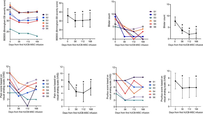
CLINICAL MEDICINE

Figure 1. Study design. (A) Flow chart for clinical trial and treatment use (expanded access to investigational drugs for treatment use). (B) Study design for
hUCB-MSC treatment and evaluation.

                                   decreased by 5.6 points (95% CI, –7.39 to –3.86) and 5.4 points (95% CI, –8.14 to –2.61), respectively.
                                   Blister count and the ratio of blister area to body surface area also decreased by 4 points (95% CI, –6.74
                                   to –1.26) and 2 points (95% CI, –4.02 to –0.06), respectively, at day 56 compared with baseline. After
                                   day 56, these clinical effects of hUCB-MSCs were either maintained or slightly attenuated over time
                                   until day 168 (Figure 3 and Supplemental Figure 1). Chronic nonhealing wounds in RDEB are asso-
                                   ciated with decreased quality of life (QOL) and increased risk of cutaneous squamous cell carcinoma
                                   (cSCC). We evaluated the effect of hUCB-MSCs on the healing of chronic open wounds that were
                                   unhealed for at least 12 weeks with wound size >100 cm2, as defined in a previous study (13). One
                                   pediatric (subject 4) and one adult (subject 6) subject each had 2 chronic open wounds. Of the 4 chron-
                                   ic wounds from 2 subjects, 2 wounds (1 from subject 4 and 1 from subject 6) (50%) showed a 50% or
                                   greater reduction in wound size compared with baseline at day 56. Of these 2 wounds, only 1 remained
                                   at least 50% healed by day 112.
                                        hUCB-MSC treatment resulted in a substantial mean reduction in pain (−3 points on visual analogue
                                   scale [VAS] score, 95% CI, –4.76 to –1.24) and itch (−2 points on VAS score, 95% CI, –3.76 to –0.24) from
                                   baseline to day 56. Mean VAS scores for pruritus were maintained by day 168, while pain VAS scores
                                   showed a gradual increase over time (Figure 3). At day 56, QOL, as assessed by a QOL in EB questionnaire
                                   (QOLEB), was improved by 6.2 points (95% CI, –8.69 to –3.65). The baseline and mean change from the
                                   baseline for the secondary outcome data are summarized in Supplemental Table 5. As shown in Supplemen-
                                   tal Figure 2, age subgroup analyses (children vs. adults) showed no significant between-group differences in
                                   the secondary outcomes, including BEBSS, TBSA, blister count and area, itch and pain scores, and QOLEB.
                                        Molecular assays for C7 in skin. Then we evaluated whether systemic infusions of hUCB-MSCs could
                                   restore C7 and anchoring fibrils in RDEB skin by immunofluorescence staining and transmission electron
                                   microscopy (TEM) analysis of skin of patients before and after treatment. On day 56, 1 patient (subject 1)
                                   showed an increase in C7 expression levels at the DEJ, as assessed by mean fluorescence intensity (MFI)
                                   compared with baseline, while others (subjects 2–6) showed no significant changes in C7 expression in
                                   skin after MSC treatment (Figure 4). No obvious differences in anchoring fibril structure or distribution
                                   were observed between baseline and day 56 in all 6 patients, as assessed by TEM (data not shown).
                                        Changes in skin infiltration of macrophages and mast cells. Macrophages have a central role in maintaining
                                   tissue homeostasis and repair. Classic proinflammatory (M1) and alternatively activated, antiinflammato-
                                   ry (M2) macrophages exhibit distinct phenotypes and functions (25, 26). Previous studies indicated that
                                   MSCs can promote M2 polarization of tissue macrophages, contributing to tissue regeneration (27–29).
                                   Therefore, we analyzed the phenotypes of macrophages in the skin of patients with RDEB before and after
                                   hUCB-MSC treatment. The number of CD68+ total macrophages was higher in the skin of patients with
                                   RDEB at baseline than in healthy controls. Intravenous administration of hUCB-MSCs did not affect the
                                   density of CD68+ total macrophages but significantly increased macrophages expressing CD206, a marker

JCI Insight 2021;6(2):e143606 https://doi.org/10.1172/jci.insight.143606                                                                                    3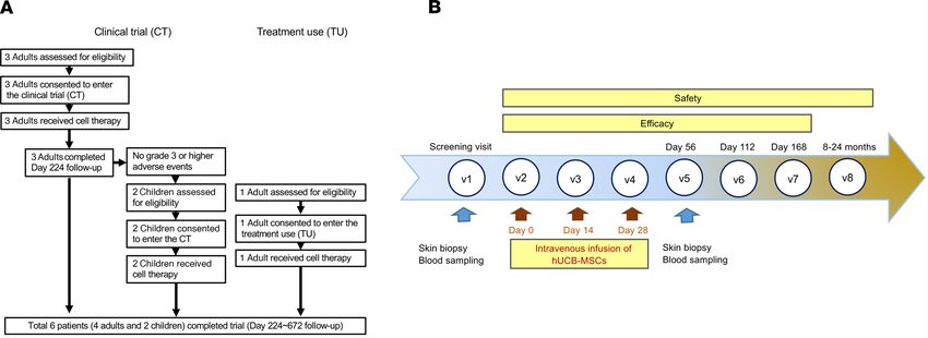
CLINICAL MEDICINE

Table 1. Demographics and clinical characteristics of 6 patients with RDEB

Subject                       1                    2                    3                     4                     5                    6
Sex/age in yr            F/60                F/25               M/21                 F/13                  F/8                          F/28
Race (ethnicity)    Asian (Korean)      Asian (Korean)     Asian (Korean)      Asian (Korean)        Asian (Korean)               Asian (Korean)
                     c.3631C > T,      c.3139+12G > A,      c.2005C > T,         c.3631C > T,                                       c.2922+2T >
                                                                                                  c.7371insA, exon 96;
                   p.Gln1211*, exon   intron 23; c.5188C p.Arg669*, exon     p.Gln1211*, exon 27;                                   G, intron 22;
COL7A1 mutations                                                                                   c.2318_2319delCT,
                   27; c.8569G > T,    > T, p.Arg1730*,   15; c.8569G > T, c.3717_3721delTACTC,                                   c.3139+12G > A,
                                                                                                         exon 18
                 p.Glu2857*, exon 116      exon 58      p.Glu2857*, exon 116       exon 27                                            intron 23
                                         Moderate to        Moderate to                               Moderate to
Phenotype               Severe                                                      Severe                                            Severe
                                            severe             severe                                    severe
C7 expression at
                  Barely detectable Barely detectable         Reduced           Undetectable         Undetectable                  Undetectable
the DEJ (DIF)
Circulating
autoantibodies         Negative            Negative           Negative            Negative              Negative                     Negative
against C7 (IIF)
                                                                                                                                 Mitten deformity
                      Mitten deformity,                                                                                         (s/p hand surgery),
Major clinical                           Mitten deformity Esophageal stricture        Mitten deformity       Mitten deformity
                      cataract, corneal                                                                                        esophageal stricture
features                                (s/p hand surgery) (s/p balloon dilation)    (s/p hand surgery)     (s/p hand surgery)
                          erosions                                                                                                  (s/p balloon
                                                                                                                                      dilation)
Race and ethnicity were classified by investigators. C7, type VII collagen; DEJ, dermoepidermal junction; DIF, direct immunofluorescence test; F,
female; IIF, indirect immunofluorescence test; M, male; s/p, status after operation.

                                  of M2 macrophages, in RDEB skin at day 56 (Figure 5). Mast cells play a central role in neuroinflamma-
                                  tory pain and itch (30, 31). Baseline skin biopsies of patients with RDEB showed a significant increase of
                                  mast cell infiltration compared with normal human skin, but mast cell infiltration was significantly reduced
                                  56 days after hUCB-MSC treatment (Figure 5).
                                       Changes in systemic inflammatory markers and neuropeptides. Chronic wounds in RDEB trigger system-
                                  ic inflammation that may contribute to multiple-organ damage (2, 3, 32–34). Since MSCs have potent
                                  immunomodulatory capacities, we investigated the effect of hUCB-MSC infusion on serum inflammatory
                                  markers in patients with RDEB. Serum levels of CRP fluctuated in individual patients over time, but no
                                  significant change in the mean CRP values was observed 56 days after hUCB-MSC treatment (data not
                                  shown). Additionally, baseline serum levels of proinflammatory cytokines, IL-1β and IL-6, were elevated in
                                  patients with RDEB compared with those in healthy controls, but these levels were not significantly altered
                                  by hUCB-MSC treatment on day 56 (Figure 6).
                                       Given the remarkable efficacy of hUCB-MSC treatment in reducing pain and itch in patients with
                                  RDEB in this study, we also analyzed the changes in serum levels of neuropeptides. Baseline serum levels
                                  of substance P were significantly higher in patients with RDEB compared with age-matched healthy con-
                                  trol values, and, notably, substance P levels were significantly reduced 56 days after hUCB-MSC treatment.
                                  Serum calcitonin gene–related peptide (CGRP) levels were also higher in patients with RDEB than in
                                  healthy controls, and these levels were reduced from baseline after hUCB-MSC treatment (P = 0.06), but
                                  these changes were not statistically significant (Figure 6).

                                  Discussion
                                  This open-label, phase I/IIa clinical trial shows that 3 repeated intravenous administrations of allogeneic
                                  hUCB-MSCs are well tolerated and potentially provide clinical benefits by reducing disease severity, dis-
                                  ease-affected body area, blister count, and pain and itch and improving QOL in children and adults with
                                  moderate-to-severe or severe RDEB. This study is meaningful in that it is the first clinical trial to our knowl-
                                  edge to apply MSCs derived from UCB, systemically, to patients with RDEB.
                                       Three separate intravenous infusions of hUCB-MSCs did not cause serious AEs. A previous clini-
                                  cal trial of BM-MSCs in 10 adult patients with RDEB reported development of cSCC in 2 participants
                                  about 6–7 months after the injections (14), suggesting careful monitoring of this potential complication,
                                  particularly in adult patients. In this trial, of the 4 adult patients, 1 patient (subject 6) was followed up
                                  for 16 months, and the remaining 3 patients (subjects 1, 2, and 3) were followed up for up to 24 months;

JCI Insight 2021;6(2):e143606 https://doi.org/10.1172/jci.insight.143606                                                                            4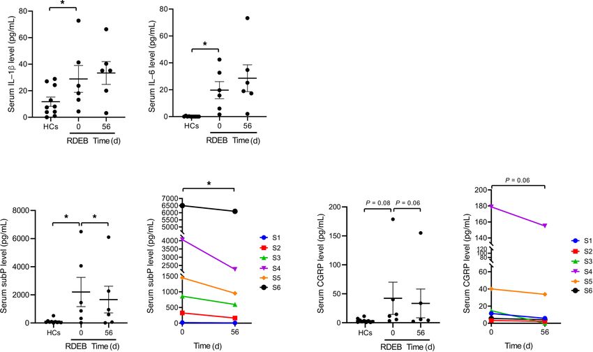
CLINICAL MEDICINE

                                 Figure 2. Marked reduction in erythema and erosions after hUCB-MSC treatment. Photographs of a pediatric (A, sub-
                                 ject 4) and an adult patient with RDEB (B, subject 1) at baseline and after 3 repeated injections of hUCB-MSCs.

                                 there was no development of cSCC during these follow-up periods. However, long-term follow-up data
                                 for more patients is needed to accurately evaluate the potential relationship between allogeneic hUCB-
                                 MSCs therapy and the risk of cSCC in RDEB.
                                      Although the primary objective was to assess safety, our data provide evidence of the potential
                                 efficacy of hUCB-MSC therapy in various clinical aspects of RDEB. hUCB-MSC infusions signifi-
                                 cantly reduced disease severity, as assessed by using BEBSS, affected body surface area, blister count,
                                 and blister area, with a maximal effect at day 56 in most patients. Over time, these clinical effects of
                                 hUCB-MSCs were progressively attenuated, but some patients showed sustained improvement up to
                                 day 168. With regard to wound healing, 50% (2 of 4) of large open wounds that were present for at
                                 least 12 weeks achieved 50% or greater healing by 56 days after treatment. Despite the small number of
                                 available chronic large wounds and the lack of control wounds, based on a previous report that indicat-
                                 ed that a 50% reduction in chronic RDEB wounds is clinically meaningful in terms of improvement in
                                 patient-reported outcomes (13), our results indicate that hUCB-MSC therapy exerts beneficial effects
                                 on wound healing in RDEB. In addition to the improvement of cutaneous lesions, intravenous admin-
                                 istration of hUCB-MSCs also relieved the symptom of dysphagia in 1 patient (subject 3), allowing the
                                 scheduled balloon dilation for esophageal stricture to be delayed.
                                      Recalcitrant pain and pruritus are among the most bothersome symptoms of RDEB (35–38). Pain in
                                 severe generalized RDEB is often very severe in that it does not respond well to potent opioid analgesics,
                                 and its intensity was shown to be greater than in postherpetic neuralgia (39). RDEB also causes severe pru-
                                 ritus that is thought to be associated with cutaneous inflammation secondary to barrier disruption, wound
                                 healing processes, and dysregulated activity of epidermal nerve fibers (38).
                                      In this study, hUCB-MSCs markedly reduced pain and pruritus in patients with RDEB by reducing
                                 average VAS scores by 3 cm and 2 cm on day 56, respectively. Given that the minimum important differ-
                                 ence for clinical improvement of chronic pain or pruritus has shown to be 2 to 3 cm on the VAS score (40,
                                 41), our data suggest that hUCB-MSC treatment is effective in achieving a clinically relevant improvement
                                 in pain and pruritus in RDEB that may lead to improved QOL.
                                      When comparing the clinical efficacy of hUCB-MSCs with that of BM-MSCs in RDEB, the mean
                                 differences in BEBSS and QOLEB scores at day 56 in our study were 5.6 points (95% CI, –7.39 to
                                 –3.86) and 6.2 points (95% CI, –8.69 to –3.65), which were comparable to those in previous studies
                                 using BM-MSCs in children (mean difference of BEBSS at day 60 was 5.2, QOLEB score was 4.4) and
                                 adults (mean difference of BEBSS at day 60 was 1.61, QOLEB score was 3.13) (8, 14). These findings
                                 indicate that hUCB-MSCs provide comparable therapeutic effects to BM-MSCs in improving disease
                                 severity and QOL in patients with RDEB. Regarding the itch and pain outcome, it is difficult to directly
                                 compare the effect of hUCB-MSCs with that of BM-MSCs because of the different measurement tools

JCI Insight 2021;6(2):e143606 https://doi.org/10.1172/jci.insight.143606                                                                         5
CLINICAL MEDICINE
Figure 3. Systemic treatment with hUCB-MSCs improved clinical symptoms in patients with RDEB. The time course of changes in disease severity (assessed
by Birmingham Epidermolysis Bullosa Severity Score [BEBSS]), blister count, visual analog scale (VAS) pain score, and VAS pruritus score was assessed through-
out the trial. For each parameter, a graphical representation of mean score per visit with range per visit was added. Two-tailed Student’s t test was performed
for all the comparisons (n = 6). *P < 0.05. S, subject. Values are shown as the mean ± SEM.
in each study and the lack of data on pain in the previous study using BM-MSCs in adult patients. The
results of our trial show that hUCB-MSCs effectively ameliorate pain as well as pruritus in both chil-
dren and adults with RDEB.
When comparing the clinical efficacy of allogeneic MSCs in pediatric and adult patients with RDEB,
previous studies reported a better clinical efficacy of BM-MSCs in children (mean difference of BEBSS at
day 60 was 5.2) than in adults (mean difference of BEBSS at day 60 was 1.61) (8, 14), which was speculated
to be associated with more severe systemic inflammation and scarring in adults patients with RDEB. In
contrast, the therapeutic efficacy of hUCB-MSCs in this trial (mean difference of BEBSS at day 56 was
5.13 in children and 7.18 in adults) was similar in children and adults. Considering that the number of cells
administered per kilogram of patient’s body weight was lower in children (3 infusions, each dose 1 × 10⁶ to
2 × 10⁶ cells/kg) than in adults (3 infusions, each dose 3 × 10⁶ cells/kg) in this trial, additional clinical data
are needed to accurately compare the effects of hUCB-MSCs in pediatric and adult patients with RDEB.
Mechanistically, systemic treatment with hUCB-MSCs did not restore the expression of C7 and
anchoring fibril at the basement membrane in the skin of most patients, except for 1, who showed increased
C7 expression on day 56. These findings are consistent with previous clinical trials of systemic adminis-
tration of BM-MSCs (8, 9, 14) and suggest that the therapeutic benefits of hUCB-MSCs are not primarily
caused by the recovery of C7 expression.
The mechanisms underlying hUCB-MSC–mediated therapeutic effects on RDEB are still unknown. To
understand their mechanisms of action, we further assessed the changes in blood biomarkers of inflamma-
tion and innate immune cells infiltration in the skin following hUCB-MSC treatment.
Patients with RDEB showed higher serum levels of CRP and proinflammatory cytokines, IL-1β and
IL-6, compared with healthy controls, suggesting systemic inflammation in severe generalized RDEB.
Despite the reductions in disease severity and cutaneous erythema, serum levels of CRP, IL-1β, and IL-6
showed no significant change after hUCB-MSC treatment compared with baseline, suggesting that these
inflammatory molecules are not suitable biomarkers for monitoring therapeutic response to hUCB-MSCs.
JCI Insight 2021;6(2):e143606 https://doi.org/10.1172/jci.insight.143606 6CLINICAL MEDICINE
Figure 4. Systemic treatment with hUCB-MSCs does not significantly affect the expression levels of C7 at the DEJ
in most patients, except for in 1 patient (subject 1), who showed an increase in C7 expression at day 56. (A) Repre-
sentative immunofluorescence staining for type VII collagen (C7) using LH7.2, a monoclonal antibody that recognizes
the NC1 domain of C7, on skin biopsy samples obtained before treatment (baseline) and at day 56 from patients with
RDEB (subjects 1 and 5) receiving hUCB-MSC treatment. Scale bars: 20 μm. White arrows indicate C7 expression at the
dermoepidermal junction (DEJ). (B) The intensity of staining for C7 expression along the DEJ was morphometrically
quantitated as MFI using ImageJ (NIH). Values are shown as the mean ± SEM.
CRP and IL-6 are markers of acute phase response, and fluctuating CRP values in individual patients might
reflect the dynamic inflammatory status in patients with RDEB. Our findings are consistent with those of a
prior clinical trial of BM-MSCs in patients with RDEB, which reported that inflammatory molecules were
generally unchanged, but high mobility group box-1 was significantly decreased after treatment (14, 32).
To date, little is known about the pathophysiological mechanism of pain and pruritus in RDEB, but recent
study found a decreased nerve fiber density and increased number of activated mast cells in skin of patients
with RDEB, indicating neuropathic pain and itch (42, 43). Sensory nerve-derived neuropeptides, substance P,
and CGRP participate in neuroimmune crosstalk, thereby leading to neurogenic inflammation, neuropathic
pain, and itch (30, 31). Moreover, the substance P-neurokinin 1 receptor antagonists have been reported to
effectively reduce pruritus in patients with prurigo (44), cutaneous T cell lymphoma (45), and EB (46).
Interestingly, serum substance P levels were significantly higher and serum CGRP levels tended to be
higher in patients with RDEB compared with those in healthy controls. In addition, serum substance P and
CGRP levels were reduced after hUCB-MSC treatment. Consistent with a previous study (42), increased
numbers of mast cells were detected in the skin of patients with RDEB compared with healthy skin at
baseline. Of note, infiltration of mast cells was substantially reduced after hUCB-MSC treatment. These
findings suggest the possible role of substance P and mast cells in the neuropathic pain and itch in patients
with RDEB. Furthermore, the effective attenuation of pain and pruritus in hUCB-MSC–treated patients
with RDEB could be due to the inhibition of substance P levels and mast cell activation.
JCI Insight 2021;6(2):e143606 https://doi.org/10.1172/jci.insight.143606 7CLINICAL MEDICINE
Figure 5. hUCB-MSC treatment modulates macrophage phenotype and mast cell infiltration in skin from patients with RDEB. (A) Representative immu-
nofluorescence staining for total macrophages (CD68), CD206+ macrophages, and mast cells (c-kit) on skin biopsy samples before treatment (baseline) and
at day 56 for 6 matched pairs of patients with RDEB receiving hUCB-MSC treatment. Scale bars: 50 μm. (B) Mean total numbers of skin-infiltrating cells
in biopsies from healthy controls (HCs) and RDEB skin at day 0 and at day 56 following hUCB-MSC treatment. By day 56, hUCB-MSC treatment markedly
increased CD206+ macrophage counts and reduced mast cell counts. Values are shown as the mean ± SEM. Wilcoxon’s signed-rank test was performed for
all the comparisons (n = 6). ***P < 0.001. S, subject.
Another interesting aspect of this work is the evaluation of changes in macrophage phenotype fol-
lowing hUCB-MSC treatment. Macrophages play an important role in immune modulation, tissue repair,
and fibrosis (25, 26). In this study, we found that hUCB-MSC treatment did not alter the number of total
macrophages but markedly increased M2 macrophage infiltration in the skin of patients with RDEB. These
findings are consistent with observations from a preclinical study of human UCB-derived nonhemato-
poietic stromal stem cells in RDEB mouse model (16), supporting that hUCB-MSC therapy–induced M2
polarization of tissue macrophages also occurs in patients with RDEB. The increase in these prorepair or
alternatively activated M2 macrophages might contribute to accelerated wound healing and the resolution
of inflammation following hUCB-MSC treatment; however, further studies are necessary to elucidate the
functional significance of macrophage M2 polarization.
In addition to studies of allogeneic MSCs from UCB or bone marrow, there has been an ongoing
clinical trial using allogenic ABCB5-expressing MSCs (ABCB5+ MSCs) in patients with RDEB (Clini-
calTrials.gov NCT03529877) since January 2019. Recently, human dermal ABCB5+ MSCs have emerged
as a promising novel therapeutic candidate for the treatment of various incurable diseases, with their
JCI Insight 2021;6(2):e143606 https://doi.org/10.1172/jci.insight.143606 8CLINICAL MEDICINE
Figure 6. hUCB-MSC treatment reduces serum substance P levels in
patients with RDEB. Serum levels of inflammatory cytokines (IL-1β
and IL-6) and neuropeptides (substance P and CGRP) were assessed
in healthy controls (HCs) and patients with RDEB (n = 6) at baseline
and at day 56 following hUCB-MSC treatment. Values are shown as
the mean ± SEM. Wilcoxon’s signed-rank test was used to assess the
statistical difference between the repeated measurements in the same
patient. *P < 0.05. S, subject.
immunomodulatory effects and safety (47). In addition, there is growing evidence that MSC-derived
extracellular vesicles augment the therapeutic potential of MSCs in various pathways (48, 49). Especially
in RDEB, MSC-derived extracellular vesicles can support the transport of C7 within the extracellular
space and provide fibroblasts with mRNA encoding C7 (50). Taken together, in addition to hUCB-MSCs
and BM-MSCs, human dermal ABCB5+ MSCs or MSC-derived extracellular vesicles also can be alterna-
tive therapeutic candidates in the field of cell therapy for RDEB.
The limitations of this open-label study included the small number of patients and the lack of a control
placebo-treated arm.
In conclusion, allogeneic hUCB-MSCs were well tolerated when administered intravenously 3 times
in both pediatric and adult patients with RDEB. hUCB-MSC therapy reduced disease severity, with sig-
nificant improvements noted in erythema in the affected area, blister count, pain, pruritus, and QOL. In
addition, transient clinical benefits of allogenic hUCB-MSCs were observed, with a maximal efficacy at
56–112 days after treatment and a gradual attenuation of these clinical benefits through day 168. In the
future, larger clinical trials are needed to investigate the optimal dosage, number of injections, differential
efficacy of different tissue-derived MSCs, and the long-term safety of allogeneic MSC therapy for RDEB.
Methods
Patients, study design, and procedures. This phase I/IIa, single-center, nonrandomized, open-label trial to eval-
uate the safety and efficacy of hUCB-MSCs for patients with RDEB was conducted at Gangnam Severance
Hospital. The diagnosis of RDEB was made by immunofluorescence antigen mapping, TEM, and muta-
tion analysis of the COL7A1 gene. Detailed criteria for patient recruitment are described in Supplemental
Table 1. Patients who provided informed consent were screened within 4 weeks before the start of the cell
therapy. The visit schedule consisted of a 4-month run-in period that included a screening visit and an
enrollment visit, 3 administrations of hUCB-MSCs, and an 8- to 24-month follow-up period (Supplemental
Table 2). Patients received 3 separate intravenous injections of hUCB-MSCs every 2 weeks and then were
assessed at days 56, 112, and 168 and 8–24 weeks after treatment. Following the first administration of
hUCB-MSCs, the patients remained hospitalized for 24 hours for observation of possible AEs. Peripheral
blood samples were obtained at each visit for safety laboratory tests and biomarkers analysis. Skin biopsy
JCI Insight 2021;6(2):e143606 https://doi.org/10.1172/jci.insight.143606 9CLINICAL MEDICINE
samples obtained at visits 1 and 5 were examined for changes in C7 and anchoring fibril expression by
immunofluorescence staining and TEM, respectively, and skin infiltration of immune cells after treatment.
Study design, with schedules, is described in Supplemental Table 2.
Production of hUCB-MSCs. hUCB-MSCs were manufactured and expanded according to good manufac-
turing practice (GMP) regulations. hUCB-MSCs from UCB of healthy donors were isolated and expanded
with the KSB-3 Complete Medium Kit (Kangstem Biotech Co. Ltd.) at the GMP facility of Kangstem
Biotech Co. Ltd. The manufactured cells were confirmed to meet the quality control criteria approved by
the Ministry of Food and Drug Safety.
Outcome measures. The primary endpoints of the investigation were the safety and tolerability of 3 sep-
arate intravenous administrations of hUCB-MSCs. Safety was assessed through the monitoring of AEs,
laboratory assessments, vital signs, electrocardiograms, and abbreviated physical examinations at each visit
during the 8- to 24-month period after treatment. Secondary efficacy endpoints included (a) disease severity
scores assessed by BEBSS and TBSA affected by EB; (b) wound assessment by clinical photograph, blister
count, and the ratio of blister area to body surface area; (3) VAS for pain and pruritus; and (4) QOLEB
during the 6-month period after treatment compared with those in the screening period.
Immunofluorescence staining and TEM analysis. Frozen skin tissues from the patients were sectioned at
5 μm and stained with primary antibodies, including mouse monoclonal [LH7.2] antibodies against C7
(ab6312; Abcam), mouse monoclonal antibodies against CD206 (321102, Biolegend), mouse monoclonal
antibodies against CD68 (ab955, Abcam), and rabbit polyclonal antibodies against c-kit (A4502, Dako).
Alexa Fluor 488–conjugated rabbit anti-mouse IgG and goat anti-rabbit IgG (both from Thermo Fisher
Scientific) were used as secondary antibodies. Sections were stained with DAPI (Thermo Fisher Scientific).
Images were captured using an LSM 780 confocal microscope (Carl Zeiss). Negative controls omitting the
primary antibody were also performed (data not shown). The skin tissue sections were fixed in Karnovsky’s
fixative and examined under a TEM (H-7600, Hitachi).
MFI. MFI was calculated for each immunofluorescence stained image for C7 using ImageJ (NIH). Five
measurements were taken at regular intervals using 8 × 8 pixels every 100 pixels along the dermal-epider-
mal junction. The values are presented as mean ± SEM.
IIF. The detection of circulating anti-C7 autoantibodies was performed in all patients by an IIF study
performed on salt-split normal human skin substrate. To evaluate the presence of anti-C7 antibodies,
patient serum was obtained at baseline, day 56, day 112, and day 168 for evaluation of anti-C7 antibodies
by salt-split IIF.
Serum biomarkers measurement. Levels of biomarkers were measured in pretreatment and day 56 serum
samples. IL-1β (Human IL-1β/IL-1F2 Quantikine ELISA Kit, DLB50, R&D Systems), IL-6 (Quantikine
ELISA Kit, D6050, R&D Systems), substance P (Substance P Parameter Assay Kit, KGE007, R&D Sys-
tems), and CGRP (human CGRP kit, A05481.96, Bertin Pharma) were quantified by individual compet-
itive ELISAs according to the manufacturer’s instructions. The normal reference range for the proinflam-
matory cytokines and neuropeptides was defined from a population of 10 healthy subjects ranging from 18
to 50 years of age.
Statistics. In this trial, 6 patients were enrolled and completed the follow-up. For the secondary out-
comes (clinical parameters), the mean differences from baseline were analyzed, together with a P value and
a 95% CI (2-tailed paired t test). For the serum biomarkers and the number of cell infiltrates in the skin,
Wilcoxon’s signed-rank test was used in a statistical analysis to compare the paired samples of patients
before and at different time points during treatment. Two-tailed Student’s t test was used for comparing the
secondary outcomes (clinical parameters) among age subgroups. Statistical analyses were performed using
Prism 8 (GraphPad Prism). Statistical significance was defined as P < 0.05.
Study approval. All methods and procedures associated with this study were approved by the institution-
al review board (no. 3-2015-0285) of Yonsei University College of Medicine and were performed in com-
pliance with the Declaration of Helsinki and good clinical practice, as defined under the KFDA regulations
and the International Conference on Harmonisation guidelines. Prior to inclusion in this study, written
informed consent was received from all participants or their guardians in case of pediatric patients. Written
informed consent was provided for use of the patient photographs. This clinical trial began in October
2016 after being approved by the institutional review board of Gangnam Severance Hospital in December
2015 and by the KFDA in June 2016. This study was retrospectively registered on Clinicaltrials.gov, as
institutional review board and KFDA approval is sufficient to initiate a clinical trial and registration on
JCI Insight 2021;6(2):e143606 https://doi.org/10.1172/jci.insight.143606 10CLINICAL MEDICINE
Clinicaltrials.gov is not mandatory in Korea. Our study includes all information, including demographics
and results, for all patients.
Author contributions
SCK and SEL designed the study and performed patient enrollment and patient care. SEL, SJL, and SCK
prepared the manuscript. SEL, SJL, and SEK acquired data and tissue and performed laboratory experi-
mentation and biostatistical analysis. KK and BC provided financial support. KR provided the investiga-
tional product (hUCB-MSCs).
Acknowledgments
This work was funded by Daewoong Pharmaceutical Co. Ltd. Kangstem Biotech Co. Ltd. provided
hUCB-MSCs.
Editor’s note: This article includes data from a retrospectively registered clinical trial. Publication of these
data was permitted because approval for these studies was received from the Institutional Review Board of
Gangnam Severance Hospital and from the Korean Food & Drug Administration prior to initiation of the trial.
Address correspondence to: Sang Eun Lee, Department of Dermatology, Gangnam Severance Hospital,
Yonsei University College of Medicine, 211 Eonjuro, Gangnam-gu, Seoul 06273, South Korea. Phone:
82.2.2019.3360; Email: jennifer823@yuhs.ac. Or to: Soo-Chan Kim, Department of Dermatology,
Yongin Severance Hospital, Yonsei University College of Medicine, Yongin 16995, South Korea. Phone:
82.31.5189.8760; Email: KIMSC@yuhs.ac.
1. Has C, et al. Consensus reclassification of inherited epidermolysis bullosa and other disorders with skin fragility. Br J Dermatol.
2020;183(4):614–627.
2. Fine JD, Mellerio JE. Extracutaneous manifestations and complications of inherited epidermolysis bullosa: part I. Epithelial
associated tissues. J Am Acad Dermatol. 2009;61(3):367–384.
3. Fine JD, Mellerio JE. Extracutaneous manifestations and complications of inherited epidermolysis bullosa: part II. Other
organs. J Am Acad Dermatol. 2009;61(3):387–402.
4. Feinstein JA, et al. Assessment of the timing of milestone clinical events in patients with epidermolysis bullosa from North
America. JAMA Dermatol. 2019;155(2):196–203.
5. Wagner JE, et al. Bone marrow transplantation for recessive dystrophic epidermolysis bullosa. N Engl J Med. 2010;363(7):629–639.
6. Petrof G, et al. Fibroblast cell therapy enhances initial healing in recessive dystrophic epidermolysis bullosa wounds: results of a
randomized, vehicle-controlled trial. Br J Dermatol. 2013;169(5):1025–1033.
7. Venugopal SS, et al. A phase II randomized vehicle-controlled trial of intradermal allogeneic fibroblasts for recessive dystrophic
epidermolysis bullosa. J Am Acad Dermatol. 2013;69(6):898–908.
8. Petrof G, et al. Potential of systemic allogeneic mesenchymal stromal cell therapy for children with recessive dystrophic epider-
molysis bullosa. J Invest Dermatol. 2015;135(9):2319–2321.
9. El-Darouti M, et al. Treatment of dystrophic epidermolysis bullosa with bone marrow non-hematopoeitic stem cells: a random-
ized controlled trial. Dermatol Ther. 2016;29(2):96–100.
10. Ganier C, et al. Intradermal injection of bone marrow mesenchymal stromal cells corrects recessive dystrophic epidermolysis
bullosa in a xenograft model. J Invest Dermatol. 2018;138(11):2483–2486.
11. Liao Y, et al. Cord blood-derived stem cells suppress fibrosis and may prevent malignant progression in recessive dystrophic epi-
dermolysis bullosa. Stem Cells. 2018;36(12):1839–1850.
12. Mellerio JE, Uitto J. Meeting report: the first global congress on epidermolysis bullosa, EB2020 London: toward treatment and
cure. J Invest Dermatol. 2020;140(9):1681–1687.
13. Eichstadt S, et al. Phase 1/2a clinical trial of gene-corrected autologous cell therapy for recessive dystrophic epidermolysis
bullosa. JCI Insight. 2019;4(19):130554.
14. Rashidghamat E, et al. Phase I/II open-label trial of intravenous allogeneic mesenchymal stromal cell therapy in adults with
recessive dystrophic epidermolysis bullosa. J Am Acad Dermatol. 2020;83(2):447–454.
15. Liao Y, et al. Human cord blood-derived unrestricted somatic stem cells promote wound healing and have therapeutic potential
for patients with recessive dystrophic epidermolysis bullosa. Cell Transplant. 2014;23(3):303–317.
16. Liao Y, et al. Rescue of the mucocutaneous manifestations by human cord blood derived nonhematopoietic stem cells in a
mouse model of recessive dystrophic epidermolysis bullosa. Stem Cells. 2015;33(6):1807–1817.
17. Zhang X, et al. Isolation and characterization of mesenchymal stem cells from human umbilical cord blood: reevaluation of
critical factors for successful isolation and high ability to proliferate and differentiate to chondrocytes as compared to mesenchy-
mal stem cells from bone marrow and adipose tissue. J Cell Biochem. 2011;112(4):1206–1218.
18. Heo JS, et al. Comparison of molecular profiles of human mesenchymal stem cells derived from bone marrow, umbilical cord
blood, placenta and adipose tissue. Int J Mol Med. 2016;37(1):115–125.
19. Olson AL, McNiece IK. Novel clinical uses for cord blood derived mesenchymal stromal cells. Cytotherapy. 2015;17(6):796–802.
20. Berglund S, et al. Advances in umbilical cord blood cell therapy: the present and the future. Expert Opin Biol Ther.
JCI Insight 2021;6(2):e143606 https://doi.org/10.1172/jci.insight.143606 11CLINICAL MEDICINE
2017;17(6):691–699.
21. Huang X, et al. Past, present, and future efforts to enhance the efficacy of cord blood hematopoietic cell transplantation.
F1000Res. 2019;8:F1000 Faculty Rev-1833.
22. Kern S, et al. Comparative analysis of mesenchymal stem cells from bone marrow, umbilical cord blood, or adipose tissue. Stem
Cells. 2006;24(5):1294–1301.
23. Jin HJ, et al. Comparative analysis of human mesenchymal stem cells from bone marrow, adipose tissue, and umbilical cord
blood as sources of cell therapy. Int J Mol Sci. 2013;14(9):17986–18001.
24. Thompson M, et al. Cell therapy with intravascular administration of mesenchymal stromal cells continues to appear safe: an
updated systematic review and meta-analysis. EClinicalMedicine. 2020;19:100249.
25. Mantovani A, et al. Macrophage plasticity and polarization in tissue repair and remodelling. J Pathol. 2013;229(2):176–185.
26. Wynn TA, Vannella KM. Macrophages in tissue repair, regeneration, and fibrosis. Immunity. 2016;44(3):450–462.
27. Vasandan AB, et al. Human mesenchymal stem cells program macrophage plasticity by altering their metabolic status via a
PGE2-dependent mechanism. Sci Rep. 2016;6:38308.
28. He X, et al. MSC-derived exosome promotes M2 polarization and enhances cutaneous wound healing. Stem Cells Int.
2019;2019:7132708.
29. Jin L, et al. Mesenchymal stem cells promote type 2 macrophage polarization to ameliorate the myocardial injury caused by
diabetic cardiomyopathy. J Transl Med. 2019;17(1):251.
30. Gupta K, Harvima IT. Mast cell-neural interactions contribute to pain and itch. Immunol Rev. 2018;282(1):168–187.
31. Siiskonen H, Harvima I. Mast cells and sensory nerves contribute to neurogenic inflammation and pruritus in chronic skin
inflammation. Front Cell Neurosci. 2019;13:422.
32. Petrof G, et al. Serum levels of high mobility group box 1 correlate with disease severity in recessive dystrophic epidermolysis
bullosa. Exp Dermatol. 2013;22(6):433–435.
33. Annicchiarico G, et al. Proinflammatory cytokines and antiskin autoantibodies in patients with inherited epidermolysis bullosa.
Medicine (Baltimore). 2015;94(42):e1528.
34. Esposito S, et al. Autoimmunity and cytokine imbalance in inherited epidermolysis bullosa. Int J Mol Sci. 2016;17(10):E1625.
35. van Scheppingen C, et al. Main problems experienced by children with epidermolysis bullosa: a qualitative study with
semi-structured interviews. Acta Derm Venereol. 2008;88(2):143–150.
36. Goldschneider KR, Lucky AW. Pain management in epidermolysis bullosa. Dermatol Clin. 2010;28(2):273–282.
37. Snauwaert JJ, et al. Burden of itch in epidermolysis bullosa. Br J Dermatol. 2014;171(1):73–78.
38. Papanikolaou M, et al. Prevalence, pathophysiology and management of itch in epidermolysis bullosa [published online August
18, 2020]. Br J Dermatol. https://doi.org/10.1111/bjd.19496.
39. Jeon IK, et al. Quality of life and economic burden in recessive dystrophic epidermolysis bullosa. Ann Dermatol. 2016;28(1):6–14.
40. Lee JS, et al. Clinically important change in the visual analog scale after adequate pain control. Acad Emerg Med.
2003;10(10):1128–1130.
41. Reich A, et al. Itch assessment with visual analogue scale and numerical rating scale: determination of minimal clinically
important difference in chronic itch. Acta Derm Venereol. 2016;96(7):978–980.
42. Mack MR, et al. Peripheral neuro-immune pathology in recessive dystrophic epidermolysis bullosa. J Invest Dermatol.
2015;135(4):1193–1197.
43. von Bischhoffshausen S, et al. Recessive dystrophic epidermolysis bullosa results in painful small fibre neuropathy. Brain.
2017;140(5):1238–1251.
44. Agelopoulos K, et al. Neurokinin 1 receptor antagonists exhibit peripheral effects in prurigo nodularis including reduced
ERK1/2 activation. J Eur Acad Dermatol Venereol. 2019;33(12):2371–2379.
45. Kwatra SG, et al. Effects of neuroimmune axis modulation by aprepitant on antipruritic and global disease severity in patients
with cutaneous T-cell lymphoma. Br J Dermatol. 2018;178(5):1221–1222.
46. Chiou AS, et al. Phase 2 trial of a neurokinin-1 receptor antagonist for the treatment of chronic itch in patients with epidermol-
ysis bullosa: a randomized clinical trial. J Am Acad Dermatol. 2020;82(6):1415–1421.
47. Tappenbeck N, et al. In vivo safety profile and biodistribution of GMP-manufactured human skin-derived ABCB5-positive mes-
enchymal stromal cells for use in clinical trials. Cytotherapy. 2019;21(5):546–560.
48. Park K-S, et al. Enhancement of therapeutic potential of mesenchymal stem cell-derived extracellular vesicles. Stem Cell Res
Ther. 2019;10(1):288.
49. Qiu G, et al. Functional proteins of mesenchymal stem cell-derived extracellular vesicles. Stem Cell Res Ther. 2019;10(1):359.
50. McBride JD, et al. Dual mechanism of type VII collagen transfer by bone marrow mesenchymal stem cell extracellular vesicles
to recessive dystrophic epidermolysis bullosa fibroblasts. Biochimie. 2018;155:50–58.
JCI Insight 2021;6(2):e143606 https://doi.org/10.1172/jci.insight.143606 12You can also read