Hormonal and Molecular Regulation of Phallus Differentiation in a Marsupial Tammar Wallaby - MDPI
←
→
Page content transcription
If your browser does not render page correctly, please read the page content below
G C A T
T A C G
G C A T
           genes
Review
Hormonal and Molecular Regulation of Phallus
Differentiation in a Marsupial Tammar Wallaby
Yu Chen 1,2, *     and Marilyn B. Renfree 2, *
 1   Department of Molecular Genetics and Microbiology, University of Florida, Gainesville, FL 32603, USA
 2   School of BioSciences, The University of Melbourne, Parkville, VIC 3010, Australia
 *   Correspondence: yuchen2@ufl.edu (Y.C.); m.renfree@unimelb.edu.au (M.B.R.)

 Received: 9 December 2019; Accepted: 14 January 2020; Published: 16 January 2020

 Abstract: Congenital anomalies in phalluses caused by endocrine disruptors have gained a great deal
 of attention due to its annual increasing rate in males. However, the endocrine-driven molecular
 regulatory mechanism of abnormal phallus development is complex and remains largely unknown.
 Here, we review the direct effect of androgen and oestrogen on molecular regulation in phalluses using
 the marsupial tammar wallaby, whose phallus differentiation occurs after birth. We summarize and
 discuss the molecular mechanisms underlying phallus differentiation mediated by sonic hedgehog
 (SHH) at day 50 pp and phallus elongation mediated by insulin-like growth factor 1 (IGF1) and
 insulin-like growth factor binding protein 3 (IGFBP3), as well as multiple phallus-regulating genes
 expressed after day 50 pp. We also identify hormone-responsive long non-coding RNAs (lncRNAs)
 that are co-expressed with their neighboring coding genes. We show that the activation of SHH
 and IGF1, mediated by balanced androgen receptor (AR) and estrogen receptor 1 (ESR1) signalling,
 initiates a complex regulatory network in males to constrain the timing of phallus differentiation and
 to activate the downstream genes that maintain urethral closure and phallus elongation at later stages.

 Keywords: lncRNA; WGCNA; marsupial; androstanediol; RNAseq; IGF1; SHH; oestrogen;
 castration; phallus

1. Introduction
      The marsupial tammar wallaby has been used as a molecular research model to study sex
determination and sexual differentiation for decades. It is a unique model to investigate sex-related
molecular regulation due to its extended period of postnatal sexual differentiation. In the tammar,
testicular differentiation occurs from two days after birth, while ovarian differentiation does not begin
until day eight postpartum (pp) (reviewed in [1]). Although the genital tubercle is detected about two
days before birth [2], sexually dimorphic phallus differentiation does not begin until day 50 pp [3]
(Figure 1). After day 50 pp, the anogenital distance in males becomes longer than that in females [2–5].
The male phallus elongates faster and the urethra begins to fuse along the ventral midline, while the
female urethra remains unfused [2–5]. By day 150 pp, the urethral meatus has reached the glan penis
in males, whereas in females, the urethra remains open [3,5].

Genes 2020, 11, 106; doi:10.3390/genes11010106                                    www.mdpi.com/journal/genes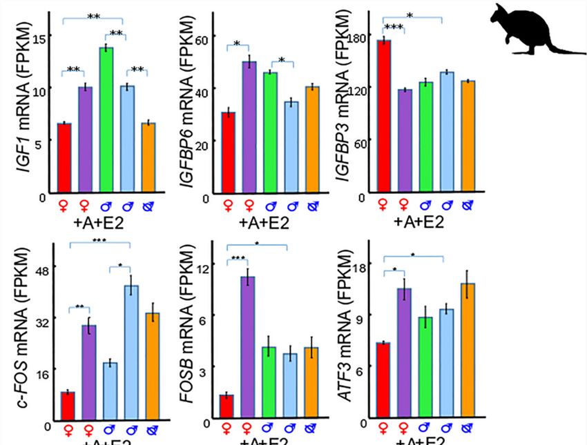
Genes 2020, 11, 106 2 of 14
Genes 2020, 11, 106 2 of 14
Figure
Figure 1. 1.The
Thetimeline
timelineofofprostate
prostate differentiation,
differentiation, phallus
phallus development,
development,the theandrogen
androgenimprinting
imprinting
window, the androgen sensitive phase, and the insulin-like growth factor 1 (IGF1)
window, the androgen sensitive phase, and the insulin-like growth factor 1 (IGF1) dependentdependent phase
phase in
in the tammar
the tammar wallaby.wallaby.
TheThe developmentofofmale
development maletammar
tammar phallus
phallus isis androgen
androgen dependent
dependent[5,6],[5,6],like
likethat
thatofof
eutherian
eutherian
mammals. In males, there is an increase of testicular testosterone from birth
mammals. In males, there is an increase of testicular testosterone from birth to day 40 pp [7]. However, to day 40 pp [7].
However,
testicular testicular testosterone
testosterone concentration concentration
falls sharplyfallsafter
sharply
dayafter daybut
40 pp, 40 pp, but is unmeasurable
is unmeasurable in
in ovaries,
ovaries, and plasma levels do not differ between the sexes up to day 50 [3,5,7,8]. There is a critical
and plasma levels do not differ between the sexes up to day 50 [3,5,7,8]. There is a critical androgen
androgen imprinting window (window of androgen sensitivity or androgen programming window)
imprinting window (window of androgen sensitivity or androgen programming window) between
between days 25 to 30 pp, first described in the tammar in 2004 and then identified in rats and humans
days 25 to 30 pp, first described in the tammar in 2004 and then identified in rats and humans [5,9–12].
[5,9–12]. Altering androgen concentrations by castration of male young or treatment of female young
Altering androgen concentrations by castration of male young or treatment of female young with the
with the potent androgen androstanediol during this programming window contributes to abnormal
potent androgen androstanediol during this programming window contributes to abnormal phallus
phallus development, including hypospadias or phallus sex reversal [5,6]. Interestingly, although
development,
androgen controls includingbothhypospadias
urethral closureor phallus sex reversal
and phallus [5,6].the
elongation, Interestingly, although androgen
molecular regulation behind
controls both urethral closure and phallus elongation, the molecular
these two phases can differ. When males are castrated at day 25 pp, their phalluses are regulation behind
feminizedthese
andtwo
phases can differ.
the urethra remainsWhen males[5].
unfused areWhen
castrated
malesat are
daycastrated
25 pp, their
at dayphalluses
40 pp orare at feminized
day 80 pp,and theirthe
urethra remains unfused [5]. When males are castrated at day 40 pp or at day
phalluses become shorter but the treatment has no effect on urethral closure [5]. These results indicate80 pp, their phalluses
become shorterclosure
that urethral but theistreatment
regulatedhas no effect
by the on urethral
androgen priming,closure
whereas [5].phallus
Theseelongation
results indicate
requiresthat
urethral
constantclosure is regulated
or increasing levels byof
the androgenAlthough
androgen. priming,several
whereas phallus
phallus elongation
regulating genesrequires
in theconstant
tammar or
increasing
have been levels of androgen.
reported, there has Although
been lessseveral phallus
attention on regulating
the signallinggenes in the tammar
pathways during have been
phallus
development.
reported, there has been less attention on the signalling pathways during phallus development.
In In
thisthis paper,
paper, wewereview
reviewthe themolecular
molecular regulation
regulation of of androgen
androgenpriming
primingononsonic sonichedgehog
hedgehog (SHH),
(SHH),
insulin-like
insulin-like growth
growth factor1 1(IGF1)
factor (IGF1)and
andlong
long non-coding
non-coding RNA RNA(lncRNAs),
(lncRNAs),during
duringphallus
phallus differentiation
differentiation
in in
thethe tammar.First,
tammar. First,we
we present
present the
the molecular
molecularmechanism
mechanism that initiates
that the the
initiates phallus
phallusdifferentiation at
differentiation
day 50 pp and, then, the regulatory mechanism of phallus elongation
at day 50 pp and, then, the regulatory mechanism of phallus elongation at day 90 pp. We also at day 90 pp. We also identify
hormonal
identify responsive
hormonal lncRNAs
responsive during
lncRNAs phallus
during development
phallus development in the tammar
in the tammar andand describe
describetheir
their
relationship to their neighboring coding genes.
relationship to their neighboring coding genes.
2. A
2. A Unique
Unique Androgen-SensitiveRegulation
Androgen-Sensitive Regulation Network
Network ofof Sonic
SonicHedgehog
Hedgehog(SHH)(SHH)
In the tammar, SHH expression remains low in males when testicular testosterone is high, but
In the tammar, SHH expression remains low in males when testicular testosterone is high, but
increases after the content of testosterone (ng/mg protein) in the testes drops [7,9]. Similarly, in
increases after the content of testosterone (ng/mg protein) in the testes drops [7,9]. Similarly, in phallus
phallus transcriptome data (Figure 2), SHH expression increases after removing the testes, but
transcriptome data (Figure 2), SHH expression increases after removing the testes, but decreases in
decreases in female phalluses when given androgen [13]. The negative association between SHH
female phalluses when given androgen [13]. The negative association between SHH expression and
expression and androgen is also seen in a lymph node carcinoma of the prostate (LNCaP) cell line
androgen is also seen in a lymph node carcinoma of the prostate (LNCaP) cell line [14]. Through steroid
[14]. Through steroid treatment and RNA-sequencing (RNA-seq) data analysis in the tammar, a
treatment
numberand RNA-sequencing
of genes are shown to(RNA-seq) data expression
have a similar analysis in pattern
the tammar, a number
to that of SHH. of genes
SHH, Wntarefamily
shown
to have
member a similar expression
5A (WNT5A), pattern
and MAF to that
BZIP of SHH.factor
transcription SHH,BWnt family
(MAFB) aremember 5A (WNT5A),
all downregulated in and MAF
female
BZIP transcription factor B
phalluses by androgen treatment at day 50 pp, but are upregulated after castration in males [13,15], at
(MAFB) are all downregulated in female phalluses by androgen treatment
day 50 pp,
while but aregrowth
fibroblast upregulated after
factor 10 castration
(FGF10) in males [13,15],
is upregulated while fibroblast
by androgen treatment, growth factor 10 (FGF10)
but downregulated
is upregulated
after castrationbyin androgen
males [15].treatment, but downregulated after castration in males [15].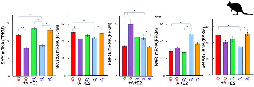
Genes 2020, 11, 106                                                                                                                3 of 14
   Genes 2020, 11, 106                                                                                                          3 of 14

      Figure
         Figure   Gene
               2. 2. Gene expressionofofSHH,
                         expression        SHH,WNT5A,
                                                  WNT5A, MAFB,       FGF10, and
                                                            MAFB, FGF10,            WIF1ininphalluses
                                                                              andWIF1         phallusesatat dayday5050
                                                                                                                     pp.pp.  SHH,
                                                                                                                          SHH,
      WNT5A,
         WNT5A,   and  MAFB
                     and MAFBexpression
                                  expressionisishigher
                                                  higher in
                                                          in female
                                                              female phalluses
                                                                      phalluses andandincreases
                                                                                         increasesininmale
                                                                                                       malephalluses
                                                                                                                 phalluses    after
                                                                                                                           after
      castration.
         castration. SHH
                      SHHisisupregulated
                               upregulatedby   byoestrogen
                                                  oestrogen treatment
                                                              treatment inin males
                                                                             malesbut butdownregulated
                                                                                           downregulated     inin  females
                                                                                                                 females      after
                                                                                                                           after
      adiol
         adiol  treatment.WNT5A
             treatment.    WNT5A is   is downregulated
                                         downregulated ininfemales
                                                                females   after
                                                                       after     adiol
                                                                             adiol      treatment.
                                                                                    treatment.  BothBoth
                                                                                                      FGF10   FGF10   and are
                                                                                                                 and WIF1    WIF1
      arehigher
           higherininmale
                       malephalluses
                              phallusesatatday day5050pp.
                                                       pp.FGF10
                                                             FGF10is isupregulated
                                                                         upregulated   bybyadiol treatment
                                                                                             adiol  treatment    and  WIF1
                                                                                                                    and   WIF1 is is
         downregulatedbybyoestrogen
      downregulated             oestrogentreatment.
                                            treatment. A:
                                                        A: adiol,
                                                            adiol, E: oestrogen,       p-value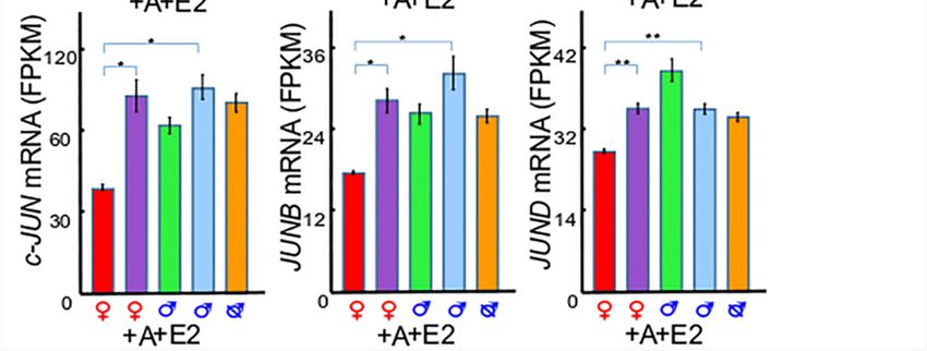
Genes 2020, 11, 106                                                                                    4 of 14

goes down, FGF10 increases, presumably to maintain phallus elongation at later stages, as seen in
mice [27–29].

2.4. The SHH Switch
      Sonic Hedgehog is negatively regulated by androgen in the tammar, which is unusual as compared
with eutherian mammals. SHH levels transiently increase when testicular testosterone drops at around
day 40 pp [9]. After day 50 pp, there is no significant difference in plasma testosterone, plasma
dihydrotestosterone, and adrenal testosterone between males and females up until day 150 pp [7,8].
However, there are increased levels of the potent androgen androstanediol [5,30] which appears to be
critical to maintain phallus elongation and urethral closure after day 50 pp in the tammar.
      Sexually dimorphic structures differentiate post-natally in marsupials and over a long time period.
Prostate differentiation in the tammar begins at day 25 pp in males [31], while the phallus does
not become sexually dimorphic until day 50 to 60 pp. This is in marked contrast to humans, mice
and rats in which phallus differentiation begins synchronously with prostatic, ductal, and testicular
androgen production. During pregnancy in humans, the prostate and penis differentiate at about
10 weeks [32–36], at 16.5 to 17.5 days in mice [37–41], and at 17 to 19 days of gestation in rats [42,43]. The
unique SHH increase might be a regulatory mechanism to constrain the onset of phallus dimorphism
up to day 50 to 60 pp in the tammar and switch it on in the males at this time.
      SHH and IGF signalling have a synergistic relationship to induce proliferation in multiple tissues
in mice [44–46]. In addition, SHH-induced proliferation is inhibited by the anti-IGFR1 blocking
antibody, cixutumumab (IMC-A12) [44]. IGF2 binds to the IGFR1 [47], and since hepatic IGF2 in the
tammar is highest in males from day 50 to 70 pp [16], it may have a similar relationship with SHH
signalling at days 50 to 60 pp in tammar phalluses to regulate SHH-induced proliferation.

3. Insulin-Like Growth Factor 1 (IGF1) in Phallus Growth and Urethral Closure
     Laron syndrome (OMIM ID #262500), also known as growth hormone (GH) insensitivity syndrome,
affects phallus growth and leads to micro-penis [48]. Without GH, IGF1 is not secreted at sufficiently high
levels, so IGF1 treatment in human patients can reverse the micro-penis seen in Laron syndrome [49,50].
The lifespan of IGF1 and its pathway activity is affected by the insulin-like growth factor binding
proteins (IGFBPs) [51–53]. However, the interplay between IGF1 and IGFBPs in phallus development
has not been thoroughly investigated in eutherian mammals. Here, we review the role of the IGF
network by using RNA-seq analysis and co-expression analysis in phalluses with a tammar as a model.

3.1. IGF1 and Insulin-Like Growth Factor Binding Protein 6 (IGFBP6)
     Both IGF1 and IGFBP6 are upregulated by androgen and oestrogen treatment in tammar
phalluses [15] (Figure 3). Such androgenic and oestrogenic dependency of IGF1 is also seen in
eutherian mammals. For instance, testosterone increases IGF1 in bovine muscle satellite cells [54],
rat uterine tissue [55], and human prostate cancer cell lines [56,57]. Oestrogen also increases IGF1
expression in the primate cerebral cortex [58] and in the mouse uterus [59]. Similarly, IGFBP6 decreases
in rat epididymides after blocking dihydrotestosterone (DHT) synthesis [60] and is upregulated in
prostate cancer cells after treatment with diethylstilbestrol (DES), a synthetic oestrogen [61]. However,
the detailed mechanisms remain unknown.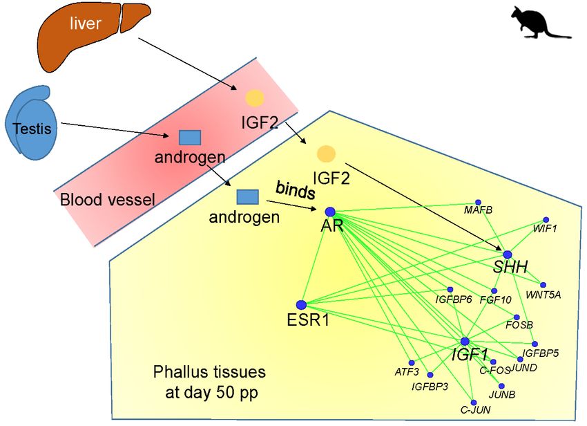
Genes 2020, 11, 106                                                                                                        5 of 14

      Genes 2020, 11, 106                                                                                            5 of 14

      Figure Figure  3. Gene
               3. Gene       expression
                         expression  of of IGF1,
                                        IGF1,     IGFBP3,IGFBP6,
                                                IGFBP3,   IGFBP6, and
                                                                   andActivator
                                                                        ActivatorProtein
                                                                                    Protein1 (AP-1) genes
                                                                                              1 (AP-1)     in phalluses
                                                                                                        genes   in phalluses
             at day 50 pp. Both IGF1 and IGFBP6 are upregulated by adiol and oestrogen treatment. IGF1 is higher
      at day 50 pp. Both IGF1 and IGFBP6 are upregulated by adiol and oestrogen treatment. IGF1 is higher
             in normal male phalluses at day 50 pp and is downregulated in males after castration. IGFBP3 is
      in normal male phalluses at day 50 pp and is downregulated in males after castration. IGFBP3 is higher
             higher in female phalluses at day 50 pp and is downregulated in females after adiol treatment. All six
      in female phalluses at day 50 pp and is downregulated in females after adiol treatment. All six AP-1
             AP-1 genes (C-FOS, FOSB, ATF3, c-JUN, JUNB, and JUND) are higher in male phalluses and
              (C-FOS, FOSB,
      genesupregulated          ATF3, c-JUN, JUNB, and JUND) are higher in male phalluses and upregulated
                           by adiol treatment. C-FOS is downregulated by oestrogen treatment. A: adiol, E:
      by adiol   treatment.   C-FOS   is downregulated
             oestrogen, FPKM: Fragments                     by oestrogen
                                            per kilobase million; *: p-valuetreatment.    A: adiol,
                                                                             < 0.05, **: p-value     E: oestrogen,
                                                                                                 < 0.005, ***: p-valueFPKM:
                                                                                                                       <
      Fragments      per kilobase
             0.001. Figure          from [15].*: p-value < 0.05, **: p-value < 0.005, ***: p-value < 0.001. Figure
                           redrawnmillion;
      redrawn from [15].
       3.2. IGF1 and Insulin-Like Growth Factor Binding Protein 3 (IGFBP3)
3.2. IGF1 and Insulin-Like Growth Factor Binding Protein 3 (IGFBP3)
            In contrast to IGF1, IGFBP3 expression is higher in female phalluses than male phalluses at day
      In
       50 contrast
           pp, day 90    IGF1,
                     to pp,  andIGFBP3    expression
                                  at day 150   pp, and is downregulated
                                                             higher in female     phalluses
                                                                              in female       than after
                                                                                         phalluses  maleandrogen
                                                                                                           phalluses at
day 50 treatment
          pp, day[15]
                    90 (Figure
                        pp, and3).atAday
                                      similar
                                          150response
                                               pp, andisisalso   found in eutherian
                                                              downregulated          mammals
                                                                                in female       in whichafter
                                                                                            phalluses     IGFBP3 is
                                                                                                              androgen
       downregulated
treatment    [15] (Figurein prostate cancerresponse
                            3). A similar    cells afteristreatment of androgen
                                                            also found            [57,62,63]
                                                                        in eutherian         or synthetic
                                                                                       mammals            androgen
                                                                                                   in which   IGFBP3 is
       [63]. The opposing
downregulated                 expression
                    in prostate           of IGF1
                                 cancer cells afterand     IGFBP3ofsuggests
                                                     treatment      androgen that  IGFBP3 may
                                                                                [57,62,63]       be the agent
                                                                                           or synthetic        that [63].
                                                                                                          androgen
       inhibits female phallus development by negative regulation of cell proliferation, as seen in many
The opposing expression of IGF1 and IGFBP3 suggests that IGFBP3 may be the agent that inhibits
       other studies [57,64–68]. Thus, IGF1 may be responsible for maintaining normal male phallus growth
female phallus development by negative regulation of cell proliferation, as seen in many other
       at later stages.
studies [57,64–68]. Thus, IGF1 may be responsible for maintaining normal male phallus growth at
later stages.
       3.3. IGF1 and Activator Protein 1 (AP-1)
          The transcription
3.3. IGF1 and               of IGF1
              Activator Protein     is regulated by the DNA binding of Activator Protein 1 (AP-1) complex
                                 1 (AP-1)
      [69]. Interestingly, both IGF1 and AP-1 genes are higher in males and increase in female phalluses
     The
     after transcription of IGF1
           androgen treatment      is tammar
                              in the   regulated
                                              [15] by  the 3).
                                                   (Figure DNA     binding
                                                               Similar       of Activator
                                                                       androgen sensitivity isProtein  1 in
                                                                                               also seen (AP-1)
complex [69]. Interestingly, both IGF1 and AP-1 genes are higher in males and increase in female
phalluses after androgen treatment in the tammar [15] (Figure 3). Similar androgen sensitivity is also
seen in other studies. For example, Fos proto-oncogene, AP-1 transcription factor subunit (c-Fos), and
Genes 2020, 11, 106 6 of 14
Genes 2020, 11, 106 6 of 14
activating transcription factor 3 (ATF3) are induced by androgen in the rat hippocampus [70] and in
humanother studies.
prostate For cells
cancer example, Fos proto-oncogene,
[71], respectively. AP-1 transcription
Since androgen treatmentfactor subunit
induces (c-Fos),
phallus and [5]
elongation
activating transcription factor 3 (ATF3) are induced by androgen in the rat hippocampus [70] and
and urethral closure [13] in the tammar, it is likely that the AP-1 genes, under the regulation of androgen in
human prostate cancer cells [71], respectively. Since androgen treatment induces
control cell proliferation in phalluses, as it does in other cells (reviewed in [72]). phallus elongation
[5] and urethral closure [13] in the tammar, it is likely that the AP-1 genes, under the regulation of
androgen
3.4. IGF1 control cell
and Urethral proliferation in phalluses, as it does in other cells (reviewed in [72]).
Closure
Several
3.4. IGF1 hypotheses are proposed to explain the mechanism of urethral closure. One of the
and Urethral Closure
hypotheses is that the proliferation of cells in urorectal septum (URS) contributes to the urethral
Several hypotheses are proposed to explain the mechanism of urethral closure. One of the
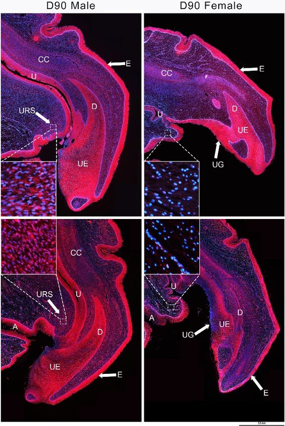
closure [73–75]. is
hypotheses Interestingly, IGF1 is localized
that the proliferation of cells ininurorectal
the mesenchyme
septum (URS)of the URS onlytointhe
contributes male phalluses
urethral
at dayclosure
90 pp,[73–75].
but is absent in that of female phalluses [15] (Figure 4). Proliferating cell
Interestingly, IGF1 is localized in the mesenchyme of the URS only in male phalluses nuclear antigen
(PCNA), a marker for cell proliferation, shows similar localization [15] (Figure 4). The
at day 90 pp, but is absent in that of female phalluses [15] (Figure 4). Proliferating cell nuclear antigen importance
of IGF1 at thisa marker
(PCNA), time is forfurther supported shows
cell proliferation, by an similar
earlierlocalization
study in the [15]tammar that
(Figure 4). shows
The that hepatic
importance of
IGF1 atofthis
expression IGF2 time is further supported
is significantly higher inbymales
an earlier
thanstudy in the
females tammar
at day thatabout
70 pp, showsthethat
timehepatic
that male
expression
and female of IGF2 become
phalluses is significantly higher
sexually in males than
dimorphic [16].females
WhileatIGF2
day 70 pp, aboutfrom
decreases the time
daythat
70 male
and is no
longer sexually dimorphic by day 100, hepatic and plasma levels of IGF1 significantly increase no
and female phalluses become sexually dimorphic [16]. While IGF2 decreases from day 70 and is in both
longer sexually dimorphic by day 100, hepatic and plasma levels of IGF1 significantly increase in
sexes from day 90 pp to day 250 of pouch life [16]. These data suggest for the first time that urethral
both sexes from day 90 pp to day 250 of pouch life [16]. These data suggest for the first time that
closure may involve IGF1-mediated cell proliferation specifically in male URS.
urethral closure may involve IGF1-mediated cell proliferation specifically in male URS.
Figure
Figure 4. IGF1
4. IGF1 andand proliferating
proliferating cellnuclear
cell nuclear antigen
antigen (PCNA)
(PCNA)distribution in phalluses
distribution at day
in phalluses at 90
daypp.90 pp.
In both male and female phalluses, IGF1 (top) and PCNA (bottom) are expressed in epithelial cells
In both male and female phalluses, IGF1 (top) and PCNA (bottom) are expressed in epithelial cells
and in the corpora cavernosa. However, IGF1 and nuclear PCNA are found only in the URS of male
and in the corpora cavernosa. However, IGF1 and nuclear PCNA are found only in the URS of male
phalluses (see insets). CC: corpus cavernosum, D: diverticulum, E: epithelium, U: urethra, UE:
phalluses (see insets). CC: corpus cavernosum, D: diverticulum, E: epithelium, U: urethra, UE: urethral
urethral epithelium, UG: urethra groove, URS: urorectal septum, red staining: IGF1 (top) and PCNA
epithelium, UG:
(bottom), andurethra groove,DAPI
blue staining: URS:(4′,6-diamidino-2-fenilindol).
urorectal septum, red staining: IGF1
Scale bar, 1.0(top) and PCNA
mm. Figures from (bottom),
[15].
and blue staining: DAPI (40 ,6-diamidino-2-fenilindol). Scale bar, 1.0 mm. Figures from [15].Genes 2020, 11, 106 7 of 14
3.5. IGF1 Dependent Phallus Growth
A study conducted by Leihy et al., 2004 demonstrated for the first time an androgen sensitive
phase during phallus elongation between days 20 and 40 pp in the tammar [5]. Removing testes
in males before day 120 pp reduced phallus length while applying androgen treatment in females
before day 120 pp enhances phallus elongation, but has no effect on urethral closure [5]. However, as
mentioned before, there is no significant difference in plasma testosterone between male and female
at least up to day 50 pp [7,8]. Thus, there must be another regulatory network that is activated by
the earlier androgen window of sensitivity to maintain the phallus elongation after day 50 pp. SHH
appears to be the key switch that initiates the expression of potential regulatory genes. These may
include IGF1, IGFBP3, FGF10, fibroblast growth factor receptor 2 (FGFR2IIIb), Eph-related receptor tyrosine
kinase ligand 5 (EFNB2), MAFB, and distal-less homeobox 5 (DLX5). The balance between IGF1 and
IGFBP3 could be important in regulating phallus elongation and urethral closure. FGF10, FGFR2IIIb,
EFNB2, MAFB, and DLX5 may also involve phallus elongation, since they are significantly higher in
male phalluses at day 90 pp [15] and have a conserved localization in urethral epithelium, as seen in
mice [28,37,38,76–78]. In addition, these genes appear to be important to maintain cell proliferation
and survival [79–85] during male phallus development in mice [23,29,86,87].
4. Co-Expression Network and Hormonally Responsive Long Non-Coding RNAs
Our RNA-seq dataset consists of five different treatment groups with 5 replicates for each group,
which makes it hard to interpret with differential expression (DE) analysis. We used weighted genome
co-expression network analysis (WGCNA) to find co-expressed genes. It is also a good way to identify
lncRNAs as most of them have extremely low sequence conservation, making them difficult to identify
cross species with alignment. In our previous paper, we set up a pipeline by combining WGCNA, DE
analysis, and the location of lncRNAs and identified the following three coding gene-neighboring
lncRNAs: lnc-RSPO4, lnc-BMP5, and lnc-ZBTB16 [88].
4.1. IGF1, Androgen Receptor (AR), and ESR1 Co-Expression Network
IGF1 is considered as a hub gene in its co-expression network due to its high correlation with a large
number of protein-coding genes and lncRNAs. Within the IGF1 co-expression network, both IGFBP5,
an IGF signalling regulator (reviewed in [89]) that inhibits SHH-induced proliferation in cerebellar
granule cells in mice [44], and FGF10, a phallus regulating gene in mice and the tammar [15,28,29,77,78],
have a high correlation (R ≥ 0.8) with IGF1 [88] (Figure 5). IGF1 is also co-expressed with multiple genes
that may have a role in regulating reproductive development (Figure 5). For instance, it is co-expressed
with other IGF family members, including insulin like growth factor 2 binding protein (IGF2BP) 1–3,
insulin like 5 (INSL5), and IGFBP7. Apart from FGF10, IGF1 is also associated with FGF11, FGF13, and
tyrosine-protein kinase receptor EPH-2 (EPHB1). Two receptors, frizzled class receptor 4 (FZD4) and FZD9,
in the WNT signalling pathway show high association with IGF1. Interestingly, IGF1 is co-expressed
with zinc finger and BTB domain containing 20 (ZBTB20), whose mutation causes micro-penis [90]. We
also find mutations of several kinesin family members (KIF), such as KIF1A, KIF1B and KIF7, that are
associated with IGF1 and can also induce an abnormal phallus phenotype in human [91,92]. These
data further confirm the importance of IGF1 in regulating phallus development in the tammar.Genes 2020, 11, 106 8 of 14
Genes 2020, 11, 106 8 of 14
Figure 5. IGF1, androgen receptor (AR), and ESR1 co-expression network. IGF1 co-expressed coding
Figure 5. IGF1, androgen receptor (AR), and ESR1 co-expression network. IGF1 co-expressed coding
genes (selected based on correlation and reference review) and predicted co-regulatory long non-coding
genes (selected based on correlation and reference review) and predicted co-regulatory long non-
RNAs (R > 0.9). Figure redrawn from [88].
coding RNAs (R > 0.9). Figure redrawn from [88].
In mice, oestrogen signalling clearly has a regulatory role in phallus development [93,94], as we
In mice, oestrogen signalling clearly has a regulatory role in phallus development [93,94], as we
have found in the tammar [13,15,88]. In the tammar co-expression network, about 50% of estrogen
have found in the tammar [13,15,88]. In the tammar co-expression network, about 50% of estrogen
receptor 1 (ESR1) co-expressed coding genes and lncRNAs are also associated with AR [88], suggesting
receptor 1 (ESR1) co-expressed coding genes and lncRNAs are also associated with AR [88],
an interaction between androgen receptor (AR) signalling and ESR1 signalling during tammar phallus
suggesting an interaction between androgen receptor (AR) signalling and ESR1 signalling during
development [88]. However, those lncRNAs could have other genetic targets because none of them
tammar phallus development [88]. However, those lncRNAs could have other genetic targets because
were located within 100 kb upstream or downstream of IGF1, AR, and ESR1.
none of them were located within 100 kb upstream or downstream of IGF1, AR, and ESR1.
4.2. lnc-RSPO4, lnc-BMP5, and lnc-ZBTB16
4.2. lnc-RSPO4, lnc-BMP5, and lnc-ZBTB16
We identified three novel lncRNAs using our pipeline. Lnc-RSPO4 is co-expressed with roof
We identified
plate-specific three(RSPO4)
spondin-4 novel lncRNAs using Both
coding gene. our pipeline.
RSPO4 and Lnc-RSPO4
lnc-RSPO4 is co-expressed
are downregulatedwith roof
in
plate-specific spondin-4 (RSPO4) coding gene. Both RSPO4 and lnc-RSPO4
tammar female phalluses after androgen treatment [88], in a similar expression pattern to that of SHH are downregulated in
tammar
and WNT5Afemale
[13].phalluses after androgen
Interestingly, RSPO4 is atreatment
ligand of[88], in a similar
leucine-rich expression
repeat containing pattern to that of SHH
G protein-coupled
and WNT5A [13]. Interestingly, RSPO4 is a ligand of leucine-rich repeat
receptor (LGR) 4–6 receptors that potentiate WNT signalling [95–98]. Thus, RSPO4 and lnc-RSPO4containing G protein-coupled
receptor
could also(LGR) 4–6 receptors
be involved that potentiate
in the molecular WNTmediated
regulation signalling by[95–98].
SHH and Thus,
WNT5A RSPO4 and lnc-RSPO4
signalling during
could also be involved
tammar phallus development. in the molecular regulation mediated by SHH and WNT5A signalling during
tammar phallus development.
Both lnc-BMP5 and lnc-ZBTB16 are downregulated in tammar male phalluses after oestrogen
Both [88].
treatment lnc-BMP5 andalso
They are lnc-ZBTB16 are downregulated
co-expressed in tammar
with bone morphogenetic male(BMP5)
protein phalluses
andafter oestrogen
zinc finger and
treatment [88]. They are also co-expressed with bone morphogenetic protein
BTB domain containing 16 (ZBTB16), respectively, in our co-expression network [88]. Interestingly, (BMP5) and zinc finger and
BTB domain containing 16 (ZBTB16), respectively, in our co-expression
Bmp5 is downregulated by flutamide, an androgen signalling inhibitor during phallus development network [88]. Interestingly,
Bmp5
in miceis[93].
downregulated
Mutation ofbyZBTB16 flutamide, an androgen
induces signalling
micro-penis [99,100],inhibitor
which during
is similarphallus
to thedevelopment
phenotype
observed after oestrogen treatment in the tammar [13]. These data show that there is aphenotype
in mice [93]. Mutation of ZBTB16 induces micro-penis [99,100], which is similar to the complex
observed after
regulatory system oestrogen
of lncRNAs treatment
duringin the tammar
phallus [13]. These
development mediateddata by
show that there
hormonal is a complex
signalling.
regulatory system of lncRNAs during phallus development mediated by hormonal signalling.
5. Conclusions and Future Directions
5. Conclusions and Future Directions
Tammar phallus development is under the regulation of a complex molecular network mediated
Tammarhormones.
by endocrine phallus development
This reviewisdescribes
under thetwo regulation of a complex molecular
endocrine-mediated networks, network
the SHH mediated
network
and the IGF1 network, which may act as molecular switches to constrain and decide male network
by endocrine hormones. This review describes two endocrine-mediated networks, the SHH phallus
and the IGF1(Figure
development network,6). which may actanalysis
The RNA-seq as molecular
identifiesswitches
two sets to of
constrain and decide
genes, including male phallus
WNT5A, MAFB,
development
RSPO4, (Figure
lnc-RSPO4, 6). The
FGF10, WIF1RNA-seq analysis
and AP-1, FGF10,identifies
IGFBP3,two sets ofIGFBP5,
IGFBP6, genes, including
EFNB2, that WNT5A, MAFB,
interact with
RSPO4, lnc-RSPO4, FGF10, WIF1 and AP-1, FGF10, IGFBP3, IGFBP6, IGFBP5, EFNB2, that interact
with SHH and IGF1, respectively, in the tammar phalluses at day 50 pp. Interestingly, due to theGenes 2020, 11, 106 9 of 14
Genes 2020, 11, 106 9 of 14
SHH and IGF1, respectively, in the tammar phalluses at day 50 pp. Interestingly, due to the negative
association between androgen
negative association and SHH transcription,
between androgen an SHH switch
and SHH transcription, could
an SHH be a could
switch uniqueberegulatory
a unique
mechanism in the tammar
regulatory mechanism to constrain
in the tammar tothe timing of
constrain phallus
the timingdifferentiation.
of phallus differentiation.
Figure Summaryofof
6. Summary
Figure 6. IGF2-SHH
IGF2-SHH and
and androgen-IGF1
androgen-IGF1 signalling
signalling networks.
networks. The activation
The activation of
of IGF2-
IGF2-SHH and androgen-IGF1 signalling networks initiate urethral closure in males, whereas
SHH and androgen-IGF1 signalling networks initiate urethral closure in males, whereas in females, in
females, non-activation of those two signalling networks results in an unfused
non-activation of those two signalling networks results in an unfused urethra. urethra.
The
The molecular
molecular regulatory
regulatory network
network thatthat maintains
maintains phallus
phallus growth
growth after
after day
day 5050 pp
pp consists
consists of
of
another set of genes, including IGF1, IGFBP3, FGF10, FGFR2IIIb, EFNB2,
another set of genes, including IGF1, IGFBP3, FGF10, FGFR2IIIb, EFNB2, MAFB, and DLX5. The MAFB, and DLX5. The
increased
increased level
levelofofthose
thosegenes maymay
genes be initiated and enhanced
be initiated by activation
and enhanced of two endocrine-mediated
by activation of two endocrine-
SHH and IGF1 switches in males, resulting in a phallus with complete
mediated SHH and IGF1 switches in males, resulting in a phallus with complete urethral closure
urethralandclosure
elongated
and
shaft. In addition, it is likely that urethral closure is mediated by the activation of IGF1
elongated shaft. In addition, it is likely that urethral closure is mediated by the activation of IGF1 signalling in
the male urorectal
signalling in the maleseptum.
urorectal septum.
Co-expression analysis to
Co-expression analysis toidentify
identifynovel
novelhormone-responsive
hormone-responsive lncRNAs,
lncRNAs, suchsuch as lnc-BMP5,
as lnc-BMP5, lnc-
lnc-RSPO4, and lnc-ZBTB16, in the tammar phalluses reveals complex regulatory
RSPO4, and lnc-ZBTB16, in the tammar phalluses reveals complex regulatory networks of IGF1, AR, networks of IGF1, AR,
and ESR1 that associate with multiple hormone-responsive coding genes and
and ESR1 that associate with multiple hormone-responsive coding genes and lncRNAs during lncRNAs during tammar
phallus
tammardevelopment. The data also
phallus development. The indicate
data alsoa potential
indicate ainterplay
potentialbetween
interplayARbetween
and ESR1 ARsignalling.
and ESR1
Taken together, the activation of the SHH switch and IGF1 switch, mediated by the balance
signalling.
between
TakenARtogether,
and ESR1 thesignalling,
activationinitiate
of the aSHH
complex
switchregulatory
and IGF1network
switch, in males tobyconstrain
mediated the
the balance
timing of phallus differentiation and to activate the downstream genes that maintain
between AR and ESR1 signalling, initiate a complex regulatory network in males to constrain the urethral closure
and phallus
timing elongation
of phallus at later stage.
differentiation and to activate the downstream genes that maintain urethral closure
and phallus
Author elongation
Contributions: at later stage. draft preparation, Y.C. and M.B.R.; writing—review and editing, Y.C.
Writing—original
and M.B.R. All authors have read and agreed to the published version of the manuscript.
Author Contributions: Writing—original draft preparation, Y.C. and M.B.R.; writing—review and editing, Y.C.
Funding: This study was supported by grants from the National Health and Medical Research Council of Australia.
and M.B.R. All authors have read and agreed to the published version of the manuscript.
Acknowledgments: We thank members of the Wallaby research group (Walgroup) for assistance when handling
Funding:We
animals. This
alsostudy
thankwas supported
members by grants
of RIKEN and NIGfrom
forthe
theNational
technicalHealth
supportand Medical Research Council of
on NGS.
Australia.
Acknowledgments: We thank members of the Wallaby research group (Walgroup) for assistance when handling
animals. We also thank members of RIKEN and NIG for the technical support on NGS.Genes 2020, 11, 106 10 of 14
Conflicts of Interest: The authors declare no conflict of interest. The funders had no role in the design of the
study; in the collection, analyses, or interpretation of data; in the writing of the manuscript, or in the decision to
publish the results.
References
1. Renfree, M.B.; Pask, A.J.; Shaw, G. Sexual development of a model marsupial male. Aust. J. Zool. 2006, 54,
151–158. [CrossRef]
2. Renfree, M.B.; Short, R.V.; Shaw, G. Sexual differentiation of the urogenital system of the fetal and neonatal
tammar wallaby, Macropus eugenii. Anat. Embryol. 1996, 194, 111–134. [CrossRef] [PubMed]
3. Leihy, M.W.; Shaw, G.; Wilson, J.D.; Renfree, M.B. Development of the penile urethra in the tammar wallaby.
Sex. Dev. 2011, 5, 241–249. [CrossRef]
4. Butler, C.M.; Shaw, G.; Renfree, M.B. Development of the penis and clitoris in the tammar wallaby, Macropus
eugenii. Anat. Embryol. 1999, 199, 451–457. [CrossRef]
5. Leihy, M.W.; Shaw, G.; Wilson, J.D.; Renfree, M.B. Penile development is initiated in the tammar wallaby
pouch young during the period when 5α-androstane-3α, 17β-diol is secreted by the testes. Endocrinology
2004, 145, 3346–3352. [CrossRef]
6. Renfree, M.B.; Chew, K.Y.; Shaw, G. Inducing sex reversal of the urogenital system of marsupials. Differentiation
2014, 87, 23–31. [CrossRef]
7. Renfree, M.B.; Wilson, J.D.; Short, R.V.; Shaw, G.; George, F.W. Steroid hormone content of the gonads of the
tammar wallaby during sexual differentiation. Biol. Reprod. 1992, 47, 644–647. [CrossRef]
8. Wilson, J.D.; George, F.W.; Shaw, G.; Renfree, M.B. Virilization of the male pouch young of the tammar
wallaby does not appear to be mediated by plasma testosterone or dihydrotestosterone. Biol. Reprod. 1999,
61, 471–475. [CrossRef] [PubMed]
9. Chew, K.Y.; Pask, A.J.; Hickford, D.; Shaw, G.; Renfree, M.B. A dual role for SHH during phallus development
in a marsupial. Sex. Dev. 2014, 8, 166–177. [CrossRef] [PubMed]
10. Welsh, M.; Saunders, P.T.; Fisken, M.; Scott, H.M.; Hutchison, G.R.; Smith, L.B.; Sharpe, R.M. Identification
in rats of a programming window for reproductive tract masculinization, disruption of which leads to
hypospadias and cryptorchidism. J. Clin. Investig. 2008, 118, 1479–1490. [CrossRef]
11. Welsh, M.; Suzuki, H.; Yamada, G. The masculinization programming window. Endocr. Dev. 2014, 27, 17–27.
[PubMed]
12. Welsh, M.; MacLeod, D.J.; Walker, M.; Smith, L.B.; Sharpe, R.M. Critical androgen-sensitive periods of rat
penis and clitoris development. Int. J. Androl. 2010, 33, e144–e152. [CrossRef] [PubMed]
13. Chen, Y.; Yu, H.; Pask, A.J.; Fujiyama, A.; Suzuki, Y.; Sugano, S.; Shaw, G.; Renfree, M.B. Hormone-responsive
genes in the SHH and WNT/β-catenin signaling pathways influence urethral closure and phallus growth.
Biol. Reprod. 2018, 99, 806–816. [CrossRef] [PubMed]
14. Sirab, N.; Terry, S.; Giton, F.; Caradec, J.; Chimingqi, M.; Moutereau, S.; Vacherot, F.; Taille, A.D.L.;
Kouyoumdjian, J.C.; Loric, S. Androgens regulate hedgehog signalling and proliferation in
androgen-dependent prostate cells. Int. J. Cancer 2012, 131, 1297–1306. [CrossRef] [PubMed]
15. Chen, Y.; Yu, H.; Pask, A.J.; Fujiyama, A.; Suzuki, Y.; Sugano, S.; Shaw, G.; Renfree, M.B. Effects of androgen
and oestrogen on the IGF pathways controlling phallus growth. Reproduction 2019, 157, 1–12. [CrossRef]
[PubMed]
16. Menzies, B.R.; Shaw, G.; Fletcher, T.P.; Pask, A.J.; Renfree, M.B. Maturation of the growth axis in marsupials
occurs gradually during post-natal life and over an equivalent developmental stage relative to eutherian
species. Mol. Cell. Endocrinol. 2012, 349, 189–194. [CrossRef]
17. Miyagawa, S.; Satoh, Y.; Haraguchi, R.; Suzuki, K.; Iguchi, T.; Taketo, M.M.; Nakagata, N.; Matsumoto, T.;
Takeyama, K.-I.; Kato, S. Genetic interactions of the androgen and Wnt/β-catenin pathways for the
masculinization of external genitalia. Mol. Endocrinol. 2009, 23, 871–880. [CrossRef]
18. Miyagawa, S.; Moon, A.; Haraguchi, R.; Inoue, C.; Harada, M.; Nakahara, C.; Suzuki, K.; Matsumaru, D.;
Kaneko, T.; Matsuo, I. Dosage-dependent hedgehog signals integrated with Wnt/β-catenin signaling regulate
external genitalia formation as an appendicular program. Development 2009, 136, 3969–3978. [CrossRef]
19. Seifert, A.W.; Zheng, Z.; Ormerod, B.K.; Cohn, M.J. Sonic hedgehog controls growth of external genitalia by
regulating cell cycle kinetics. Nat. Commun. 2010, 1, 23. [CrossRef]Genes 2020, 11, 106 11 of 14
20. Kawano, Y.; Kypta, R. Secreted antagonists of the Wnt signalling pathway. J. Cell Sci. 2003, 116, 2627–2634.
[CrossRef]
21. Hsieh, J.-C.; Kodjabachian, L.; Rebbert, M.L.; Rattner, A.; Smallwood, P.M.; Samos, C.H.; Nusse, R.; Dawid, I.B.;
Nathans, J. A new secreted protein that binds to Wnt proteins and inhibits their activites. Nature 1999, 398,
431–436. [CrossRef] [PubMed]
22. Ng, R.C.; Matsumaru, D.; Ho, A.S.; Garcia-Barceló, M.; Yuan, Z.; Smith, D.; Kodjabachian, L.; Tam, P.K.;
Yamada, G.; Lui, V.C. Dysregulation of Wnt inhibitory factor 1 (Wif1) expression resulted in aberrant
wnt-β-catenin signaling and cell death of the cloaca endoderm, and anorectal malformations. Cell Death Differ.
2014, 21, 978–989. [CrossRef] [PubMed]
23. Suzuki, K.; Numata, T.; Suzuki, H.; Raga, D.D.; Ipulan, L.A.; Yokoyama, C.; Matsushita, S.; Hamada, M.;
Nakagata, N.; Nishinakamura, R.; et al. Sexually dimorphic expression of Mafb regulates masculinization of
the embryonic urethral formation. Proc. Natl. Acad. Sci. USA 2014, 111, 16407–16412. [CrossRef] [PubMed]
24. Matsushita, S.; Suzuki, K.; Ogino, Y.; Hino, S.; Sato, T.; Suyama, M.; Matsumoto, T.; Omori, A.; Inoue, S.;
Yamada, G. Androgen regulates Mafb expression through its 3’ UTR during mouse urethral masculinization.
Endocrinology 2015, 157, 844–857. [CrossRef]
25. Matsushita, S.; Suzuki, K.; Murashima, A.; Kajioka, D.; Acebedo, A.R.; Miyagawa, S.; Haraguchi, R.;
Ogino, Y.; Yamada, G. Regulation of masculinization: Androgen signalling for external genitalia development.
Nat. Rev. Urol. 2018, 15, 358. [CrossRef]
26. Bellusci, S.; Grindley, J.; Emoto, H.; Itoh, N.; Hogan, B. Fibroblast growth factor 10 (FGF10) and branching
morphogenesis in the embryonic mouse lung. Development 1997, 124, 4867–4878.
27. Haraguchi, R.; Suzuki, K.; Murakami, R.; Sakai, M.; Kamikawa, M.; Kengaku, M.; Sekine, K.; Kawano, H.;
Kato, S.; Ueno, N. Molecular analysis of external genitalia formation: The role of fibroblast growth factor
(FGF) genes during genital tubercle formation. Development 2000, 127, 2471–2479.
28. Petiot, A.; Perriton, C.L.; Dickson, C.; Cohn, M.J. Development of the mammalian urethra is controlled by
Fgfr2-IIIb. Development 2005, 132, 2441–2450. [CrossRef]
29. Satoh, Y.; Haraguchi, R.; Wright, T.J.; Mansour, S.L.; Partanen, J.; Hajihosseini, M.K.; Eswarakumar, V.P.;
Lonai, P.; Yamada, G. Regulation of external genitalia development by concerted actions of FGF ligands and
FGF receptors. Anat. Embryol. 2004, 208, 479–486. [CrossRef]
30. Shaw, G.; Renfree, M.B.; Leihy, M.W.; Shackleton, C.H.; Roitman, E.; Wilson, J.D. Prostate formation in a
marsupial is mediated by the testicular androgen 5α-androstane-3α, 17β-diol. Proc. Natl. Acad. Sci. USA
2000, 97, 12256–12259. [CrossRef]
31. Shaw, G.; Renfree, M.B.; Short, R.V. Experimental manipulation of sexual differentiation in wallaby pouch
young treated with exogenous steroids. Development 1988, 104, 689–701. [PubMed]
32. Kurzrock, E.A.; Baskin, L.S.; Cunha, G.R. Ontogeny of the male urethra: Theory of endodermal differentiation.
Differentiation 1999, 64, 115–122. [CrossRef] [PubMed]
33. Baskin, L.; Lee, Y.; Cunha, G. Neuroanatomical ontogeny of the human fetal penis. Br. J. Urol. 1997, 79,
628–640. [CrossRef]
34. Baskin, L.; Shen, J.; Sinclair, A.; Cao, M.; Liu, X.; Liu, G.; Isaacson, D.; Overland, M.; Li, Y.; Cunha, G.R.
Development of the human penis and clitoris. Differentiation 2018, 103, 74–85. [CrossRef] [PubMed]
35. Lowsley, O.S. The development of the human prostate gland with reference to the development of other
structures at the neck of the urinary bladder. Am. J. Anat. 1912, 13, 299–349. [CrossRef]
36. Kellokumpu-Lehtinen, P.; Santti, R.; Pelliniemi, L. Correlation of early cytodifferentiation of the human fetal
prostate and leydig cells. Anat. Rec. 1980, 196, 263–273. [CrossRef]
37. Perriton, C.L.; Powles, N.; Chiang, C.; Maconochie, M.K.; Cohn, M.J. Sonic hedgehog signaling from the
urethral epithelium controls external genital development. Dev. Biol. 2002, 247, 26–46. [CrossRef]
38. Haraguchi, R.; Mo, R.; Hui, C.; Motoyama, J.; Makino, S.; Shiroishi, T.; Gaffield, W.; Yamada, G. Unique
functions of sonic hedgehog signaling during external genitalia development. Development 2001, 128,
4241–4250.
39. Liu, G.; Liu, X.; Shen, J.; Sinclair, A.; Baskin, L.; Cunha, G.R. Contrasting mechanisms of penile urethral
formation in mouse and human. Differentiation 2018, 101, 46–64. [CrossRef]
40. Cunha, G.R.; Donjacour, A.A.; Cooke, P.S.; Mee, H.; Bigsby, R.M.; Higgins, S.J.; Sugimura, Y. The endocrinology
and developmental biology of the prostate. Endocr. Rev. 1987, 8, 338–362. [CrossRef]Genes 2020, 11, 106 12 of 14
41. Timms, B.G.; Mohs, T.J.; Didio, L.J. Ductal budding and branching patterns in the developing prostate. J. Urol.
1994, 151, 1427–1432. [CrossRef]
42. Price, D. Normal development of the prostate and seminal vesicles of the rat with a study of experimental
postnatal modifications. Am. J. Anat. 1936, 60, 79–127. [CrossRef]
43. Inomata, T.; Eguchi, Y.; Nakamura, T. Development of the external genitalia in rat fetuses. Exp. Anim. 1985,
34, 439–444. [CrossRef]
44. Fernandez, C.; Tatard, V.M.; Bertrand, N.; Dahmane, N. Differential modulation of sonic-hedgehog-induced
cerebellar granule cell precursor proliferation by the IGF signaling network. Dev. Neurosci. 2010, 32, 59–70.
[CrossRef]
45. Rao, G.; Pedone, C.A.; del Valle, L.; Reiss, K.; Holland, E.C.; Fults, D.W. Sonic hedgehog and insulin-like
growth factor signaling synergize to induce medulloblastoma formation from nestin-expressing neural
progenitors in mice. Oncogene 2004, 23, 6156. [CrossRef] [PubMed]
46. Pirskanen, A.; Kiefer, J.C.; Hauschka, S.D. IGFs, insulin, Shh, bFGF, and TFG-β1 interact synergistically to
promote somite myogenesis in vitro. Dev. Biol. 2000, 224, 189–203. [CrossRef] [PubMed]
47. Andersen, M.; Nørgaard-Pedersen, D.; Brandt, J.; Pettersson, I.; Slaaby, R. IGF1 and IGF2 specificities to
the two insulin receptor isoforms are determined by insulin receptor amino acid 718. PLoS ONE 2017, 12,
e0178885. [CrossRef] [PubMed]
48. Laron, Z.; Pertzelan, A.; Mannheimer, S. Genetic pituitary dwarfism with high serum concentation of growth
hormone—A new inborn error of metabolism? Isr. J. Med. Sci. 1966, 2, 152–155.
49. Laron, Z.; Klinger, B. Effect of insulin-like growth factor-I treatment on serum androgens and testicular and
penile size in males with Laron syndrome (primary growth hormone resistance). Eur. J. Endocrinol. 1998,
138, 176–180. [CrossRef]
50. Levy, J.; Husmann, D. Micropenis secondary to growth hormone deficiency: Does treatment with growth
hormone alone result in adequate penile growth? J. Urol. 1996, 156, 214–216. [CrossRef]
51. Stewart, C.E.; Bates, P.C.; Calder, T.A.; Woodall, S.M.; Pell, J.M. Potentiation of insulin-like growth factor-I
(IGF-I) activity by an antibody: Supportive evidence for enhancement of IGF-I bioavailability in vivo by IGF
binding proteins. Endocrinology 1993, 133, 1462–1465. [CrossRef] [PubMed]
52. Kalus, W.; Zweckstetter, M.; Renner, C.; Sanchez, Y.; Georgescu, J.; Grol, M.; Demuth, D.; Schumacher, R.;
Dony, C.; Lang, K.; et al. Structure of the IGF-binding domain of the insulin-like growth factor-binding
protein-5 (IGFBP-5): Implications for IGF and IGF-I receptor interactions. EMBO J. 1998, 17, 6558–6572.
[CrossRef] [PubMed]
53. Clemmons, D.R. Role of IGF binding proteins in regulating metabolism. Trends Endocrinol. Metab. 2016, 27,
375–391. [CrossRef] [PubMed]
54. Kamanga-Sollo, E.; Pampusch, M.; Xi, G.; White, M.; Hathaway, M.; Dayton, W. IGF-I mRNA levels in bovine
satellite cell cultures: Effects of fusion and anabolic steroid treatment. J. Cell. Physiol. 2004, 201, 181–189.
[CrossRef]
55. Sahlin, L.; Norstedt, G.; Eriksson, H. Androgen regulation of the insulin-like growth factor-I and the estrogen
receptor in rat uterus and liver. J. Steroid Biochem. Mol. Biol. 1994, 51, 57–66. [CrossRef]
56. Arnold, J.T.; Le, H.; McFann, K.K.; Blackman, M.R. Comparative effects of DHEA vs. testosterone,
dihydrotestosterone, and estradiol on proliferation and gene expression in human LNCaP prostate cancer
cells. Am. J. Physiol. Endocrinol. Metab. 2005, 288, E573–E584. [CrossRef]
57. Le, H.; Arnold, J.T.; McFann, K.K.; Blackman, M.R. DHT and testosterone, but not DHEA or E2, differentially
modulate IGF-I, IGFBP-2, and IGFBP-3 in human prostatic stromal cells. Am. J. Physiol.-Endocrinol. Metab.
2006, 290, E952–E960. [CrossRef]
58. Cheng, C.M.; Cohen, M.; Wang, J.; Bondy, C.A. Estrogen augments glucose transporter and IGF1 expression
in primate cerebral cortex. FASEB J. 2001, 15, 907–915. [CrossRef]
59. Hewitt, S.C.; Li, Y.; Li, L.; Korach, K.S. Estrogen-mediated regulation of IGF1 transcription and uterine
growth involves direct binding of estrogen receptor α to estrogen-responsive elements. J. Biol. Chem. 2010,
285, 2676–2685. [CrossRef]
60. Henderson, N.A.; Cooke, G.M.; Robaire, B. Region-specific expression of androgen and growth factor
pathway genes in the rat epididymis and the effects of dual 5α-reductase inhibition. J. Endocrinol. 2006, 190,
779–791. [CrossRef]Genes 2020, 11, 106 13 of 14
61. Koike, H.; Ito, K.; Takezawa, Y.; Oyama, T.; Yamanaka, H.; Suzuki, K. Insulin-like growth factor binding
protein-6 inhibits prostate cancer cell proliferation: Implication for anticancer effect of diethylstilbestrol in
hormone refractory prostate cancer. Br. J. Cancer 2005, 92, 1538–1544. [CrossRef] [PubMed]
62. Kojima, S.; Mulholland, D.J.; Ettinger, S.; Fazli, L.; Nelson, C.C.; Gleave, M.E. Differential regulation of IGFBP-3
by the androgen receptor in the lineage-related androgen-dependent LNCaP and androgen-independent
C4-2 prostate cancer models. Prostate 2006, 66, 971–986. [CrossRef] [PubMed]
63. Peng, L.; Wang, J.; Malloy, P.J.; Feldman, D. The role of insulin-like growth factor binding protein-3 in the
growth inhibitory actions of androgens in LNCaP human prostate cancer cells. Int. J. Cancer 2008, 122,
558–566. [CrossRef]
64. Grimberg, A.; Cohen, P. Role of insulin-like growth factors and their binding proteins in growth control and
carcinogenesis. J. Cell. Physiol. 2000, 183, 1–9. [CrossRef]
65. Pollak, M. Insulin-like growth factor physiology and cancer risk. Eur. J. Cancer 2000, 36, 1224–1228. [CrossRef]
66. Duan, C.; Xu, Q. Roles of insulin-like growth factor (IGF) binding proteins in regulating IGF actions.
Gen. Comp. Endocrinol. 2005, 142, 44–52. [CrossRef]
67. Schmid, C.H.; Rutishauser, J.; Schläpfer, I.; Froesch, E.R.; Zapf, J. Intact but not truncated insulin-like growth
factor binding protein-3 (IGFBP-3) blocks IGF I-induced stimulation of osteoblasts: Control of IGF signalling
to bone cells by IGFBP-3-specific proteolysis? Biochem. Biophys. Res. Commun. 1991, 179, 579–585. [CrossRef]
68. Valentinis, B.; Bhala, A.; DeAngelis, T.; Baserga, R.; Cohen, P. The human insulin-like growth factor (IGF)
binding protein-3 inhibits the growth of fibroblasts with a targeted disruption of the Igf-1 receptor gene.
Mol. Endocrinol. 1995, 9, 361–367.
69. Umayahara, Y.; Kawamori, R.; Watada, H.; Imano, E.; Iwama, N.; Morishima, T.; Yamasaki, Y.; Kajimoto, Y.;
Kamada, T. Estrogen regulation of the insulin-like growth factor 1 gene transcription involves an AP-1
enhancer. J. Biol. Chem. 1994, 269, 16433–16442.
70. Kerr, J.; Beck, S.; Handa, R. Androgens selectively modulate C-Fos messenger RNA induction in the rat
hippocampus following novelty. Neuroscience 1996, 74, 757–766. [CrossRef]
71. Pelzer, A.E.; Bektic, J.; Haag, P.; Berger, A.P.; Pycha, A.; Schäfer, G.; Rogatsch, H.; Horninger, W.; Bartsch, G.;
Klocker, H. The expression of transcription factor activating transcription factor 3 in the human prostate and
its regulation by androgen in prostate cancer. J. Urol. 2006, 175, 1517–1522. [CrossRef]
72. Angel, P.; Karin, M. The role of Jun, Fos and the AP-1 complex in cell-proliferation and transformation.
Biochim. Biophys. Acta Rev. Cancer 1991, 1072, 129–157. [CrossRef]
73. Seifert, A.W.; Harfe, B.D.; Cohn, M.J. Cell lineage analysis demonstrates an endodermal origin of the distal
urethra and perineum. Dev. Biol. 2008, 318, 143–152. [CrossRef] [PubMed]
74. Seifert, A.W.; Bouldin, C.M.; Choi, K.S.; Harfe, B.D.; Cohn, M.J. Multiphasic and tissue-specific roles of
sonic hedgehog in cloacal septation and external genitalia development. Development 2009, 136, 3949–3957.
[CrossRef]
75. Hyuga, T.; Suzuki, K.; Acebedo, A.R.; Hashimoto, D.; Kajimoto, M.; Miyagawa, S.; Enmi, J.-I.; Yoshioka, Y.;
Yamada, G. Regulatory roles of epithelial-mesenchymal interaction (EMI) during early and androgen
dependent external genitalia development. Differentiation 2019, 110, 29–35. [CrossRef] [PubMed]
76. Dravis, C.; Yokoyama, N.; Chumley, M.J.; Cowan, C.A.; Silvany, R.E.; Shay, J.; Baker, L.A.; Henkemeyer, M.
Bidirectional signaling mediated by ephrin-B2 and EphB2 controls urorectal development. Dev. Biol. 2004,
271, 272–290. [CrossRef] [PubMed]
77. Gredler, M.L.; Seifert, A.W.; Cohn, M.J. Tissue-specific roles of Fgfr2 in development of the external genitalia.
Development 2015, 142, 2203–2212. [CrossRef]
78. Harada, M.; Omori, A.; Nakahara, C.; Nakagata, N.; Akita, K.; Yamada, G. Tissue-specific roles of FGF
signaling in external genitalia development. Dev. Dyn. 2015, 244, 759–773. [CrossRef]
79. Bhushan, A.; Itoh, N.; Kato, S.; Thiery, J.P.; Czernichow, P.; Bellusci, S.; Scharfmann, R. Fgf10 is essential for
maintaining the proliferative capacity of epithelial progenitor cells during early pancreatic organogenesis.
Development 2001, 128, 5109–5117.
80. Hart, A.; Papadopoulou, S.; Edlund, H. Fgf10 maintains notch activation, stimulates proliferation, and blocks
differentiation of pancreatic epithelial cells. Dev. Dyn. 2003, 228, 185–193. [CrossRef]
81. Steinberg, Z.; Myers, C.; Heim, V.M.; Lathrop, C.A.; Rebustini, I.T.; Stewart, J.S.; Larsen, M.; Hoffman, M.P.
Fgfr2b signaling regulates ex vivo submandibular gland epithelial cell proliferation and branching
morphogenesis. Development 2005, 132, 1223–1234. [CrossRef] [PubMed]Genes 2020, 11, 106 14 of 14
82. Weiler, S.; Rohrbach, V.; Pulvirenti, T.; Adams, R.; Ziemiecki, A.; Andres, A.C. Mammary epithelial-specific
knockout of the ephrin-B2 gene leads to precocious epithelial cell death at lactation. Dev. Growth Differ. 2009,
51, 809–819. [CrossRef] [PubMed]
83. Bendall, A.J.; Hu, G.; Levi, G.; Abate-Shen, C. Dlx5 regulates chondrocyte differentiation at multiple stages.
Int. J. Dev. Biol. 2003, 47, 335–344. [PubMed]
84. Yu, H.; Jiang, H.; Xu, D.; Jin, J.; Zhao, Z.; Ma, Y.; Liang, J. Transcription factor MafB promotes hepatocellular
carcinoma cell proliferation through up-regulation of cyclin D1. Cell. Physiol. Biochem. 2016, 39, 700–708.
[CrossRef]
85. Shaulian, E.; Karin, M. Ap-1 in cell proliferation and survival. Oncogene 2001, 20, 2390–2400. [CrossRef]
86. Egea, J.; Klein, R. Bidirectional Eph–ephrin signaling during axon guidance. Trends Cell Biol. 2007, 17,
230–238. [CrossRef]
87. Suzuki, K.; Haraguchi, R.; Ogata, T.; Barbieri, O.; Alegria, O.; Vieux-Rochas, M.; Nakagata, N.; Ito, M.;
Mills, A.A.; Kurita, T. Abnormal urethra formation in mouse models of split-hand/split-foot malformation
type 1 and type 4. Eur. J. Hum. Genet. 2008, 16, 36–44. [CrossRef]
88. Chen, Y.; Kuroki, Y.; Shaw, G.; Pask, A.J.; Yu, H.; Toyoda, A.; Fujiyama, A.; Renfree, M.B. Androgen
and oestrogen affect the expression of long non-coding rnas during phallus development in a marsupial.
Non-Coding RNA 2019, 5, 3. [CrossRef]
89. Lindsey, R.C.; Rundle, C.H.; Mohan, S. Role of IGF-I and EFN-EPH signaling in skeletal metabolism.
J. Mol. Endocrinol. 2018, 61, T87–T102. [CrossRef]
90. Lovrecic, L.; Rudolf, G.; Veble, A.; Peterlin, B. A new case of rare proximal 3q13 interstitial deletion. Open Med.
2011, 6, 625–630. [CrossRef]
91. Walsh, D.M.; Shalev, S.A.; Simpson, M.A.; Morgan, N.V.; Gelman-Kohan, Z.; Chemke, J.; Trembath, R.C.;
Maher, E.R. Acrocallosal syndrome: Identification of a novel KIF7 mutation and evidence for oligogenic
inheritance. Eur. J. Med. Genet. 2013, 56, 39–42. [CrossRef] [PubMed]
92. Chen, C.-P.; Lin, C.-J.; Chang, T.-Y.; Chern, S.-R.; Wu, P.-S.; Chen, Y.-T.; Su, J.-W.; Lee, C.-C.; Chen, L.-F.;
Wang, W. Prenatal diagnosis of ring chromosome 2 with lissencephaly and 2p25.3 and 2q37.3 microdeletions
detected using array comparative genomic hybridization. Gene 2013, 519, 164–168. [CrossRef] [PubMed]
93. Zheng, Z.; Armfield, B.A.; Cohn, M.J. Timing of androgen receptor disruption and estrogen exposure
underlies a spectrum of congenital penile anomalies. Proc. Natl. Acad. Sci. USA 2015, 112, E7194–E7203.
[CrossRef] [PubMed]
94. Govers, L.C.; Phillips, T.R.; Mattiske, D.M.; Rashoo, N.; Black, J.R.; Sinclair, A.; Baskin, L.S.; Risbridger, G.P.;
Pask, A.J. A critical role for estrogen signaling in penis development. FASEB J. 2019, 33, fj-201802586RR.
[CrossRef]
95. Carmon, K.S.; Gong, X.; Lin, Q.; Thomas, A.; Liu, Q. R-spondins function as ligands of the orphan receptors
LGR4 and LGR5 to regulate Wnt/β-catenin signaling. Proc. Natl. Acad. Sci. USA 2011, 108, 11452–11457.
[CrossRef]
96. Carmon, K.S.; Gong, X.; Yi, J.; Thomas, A.; Liu, Q. RSPO–LGR4 functions via IQGAP1 to potentiate Wnt
signaling. Proc. Natl. Acad. Sci. USA 2014, 111, E1221–E1229. [CrossRef]
97. Chen, P.-H.; Chen, X.; Lin, Z.; Fang, D.; He, X. The structural basis of R-spondin recognition by LGR5 and
RNF43. Genes Dev. 2013, 27, 1345–1350. [CrossRef]
98. Kim, K.-A.; Zhao, J.; Andarmani, S.; Kakitani, M.; Oshima, T.; Binnerts, M.E.; Abo, A.; Tomizuka, K.;
Funk, W.D. R-Spondin proteins: A novel link to β-catenin activation. Cell Cycle 2006, 5, 23–26. [CrossRef]
99. Wieczorek, D.; Köster, B.; Gillessen-Kaesbach, G. Absence of thumbs, A/hypoplasia of radius, hypoplasia of
ulnae, retarded bone age, short stature, microcephaly, hypoplastic genitalia, and mental retardation. Am. J.
Med. Genet. Part A 2002, 108, 209–213. [CrossRef]
100. Fischer, S.; Kohlhase, J.; Böhm, D.; Schweiger, B.; Hoffmann, D.; Heitmann, M.; Horsthemke, B.; Wieczorek, D.
Biallelic loss of function of the promyelocytic leukaemia zinc finger (PLZF) gene causes severe skeletal
defects and genital hypoplasia. J. Med. Genet. 2008, 45, 731–737. [CrossRef]
© 2020 by the authors. Licensee MDPI, Basel, Switzerland. This article is an open access
article distributed under the terms and conditions of the Creative Commons Attribution
(CC BY) license (http://creativecommons.org/licenses/by/4.0/).You can also read