Cortical Areas Associated With Multisensory Integration Showing Altered Morphology and Functional Connectivity in Relation to Reduced Life Quality ...
←
→
Page content transcription
If your browser does not render page correctly, please read the page content below
ORIGINAL RESEARCH
                                                                                                                                             published: 16 August 2021
                                                                                                                                      doi: 10.3389/fnhum.2021.717130

                                           Cortical Areas Associated With
                                           Multisensory Integration Showing
                                           Altered Morphology and Functional
                                           Connectivity in Relation to Reduced
                                           Life Quality in Vestibular Migraine
                                           Xia Zhe 1† , Li Chen 2† , Dongsheng Zhang 1 , Min Tang 1 , Jie Gao 1 , Kai Ai 3 , Weijun Liu 4 ,
                                           Xiaoyan Lei 1 and Xiaoling Zhang 1 *
                                           1
                                             Department of MRI, Shaanxi Provincial People’s Hospital, Xi’an, China, 2 Department of Neurology, Shaanxi Provincial
                                           People’s Hospital, Xi’an, China, 3 Department of Clinical Science, Philips Healthcare, Xi’an, China, 4 Consumables and
                                           Reagents Department, Shaanxi Provincial People’s Hospital, Xi’an, China

                                           Background: Increasing evidence suggests that the temporal and parietal lobes are
                            Edited by:     associated with multisensory integration and vestibular migraine. However, temporal
                      Long-Biao Cui,
    People’s Liberation Army General       and parietal lobe structural and functional connectivity (FC) changes related to vestibular
                      Hospital, China      migraine need to be further investigated.
                      Reviewed by:         Methods: Twenty-five patients with vestibular migraine (VM) and 27 age- and sex-
                           Chao He,
General Hospital of Northern Theater       matched healthy controls participated in this study. Participants completed standardized
                   Command, China          questionnaires assessing migraine and vertigo-related clinical features. Cerebral cortex
                 Marianne Dieterich,
                                           characteristics [i.e., thickness (CT), fractal dimension (FD), sulcus depth (SD), and the
   LMU Munich University Hospital,
                           Germany         gyrification index (GI)] were evaluated using an automated Computational Anatomy
                   *Correspondence:        Toolbox (CAT12). Regions with significant differences were used in a seed-based
                         Xiaoling Zhang    comparison of resting-state FC conducted with DPABI. The relationship between
                     zxl.822@163.com
                                           changes in cortical characteristics or FC and clinical features was also analyzed in the
    †
        These authors have contributed     patients with VM.
                   equally to this work
                   Specialty section:
                                           Results: Relative to controls, patients with VM showed significantly thinner CT in the
         This article was submitted to     bilateral inferior temporal gyrus, left middle temporal gyrus, and the right superior parietal
       Brain Imaging and Stimulation,
                                           lobule. A shallower SD was observed in the right superior and inferior parietal lobule. FD
                a section of the journal
    Frontiers in Human Neuroscience        and GI did not differ significantly between the two groups. A negative correlation was
                                           found between CT in the right inferior temporal gyrus, as well as the left middle temporal
             Received: 30 May 2021
                                           gyrus, and the Dizziness Handicap Inventory (DHI) score in VM patients. Furthermore,
             Accepted: 26 July 2021
           Published: 16 August 2021       patients with VM exhibited weaker FC between the left inferior/middle temporal gyrus
                             Citation:     and the left medial superior frontal gyrus, supplementary motor area.
   Zhe X, Chen L, Zhang D, Tang M,
                                           Conclusion: Our data revealed cortical structural and resting-state FC abnormalities
Gao J, Ai K, Liu W, Lei X and Zhang X
    (2021) Cortical Areas Associated       associated with multisensory integration, contributing to a lower quality of life.
        With Multisensory Integration      These observations suggest a role for multisensory integration in patients with VM
   Showing Altered Morphology and
Functional Connectivity in Relation to
                                           pathophysiology. Future research should focus on using a task-based fMRI to measure
   Reduced Life Quality in Vestibular      multisensory integration.
                             Migraine.
  Front. Hum. Neurosci. 15:717130.         Keywords: vestibular migraine, cortical surface, surface-based morphometry, temporal lobe, parietal lobe,
  doi: 10.3389/fnhum.2021.717130           multisensory integration

Frontiers in Human Neuroscience | www.frontiersin.org                              1                                        August 2021 | Volume 15 | Article 717130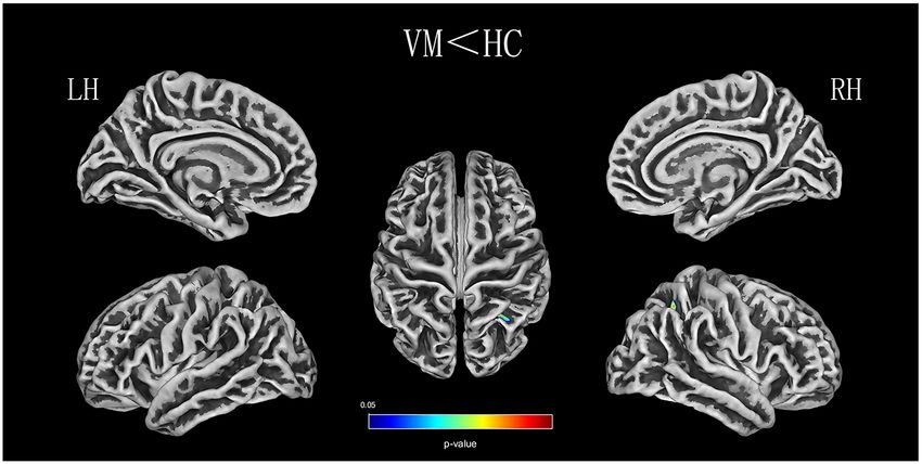
Zhe et al.                                                                                                         VM and Multisensory Integration

INTRODUCTION                                                              Russo et al. (2014) demonstrated that abnormal thalamic
                                                                          function is involved in central vestibular processing. Teggi et al.
Vestibular migraine (VM) is considered to be the most common              (2016) reported activation of brain areas related to integrating
central cause of episodic vertigo, manifesting as moderately to           visual and vestibular cues in patients with VM undergoing
severely intense vestibular symptoms and a migraine history. In           fMRI during visual stimulation in vertigo-free periods. A
2012, the International Headache Society and the Ba’ra’ny Society         fMRI study to observe treatment effectiveness found ALFF
proposed criteria for diagnosing VM as a disease entity (Lempert          values in the left posterior cerebellum of patients with VM
et al., 2012). An estimated 2.7% of adults suffer from vestibular         increased significantly after 1 month of vestibular rehabilitation
migraines according to a recent population-based survey in the            training (Liu et al., 2020). Functional imaging demonstrated
United States (Formeister et al., 2018). Women suffer from VM             the cerebellum can improve vestibular functioning through
2 to 3 times more frequently than do men (Neuhauser et al.,               a vestibular compensation mechanism. Recently, Wang et al.
2001; Lempert and Neuhauser, 2009). VM is a disabling disorder            (2021) evaluated resting-state FC alterations in patients with VM
that results in a significant burden on healthcare. Therefore, it         during the interictal period. Although several fMRI studies have
is important to understand the pathophysiology of VM to help              focused on ALFF or FC, no studies have explored FC alterations
develop treatment plans for patients.                                     based on cortical structural abnormalities in patients with VM.
    Previous studies indicate that the temporal lobe and the                  Given that previous findings have shown that changes in the
parietal lobe are associated with multisensory integration and            gray-matter volume of the temporal and parietal lobes are related
vestibular processing (Obermann et al., 2014; Komeilipoor et al.,         to multisensory vestibular processing in VM (Obermann et al.,
2017; Messina et al., 2017; Oh et al., 2018). Patients with VM            2014; Messina et al., 2017; Zhe et al., 2020), we hypothesized that
have activity in the temporal and parietal lobes during VM                alterations in cerebral cortex characteristics in patients with VM
attacks (Shin et al., 2014). Several voxel-based morphometric             could be located in brain regions associated with multisensory
(VBM) studies have reported that patients with VM exhibit                 vestibular processing. And we also hypothesized that changes in
gray matter (GM) volume abnormalities in temporal lobe                    cortical regions were accompanied by changes in FC. Therefore,
regions, including the superior temporal gyrus, middle temporal           in the current study, we used a whole-brain SBM technique
gyrus, and inferior temporal gyrus, as well as the parietal lobe          to evaluate cortical surface characteristics in VM patients,
(Obermann et al., 2014; Messina et al., 2017). Together, these            during an interictal period, compared with healthy controls.
findings strongly suggest that structural abnormalities in the            Furthermore, we used seed-based FC to investigate whether
temporal lobe are involved in multisensory integration, including         cortical regions with structural abnormalities also exhibit FC
visual, auditory, tactile, and vestibular processing (Beauchamp,          alterations in a patient with VM. Additionally, we assessed the
2005; Amedi et al., 2007). It is unclear, however, whether                relationships between brain morphological or FC changes and
cerebral cortex characteristics alter multisensory integration            clinical parameters.
and vestibular processing in brain areas during VM attacks.
Surface-based morphometry (SBM) can focus on cortical                     MATERIALS AND METHODS
structural characteristics, yielding more specific information
about neurological development as well as changes in cortical             Subjects
function related to thinning of the cortex (Panizzon et al., 2009;        Patients were recruited from the vertigo and dizziness outpatient
Yotter et al., 2011a; Dahnke et al., 2013). Compared with VBM,            service center of the Shaanxi Provincial People’s Hospital
SBM has been shown to be more sensitive and precise for                   in China between January 2016 and October 2020, who
detecting gray-matter atrophy, and it uses a completely automatic         were diagnosed with VM by a neurologist based on the
method, which provides the basis for projection-based thickness           International Classification of Headache Disorder 3rd edition
(PBT) measurement in order to obtain a local measure of GM                criteria (Lempert et al., 2012). Twenty-five right-handed
within the cortex (Lemaitre et al., 2012; Dahnke et al., 2013). The       patients with VM (21 without aura and four with aura)
SBM approach has been frequently used as a research method                and 27 healthy controls participated in this study. Patients
to assess cortical surface characteristics in migraine and other          were excluded if they had a history of other neurologic,
vestibular disorders (Komaromy et al., 2019; Nigro et al., 2019;          psychiatric, audiovestibular, or systemic disorders. All patients
Lai et al., 2020). However, no study to date has investigated the         in a symptom-free interval underwent a routine neurologic
pattern of cerebral cortex characteristics and their changes in           and neuro-otological examination, as well as MRI scanning,
relation to the clinical features of VM.                                  which were performed on the same day. No peripheral
    Resting-state functional connectivity (FC) provides a                 vestibular dysfunction was found in videonystagmography
powerful method to investigate the FC among brain regions,                (VNG) recordings. The clinical symptoms of each patient were
detecting the synchronized blood oxygen level-dependent                   assessed using a Visual Analog Scale (VAS; 0 = no pain;
(BOLD) signals from the seed region to the whole brain so as              10 = worst possible pain), the Migraine Disability Assessment
to locate highly correlated areas with similar characteristics            Scale (MIDAS), the Headache Impact Test-6 (HIT-6), and
(Xu et al., 2019; Niu et al., 2020). However, previous functional         the Dizziness Handicap Inventory (DHI) using face-to-face
magnetic resonance imaging (fMRI) studies have used the                   interviews with a standardized questionnaire and questions
amplitude of low-frequency fluctuation (ALFF) during external             (Sauro et al., 2010; Hawker et al., 2011; Balci et al., 2018). Eight
stimulation to assess functional changes in patients with VM.             of the patients with VM were treated with migraine-preventive

Frontiers in Human Neuroscience | www.frontiersin.org                 2                                  August 2021 | Volume 15 | Article 717130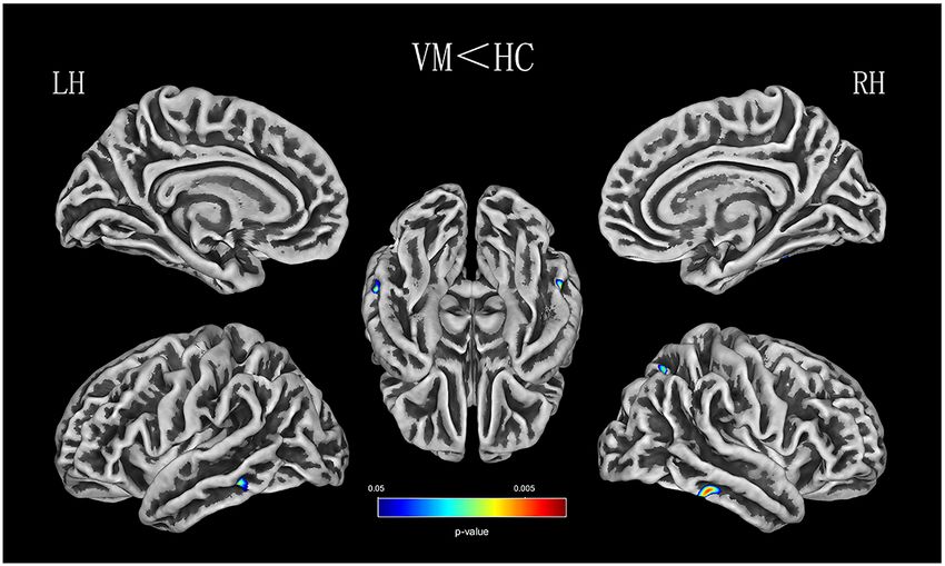
Zhe et al.                                                                                                               VM and Multisensory Integration

medications and nonsteroidal analgesics. Most patients (n = 17)           registration were conducted. In addition, CAT12 allows the
did not take any medication regularly.                                    estimation of other morphological indices of fractal dimension
   The 27 age-, sex- and handedness-matched healthy controls              (FD), sulcus depth (SD), and gyrification index (GI), which
were from the community. The exclusion criteria were: migraine;           were also calculated for each participant with default parameter
chronic pain; previous vestibular neuritis; Meniere’s disease;            settings. The calculation of CT, FD, SD, and GI was performed
secondary somatoform vertigo; drug abuse; neurologic, mental              in subject native surface space. The images of cerebral cortex
or systemic disorders; ischemic or hemorrhagic stroke; or severe          characteristics were checked for homogeneity. As all the
head trauma. All subjects had no structural abnormalities or              images had high correlation values (>0.85), none of them
white matter (WM) lesions in T2-weighted or FLAIR imaging.                had to be discarded. Finally, the CT images were smoothed
This study was approved by the Ethics Committee of the Shaanxi            using a Gaussian kernel with a full width at half maximum
Provincial People’s Hospital. All participants provided written           (FWHM) of 15 mm, and three other surface parameters
informed consent before entering the study.                               were smoothened with an isotropic 20 mm FWHM Gaussian
                                                                          kernel.
Imaging Data Acquisition                                                      All functional images were preprocessed using Data
All the images were obtained using a 3.0 T Philips Ingenia                Processing and Analysis for Brain Imaging 3.02 , which is
scanner with a 16-channel phased-array head coil. A                       based on Statistical Parametric Mapping 123 . First, the first
high-resolution three-dimensional (3D) magnetization-prepared             10 volumes were removed to allow subjects to adapt to the
rapid-acquisition gradient echo (MPRAGE) T1-weighted (T1w)                magnetic field. Second, slice timing correction was performed
sequence covering the whole brain (332 sagittal slices) was               to correct for the inter-slice time delay within each volume.
collected. The acquisition parameters were: repetition time               Third, head motion >1.5 mm and translation >1.5◦ of rotation in
(TR) = 1,900 ms; echo time (TE) = 2.26 ms; inversion time                 any direction were excluded. Images were spatially normalized
(TI) = 900 ms; flip angle (FA) = 9◦ ; matrix = 256 × 256; field           into MNI space using a standard EPI template provided by
of view = 220 × 220 mm; and 1.00 mm slice thickness with                  SPM12 and resliced into a voxel size of 3 × 3 × 3 mm. Finally,
no interslice gap. Resting-state functional BOLD images were              data were spatially smoothed using a 6-mm FWHM Gaussian
scanned using gradient echo-planar imaging with the following             kernel.
parameters: repetition time = 2,000 s; echo time = 30 ms;                     Seed-based FC analysis was performed with seeds from the
slices = 34; slice thickness = 4 mm; slice gap = 0 mm; field of           SBM findings. Seeds were defined as 3-mm-radius spheres
view = 230 × 230 mm; matrix = 128 × 128; flip angle = 90◦ ; and           centered on the peak voxel for the CT and SD clusters showing
200 volumes. For the resting-state scan, all subjects were asked          between-group differences. The averaged time-course of each
to keep their eyes closed and their minds calm, and to stay awake         seed area was extracted, and Pearson’s correlation (r) was used
throughout the scan. After the scan, subjects were asked whether          to calculate the FC between the extracted time-courses and the
or not they remained awake during the entire procedure.                   time-courses of the entire brain in a voxel-wise manner. The
                                                                          individual r-maps were normalized to Z-maps using Fisher’s Z-
Image Processing                                                          transformation.
Structural images were processed using CAT121 and SPM12 run
in MATLAB R2014b (The MathWorks, Inc.). CAT12 provides                    Statistical Analysis
a volume-based method for estimating regional thickness (CT)              Demographic and Clinical Data
without extensive reconstruction of the cortical surface and has          A two-sample t-test was used to estimate the differences in age,
been shown to be a fast and reliable alternative to FreeSurfer            sex, and years of education between the VM and healthy control
(Paul et al., 2017; Seiger et al., 2018). Moreover, CAT12 is              groups. The statistical significance level was set at P < 0.05.
a fully automated method that allows the measurement of                   These statistical analyses were performed using the SPSS software
the whole brain cortical surface. For each participant, the               package (version 22.0).
processing pipeline included bias-field, noise removal, skull
stripping, and segmentation into GM, WM, and cerebrospinal
                                                                          Cortical Surface Characteristics Analysis
                                                                          Cortical surface characteristics were compared between the
fluid (CSF). The images were finally normalized to MNI
                                                                          VM patients and healthy controls using two-sample t-tests
space, which uses diffeomorphic anatomical registration using
                                                                          in CAT12 with age and sex as covariates. Family-wise error
exponentiated Lie algebra (DARTEL) to a 1.5 mm isotropic
                                                                          (FWE) correction was performed to correct for multiple
adult template (Ashburner, 2007). Here, the CT evaluation and
                                                                          comparisons; P < 0.05 was considered statistically significant.
reconstruction of the central surface were performed in one step,
                                                                          Then, the surviving clusters were reported. Finally, based on
based on the PBT method (Dahnke et al., 2013). Importantly,
                                                                          the Desikan–Killiany (DK40) atlas (Desikan et al., 2006), we
the PBT allows the appropriate handing of partial volume
                                                                          extracted the mean cortical surface characteristics (CT, SD, GI,
information, sulcal blurring, and sulcal asymmetries without
                                                                          and FD) from the above mentioned significant clusters. Partial
explicit sulcus reconstruction (Dahnke et al., 2013). After the
                                                                          correlations adjusted for age and sex were used to analyze
initial surface reconstruction, topology correction (Yotter et al.,
                                                                          differences between the cortical surface characteristics of these
2011b), spherical mapping (Yotter et al., 2011a), and spherical
                                                                          2 http://rfmri.org/dpabi
1 http://dbm.neuro.uni-jena.de/cat12                                      3 http://www.fil.ion.ucl.ac.uk/spm

Frontiers in Human Neuroscience | www.frontiersin.org                 3                                        August 2021 | Volume 15 | Article 717130
Zhe et al. VM and Multisensory Integration
TABLE 1 | Demographic and clinical characteristics of patients.
Characteristics VM (n = 27) HC (n = 25) P value
Mean ± SD Mean ± SD
Sex (female/male) 27/4 25/4 0.91
Age (years) 38.22 ± 10.58 37.28 ± 11.45 0.76
Education (years) 13.89 ± 3.61 14.40 ± 2.48 0.56
Disease duration (years) 9.15 ± 7.58
Headache frequency (number) 6.67 ± 4.93
VAS 4.74 ± 2.75
MIDAS 54.33 ± 53.13
HIT-6 51.56 ± 19.94
DHI 48.93 ± 16.43
Note: VM, vestibular migraine; HC, healthy control; VAS, Visual Analog Scale (0 = no pain, 10 = worst possible pain); MIDAS, Migraine Disability Assessment Scale; HIT-6, Headache
Impact Test-6; DHI, Dizziness Handicap Inventory.
TABLE 2 | Decreased CT in various brain regions in patients with VM.
Brain regions Peak MNI Cluster voxels T Z P
R Inferior temporal gyrus 63 −36 −22 149 5.38 4.74 0.000
Superior parietal lobule 24 −63 50 68 5.06 4.60 0.000
L Inferior temporal gyrus −56 −38 −14 102 5.19 4.51 0.000
Middle temporal gyrus −56 −38 −14 102 5.19 4.51 0.000
Note: MNI, Montreal Neurological Institute; R, right; L, Left; CT, thickness; VM, vestibular migraine.
altered regions and clinical indices (including the VAS score,
disease duration, attack frequency, MIDAS score, HIT-6 score,
and DHI score). The significance threshold was set at P < 0.05.
Seed-Based FC Analysis
A comparison of FC between groups was performed using a
two-sample t-test within DPABI, with age and sex as covariates.
Correction for multiple comparisons was performed using a
Gaussian random field at P < 0.05 (voxel P < 0.001). Then, the
surviving clusters were reported.
Finally, we extracted the average Z-values for each region
with significant differences and performed a partial correlation
analysis with patients’ clinical parameters using SPSS 22.0,
controlling for age and sex. The significance threshold was set
FIGURE 1 | CT (thickness) analysis results of patients with vestibular
at P < 0.05.
migraine (VM) compared with healthy controls [P < 0.05, family-wise error
(FWE)-corrected].
RESULTS
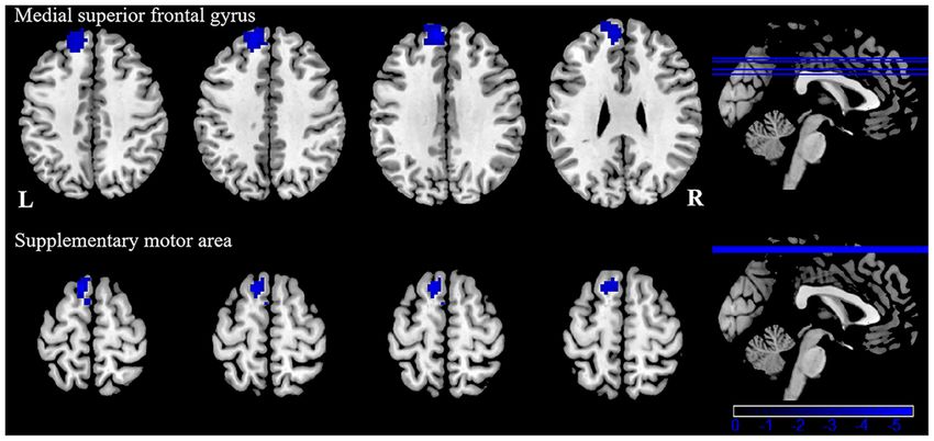
FD. In the VM patients, a significant negative correlation was
Demographic and Clinical Data found between DHI scores and the CT of the right inferior
There were no significant differences between the VM patients temporal gyrus (r = −0.542; P = 0.005; Figure 3A) and left middle
and healthy controls in age, sex, or years of education. The temporal gyrus (r = −0.553; P = 0.004; Figure 3B). No correlation
results are summarized in Table 1. VM patients suffered from was found between abnormal SD and disease duration, attack
a moderate and severe migraine burden with a mean VAS score frequency, VAS score, MIDAS score, HIT-6 score, or DHI score
of 4.74 ± 2.75, mean HIT-6 score of 51.56 ± 19.94, and mean in the VM patients.
MIDAS score of 54.33 ± 53.13. Their scores on the vertigo scale
were moderate with a mean DHI score of 48.93 ± 16.43. Seed-Based FC Results
Patients with VM showed significantly weaker FC between
Cortical Surface Characteristics Results the left inferior/middle temporal gyrus and the left superior
Relative to healthy comparison subjects, the VM patients showed frontal gyrus, supplementary motor area (Table 4, Figure 4).
significantly thinner CT in the bilateral inferior temporal gyrus, There were no significant group differences in FC with other
left middle temporal gyrus, and right superior parietal lobule seed regions (right inferior temporal gyrus, right superior, and
(Table 2, Figure 1). Reduced SD was found in the right superior inferior parietal lobule). No significant correlation was observed
and inferior parietal lobule (Table 3, Figure 2). There was no between FC alterations and clinical characteristics in patients
significant intergroup difference for surface parameters GI and with VM.
Frontiers in Human Neuroscience | www.frontiersin.org 4 August 2021 | Volume 15 | Article 717130Zhe et al. VM and Multisensory Integration
TABLE 3 | Decreased SD in various brain regions in patients with VM.
Brain regions Peak MNI Cluster voxels T Z P
R Superior parietal lobule 28 −51 49 382 5.02 4.48 0.000
Inferior parietal lobule 32 −60 47 382 4.91 4.39 0.000
Note: MNI, Montreal Neurological Institute; R, right; SD, sulcus depth.
FIGURE 2 | SD (sulcus depth) analysis results of patients with VM compared with healthy controls (P < 0.05, FWE-corrected).
FIGURE 3 | Correlation between the CT of the inferior (A) and middle temporal gyrus (B) and dizziness handicap inventory (DHI) score in patients with VM
(P < 0.05).
DISCUSSION VM and healthy controls, as well as associations between cortical
surface characteristics or FC and clinical variables. Compared
As far as we know, our study is the first to directly investigate with healthy controls, we found that patients with VM had
cortical surface characteristics and FC changes in patients with decreased CT and SD in certain areas, including multisensory
Frontiers in Human Neuroscience | www.frontiersin.org 5 August 2021 | Volume 15 | Article 717130Zhe et al. VM and Multisensory Integration
TABLE 4 | Abnormal functional connectivity of the left inferior/middle temporal gyrus in patients with VM.
Seed points Brain region BA Peak MNI Cluster voxels T
L Inferior/middle temporal gyrus L Medial superior frontal gyrus 9 −9 51 33 248 −5.54
L Supplementary motor area 6 −9 18 69 75 −5.12
Note: MNI, Montreal Neurological Institute; R, right.
FIGURE 4 | Functional connectivity (FC) analysis results for patients with VM compared with healthy controls (P < 0.05, GRF-corrected).
integration and vestibular processing regions. Additionally, we reported that the metabolism of the temporoparietal-insular
found that DHI scores and CT were significantly correlated in areas increased during a VM attack (Fasold et al., 2002). These
the right inferior temporal gyrus and the left middle temporal results suggest that some modification of structural covariance
gyrus. Using the clusters derived from the SBM analysis patterns in the temporal lobe is involved in pain-processing
as seed regions, we found significantly weaker FC between and multisensory integration (Moulton et al., 2011). The VM
the left inferior/middle temporal gyrus and the left medial patients in that study also showed reduced CT in multiple areas
superior frontal gyrus, supplementary motor area. Our data of the temporal lobe, including the inferior and middle gyrus,
confirmed our hypothesis that VM patients have abnormalities in compared with healthy controls, which is in line with the findings
cortical surface characteristics related to multisensory vestibular of several previous studies. Obermann et al. (2014) found that
processing and that changes in cortical regions are accompanied gray-matter volume was decreased in the inferior temporal
by changes in FC. gyrus, middle temporal gyrus, and the superior temporal gyrus,
The temporal lobe has been recognized as a region associated the middle cingulate, dorsolateral prefrontal, insular, parietal,
with multisensory integration, which involves auditory, and occipital cortices (Obermann et al., 2014). These structurally
olfactory, vestibular, and visual senses and the perception abnormal brain areas in patients with VM are involved in
of spoken and written language (Kiernan, 2012). A number multisensory vestibular control, as well as pain processing and
of studies on patients with migraine have found that CT was central vestibular compensation. In contrast, a recent VBM
thinning in the temporal lobe, suggesting that the temporal study found an increase in the temporal lobe, frontal lobe, and
lobe plays an important role in the regulation of pain (Coppola occipital lobe in VM patients compared with healthy controls
et al., 2017; Jia and Yu, 2017). Several studies have demonstrated (Messina et al., 2017). These inconsistent findings might be due
greater interregional CT correlations in patients with migraine, to differences in sample size, attack frequency, medication status,
specifically over temporal regions (Chong et al., 2020). Schwedt and data acquisition and processing in the various studies.
et al. (2015) found that temporal pole correlations distinguished The inferior temporal gyrus is related to visual processing
groups of migraine patients from healthy controls. An fMRI (Naito et al., 2003). There is some evidence that decreased CT
study of 12 right-handed patients with VM, which used cold in the inferior temporal gyrus might contribute to abnormalities
caloric stimulation, found a typical pattern of BOLD signals in multisensory integration of visual processing, such as
in temporal-parietal areas in the interictal interval, including amplifying vision (photophobia), hearing (phonophobia), or
patients with migraine without aura and healthy controls (Russo olfactory stimuli, which may induce an attack of VM. Repeated
et al., 2014). A recent functional imaging study of two patients VM attacks over time that seem to lead to an alteration of
Frontiers in Human Neuroscience | www.frontiersin.org 6 August 2021 | Volume 15 | Article 717130Zhe et al. VM and Multisensory Integration
multisensory integration of visual processing structures may network involved in pain and vestibular processing (Dieterich
provide an explanation as to why most VM patients have and Brandt, 2008). The parietal lobule has been implicated in
increased sensitivity to visual, auditory, and olfactory stimuli VM, and some VBM studies on patients with VM have reported
during VM attacks. Previous studies have also suggested that the a lower gray-matter volume in the parietal lobes of such patients
middle temporal gyrus plays a key role in interconnecting with compared with controls (Obermann et al., 2014; Zhe et al., 2020).
other multisensory cortical areas, and it is deemed to form a A recent fMRI study of two VM patients reported increased
multisensory integrative network (Helmchen et al., 2014). The activity in the inferior parietal lobule during visual stimulation
middle temporal gyrus, inferior temporal gyrus, and superior in a vertigo-free period (Teggi et al., 2016). Correlation analysis
temporal gyrus, and the lateral temporal lobe play a role in also revealed that decreased gray-matter volume in the parietal
the underlying connection between migraine and the vestibular lobe is associated with illness duration and headache intensity in
system (Rocca et al., 2006; Schwedt et al., 2013; Helmchen patients with VM (Obermann et al., 2014). These studies indicate
et al., 2014). The middle temporal gyrus belongs to the temporal that cortical abnormalities of the parietal lobe are involved in
perisylvian vestibular cortex, which is particularly sensitive for nociception and multisensory vestibular control. Our findings in
dizziness (Kahane et al., 2010). Furthermore, CT in the inferior VM patients implicate the parietal lobe in the modulation of pain
and middle temporal gyrus is negatively correlated with the perception and dysfunction of sensory integration.
severity of vertigo in VM patients. The DHI was used to In order to assess if these cortical structural abnormalities
evaluate the self-perceived handicapping effects of dizziness, also exhibited FC alterations, we performed a resting-state
which is related to the physical, emotional, and functional FC study. Our results showed decreased FC between the left
aspects of patients. Vertigo attacks result in subjective spatial inferior/middle temporal gyrus and left medial superior frontal
orientation errors, surrounding environment spiraling around, gyrus supplementary motor area in patients with VM, compared
and complaining of imbalance in patients. That may lead to to healthy controls. The medial superior frontal gyrus is not
patients who usually dare not to attempt daily activities, and only involved in emotional responses and feelings of pain, but
experience obvious anxiety and depression which reflect the also in memory, attention responses, and cognitive reactions
degree of vertigo. In the present study, a mean DHI score of related to pain (Bluhm et al., 2007). The study confirmed
48.93 points was obtained. In patients with VM, as the DHI that the superior frontal gyrus is involved in the integration
score increased, there was a decrease in life quality scales showing of somatosensory and vestibular information (Klingner et al.,
moderate disability in DHI. However, we did not evaluate 2016). Our research indicates that the endogenous analgesic
symptoms of depression and anxiety. Thus, it is not clear whether mechanism of VM patients is adjusted because some long–term
anxiety or depression is associated with DHI in patients with VM. migraine and vertigo attacks occur, altering the emotional
Future studies should clarify this. Correlations revealing a CT response to pain, or reducing pain perception and cognition,
decrease in the temporal lobe was associated with an increased which can reduce the input of pain signals. Anatomically, the
subjective intensity of vertigo in VM, which indicated that the SMA is located in the dorsomedial frontal cortex, which is
temporal lobe is involved in the pathophysiology of patients with involved in executive control (Aron and Roldrack, 2006; Li
VM and is associated with the daily life of the patient. Therefore, et al., 2006), pain anticipation (Koyama et al., 2005; Rainville
CT reduction in the inferior and middle temporal gyrus is a et al., 2009), and an affective component of pain (Apkarian
potentially valuable morphological characteristic, which might et al., 2005; Geha et al., 2008). Our results indicate that the
result in central vestibular syndromes that manifest along with FC decrease between the left inferior/middle temporal gyrus
vertigo and dizziness. Based on all of the above discussion, our and left supplementary motor area may be related to deficits in
results indicate that long–term and high-frequency headaches affective pain modulation and affective pain response inhibition.
and vertigo attacks may lead to reduced CT in multisensory In addition, the SMA is associated with auditory processing
integrative and vestibular processing areas in VM, reflecting (Lima et al., 2016). Phonophobia is reported in about half of
abnormal brain structure due to the effects of brain disease. This patients with VM during a vertigo attack (Neuhauser et al., 2006).
has profound implications for our understanding of multisensory Exposure to noise may cause generalized discomfort and increase
integrative networks in patients with VM. the pain and vertigo of the patients with VM. Recurrent VM
The second finding of the current study is decreased CT in attacks may ultimately result in FC alterations associated with
the superior parietal lobule. Furthermore, we found lower SD auditory processing. Thus, the FC changes may serve as a possible
in the inferior and superior parietal lobule in patients with VM explanation for phonophobia when vertigo occurs (Wei et al.,
compared with healthy controls. Other studies proposed that 2020), which provides a new clue for therapy for this syndrome.
the parietal lobule is chiefly involved in discriminating sensory The present study has several limitations. First, our study
features of pain (Hofbauer et al., 2001; Oshiro et al., 2007, was conducted with a relatively small sample. Our study should
2009). The superior parietal lobule, as a part of the parietofrontal have included a larger sample that was more representative of
network, has been found to be related to the perceptual matrix a pathological population, which would help to assure greater
of pain (Garcia-Larrea and Peyron, 2013). It also contains major reproducibility of its results. Second, subgroup analyses of
parts of the sensory cortex that are involved in spatial orientation migraine (migraine without aura and migraine with aura) were
and sensory information processing and interpretation (Kamali not performed. To better elucidate the cortical morphological
et al., 2014). Studies have confirmed that the inferior parietal difference between them, future studies should compare the
lobe belongs to part of the multisensory vestibular cortical two types of VM, and it would be valuable to recruit a larger
Frontiers in Human Neuroscience | www.frontiersin.org 7 August 2021 | Volume 15 | Article 717130Zhe et al. VM and Multisensory Integration
sample size to be able to divide participants into the subgroups DATA AVAILABILITY STATEMENT
‘‘migraine without aura’’ and ‘‘migraine with aura.’’ Third, we
did not examine subcortical and brain stem structures in this The original contributions presented in the study are included in
study; therefore, future studies need to be designed to include the article, further inquiries can be directed to the corresponding
both. Fourth, because the sample size was relatively small, the author.
correlation analysis was not strictly conducted with Bonferroni
corrections. Fifth, we did not use an experimental task to ETHICS STATEMENT
measure multisensory integration. Finally, we did not evaluate
symptoms of depression and anxiety, although previous research The studies involving human participants were reviewed
has reported that patients with VM have high levels of depression and approved by the Ethics Committee of the Shaanxi
and anxiety (Kim et al., 2016). The burden of symptomatology Provincial People’s Hospital. The patients/participants
can affect cortical morphology. Assessment of depression and provided their written informed consent to participate in
anxiety scores should be performed in future studies. this study.
CONCLUSION AUTHOR CONTRIBUTIONS
In conclusion, we evaluated cortical structural and FC alterations XZhe drafted the manuscript, designed the experiment, and
in patients with VM using SBM and resting-state FC analyses, performed the statistical analysis. LC undertook clinical
compared with healthy controls. CT and SD abnormalities were parameters assessments. MT, JG, XL, and DZ collected the data.
detected in the temporal lobe and parietal lobe. Furthermore, KA and WL provided technical support. XZha made study
patients with VM displayed decreased FC between the left supervision or coordination. All authors contributed to the
inferior/middle temporal gyrus and the left superior frontal article and approved the submitted version.
gyrus, supplementary motor area. These regions are known to
be involved in multisensory integration, vestibular processing, FUNDING
and pain modulation, contributing to a lower quality of life.
This research was supported by the Social Development Science
These findings will promote our understanding of the underlying
and Technology Research Project of Shaanxi Province of China
mechanism of VM, but so far an experimental task to measure
(2021SF-290).
multisensory integration in patients with VM has not been
used. Future studies should identify brain areas associated with
multisensory integration using a task-based fMRI. Moreover, ACKNOWLEDGMENTS
further studies focusing on anxiety and depression are needed,
which are bound to shed light on emotional states in patients We would like to thank all patients and healthy controls for their
with VM. willingness to participate in the present study.
REFERENCES Chong, C. D., Aguilar, M., and Schwedt, T. J. (2020). Altered hypothalamic region
covariance in migraine and cluster headache: a structural MRI study. Headache
Amedi, A., Stern, W. M., Camprodon, J. A., Bermpohl, F., Merabet, L., 60, 553–563. doi: 10.1111/head.13742
Rotman, S., et al. (2007). Shape conveyed by visual-to-auditory sensory Coppola, G., Petolicchio, B., Di Renzo, A., Tinelli, E., Di Lorenzo, C.,
substitution activates the lateral occipital complex. Nat. Neurosci. 10, 687–689. Parisi, V., et al. (2017). Cerebral gray matter volume in patients with
doi: 10.1038/nn1912 chronic migraine: correlations with clinical features. J. Headache Pain 18:115.
Apkarian, A. V., Bushnell, M. C., Treede, R. D., and Zubieta, J. K. (2005). Human doi: 10.1186/s10194-017-0825-z
brain mechanisms of pain perception and regulation in health and disease. Eur. Dahnke, R., Yotter, R. A., and Gaser, C. (2013). Cortical thickness and central
J. Pain 9, 463–484. doi: 10.1016/j.ejpain.2004.11.001 surface estimation. Neuroimage 65, 336–348. doi: 10.1016/j.neuroimage.2012.
Aron, A. R., and Roldrack, R. A. (2006). Cortical and subcortical contributions to 09.050
stop signal response inhibition: role of the subthalamic nucleus. J. Neurosci. 26, Desikan, R. S., Segonne, F., Fischl, B., Quinn, B. T., Dickerson, B. C., Blacker, D.,
2424–2433. doi: 10.1523/JNEUROSCI.4682-05.2006 et al. (2006). An automated labeling system for subdividing the human cerebral
Ashburner, J. (2007). A fast diffeomorphic image registration cortex on MRI scans into gyral based regions of interest. Neuroimage 31,
algorithm. Neuroimage 38, 95–113. doi: 10.1016/j.neuroimage.2007. 968–980. doi: 10.1016/j.neuroimage.2006.01.021
07.007 Dieterich, M., and Brandt, T. (2008). Functional brain imaging of peripheral and
Balci, B., Senyuva, N., and Akdal, G. (2018). Definition of balance and cognition central vestibular disorders. Brain 131, 2538–2552. doi: 10.1093/brain/awn042
related to disability levels in vestibular migraine patients. Noro. Psikiyatr Ars. Fasold, O., Von Brevern, M., Kuhberg, M., Ploner, C. J., Villringer, A., Lempert, T.,
55, 9–14. doi: 10.29399/npa.12617 et al. (2002). Human vestibular cortex as identified with caloric stimulation
Beauchamp, M. S. (2005). See me, hear me, touch me: multisensory integration in functional magnetic resonance imaging. Neuroimage 17, 1384–1393.
in lateral occipital-temporal cortex. Curr. Opin. Neurobiol. 15, 145–153. doi: 10.1006/nimg.2002.1241
doi: 10.1016/j.conb.2005.03.011 Formeister, E. J., Rizk, H. G., Kohn, M. A., and Sharon, J. D. (2018). The
Bluhm, R. L., Miller, J., Lanius, R. A., Osuch, E. A., Boksman, K., Neufeld, R. W., epidemiology of vestibular migraine: a population-based survey study. Otol.
et al. (2007). Spontaneous low-frequency fluctuations in the BOLD signal in Neurotol. 39, 1037–1044. doi: 10.1097/MAO.0000000000001900
schizophrenic patients: anomalies in the default network. Schizophr. Bull. 33, Garcia-Larrea, L., and Peyron, R. (2013). Pain matrices and neuropathic pain
1004–1012. doi: 10.1093/schbul/sbm052 matrices: a review. Pain 154, S29–S43. doi: 10.1016/j.pain.2013.09.001
Frontiers in Human Neuroscience | www.frontiersin.org 8 August 2021 | Volume 15 | Article 717130Zhe et al. VM and Multisensory Integration
Geha, P. Y., Baliki, M. N., Harden, R. N., Bauer, W. R., Parrish, T. B., and Liu, L., Hu, X., Zhang, Y., Pan, Q., Zhan, Q., Tan, G., et al. (2020). Effect
Apkarian, A. V. (2008). The brain in chronic CRPS pain: abnormal gray-white of vestibular rehabilitation on spontaneous brain activity in patients with
matter interactions in emotional and autonomic regions. Neuron 60, 570–581. vestibular migraine: a resting-state functional magnetic resonance imaging
doi: 10.1016/j.neuron.2008.08.022 study. Front. Hum. Neurosci. 14:227. doi: 10.3389/fnhum.2020.00227
Hawker, G. A., Mian, S., Kendzerska, T., and French, M. (2011). Measures of Messina, R., Rocca, M. A., Colombo, B., Teggi, R., Falini, A., Comi, G., et al. (2017).
adult pain: visual analog scale for pain (VAS Pain), numeric rating scale for Structural brain abnormalities in patients with vestibular migraine. J. Neurol.
pain (NRS Pain), mcgill pain questionnaire (MPQ), short-form mcgill pain 264, 295–303. doi: 10.1007/s00415-016-8349-z
questionnaire (SF-MPQ), chronic pain grade scale (CPGS), short form-36 Moulton, E. A., Becerra, L., Maleki, N., Pendse, G., Tully, S., and Hargreaves, R.,
bodily pain scale (SF-36 BPS) and measure of intermittent and constant et al. (2011). Painful heat reveals hyperexcitability of the temporal
osteoarthritis pain (ICOAP). Arthritis Care Res. (Hoboken) 63, S240–S252. pole in interictal and ictal migraine states. Cereb. Cortex 21, 435–448.
doi: 10.1002/acr.20543 doi: 10.1093/cercor/bhq109
Helmchen, C., Ye, Z., Sprenger, A., and Munte, T. F. (2014). Changes in Naito, Y., Tateya, I., Hirano, S., Inoue, M., Funabiki, K., Toyoda, H., et al. (2003).
resting-state fMRI in vestibular neuritis. Brain Struct. Funct. 219, 1889–1900. Cortical correlates of vestibulo-ocular reflex modulation: a PET study. Brain
doi: 10.1007/s00429-013-0608-5 126, 1562–1578. doi: 10.1093/brain/awg165
Hofbauer, R. K., Rainville, P., Duncan, G. H., and Bushnell, M. C. (2001). Cortical Neuhauser, H., Leopold, M., Von Brevern, M., Arnold, G., and Lempert, T. (2001).
representation of the sensory dimension of pain. J. Neurophysiol. 86, 402–411. The interrelations of migraine, vertigo and migrainous vertigo. Neurology 56,
doi: 10.1152/jn.2001.86.1.402 436–441. doi: 10.1212/wnl.56.4.436
Jia, Z., and Yu, S. (2017). Grey matter alterations in migraine: a systematic review Neuhauser, H. K., Radtke, A., von Brevern, M., Feldmann, M., Lezius, F., Ziese, T.,
and meta-analysis. Neuroimage Clin. 14, 130–140. doi: 10.1016/j.nicl.2017.01. et al. (2006). Migrainous vertigo: prevalence and impact on quality of life.
019 Neurology 67, 1028–1033. doi: 10.1212/01.wnl.0000237539.09942.06
Kahane, P., Hoffmann, D., Minotti, L., Minotti, L., and Berthoz, A. (2010). Nigro, S., Indovina, I., Riccelli, R., Chiarella, G., Petrolo, C., Lacquaniti, F.,
Reappraisal of the human vestibular cortex by cortical electrical stimulation et al. (2019). Reduced cortical folding in multi-modal vestibular regions in
study. Ann. Neurol. 54, 615–624. doi: 10.1002/ana.10726 persistent postural perceptual dizziness. Brain Imaging Behav. 13, 798–809.
Kamali, A., Sair, H. I., Radmanesh, A., and Hasan, K. M. (2014). Decoding doi: 10.1007/s11682-018-9900-6
the superior parietal lobule connections of the superior longitudinal Niu, X., Xu, H., Guo, C., Yang, T., Dustin, K., Gao, L., et al. (2020). Strengthened
fasciculus/arcuate fasciculus in the human brain. Neuroscience 277, 577–583. thalamoparietal functional connectivity in patients with hemifacial spasm:
doi: 10.1016/j.neuroscience.2014.07.035 a cross-sectional resting-state fMRI study. Br. J. Radiol. 93:20190887.
Kiernan, J. A. (2012). Anatomy of the temporal lobe. Epilepsy Res. Treat. doi: 10.1259/bjr.20190887
2012:176157. doi: 10.1016/j.cortex.2017.11.006 Obermann, M., Wurthmann, S., Steinberg, B. S., Theysohn, N., Diener, H. C., and
Kim, S. K., Kim, Y. B., Park, I. S., Hong, S. J., Kim, H., and Hong, S. M. (2016). Naegel, S. (2014). Central vestibular system modulation in vestibular migraine.
Clinical analysis of dizzy patients with high levels of depression and anxiety. Cephalalgia 34, 1053–1061. doi: 10.1177/0333102414527650
J. Audiol. Otol. 20, 174–178. doi: 10.7874/jao.2016.20.3.174 Oh, S. Y., Boegle, R., Ertl, M., Stephan, T., and Dieterich, M. (2018).
Klingner, C. M., Axer, H., .Brodoehl, S., and Witte, O. W. (2016). Vertigo and Multisensory vestibular, vestibular-auditory and auditory network effects
the processing of vestibular information: a review in the context of predictive revealed by parametric sound pressure stimulation. Neuroimage 176, 354–363.
coding. Neurosci. Biobehav. Rev. 71, 379–387. doi: 10.1016/j.neubiorev.2016.09. doi: 10.1016/j.neuroimage.2018.04.057
009 Oshiro, Y., Quevedo, A. S., Mchaffie, J. G., Kraft, R. A., and Coghill, R. C. (2009).
Komaromy, H., He, M., Perlaki, G., Orsi, G., Nagy, S. A., Bosnyak, E., et al. (2019). Brain mechanisms supporting discrimination of sensory features of pain: a new
Influence of hemispheric white matter lesions and migraine characteristics on model. J. Neurosci. 29, 14924–14931. doi: 10.1523/JNEUROSCI.5538-08.2009
cortical thickness and volume. J. Headache Pain 20:4. doi: 10.1186/s10194-019- Oshiro, Y., Quevedo, A. S., Mchaffie, J. G., Kraft, R. A., and Coghill, R. C. (2007).
0959-2 Brain mechanisms supporting spatial discrimination of pain. J. Neurosci. 27,
Komeilipoor, N., Cesari, P., and Daffertshofer, A. (2017). Involvement of superior 3388–3394. doi: 10.1523/JNEUROSCI.5128-06.2007
temporal areas in audiovisual and audiomotor speech integration. Neuroscience Panizzon, M. S., Fennema-Notestine, C., Eyler, L. T., Jernigan, T. L., Prom-
343, 276–283. doi: 10.1016/j.neuroscience.2016.03.047 Wormley, E., Neale, M., et al. (2009). Distinct genetic influences on
Koyama, T., Mchaffie, J. G., Laurienti, P. G., and Coghill, R. C. (2005). cortical surface area and cortical thickness. Cereb Cortex 19, 2728–2735.
The subjective experience of pain: where expectations become reality. doi: 10.1093/cercor/bhp026
Proc. Natl. Acad. Sci. U S A 102, 12950–12955. doi: 10.1073/pnas.0408 Paul, F. R., R. Schmidt, P., Dahnke, R., Biberacher, V., Beer, A., Buck, D.,
576102 et al. (2017). Volume versus surface-based cortical thickness measurements: a
Lai, K. L., Niddam, D. M., Fuh, J. L., Chen, W. T., Wu, J. C., and comparative study with healthy controls and multiple sclerosis patients. PLos
Wang, S. J. (2020). Cortical morphological changes in chronic migraine in a One 12:e0179590. doi: 10.1371/journal.pone.0179590
Taiwanese cohort: surface- and voxel-based analyses. Cephalalgia 40, 575–585. Rainville, P., Roy, M., Pich, M., Chen, J. I., and Peretz, Z. (2009). Cerebral and
doi: 10.1177/0333102420920005 spinal modulation of pain by emotions. Proc. Natl. Acad. Sci. U S A 106,
Lemaitre, H., Goldman, A. L., Sambataro, F., Verchinski, B. A., Meyer- 20900–20905. doi: 10.1073/pnas.0904706106
Lindenberg, A., Weinberger, D. R., et al. (2012). Normal age-related brain Rocca, M. A., Ceccarelli, A., Falini, A., Colombo, B., Tortorella, P., Bernasconi, L.,
morphometric changes: nonuniformity across cortical thickness, surface et al. (2006). Brain gray matter changes in migraine patients with T2-visible
area and gray matter volume. Neurobiol. Aging 33, e611.617–e611.619. lesions: a 3-T MRI study. Stroke 37, 1765–1770. doi: 10.1161/01.STR.
doi: 10.1016/j.neurobiolaging.2010.07.013 0000226589.00599.4d
Lempert, T., and Neuhauser, H. (2009). Epidemiology of vertigo, migraine and Russo, A., Marcelli, V., Esposito, F., Corvino, V., Marcuccio, L., Giannone, A., et al.
vestibular migraine. J. Neurol. 256, 333–338. doi: 10.1007/s00415-009-0149-2 (2014). Abnormal thalamic function in patients abnormal thalamic function in
Lempert, T., Olesen, J., Furman, J., Waterston, J., Seemungal, B., Carey, J., et al. patients. Neurology 82, 2120–2126. doi: 10.1212/WNL.0000000000000496
(2012). Vestibular migraine: diagnostic criteria. J. Vestib. Res. 22, 167–172. Sauro, K. M., Rose, M. S., Becker, W. J., Christie, S. N., Giammarco, R.,
doi: 10.3233/VES-2012-0453 Mackie, G. F., et al. (2010). HIT-6 and MIDAS as measures of headache
Li, C. S. H., Huang, C., Constable, R. T., and Sinha, R. (2006). Imaging disability in a headache referral population. Headache 50, 383–395.
response inhibition in a stop-signal task: neural correlates independent of doi: 10.1111/j.1526-4610.2009.01544.x
signal monitoring and post-response processing. J. Neurosci. 26, 186–192. Schwedt, T. J., Berisha, V., and Chong, C. D. (2015). Temporal lobe cortical
doi: 10.1523/JNEUROSCI.3741-05.2006 thickness correlations differentiate the migraine brain from the healthy brain.
Lima, C. F., Krishnan, S., and Scott, S. K. (2016). Roles of supplementary PLoS One 10:e0116687. doi: 10.1371/journal.pone.0116687
motor areas in auditory processing and auditory imagery. Trends Neurosci. 39, Schwedt, T. J., Schlaggar, B. L., Mar, S., Nolan, T., Coalson, R. S., Nardos, B.,
527–542. doi: 10.1016/j.tins.2016.06.003 et al. (2013). Atypical resting-state functional connectivity of affective pain
Frontiers in Human Neuroscience | www.frontiersin.org 9 August 2021 | Volume 15 | Article 717130Zhe et al. VM and Multisensory Integration regions in chronic migraine. Headache 53, 737–751. doi: 10.1111/head. Yotter, R. A., Thompson, P. M., and Gaser, C. (2011b). Algorithms to improve 12081 the reparameterization of spherical mappings of brain surface meshes. Seiger, R., Ganger, S., Kranz, G. S., Hahn, A., and Lanzenberger, R. (2018). J. Neuroimaging 21, e134–e147. doi: 10.1111/j.1552-6569.2010.00484.x Cortical thickness estimations of freesurfer and the CAT12 toolbox in patients Zhe, X., Gao, J., Chen, L., Zhang, D., Tang, M., Yan, X., et al. (2020). Altered with Alzheimer’s disease and healthy controls. J. Neuroimaging 28, 515–523. structure of the vestibular cortex in patients with vestibular migraine. Brain doi: 10.1111/jon.12521 Behav. 10:e01572. doi: 10.1002/brb3.1572 Shin, J. H., Kim, Y. K., Kim, H. J., and Kim, J. S. (2014). Altered brain metabolism in vestibular migraine: comparison of interictal and ictal findings. Cephalalgia Conflict of Interest: Author KA was employed by the company Philips Healthcare. 34, 58–67. doi: 10.1177/0333102413498940 Teggi, R., Colombo, B., Rocca, M. A., Bondi, S., Messina, R., Comi, G., et al. The remaining authors declare that the research was conducted in the absence of (2016). A review of recent literature on functional MRI and personal experience any commercial or financial relationships that could be construed as a potential in two cases of definite vestibular migraine. Neurol. Sci. 37, 1399–1402. conflict of interest. doi: 10.1007/s10072-016-2618-6 Wang, S., Wang, H., Liu, X., Yan, W., Wang, M., and Zhao, R. (2021). Publisher’s Note: All claims expressed in this article are solely those of the authors A resting-state functional MRI study in patients with vestibular migraine and do not necessarily represent those of their affiliated organizations, or those of during interictal period. Acta Neurol. Belg. 121, 1–7. doi: 10.1007/s13760-021 the publisher, the editors and the reviewers. Any product that may be evaluated in -01639-9 this article, or claim that may be made by its manufacturer, is not guaranteed or Wei, H., Chen, J., Chen, Y., Yu, Y., Zhou, G., Qu, L., et al. (2020). Impaired endorsed by the publisher. functional connectivity of limbic system in migraine without aura. Brain Imaging Behav. 14, 1805–1814. doi: 10.1007/s11682-019-00116-5 Copyright © 2021 Zhe, Chen, Zhang, Tang, Gao, Ai, Liu, Lei and Zhang. This is an Xu, H, Gao, C., Li, H., Gao, L., Zhang, M., and Wang, Y. (2019). Structural and open-access article distributed under the terms of the Creative Commons Attribution functional amygdala abnormalities in hemifacial spasm. Front. Neurol. 10:393. License (CC BY). The use, distribution or reproduction in other forums is permitted, doi: 10.3389/fneur.2019.00393 provided the original author(s) and the copyright owner(s) are credited and that the Yotter, R. A., Dahnke, R., Thompson, P. M., and Gaser, C. (2011a). Topological original publication in this journal is cited, in accordance with accepted academic correction of brain surface meshes using spherical harmonics. Hum. Brain practice. No use, distribution or reproduction is permitted which does not comply Mapp. 32, 1109–1124. doi: 10.1002/hbm.21095 with these terms. Frontiers in Human Neuroscience | www.frontiersin.org 10 August 2021 | Volume 15 | Article 717130
You can also read