Nifedipine and Amlodipine Are Associated With Improved Mortality and Decreased Risk for Intubation and Mechanical Ventilation in Elderly Patients ...
←
→
Page content transcription
If your browser does not render page correctly, please read the page content below
Open Access Original
                                          Article                                                  DOI: 10.7759/cureus.8069

                                          Nifedipine and Amlodipine Are Associated
                                          With Improved Mortality and Decreased
                                          Risk for Intubation and Mechanical
                                          Ventilation in Elderly Patients Hospitalized
                                          for COVID-19
                                          Isaac Solaimanzadeh 1

                                          1. Internal Medicine, Interfaith Medical Center, Brooklyn, USA

                                          Corresponding author: Isaac Solaimanzadeh, isolaimanzadeh@interfaithmedical.org

                                          Abstract
                                          Dihydropyridine calcium channel blockers (CCB) are typically used agents in the clinical
                                          management of hypertension. Yet, they have also been utilized in the treatment of various
                                          pulmonary disorders with vasoconstriction. Severe acute respiratory syndrome coronavirus 2
                                          (SARS-CoV-2) has been implicated in the development of vasoconstrictive, proinflammatory,
                                          and pro-oxidative effects.

                                          A retrospective review was conducted on CCB use in hospitalized patients in search of any
                                          difference in outcomes related to specific endpoints: survival to discharge and progression of
                                          disease leading to intubation and mechanical ventilation. The electronic medical records for all
                                          patients that tested positive for SARS-CoV-2 that were at or above the age of 65 and that
                                          expired or survived to discharge from a community hospital in Brooklyn, NY, between the start
                                          of the public health crisis due to the viral disease up until April 13, 2020, were included.

                                          Of the 77 patients that were identified, 18 survived until discharge and 59 expired. Seven
                                          patients from the expired group were excluded since they died within one day of presentation
                                          to the hospital. Five patients were excluded from the expired group since their age was above
                                          that of the eldest patient in the survival group (89 years old). With 65 patients left, 24 were
                                          found to have been administered either amlodipine or nifedipine (CCB group) and 41 were not
                                          (No-CCB group).

                                          Patients treated with a CCB were significantly more likely to survive than those not treated with
                                          a CCB: 12 (50%) survived and 12 expired in the CCB group vs. six (14.6%) that survived and 35
                                          (85.4%) that expired in the No-CCB treatment group (P
Categories: Internal Medicine, Infectious Disease, Pulmonology
Keywords: covid-2019, coronavirus disease (covid-19), pulmonary vasoconstriction, hypoxia, high
altitude pulmonary edema, calcium channel blockers, nifedipine, amlodipine, pulmonary vasodilation,
pulmonary artery hypertension
Introduction
Nifedipine and amlodipine are dihydropyridine calcium channel blockers (CCBs) regularly used
to treat hypertension. Yet, both medications have been utilized in the treatment of various
pulmonary disorders with vasoconstriction as well. Severe acute respiratory syndrome
coronavirus 2 (SARS-CoV-2) has been described to use the angiotensin-converting enzyme 2
(ACE2) receptor for entry into target cells expressed by the epithelial cells of the lung, leading
to vasoconstrictive, proinflammatory, and pro-oxidative effects [1]. This vasoconstriction may
play a role in the pathogenesis of the disease. Dysregulation or loss of hypoxic pulmonary
vasoconstriction is suspected in Coronavirus Disease 2019 (COVID-19) as well [2-3].
A retrospective review of patients on either nifedipine or amlodipine was conducted in search
of any difference in outcomes, including survival to discharge and progression of disease
leading to intubation and mechanical ventilation. Patients in this population were prescribed
either of these medications for the treatment of hypertension. Yet, reviewing outcomes in this
context may reveal a benefit for the treatment of COVID-19 as well.
It is important to note the difference between dihydropyridine calcium channel blockers and
non-dihydropyridines, as physiologic effects are likely not the same [4]. Whenever this article
refers to a CCB, it is referring specifically and only to either nifedipine or amlodipine.
Background
Nifedipine was found to increase pulmonary vasodilation without decreasing arterial
oxygenation or causing systemic hypotension in patients that suffer pulmonary hypertension
from a chronic airflow limitation [5]. In tandem, amlodipine taken orally also produces acute
pulmonary vasodilatation in patients with pulmonary hypertension [6]. Furthermore,
amlodipine was also found to be an effective pulmonary vasodilator in patients with chronic
obstructive pulmonary disease (COPD) with pulmonary hypertension [7]. In addition to being a
safe and effective pulmonary vasodilator in these patients, it was also shown that amlodipine
leads to an improvement in the right heart function [8].
During hypoxia, nifedipine significantly reduces pulmonary vascular resistance at both rest and
exercise and inhibits hypoxic pulmonary vasoconstriction in patients with COPD [9]. In the
same study, it was also found to substantially increase oxygen delivery during both rest and
exercise.
Moreover, in patients with normal pulmonary artery pressures, nifedipine was shown to
attenuate hypoxia-induced increases in pulmonary artery pressure and acutely dilates the
constricted vascular bed associated with hypoxia in patients with COPD [10].
Although modulated via the endothelium, the core mechanism of hypoxic pulmonary
vasoconstriction is in the smooth muscle cell [11]. The reversal of vasoconstriction with the use
of dihydropyridine calcium channel blockers may be a method to improve outcomes in COVID-
19. Nifedipine was previously observed to shift the pulmonary pressure-flow relationship to the
right and increase the dispersion of blood flow distribution at rest and during exercise -
strongly suggesting the release of hypoxic pulmonary vasoconstriction [12].
In light of this, the concomitant use of either nifedipine or amlodipine in patients hospitalized
2020 Solaimanzadeh et al. Cureus 12(5): e8069. DOI 10.7759/cureus.8069 2 of 16with COVID-19 was reviewed. Again, patients herein were treated with the calcium channel
blockers for hypertension. Yet, this review sought to discover if a mortality benefit could be
revealed in an acute illness requiring hospitalization for COVID-19.
Materials And Methods
A retrospective review of electronic medical records for all patients admitted to a community
hospital who tested positive for SARS-CoV-2, who were at or above the age of 65, and who
either expired or survived to discharge from hospital between the start of the public health
crisis due to the viral disease (earliest admission date of a patient that tested positive at this
hospital: February 27, 2020) and April 13, 2020. It is important to note that only patients with a
final disposition on the day of study conclusion were included and that many more patients still
hospitalized were not included in this review. The two groups were: (1) Treated with either
nifedipine or amlodipine as part of the CCB group or (2) not treated with either amlodipine or
nifedipine as part of the no-CCB group. Being “on” either of these medications required that
they received more than one dose.
All patients in both groups were managed by a clinical team wherein antibiotics were
administered in addition to hydroxychloroquine depending on patient consent and/or QTc
prolongation status.
Patient outcomes were assessed for survival to discharge or signed out independently against
medical advice (AMA) and expiration. Also looked at as a secondary outcome was the need for
intubation and mechanical ventilation.
Clinical co-morbidities were reviewed in addition to demographic and clinical data.
Results were analyzed for statistical significance with the use of software available on these
web pages: https://www.socscistatistics.com/tests/chisquare/default2.aspx and
https://www.socscistatistics.com/tests/fisher/default2.aspx. The chi-square test and Fisher
exact test calculator for a 2 x 2 contingency table were utilized for a statistical significance limit
of PFactor CCB Percent No-CCB Percent P-value
Mean Age 74.91 (65-89) 75.59 (65-87) NS
M 11 45.8% 21 51.2% NS
F 13 54.2% 20 48.8% NS
African American 19 79.2% 31 75.6% NS
Other 5 20.8% 10 24.4% NS
Hypertension 22 91.7% 34 82.9% NS
Diabetes 15 62.5% 23 56.1% NS
Bronchial Asthma or Chronic Obstructive Pulmonary Disease 5 20.8% 10 24.4% NS
End-Stage Renal Disease 2 8.3% 4 9.8% NS
Hyperlipidemia 2 8.3% 3 7.3% NS
Anemia 2 8.3% 6 14.6% NS
Congestive Heart Failure 2 8.3% 4 9.8% NS
Benign Prostatic Hypertrophy 2 8.3% 5 12.2% NS
History of Coronary Artery Bypass Graft 1 4.2% 2 4.9% NS
Prediabetes 1 4.2% 2 4.9% NS
History of Cancer 1 4.2% 4 9.8% NS
TABLE 1: Demographic Data and Comorbidities
CCB = Dihydropyridine Calcium Channel Blockers (Nifedipine or Amlodipine). No-CCB = Not Having Taken More Than One Dose of
Any Dihydropyridine Calcium Channel Blockers (Nifedipine or Amlodipine). NS = Not Significant.
In patients treated with a CCB, 12 (50%) survived and 12 expired, whereas only six (14.6%)
survived and 35 (85.4%) expired in the No-CCB treatment group (PCCB No-CCB
Survived to Discharge 12 6
Expired 12 35
TABLE 2: CCB Medication Treatment Group vs. Survival Status
CCB = Dihydropyridine Calcium Channel Blockers (Nifedipine or Amlodipine). No-CCB = Not Having Taken More Than One Dose of
Any Dihydropyridine Calcium Channel Blockers (Nifedipine or Amlodipine).
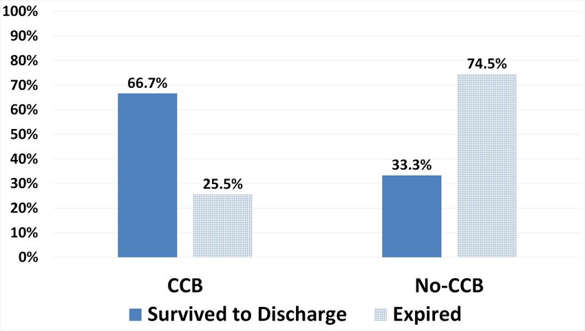
Considering this from a different perspective, 67% (12/18) of patients that survived and that
were successfully discharged from the hospital were on a CCB, whereas 74% (35/47) of patients
that expired were not on a CCB (Figure 1).
FIGURE 1: Percent Survival Vs. CCB and No-CCB Groups
CCB = Calcium Channel Blocker (Nifedipine or Amlodipine). No-CCB = Not on Calcium Channel
Blocker.
Patients treated with a CCB were significantly less likely to have undergone intubation and
mechanical ventilation. Since only one patient (4.2%) in the CCB group was intubated and
mechanically ventilated and 23 (95.8%) were not, whereas 16 (39.0%) were intubated and
mechanically ventilated and 25 (61.0%) were not in the No-CCB treatment group (PCCB No-CCB
Number of Patients Intubated and Mechanically Ventilated 1 16
Number of Patients that were NOT Intubated and Mechanically Ventilated 23 25
TABLE 3: Patients Intubated and Mechanically Ventilated Vs. CCB Medication
Treatment Group
CCB = Dihydropyridine Calcium Channel Blockers (Nifedipine or Amlodipine). No-CCB = Not having taken more than one dose of any
Dihydropyridine Calcium Channel Blockers (Nifedipine or Amlodipine).
FIGURE 2: Percent of Patients Not Intubated by CCB Group
CCB = Calcium Channel Blocker (Nifedipine or Amlodipine). No-CCB = No Calcium Channel
Blocker.
Other sample medications administered were not significantly different between groups (see
Table 4). Broad-spectrum antibiotics could be qualified with various medication regimens such
as ceftriaxone in combination with azithromycin or doxycycline, vancomycin and meropenem,
etc. Intravenous fluid comprised all patients that were administered at a minimum of 40 mL/hr
at some point during the course of hospitalization. Steroid use included any type such
as methylprednisolone, dexamethasone, or hydrocortisone.
2020 Solaimanzadeh et al. Cureus 12(5): e8069. DOI 10.7759/cureus.8069 6 of 16Intervention CCB % No-CCB % P
Broad-Spectrum Antibiotics 24 100.0% 41 100.0% NS
Intravenous Fluid 22 91.7% 33 80.5% NS
Hydroxychloroquine 20 83.3% 32 78.0% NS
Steroids 8 33.3% 12 29.3% NS
Heparin 16 66.7% 25 61.0% NS
Enoxaparin 2 8.3% 9 22.0% NS
Venodyne Boots 2 8.3% 2 4.9% NS
Apixaban 2 8.3% 1 2.4% NS
Rivaroxaban 1 4.2% 3 7.3% NS
Warfarin 1 4.2% 0 0.0% NS
No Anti-Coagulation 0 0.0% 1 2.4% NS
TABLE 4: Miscellaneous Medications Between Groups
NS = Not Significant. CCB = Calcium Channel Blocker (Nifedipine or Amlodipine). No-CCB = No Calcium Channel Blocker.
Several other factors, including vital signs and laboratory findings at initial presentation, were
compared between groups as well (Table 5). Some patients did not have specific laboratory tests
drawn; the number of patients included is indicated. Also, the first recorded pulse oximetry
measure in many patients from both groups include levels obtained after immediate placement
of oxygen supplementation was already initiated.
2020 Solaimanzadeh et al. Cureus 12(5): e8069. DOI 10.7759/cureus.8069 7 of 16CCB SD N = 24 No-CCB SD N = 41 P value
BMI 29.0 ±7.17 23 30.2 ±7.45 40 0.5341
Temp 99.6 ±1.91 24 98.8 ±1.43 41 0.0652
Pulse 92.1 ±16.79 24 100.8 ±24.73 41 0.1326
RR 19.7 ±2.06 24 21.3 ±4.92 41 0.1347
Sat 94.0 ±7.66 24 92.0 ±8.26 41 0.3488
SBP 137.5 ±27.08 24 124.6 ±22.38 41 0.0421*
DBP 77.7 ±12.05 24 72.4 ±11.61 41 0.0861
MAP 97.6 ±15.27 24 89.8 ±13.54 41 0.0359*
Hb 12.5 ±1.97 24 12.4 ±2.54 41 0.8821
GFR 49.6 ±28.49 24 36.4 ±27.6 41 0.0700
ESR 62.9 ±26.01 17 82.1 ±28.24 21 0.0371*
D-Dimer 2559.1 ±1783.93 17 5710.4 ±5735.54 32 0.0328*
LA 1.9 ±0.9741 12 4.0 ±3.5623 31 0.0557
LDH 494.3 ±195.12 17 725.2 ±376.97 30 0.0234*
CRP 135.5 ±76.67 15 175.7 ±124.79 34 0.2546
BNP 252.1 ±558.16 19 436.4 ±836 32 0.3979
IL-6 77.4 ±52.92 3 502.0 ±944.77 9 0.2290
TABLE 5: Clinical Data Between Groups at Initial Presentation to Hospital
* indicates significance at pusage is associated with significantly improved mortality in elderly patients hospitalized for
COVID-19. They also reveal that CCB usage is associated with a significantly decreased risk for
intubation and mechanical ventilation.
This study reveals the possible benefits of nifedipine and amlodipine in patients hospitalized
with COVID-19. Larger clinical studies are warranted. For those that already have hypertension
and present to hospital with elevated blood pressure, assuming no contraindications exist, it
may be fair to give preference to a CCB as first-line therapy for concomitant benefit. Further
studies of other potential therapies that function as vasodilators in the pulmonary arterial flow
should be pursued as well.
Clinical data comparisons between groups at the time of presentation were significant for
differences in systolic as well as mean arterial blood pressure. This may indicate higher levels of
hypotension or at least a lack of elevated blood pressure measures in the No-CCB group at
presentation and/or foreshadow treatment with a CCB anti-hypertensive medication.
Differences in erythrocyte sedimentation rate, D-dimer, and lactate dehydrogenase may reflect
a disparate severity of disease upon presentation. However, on the other hand, this reasoning
may be countered since results between levels of lactic acid, C-reactive protein, B-type
natriuretic peptide, and interleukin-6 were not significantly different, in addition to no
significant difference between hemoglobin and glomerular filtration rate (GFR).
It is not known if patients were taking a CCB at home or not. This may be another avenue to
explore with consideration to decipher if the severity of disease expression is diminished or
halted by CCB medication - prior to virus exposure.
Future studies may investigate different patient populations. Moreover, CCB treatment, while
monitoring blood pressure closely in patients that are suffering from COVID-19, and that do
not have underlying Hypertension, may be envisaged. Blood pressure is generally not
hypotensive in many hospitalized patients suffering from COVID -19 and can even be higher in
patients in the intensive care unit (ICU) [13].
The limitations of this study may be inherent in the small sample size, possible confounding
factors not otherwise accounted for, and even selection bias as part of prior clinical decision-
making to treat hypertension with a CCB or not. Therefore, larger and more rigorous studies
should be pursued. Prospective studies may be considered as well. Large health systems may be
in a unique position to help advance knowledge on this subject matter by conducting a more
robust retrospective review. If clinical researchers therein would peruse electronic medical
records (EMR) for similar outcomes, as described in this paper, that can aid in gaining a better
understanding of the role that CCBs may have in mitigating disease. Particular attention may
be paid, where possible, on patient adherence to outpatient medication regimens prior to acute
hospitalization. For example, if the rates of hospitalization for patients that were adherent to
CCBs were decreased, that might indicate preventative benefits. This could potentially explain
significant differences of clinical data at the initial presentation described above, but this
remains to be investigated. With the current EMR technology available at some institutions,
this may be investigated rapidly. Results obtained may yield benefits for thousands of patients
across the country and throughout the world that have not yet fallen ill but remain at risk for
contracting this disease.
The need for mechanical ventilation likely represents the continuum of progression of the
disease. It can be regarded as an intervention aimed at curtailing a trajectory towards mortality.
However, mechanical ventilation should not be considered an all-encompassing treatment
option for patients with COVID-19. In a recent study, amongst 1151 patients intubated, only 38
(3.3%) were discharged alive, with 24.5% that died and over 70% still in hospital [14]. Avoiding
2020 Solaimanzadeh et al. Cureus 12(5): e8069. DOI 10.7759/cureus.8069 9 of 16intubation with the utilization of potential countermeasures, such as with CCBs and other
vasodilators described below, can be considered and evaluated since they may aid in the
achievement of improved outcomes. When precisely during the course of the illness (early vs.
late) these medications may be most effective may also be examined. Furthermore, perhaps
beneficial effects of CCBs and other vasodilators described below can be extended to patients
that are already mechanically ventilated. Clinical studies may investigate if successful weaning
from mechanical ventilation is promoted by the use of vasodilators. Therefore, it is incumbent
upon the medical community to exert their best efforts, on behalf of the many patients at risk,
and pursue further evaluation as part of efforts to enhance clinical interventions that compel
augmentation.
Concept
This data provides an impetus to explore different approaches to the treatment of patients with
COVID-19. Focusing on vasodilatory agents may allow for an alternative treatment strategy.
Herein, improved flow via the alveolar-capillary unit may be achieved. With improved flow,
impediments to oxygenation, including inflammation and vasoconstriction, may be better
negotiated. Furthermore, blood transit improvement as a result of vasodilation may potentially
offset clot formation. Lastly, fluid accumulation or edema that can inhibit oxygenation may be
collectively reduced as well. Altogether, the improved flow may attenuate the precipitous
progression of the disease.
Virchow’s triad highlights three aspects compromising blood flow: stasis, hypercoagulability,
and endothelial injury. All three may be occurring in advanced COVID-19. Yet, the progression
to severe disease consisting of an inability to oxygenate may be the endpoint of a gradual
process. In other words, flow (or micro-perfusion) via the alveolar-capillary unit may be slowly
but surely decreasing as a result of a vicious cycle wherein inflammation secondary to viral
injury begets hypercoagulability as well as the impedance of blood flow. Clot formation
certainly lulls or wholly undermines segments of previously oxygenating pathways passing
through the alveolar-capillary unit. Moreover, inflammation by itself, enhanced by the
recruitment of cytokines, leukocytes, and the whole gamut of caustic endogenous mechanisms,
may further render viable tissue non-functional. Additionally, with inflammation comes fluid
or edema formation - this also compromises oxygen diffusion. On top of all this exists the
innate disposition or tendency for hypoxic pulmonary vasoconstriction [15]. Perhaps, vascular
inflammation also contributes to elicit reactive vasoconstriction independently. Altogether, a
microvascular process may be occurring over numerous alveolar-capillary units, which yields a
macro result. In sum, with increasing hypoxia and respiratory failure, the following challenges
are faced and each promotes the other perhaps in sequence but not necessarily: (1) Viral injury
provoking inflammation, (2) recruitment of an Immune response, (3) fluid accumulation, (4)
vasoconstriction or compromised vascular flow, and (5) hypercoagulability and clot formation.
Multi-faceted challenges are faced in various clinical cases. However, the ultimate development
of clot formation may not be applicable to many, if not most, patients upon presentation. These
patients must be distinguished as not being in a category wherein the precipitous decline just
described has already been realized. This is especially early on in the illness, since some may be
managed, improve, and recover with supplemental oxygen and conservative fluid management
or gentle fluid restriction alone. Herein, a vasodilator can be utilized from the outset since
inflammation has not been prolonged, whereby flow via the alveolar-arterial complex is still
consistent and clot formation likely has not already developed. However, in patients that have
had prolonged symptoms, a consistent deterioration of oxygen saturation and/or hypoxemia
should be evaluated with the consciousness that all five challenges may have already been
established and taken form. Other patients maybe somewhere in between.
Broader implications
2020 Solaimanzadeh et al. Cureus 12(5): e8069. DOI 10.7759/cureus.8069 10 of 16This being said, consideration of other vasodilatory agents should be pursued. These may
include phosphodiesterase inhibitors sildenafil and tadalafil, as well as acetazolamide among
others.
For example, sildenafil was shown to increase exercise capacity during severe hypoxia, as well
as reduce hypoxic pulmonary hypertension at rest and during exercise while maintaining gas
exchange and systemic blood pressure [16]. The same study also revealed that it yields an
increased maximum workload and maximum cardiac output compared with the placebo.
Beyond this, phosphodiesterase inhibitors have the added benefit of improved renal perfusion
and GFR - a valuable commodity as kidney disease is associated with the in-hospital death of
patients with COVID-19 [17,18]. Also, when not administered with nitrates, sildenafil use
resulting in hypotension, orthostatic hypotension, and syncope were found to be less than 2%
[19-20]. Tadalafil once daily was found to improved exercise capacity and reduced time to
clinical worsening in patients suffering from pulmonary arterial hypertension (PAH); offering
an alternative to Sildenafil as well [21]. Finally, combining tadalafil with acetazolamide, rather
than taking acetazolamide alone, can be an even more effective method for the prevention of
some conditions [22]. Dosing of sildenafil is less restrictive in cases of compromised renal
function.
Acetazolamide also attenuates hypoxic pulmonary vasoconstriction but has the added benefit
of increasing minute ventilation and oxygenation [23-25].
Acetazolamide, however, requires close monitoring of arterial blood gases, prior to and
following use, as treatment is contraindicated in metabolic acidosis; a condition that it can
spur. However, some of the adverse effects of acetazolamide can be avoided by reducing the
dose to compensate for age‐related reductions in renal drug clearance [26]. In any case, the
addition of sodium bicarbonate can be utilized to counteract an acid tide and may be
administered repeatedly in an alternating fashion with acetazolamide [27]. Acetazolamide also
acts to inhibit carbonic anhydrase in vascular smooth muscle and this mechanism may be
achieved by means of pH changes therein [28]. Clinical status and work of breathing must also
be monitored closely. Patients that are already on a ventilator may also stand to benefit most
from acetazolamide, as the control of various parameters may be adjusted for the optimization
of therapy. An additional asset of acetazolamide includes diuresis of fluids - many, if not most,
patients have significant crackles present on auscultation, and this likely hinders oxygenation
as well (the latter is a clinical observation) [29].
Thus, acetazolamide can provide a triple benefit: diuresis of fluid/pulmonary edema, improved
ventilation, and reversal of pulmonary vasoconstriction.
There is a caveat to all of this. That is, improvement of oxygenation and ventilation can only be
pursued if clot formation does not exist or is previously adequately treated. A recent study
found that an incidence of thrombotic complications is up to 31% of ICU patients with COVID-
19 [30]. This must be addressed as well.
For example, in patients that are early stage and without markedly elevated D-dimers,
preferably younger and not elderly, wherein crackles are grossly apparent on auscultation, the
use of a vasodilator such as acetazolamide or a CCB may potentially stave off intubation
independently. However, in an elderly patient, with markedly elevated D-dimers and little to
no crackles with clear air movement on auscultation, it may be ineffective as alveolar-capillary
units that are already clotted may harbor a formidable barrier towards improvement. Therefore,
treatment with anti-coagulation prior to vasodilator therapy should be considered in these
patients. Treating any clots or microclots first may allow for the effective flow once vasodilator
2020 Solaimanzadeh et al. Cureus 12(5): e8069. DOI 10.7759/cureus.8069 11 of 16therapy is implemented.
Context
The vasodilatory agents mentioned in this article may enhance clinical outcomes in patients
suffering from COVID-19. Yet, they should be accompanied with considerations for anti-
coagulation and anti-inflammatory agents as well, not to mention antibiotics for the
prevention of co-infection and anti-virals that may also contribute towards resolution.
Since adding a vasodilatory agent in a patient that has had prolonged disease and may have
already developed a clot or microclots may not suffice. Anti-coagulation, whether in
prophylactic or treatment doses, may aid in reducing clot formation or extension. Therefore,
combining regimens particularly in elderly patients and/or those that have had a prolonged
course may be prudent. If anti-coagulation is administered, certainly close monitoring for any
signs of bleeding must be implemented. Nonetheless, some patients may recover without anti-
coagulation as well, and clinical acumen is necessary in all circumstances.
Fluids are an important subject matter that must be appreciated within the context of overall
management, although it certainly deserves further assessment beyond this article. Generally,
in patients that have stable blood pressure, no significant clinical signs of dehydration, and
crackles on auscultation, conservative fluid management seems to be best. Ground-glass
opacities present on imaging may be reflective of fluid collection as well. In these cases,
avoiding intravenous fluid hydration and relegating to oral fluid intake as needed may decrease
the risk of excess fluid accrual in the lungs. This approach is not feasible in many clinical
situations such as where co-morbid diabetic ketoacidosis, severe dehydration, hypernatremia,
or Rhabdomyolysis occurs. Nonetheless, oxygenation impedance as a result of edema
formation is still worth taking into account in clinical management over an extended course of
hospitalization. On the other hand, patients that are dehydrated from the outset or were
previously on anti-coagulation prior to presentation and perhaps taking a CCB may have
relatively clear sounding lungs on auscultation - this is a clinical observation. Those previously
taking anti-coagulation and/or a CCB might possibly be less prone to fluid accumulation given
preemptive counters to the impedance of flow and clot formation. This may be analogous to a
pipe wherein, if flow is preserved, fluid may pass without interference. However, if it is clogged
then certainly running more water will rebound and not successfully traverse the pipe without
backing up or collecting proximally. In the former, where significant interference is not present,
fluid may be a boon to expedite the clearance of inflammation. In the latter, where viral triggers
of inflammation, down the five-step theoretical pathogenesis described above, have already
exerted influence, it may worsen clinical status. The "pipe" analogy should be further qualified
since, in our case, vascular channels may not be intact, as endothelial injury and capillary
permeability are both likely. Moreover, inflammation in itself is prone to fluid accumulation as
seen in patients with bowel wall edema following surgery, ascites in cancer, or serositis. Yet,
while abdominal fluid accumulation may be a cause for significant discomfort, even mild
pulmonary edema development may swiftly compromise oxygenation.
Finally, the use of steroids in severe cases and potentially in all hospitalized patients may aid in
alleviating inflammation but, certainly, this is under investigation. With the concomitant use of
vasodilators and anti-coagulation, the benefits of steroid use in COVID-19 may become more
apparent.
All in all, in severe cases, early utilization of one or more vasodilator agent(s), anti-coagulation,
steroids, and a diuretic (if no contraindications exist, preferably acetazolamide, given the
additional benefits mentioned above), in addition to antibiotics and potential antivirals may be
one protocol pathway to consider (Table 6).
2020 Solaimanzadeh et al. Cureus 12(5): e8069. DOI 10.7759/cureus.8069 12 of 16AND/ Vasodilator- Anti- Anti-
Vasodilator(s) AND AND Steroid AND Antibiotics AND
OR Diuretic Coagulation Viral
Nifedipine
Ceftriaxone
Extended-
Acetazolamide* Enoxaparin Methylprednisone and
Release 30-90mg Anti-
125mg PO or 1mg/kg q12- 80mg IV then azithromycin
Daily OR viral
IV q8-12hr q24 hours 40mg q12 hours or
Amlodipine 5-
doxycycline
10mg PO Daily
AND/OR
Sildenafil** 20mg
PO then 20-50mg
Q8 hours or
10mg IV q8-12 hr
TABLE 6: Sample Potential Example of a Therapeutic Approach That Can Be
Considered for Evaluation
*Consider 125-250 mg q6hr IV in mechanically ventilated patients combined with sodium bicarbonate 50 mEq q12hr to offset acid tide;
attempt weaning from the ventilator in parallel with treatment. **Consider in patients with acute kidney injury.
mg = Milligrams. mEq = Milliequivalent. kg = Kilogram. IV = Intravenous.
Therapeutic interventions herein reflect the five-step progressive course outlined above.
Patients that are otherwise stable but requiring oxygen supplementation to maintain a stable
saturation level may improve with a vasodilator such as nifedipine or amlodipine (maximizing
the dose to achieve ideal blood pressure may be of interest), prophylactic anti-coagulation,
steroids, antibiotics and antiviral therapy alone without the use of a diuretic agent. Yet, if the
clinical disease progresses and suspicion of need for invasive ventilation emerges, a diuretic
such as acetazolamide may be considered. In ventilated patients, acetazolamide may be a key to
promote weaning as described above. Frequent and repeated doses of acetazolamide may be
necessary since the inflammatory effects of the viral provocation, as well as capillary
permeability, will not vanish instantaneously and following a brief course of treatment re-
accumulation of fluid may materialize. Therefore, consistent perseverance may be a successful
strategy to quell the buildup of fluid, maintain vasodilation, and allow for conquest over time.
Indeed, all vasodilation agents may exert their benefit over a gradual time course. Just like the
decline of patients succumbing to COVID-19 occurs over an extended time course of worsening
pathogenesis, so too the vasodilation agents may mitigate the same pathogenic process over an
extended course of the viral illness. In other words, treating for just two days may not suffice
even though some clinical improvement is observed. Stopping at that point may incur a
rebound in status. Rather, a course of treatment at least five to seven days and up to two to
three weeks in severe cases may be best.
It is worth noting that a relatively minor subset of patients, tending to be elderly and frail, has
been observed to have disproportionately cold hands and feet relative to their extremities and
the rest of their body. Additionally, they may be markedly sleepy and difficult to arouse. A pulse
oximeter may fail to retrieve a saturation level when placed on their fingers and may yield a
result only after being placed on their forehead. This may reflect an underlying lack of
2020 Solaimanzadeh et al. Cureus 12(5): e8069. DOI 10.7759/cureus.8069 13 of 16perfusion to extremities and conserving mechanisms for blood distribution. These patients are
extremely ill and, although they may have stable vital signs, may be at risk for rapid
deterioration. Similar treatment may ensue, but with compromised oral intake, intravenous
fluid hydration may be needed; this may be best conducted at a gentle, gradual, and
consistent rate. This subgroup should be recognized as possibly being part of a separate group
of patients with an advanced illness that may require special attention.
Any clinician that has encountered patient presentations on a COVID unit, ICU, or emergency
department is aware that presentations of COVID-19 are disparate and each individual case is
different. Nonetheless, while correcting any concomitant disturbance (e.g. renal failure,
hyperosmolar syndrome, hypernatremia, etc.), bearing in mind the central role of pulmonary
deterioration in overall clinical demise is crucial. Ultimately, morbidity and mortality in
COVID-19 may be a result of a failure to accommodate the pathogenic sequelae of toxic viral
provocation rather than an immunologic deficiency. Therefore, aiding in the adaptation and
negotiation of a physiologic response to the transient viral insult may effectively mitigate
disease burden and promote improved outcomes. Altogether, implementing a strategy that
optimizes flow via the alveolar-capillary unit may be a progressive path forward.
Conclusions
In this small retrospective review, dihydropyridine CCBs were found to be significantly
associated with improved mortality, as well as a decreased risk for intubation and mechanical
ventilation in elderly patients hospitalized with COVID-19. Larger clinical studies are
warranted. Future studies may also elucidate results in different patient populations and
possibly reveal benefits even in mechanically ventilated patients. Consideration of treatment
with a CCB in patients who are suffering from COVID-19 and that do not have underlying
hypertension may be studied as well. Other potential therapies that function as vasodilators in
pulmonary arterial flow should be pursued. Potential benefits may outweigh the risks of
including nifedipine or amlodipine in the treatment regimens of elderly patients with
hypertension hospitalized with COVID-19.
Additional Information
Disclosures
Human subjects: Consent was obtained by all participants in this study. Animal subjects: All
authors have confirmed that this study did not involve animal subjects or tissue. Conflicts of
interest: In compliance with the ICMJE uniform disclosure form, all authors declare the
following: Payment/services info: All authors have declared that no financial support was
received from any organization for the submitted work. Financial relationships: All authors
have declared that they have no financial relationships at present or within the previous three
years with any organizations that might have an interest in the submitted work. Other
relationships: All authors have declared that there are no other relationships or activities that
could appear to have influenced the submitted work.
Acknowledgements
The authors would like to thank Dr. Baoying Lin-Chen, Director of Infection Control at
Interfaith, and the industrious members of her team who have helped prepare parts of the
database utilized to conduct this review.
References
1. Bavishi C, Maddox TM, Messerli FH: Coronavirus disease 2019 (COVID-19) infection and renin
angiotensin system blockers. JAMA Cardiol. 2020, [Epub ahead of
2020 Solaimanzadeh et al. Cureus 12(5): e8069. DOI 10.7759/cureus.8069 14 of 16print]:10.1001/jamacardio.2020.1282
2. Gattinoni L, Coppola S, Cressoni M, Busana M, Rossi S, Chiumello D: Covid-19 does not lead
to a “typical” acute respiratory distress syndrome. Am J Respir Crit Care. 2020, [Epub ahead of
print]:10.1164/rccm.202003-0817le
3. Gattinoni L, Chiumello D, Rossi S: COVID-19 pneumonia: ARDS or not? . Crit Care. 2020,
2020:154. 10.1186/s13054-020-02880-z
4. Young TE, Lundquist LJ, Chesler E, Weir K: Comparative effects of nifedipine, verapamil, and
diltiazem on experimental pulmonary hypertension. Am J Cardiol. 1983, 51:195-200.
10.1016/S0002-9149(83)80035-6
5. Muramoto A, Caldwell J, Albert RK, Lakshminarayan S, Butler J: Nifedipine dilates the
pulmonary vasculature without producing symptomatic systemic hypotension in upright
resting and exercising patients with pulmonary hypertension secondary to chronic obstructive
pulmonary disease. Am Rev Respir Dis. 1986, 132:[Epub].
6. Woodmansey PA, O'Toole L, Channer KS, et al.: Acute pulmonary vasodilatory properties of
amlodipine in humans with pulmonary hypertension. Heart. 1996, 75:171-173.
10.1136/hrt.75.2.171
7. Sajkov D, Frith PA, McEvoy RD, Wang T, Bune AJ, Alpers JA: A comparison of two long-acting
vasoselective calcium antagonists in pulmonary hypertension secondary to COPD. Chest.
1997, 111:1622-1630. 10.1378/chest.111.6.1622
8. Franz IW, van der Meyden J, Schaupp S, Tönnesmann U: The effect of amlodipine on exercise-
induced pulmonary hypertension and right heart function in patients with chronic
obstructive pulmonary disease [Article in German]. Zeitschrift für Kardiologie. 2002, 91:833-
839. 10.1007/s00392-002-0860-9
9. Kennedy TP, Michael JR, Huang CK, Kallman CH, Zahka K, Schlott W, Summer W: Nifedipine
inhibits hypoxic pulmonary vasoconstriction during rest and exercise in patients with chronic
obstructive pulmonary disease. A controlled double-blind study. Am Rev Respir Dis. 1984,
129:544-551.
10. Burghuber OC: Nifedipine attenuates acute hypoxic pulmonary vasoconstriction in patients
with chronic obstructive pulmonary disease. Respiration. 1987, 52:86-93. 10.1159/000195309
11. Rohit E, Michelakis ED, Archer SL: Hypoxic pulmonary vasoconstriction. J Appl Physiol. 2005,
98:390-403. 10.1152/japplphysiol.00733.2004
12. Agusti AG, Barberá JA, Roca J, Wagner PD, Guitart R, Rodriguez-Roisín R: Hypoxic pulmonary
vasoconstriction and gas exchange during exercise in chronic obstructive pulmonary disease.
Chest. 1990, 97:268-275. 10.1378/chest.97.2.268
13. Huang C, Wang Y, Li X, Xie X: Clinical features of patients infected with 2019 novel
coronavirus in Wuhan, China. Lancet. 2020, 17:497-506. 10.1038/s41569-020-0360-5
14. Richardson S, Hirsch JS, Narasimhan M, et al.: Presenting characteristics, comorbidities, and
outcomes among 5700 patients hospitalized with COVID-19 in the New York City area. JAMA.
2020, [Epub ahead of print]:10.1001/jama.2020.6775
15. Moudgil R, Michelakis ED, Archer SL: Hypoxic pulmonary vasoconstriction. J Appl Physiol.
2005, 98:390-403. 10.1152/japplphysiol.00733.2004
16. Ghofrani HA, Reichenberger F, Kohstall MG, et al.: Sildenafil increased exercise capacity
during hypoxia at low altitudes and at Mount Everest base camp: a randomized, double-blind,
placebo-controlled crossover trial. Ann Intern Med. 2004, 141:169-177. 10.7326/0003-4819-
141-3-200408030-00005
17. Webb DJ, Vachiery JL, Hwang LJ, Maurey JO: Sildenafil improves renal function in patients
with pulmonary arterial hypertension. Br J Clin Pharmacol. 2015, 80:235-241.
10.1111/bcp.12616
18. Li Z, Wu M, Yao J, et al.: Caution on kidney dysfunctions of COVID-19 patients . Lancet. 2020,
[Epub ahead of print]:10.2139/ssrn.3559601
19. Cheitlin MD, Hutter AM Jr, Brindis RG, et al.: ACC/AHA expert consensus document. Use of
sildenafil (Viagra) in patients with cardiovascular disease. J Am Coll Cardiol. 1999, 33:273-282.
10.1016/s0735-1097(98)00656-1
20. Zusman RM, Morales A, Glasser DB, Osterloh IH: Overall cardiovascular profile of sildenafil
citrate. American J Cardiol. 1999, 83:35-44. 10.1016/S0002-9149(99)00046-6
21. Rosenzweig EB: Tadalafil for the treatment of pulmonary arterial hypertension . Expert Opin
Pharmacother. 2010, 11:127-132. 10.1517/14656560903413542
22. Leshem E, Caine Y, Rosenberg E, Maaravi Y, Hermesh H, Schwartz E: Tadalafil and
2020 Solaimanzadeh et al. Cureus 12(5): e8069. DOI 10.7759/cureus.8069 15 of 16acetazolamide versus acetazolamide for the prevention of severe high‐altitude illness. J Travel
Med. 2012, 19:308-310. 10.1111/j.1708-8305.2012.00636.x
23. Balanos GM, Teppema LJ, Steinback CD, Brown AD, Foster GE, Leigh R, Poulin MJ:
Acetazolamide reduces hypoxic pulmonary vasoconstriction in humans . Faseb J. 2006, [Epub
ahead of print]:
24. Larson EB, Roach RC, Schoene RB, Hornbein TF: Acute mountain sickness and acetazolamide:
clinical efficacy and effect on ventilation. JAMA. 1982, 248:328-332.
10.1001/jama.1982.03330030034021
25. Gulsvik R, Skjørten I, Undhjem K, et al.: Acetazolamide improves oxygenation in patients with
respiratory failure and metabolic alkalosis. Clin Respir J. 2013, 390-396. 10.1111/crj.12025
26. Chapron DJ, Gomolin IH, Sweeney KR: Acetazolamide blood concentrations are excessive in
the elderly: propensity for acidosis and relationship to renal function. J Clin Pharmacol. 1989,
29:348-353. 10.1002/j.1552-4604.1989.tb03340.x
27. Freed SZ: The alternating use of an alkalizing salt and acetazolamide in the management of
cystine and uric acid stones. J Urol. 1975, 113:96-99. 10.1016/S0022-5347(17)59417-3
28. Puscas I, Coltau M, Baican M, Domuta G, Hecht A: Vasodilatory effect of diuretics is
dependent on inhibition of vascular smooth muscle carbonic anhydrase by a direct
mechanism of action. Drugs Exp Clin Res. 1999, 25:271-279.
29. Kassamali R, Sica DA: Acetazolamide: a forgotten diuretic agent . Cardiol Rev. 2011, 19:276-
278. 10.1097/CRD.0b013e31822b4939
30. Klok FA, Kruip MJ, van der Meer NJ, et al.: Incidence of thrombotic complications in critically
ill ICU patients with COVID-19. Thromb Res. 2020, [Epub ahead of
print]:10.1016/j.thromres.2020.04.013
2020 Solaimanzadeh et al. Cureus 12(5): e8069. DOI 10.7759/cureus.8069 16 of 16You can also read