Dysregulation of Key Proteins Associated with Sperm Motility and Fertility Potential in Cancer Patients - MDPI
←
→
Page content transcription
If your browser does not render page correctly, please read the page content below
International Journal of
            Molecular Sciences

Article
Dysregulation of Key Proteins Associated with Sperm
Motility and Fertility Potential in Cancer Patients
Manesh Kumar Panner Selvam, Renata Finelli , Saradha Baskaran                and Ashok Agarwal *
 American Center for Reproductive Medicine, Cleveland Clinic, Cleveland, OH 44195, USA;
 pannerm@ccf.org (M.K.P.S.); finelli.renata@gmail.com (R.F.); saradhabaskaran@gmail.com (S.B.)
 * Correspondence: agarwaa@ccf.org; Tel.: +1-216-444-9485

 Received: 15 July 2020; Accepted: 8 September 2020; Published: 15 September 2020

 Abstract: Cancer has adverse effects on male reproductive health. Conventional semen analysis
 does not explain the molecular changes in the spermatozoa of cancer patients. Currently,
 proteomics is being widely used to identify the fertility-associated molecular pathways affected
 in spermatozoa. The objective of this study was to evaluate the sperm proteome of patients
 with various types of cancer. Cryopreserved semen samples from patients (testicular cancer, n
 = 40; Hodgkin’s disease, n = 32; lymphoma, n = 20; leukemia, n = 17) before starting therapy
 were used for proteomic analysis, while samples from fertile donors (n = 19) were included as
 controls. The proteomic profiling of sperm was carried out by liquid chromatography-tandem
 mass spectrometry, and differentially expressed proteins involved in the reproductive processes
 were validated by Western blotting. Bioinformatic analysis revealed that proteins associated with
 mitochondrial dysfunction, oxidative phosphorylation, and Sirtuin signaling pathways were
 dysregulated in cancer patients, while oxidative phosphorylation and tricarboxylic acid cycle
 were predicted to be deactivated. Furthermore, the analysis revealed dysregulation of key proteins
 associated with sperm fertility potential and motility (NADH:Ubiquinone oxidoreductase core subunit
 S1, superoxide dismutase 1, SERPINA5, and cytochrome b-c1 complex subunit 2) in the cancer group,
 which were further validated by Western blot. Dysfunctional molecular mechanisms essential for
 fertility in cancer patients prior to therapy highlight the potential impact of cancer phenotype on
 male fertility.

 Keywords: bioinformatics; cancer; male infertility; proteomics; sperm

1. Introduction
      Cancer in reproductive age can significantly affect fertility in males, representing one of the most
common causes of infertility. The majority of people affected by cancer are >55 years old (~80%).
In 2020, a high number of people in reproductive age (15–39 years old) are predicted to be affected by
testicular cancer (6500 estimated new cases), Hodgkin’s lymphoma (2800 estimated new cases) and
non-Hodgkin’s lymphoma (4600 estimated new cases), as well as leukemia (600 estimated new cases)
in the United States [1]. As reported by the American Cancer Society, an increase in relative survival
rate has been reported for all types of cancers since the 1960s. This may be due to early diagnosis and
the availability of more effective treatments [1]. Hence, the possibility of achieving a future pregnancy
is a concern for cancer patients [2].
      Cancer therapies have gonadotoxic effects, which result in reduced semen quality and reproductive
potential [3,4]. Moreover, the exposure to radiation can also have teratogenic impact as it increases the
rate of sperm DNA mutation and the risk of genetic abnormalities in the offspring [5]. These therapies
include radio- and chemotherapy, while surgery may be the final option [6]. Hence, the cryopreservation
of semen sample is the only option for cancer patients before starting a gonadotoxic therapy to father a

Int. J. Mol. Sci. 2020, 21, 6754; doi:10.3390/ijms21186754                          www.mdpi.com/journal/ijms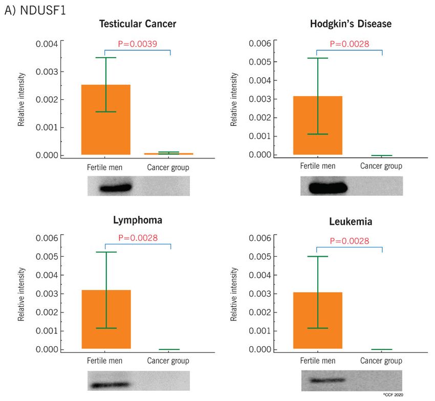
Int. J. Mol. Sci. 2020, 21, 6754                                                                                      2 of 14

biological child in the future. However, besides the impact of gonadotoxic treatments on semen quality,
preliminary studies report abnormal semen parameters, particularly sperm motility, in pre-treated
cancer patients [7–10]. A recent retrospective study conducted by Auger et al., involving 4480 male
patients of reproductive age affected by several types of cancer and systematic disease, reported
a significant decrease in sperm concentration, progressive motility and normal morphology [10].
Specifically, testicular cancer and leukemia patients showed poor semen quality, as normozoospermia
was observed only in 50.9% and 36.9% of patients, respectively [10]. Similar observations were also
reported by other investigators [11–14]. Low sperm quality was observed in patients with Hodgkin’s
disease when compared to non-Hodgkin’s lymphoma [15], while oligospermia was reported in more
than 50% of the leukemia patients [16]. However, a recent study reported that 75% of patients affected
by Hodgkin’s disease are normozoospermic prior to treatment [17]. In fact, better semen quality is
observed in patients with Hodgkin’s disease than in those with testicular cancer [8,18].
      Routine semen analysis performed in the workup of male infertility provides information regarding
the quality of sperm, however it does not help elucidate the molecular mechanisms associated with
infertility. The advent of proteomics has enabled to decipher the molecular mechanisms as well as the
subcellular changes related to the fertilization potential of spermatozoa [19,20]. Hitherto, few proteomics
studies have been conducted on semen samples of patients affected by specific types of cancer [21–24].
However, the general impact of cancer on protein expression in sperm has not been elucidated yet.
Understanding the cellular and molecular mechanisms of cancer-associated male infertility is not only
crucial for the management of male infertility, but also useful for fertility preservation in cancer patients.
Proteomic results could (1) explain the pathways negatively affecting male fertility in pre-treatment
patients, (2) help in achieving a better cryopreservation outcome in samples from cancer patients,
(3) identify protein biomarkers for cancer-associated infertility and may (4) lead to more effective
diagnosis and possible treatment of complications associated with cancer. Therefore, the main objective
of this study was to investigate the sperm proteome of cancer patients before initiating cancer therapy
in comparison with that of healthy fertile men.

2. Results

2.1. Semen Parameters
     Sperm concentration and motility are reported in Table 1 for each group. Although the average
values were within the physiological limits provided by the WHO guidelines, a significant reduction
of sperm concentration was observed in all cancer groups, while a significant decline in sperm motility
was noted in patients with testicular cancer and lymphoma when compared to the fertile donors.

           Table 1. Semen parameters of different types of cancer patients compared with fertile donors.

                    Subjects                  Sperm Concentration (106 /mL)                      Total Motility (%)
               Fertile Donors                                  81                                         71
                   (n = 19)                              (70.4–107.1)                                (56.5–70.4)
              Testicular cancer                               16 a                                       51 a
                   (n = 40)                                (9.9–29.4)                                 (35–67.5)
              Hodgkin’s disease                              26.8 a                                      60.5
                   (n = 32)                                 (13–62)                                  (41.0–74.0)
                 Leukemia                                    64.7 a                                       70
                   (n = 17)                                 (43–72)                                  (33.8–95.7)
                Lymphoma                                     55.9 a                                     43.5 a
                   (n = 20)                               (42.4–64.7)                                (24.4–55.5)
                Data are reported as median (25th–75th percentile).   a   p < 0.05 when compared to fertile donors.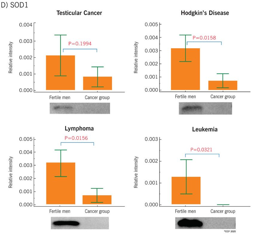
Int.Int. J. Mol.
J. Mol. Sci.Sci. 2020,
2020, 21,21, x FOR PEER REVIEW
6754 3 of3 15
of 14
Int. J. Mol. Sci. 2020, 21, x FOR PEER REVIEW 3 of 15
2.2. Protein Profile of Cancer Patients
2.2.
2.2. Protein
Protein Profile of
of Cancer
Profile profiling
CancerofPatients
Patients
Proteomic spermatozoa in cancer patients and fertile men revealed a total of 1138
Proteomic
proteins profiling
common to of
cancerspermatozoa
group
Proteomic profiling of spermatozoa in
in cancer
and fertile patients
men, with
cancer and
and fertile
fertile men
460 differentially
patients revealed
expressed
men aa total
proteins
revealed of
of 1138
(DEPs)
total 1138
proteins
proteins common to cancer group and fertile men, with 460 differentially expressed proteins (DEPs)
(Figurecommon
1). to cancer group and fertile men, with 460 differentially expressed proteins (DEPs)
(Figure 1).
(Figure 1).
Figure 1. Proteome profile of sperm in cancer group and fertile men. DEPs: differentially expressed
Figureproteins.
1. Proteome profile of sperm in cancer group and fertile men. DEPs: differentially expressed proteins.
Figure 1. Proteome profile of sperm in cancer group and fertile men. DEPs: differentially expressed
proteins.
AA comparativeanalysis
comparative analysisshowed
showed that
that 208
208 and
and 62
62 proteins
proteins were
wereunder-
under-and andover-expressed,
over-expressed,
respectively,
respectively, inin cancergroup
cancer groupcompared
compared to to fertile
fertile men,
men, while
while 182
182and
and88proteins
proteinswere werefound
found to tobebe
A comparative
uniquely expressed analysis
in showed
fertile men andthat 208 and
cancer 62 proteins
patients, wereTable
respectively. under-2 and over-expressed,
provides the list of
uniquely expressed in fertile men and cancer patients, respectively. Table 2 provides the list of uniquely
respectively,
uniquely in cancerproteins
expressed group compared
in to fertile
spermatozoa of men,patients
cancer while 182 andcompared
when 8 proteins to were
fertile found
men. to be
The
expressed proteins in spermatozoa of cancer patients when compared to fertile men. The abundance
uniquely expressed
abundance in fertile
of the proteins men and
expressed in cancer
the cancer patients,
group respectively.
and fertile men Table 2 provides
are presented the list2. of
in Figure
of the proteins expressed in the cancer group and fertile men are presented in Figure 2. Most of the
uniquely
Most ofexpressed
the proteinsproteins in spermatozoa
expressed in both groupsofwere cancer patients
present when
in very lowcompared
abundanceto fertile2).
(Figure men. The
proteins expressed in both groups were present in very low abundance (Figure 2).
abundance of the proteins expressed in the cancer group and fertile men are presented in Figure 2.
Most of the Table 2. Sperm
proteins proteins uniquely
expressed expressed in the cancer
ingroup
very compared to fertile(Figure
men.
Table 2. Sperm proteinsinuniquely
both groups were
expressed inpresent
the cancer group low abundance
compared to fertile men. 2).
S.N UniProt ID Protein Abundance
S.N UniProt
Table 2. Sperm ID uniquely Cancer
proteins vs. Protein
expressed Men group compared Abundance
in the cancer
Fertile to fertile men.
1. Q13421 mesothelin
Cancer isoform
vs. Fertile Men X1 L
S.N UniProt ID Protein Abundance
1. 2. Q6W4
Q13421× 9 mucin-6 (MUC6)
mesothelin isoform
isoform X1, partial L L
Cancer vs. Fertile MenX1
2. 3. Q13438
Q6W4 ×9 mucin-6 protein OS-9 isoform
(MUC6) isoform X1,X1 L L
1. 4. Q13421
Q02413
mesothelin
desmoglein-1
isoform X1partial
preproprotein
L
VL L
3. Q13438 protein OS-9 isoform X1
2. 5. Q6W4 ×9
P68871 mucin-6 (MUC6) isoform
hemoglobin subunit X1,betapartial VLLVL
4. Q02413 desmoglein-1 preproprotein
5. 3. 6. Q13438
O14773
P68871 protein OS-9
tripeptidyl-peptidase isoform
hemoglobin subunit X1
1 preproprotein
beta VLLVL
6. 4. 7. Q02413
O14773
Q86YZ3 desmoglein-1
tripeptidyl-peptidase preproprotein
hornerin1 preproprotein VL
VL VL
7. 5. 8. P68871
Q86YZ3
P08697 hemoglobin subunit
hornerin
alpha-2-antiplasmin beta X6
isoform VL
VL VL
8. 6. P08697
O14773 alpha-2-antiplasmin
tripeptidyl-peptidase 1
L: low; VL: very low.isoform X6
preproprotein VLVL
7. Q86YZ3 hornerin
L: low; VL: very low. VL
8. P08697 alpha-2-antiplasmin isoform X6 VL
L: low; VL: very low.
Figure2.2.Abundance
Figure Abundanceof
of sperm
sperm proteins identified
identifiedin
incancer/fertile
cancer/fertilegroups.
groups.
Figure 2. Abundance of sperm proteins identified in cancer/fertile groups.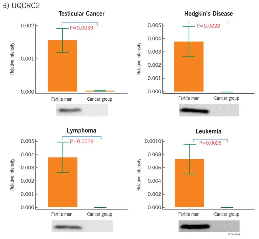
Int. J. Mol. Sci. 2020, 21, x FOR PEER REVIEW                                                                         4 of 15
Int. J. Mol. Sci. 2020, 21, 6754                                                                                       4 of 14

2.3. Canonical Pathways
2.3. Canonical Pathwaysanalysis revealed that DEPs identified in the cancer group compared to fertile
      A bioinformatic
 menAwere    involved in
        bioinformatic       the regulation
                         analysis  revealedof  theDEPs
                                            that   top canonical
                                                         identifiedpathways      (Figure
                                                                      in the cancer      3).compared
                                                                                     group   Proteins associated
                                                                                                        to fertile
 withwere
men    mitochondrial
            involved in  dysfunction,   oxidative
                           the regulation         phosphorylation,
                                           of the top  canonical pathwaysand Sirtuin
                                                                               (Figuresignaling pathways
                                                                                        3). Proteins         were
                                                                                                      associated
 dysregulated    in  cancer  group  in comparison    to fertile men    (Figure 3). Furthermore,
with mitochondrial dysfunction, oxidative phosphorylation, and Sirtuin signaling pathways were    a comparative
 analysis of two
dysregulated        sets of group
                in cancer   DEPs predicted    deactivation
                                   in comparison    to fertileofmen
                                                                 oxidative
                                                                     (Figurephosphorylation
                                                                              3). Furthermore, and  tricarboxylic
                                                                                                 a comparative
 acid (TCA)
analysis      cycle
          of two     (Table
                   sets      3). Apredicted
                        of DEPs    heat map deactivation
                                             analysis revealed     an underexpression
                                                            of oxidative                 of proteins
                                                                           phosphorylation           involved in
                                                                                              and tricarboxylic
 oxidative  phosphorylation      and the TCA   cycle (Figure   4).
acid (TCA) cycle (Table 3). A heat map analysis revealed an underexpression of proteins involved in
oxidative phosphorylation and the TCA cycle (Figure 4).

     Figure 3. Higher -log (p-value) indicates the dysfunction of the corresponding pathways due to the
      Figure 3. Higher
     involvement          -log (p-value)
                    of differential       indicates
                                     expressed        the dysfunction
                                                   proteins.            of shows
                                                               The figure   the corresponding   pathways
                                                                                   that mitochondrial      due to the
                                                                                                       dysfunction,
      involvement
     oxidative       of differential
                phosphorylation    andexpressed     proteins.
                                       Sirtuin signaling       The figure
                                                             pathway,       shows
                                                                      protein       that mitochondrial
                                                                               ubiquitination           dysfunction,
                                                                                              pathway, tricarboxylic
      oxidative
     acid         phosphorylation
           (TCA) cycle  II, isoleucine and     Sirtuin I, signaling
                                       degradation        acetyl-CoApathway,     protein
                                                                      biosynthesis         ubiquitination
                                                                                     and tRNA   charging arepathway,
                                                                                                             the top
      tricarboxylic  acid   (TCA)   cycle  II,  isoleucine
     canonical pathways affected in cancer group.             degradation   I, acetyl-CoA   biosynthesis  and  tRNA
      charging are the top canonical pathways affected in cancer group.
       Table 3. Deactivated pathways and upstream regulators affected in spermatozoa of cancer patient.

                                        Pathways                                   Z Score *
                               Oxidative Phosphorylation                              −3.46
                            Tricarboxylic acid (TCA) cycle II                         −2.45
                                 Fatty acid β-oxidation I                             −2.00
                                       Glycolysis I                                   −2.24
                                  Upstream regulators
                                         RICTOR                                       4.785
                                         KDM5A                                        3.464
                                         MAP4K4                                       3.162
                                         TRAP1                                        2.433
     * Activation or inactivation of pathways are indicated by Z score. A Z score > 2 indicates activation while a value
Int. J. Mol. Sci. 2020, 21, 6754 5 of 14
Int. J. Mol. Sci. 2020, 21, x FOR PEER REVIEW 5 of 15
4. Heat
Figure 4. Heatmap
mapillustrating
illustratingthe
theexpression
expressionof of proteins
proteins associated
associated withwith oxidative
oxidative phosphorylation
phosphorylation and
and tricarboxylic
tricarboxylic acid (TCA)
acid (TCA) cycle. cycle. Intensity
Intensity of thecorresponds
of the color color corresponds to the expression
to the expression level
level of the of the
proteins.
proteins.
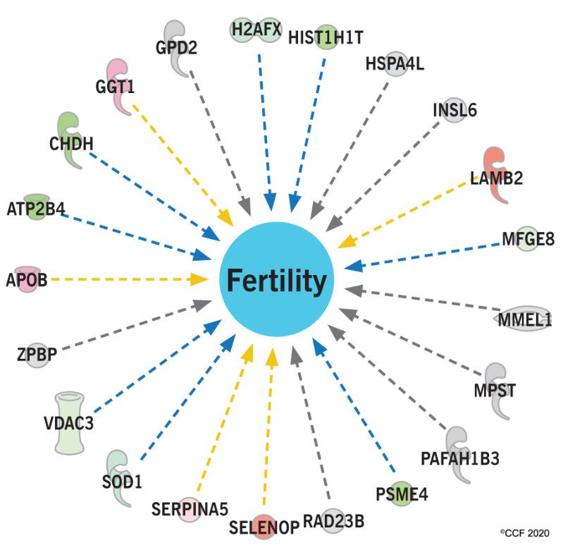
2.4. Reproductive Pathways Dysregulated in Cancer Patients
Table 3. Deactivated
Functional analysispathways
revealedand upstream
that regulators affected
the upstream in spermatozoa
regulators (RICTOR, ofKDM5A,
cancer patient.
MAP4K4,
and TRAP1) were activated due to the Pathways
aberrant expression of sperm* proteins in the cancer group
Z Score
compared to fertile men (Table 3). A pathways
Oxidative analysis showed
Phosphorylation that proteins associated with the
−3.46
sperm motility and the fertilityTricarboxylic
potential were
aciddysregulated
(TCA) cycle II in the cancer group (Figure 5A,B).
−2.45
Fatty acid β-oxidation I −2.00
2.5. Western Blot Results Glycolysis I −2.24
Upstream regulators
Four key proteins, NADH:Ubiquinone oxidoreductase core subunit S1 (NDUFS1), cytochrome b-c1
RICTOR 4.785
complex subunit 2 (UQCRC2), SERPINA5 and superoxide dismutase 1 (SOD1), were validated using
KDM5A 3.464
Western blot in different cancer types, such as testicular cancer,3.162
MAP4K4
Hodgkin’s disease, leukemia, and
lymphoma, in comparison with fertile men (Supplementary
TRAP1 Figure
2.433 S1). The analysis revealed the
underexpression
* Activation orofinactivation
NDUFS1 ofand UQCRC2
pathways in all the
are indicated bycancer
Z score.types compared
A Z score to the
> 2 indicates fertile men
activation
(Figure 6A,B). SERPINA5 was significantly overexpressed in testicular
while a value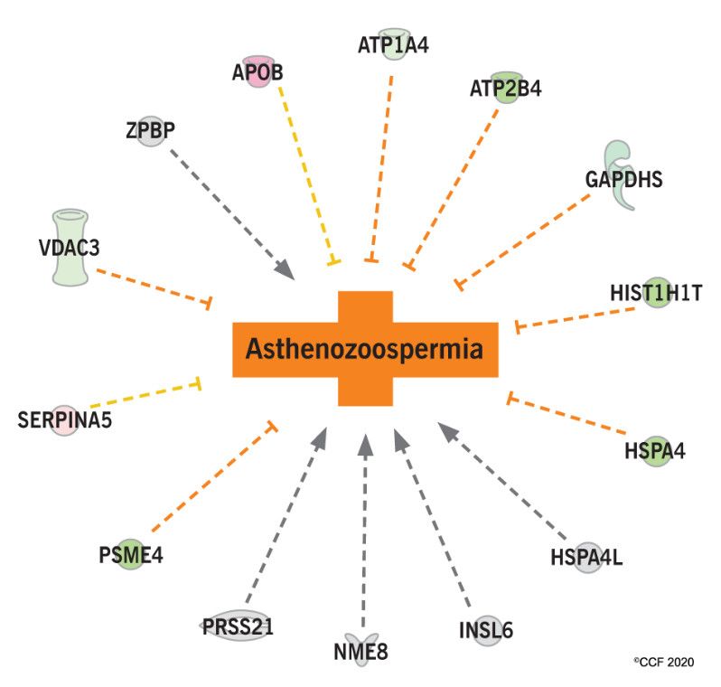
Int. J. Mol. Sci. 2020, 21, 6754                                                                            6 of 14
 Int. J. Mol. Sci. 2020, 21, x FOR PEER REVIEW                                                              6 of 15

                             (A)                                                    (B)
      Figure 5. Differentially expressed proteins associated with (A) fertility potential and (B) motility of
      sperm in the cancer group in comparison with fertile men.

2.5. Western Blot Results
     Four key proteins, NADH:Ubiquinone oxidoreductase core subunit S1 (NDUFS1), cytochrome
b-c1 complex subunit 2 (UQCRC2), SERPINA5 and superoxide dismutase 1 (SOD1), were validated
using Western blot in different cancer types, such as testicular cancer, Hodgkin’s disease, leukemia,
and lymphoma, in comparison with fertile men (Supplementary Figure S1). The analysis revealed the
underexpression of NDUFS1 and UQCRC2 in all the cancer types compared to the fertile men (Figure
6A,B). SERPINA5 was (A)       significantly overexpressed in testicular cancer,(B)      Hodgkin’s disease, and
lymphoma groups (Figure 6C), while SOD1 was underexpressed in leukemia and lymphoma cancer
     Figure 5.
     Figure   5. Differentially
                 Differentially expressed
                                expressed proteins
                                          proteins associated
                                                   associated with
                                                              with (A)
                                                                   (A) fertility
                                                                        fertility potential
                                                                                  potential and
                                                                                            and (B)
                                                                                                 (B) motility
                                                                                                     motility of
                                                                                                              of
compared    to fertile   men (Figure 6D).   In the case of Hodgkin’s      disease and testicular      cancer, SOD1
     sperm in
     sperm   in the
                 the cancer
                     cancer group
                            groupinincomparison
                                      comparisonwith
                                                 withfertile
                                                      fertilemen.
                                                             men.
expression was comparable with fertile men (Figure 6D).
2.5. Western Blot Results
     Four key proteins, NADH:Ubiquinone oxidoreductase core subunit S1 (NDUFS1), cytochrome
b-c1 complex subunit 2 (UQCRC2), SERPINA5 and superoxide dismutase 1 (SOD1), were validated
using Western blot in different cancer types, such as testicular cancer, Hodgkin’s disease, leukemia,
and lymphoma, in comparison with fertile men (Supplementary Figure S1). The analysis revealed the
underexpression of NDUFS1 and UQCRC2 in all the cancer types compared to the fertile men (Figure
6A,B). SERPINA5 was significantly overexpressed in testicular cancer, Hodgkin’s disease, and
lymphoma groups (Figure 6C), while SOD1 was underexpressed in leukemia and lymphoma cancer
compared to fertile men (Figure 6D). In the case of Hodgkin’s disease and testicular cancer, SOD1
expression was comparable with fertile men (Figure 6D).

Int. J. Mol. Sci. 2020, 21, x FOR PEER REVIEW                                                              7 of 15

     Figure
     Figure 6. Western blot
            6. Western  blot analysis
                             analysis of
                                      of (A)
                                         (A) NDUFS1
                                             NDUFS1 (B) (B) UQCRC2, (C) SERPINA5
                                                            UQCRC2, (C)          and (D)
                                                                        SERPINA5 and (D) SOD1
                                                                                         SOD1 in
                                                                                              in
     spermatozoa of cancer patients compared to fertile men.
     spermatozoa of cancer patients compared to fertile men.

3. Discussion
     During the past decade, the proteomics platform was used to profile the proteins in seminal
fluids of patients diagnosed with several types of cancers, such as prostate cancer [25–27], testicular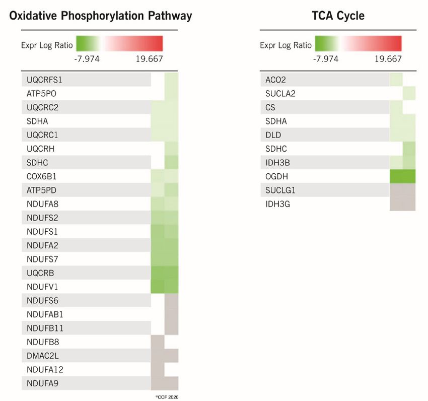
Int. J. Mol. Sci. 2020, 21, 6754                                                                     7 of 14

3. Discussion
      During the past decade, the proteomics platform was used to profile the proteins in seminal
fluids of patients diagnosed with several types of cancers, such as prostate cancer [25–27],
testicular cancer [21,22,28], Hodgkin’s disease [23], and leukemia [24]. Based on their ability to detect
the expression of thousands of proteins simultaneously, proteomics techniques are of great interest to
unravel the molecular mechanisms leading to infertility in cancer patients. In addition, these techniques
can identify putative new molecular biomarkers for diagnosis, prognosis, and therapeutics. As the
cancer treatment itself can affect male fertility potential, we analyzed the sperm proteome of cancer
patients before initiating any therapy, in order to understand the molecular pathways affected by the
cancer disease.
      Our results show an altered sperm protein profile in cancer patients. Previous proteomic studies
conducted on patients before starting any therapy reported 11 and 46 DEPs to be uniquely expressed in
normozoospermic testicular cancer patients [22,28], while 15 and 4 proteins were uniquely expressed
in asthenozoospermic patients affected by testicular cancer [22,28], suggesting that cancer might
affect sperm maturation, even if the conventional semen parameters remain unaltered. Furthermore,
an overexpression of the exosomal protein, matrix metalloproteinase 9 (MMP9), has been reported
in both normo- and asthenozoospermic cancer patients [28], in agreement with previous reports of
its increased expression in infertile men [29]. Similarly, the proteins endothelial lipase precursor,
apolipoprotein A-IV precursor, and carcinoembryonic antigen-related cell adhesion molecule 8
precursor were reported to be expressed uniquely in patients affected by Hodgkin’s disease, suggesting
an impairment of metabolic processes and pathways related to the production and synthesis of reactive
oxygen species (ROS) [23]. Our proteomic results support the above evidence and strongly suggest
that the investigated types of cancer might be responsible for the alteration of biological pathways
in sperm.
      In the current study, a total of 8 proteins were identified to be uniquely expressed in cancer
patients compared to fertile men. Their expression was reportedly altered in different types of cancer.
Mesothelin is a glycophosphatidylinositol (GPI)-anchored protein, identified as an antigen in several
human cancers affecting the pancreas, endometrium, ovary, and lung, as well as in mesothelioma and
pediatric leukemia [30]. Although it is expressed in several tissues, including testis, the inactivation of
mesothelin gene in experimental mice model did not affect the normal fertility, pregnancy and offspring
delivery [31]. This suggests that this gene might not be crucial for reproduction. The expression of
mucin isoforms has been reported in germ cells, with mucin-1 showing the highest expression in
mature spermatozoa in the testis [32]. Mucin-1 is speculated to be involved in sperm maturation
and transportation along the reproductive system [32]. In addition, it has been reportedly associated
with survival in gastric cancer patients [33], while mucin-6 isoform was associated with favorable
progression-free and cancer-specific survival in colorectal cancer [34].
      Protein OS-9 involved in the ubiquitination of misfolded proteins was identified for the first
time in a human osteosarcoma cell line [35,36], while desmoglein-1 was reported to interact with the
testis-specific isoform of Na/K ATPase in the plasma membrane of bovine sperm during capacitation [37].
Desmoglein-1 is a component of endosomes, and it was reportedly reduced in lung cancer [38].
The hemoglobin subunit beta was highly expressed in serum of ovarian cancer patients [39],
while tripeptidyl-peptidase 1 was proposed as a biomarker for colorectal and lung cancer [40,41].
Tripeptidyl-peptidase 1 is a lysosomal serine protease whose enzymatic activity has been observed in
pancreatic mucinous cysts with a high probability to develop into invasive carcinoma [42]. Moreover,
it has been also reported to be overexpressed in seminal plasma of patients with improved semen quality
after varicocelectomy, in comparison with those who did not show any improvement [43]. Hornerin,
a member of the S100 calcium-binding protein family, has been associated with the progression and
poor prognosis of hepatocellular and breast cancer [44,45]. Increased expression of alpha-2-antiplasmin,
a serine protease inhibitor, has been reported following varicocelectomy and in malignant ovarian
cancer [46,47]. In addition, it has also been proposed as a serum biomarker for the early diagnosis of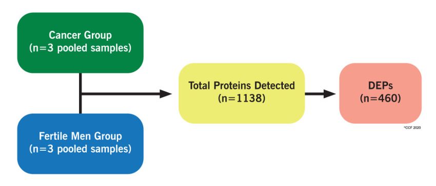
Int. J. Mol. Sci. 2020, 21, 6754                                                                       8 of 14

B-cell acute lymphoblastic leukemia [48]. The identification of these proteins in spermatozoa suggests
that common molecular mechanisms are affected in different cancer conditions, supporting the use of
these proteins as biomarkers in biological fluids other than serum such as seminal fluid.
      The differential expression pattern between cancer and control groups indicate that these proteins
could be directly involved in infertility-related pathways, suggesting their possible role as new
mediators of male infertility in cancer patients. Apart from understanding the function of specific
proteins, it is equally important to study the collective role of proteins associated with specific molecular
pathways. In the current study, molecular functions associated with mitochondria were dysregulated
in the spermatozoa of cancer patients, mainly due to the aberrant expression of the mitochondrial
proteins. Earlier proteomic studies reported that sperm mitochondrial proteins were affected in cancer
conditions such as testicular cancer [22,28] and Hodgkin’s disease [23]. Based on our proteomic
results, the oxidative phosphorylation, a process linked to mitochondrial function, was defective in the
spermatozoa of cancer patients (irrespective of the type of cancer included in this study). Moreover, heat
map analysis revealed dysregulation of a cluster of proteins involved in oxidative phosphorylation and
TCA cycle in cancer patients compared to fertile men. Western blot validation of NDUFS1 and UQCRC2
protein expression in cancer patients were in accordance with the proteomic results. NDUFS1 is
a mitochondrial membrane protein and a component of Complex I which mediates the transfer of
electrons to the respiratory chain. NDUFS1 has been reported to be under the regulation of RICTOR
in testicular cancer patients [22]. In the present study, we noticed the underexpression of NDUFS1
and the activation of upstream regulator RICTOR, which is also responsible for the maintenance
of the blood-testis-barrier [49] and the regulation of spermatogenesis [50]. This was in agreement
with previous reports, showing a lower expression of NDUFS1 in the sperm of patients affected
by non-seminoma testicular cancer before starting any therapy [21]. Therefore, underexpression
of NDUFS1 suggests that mitochondrial function is compromised in cancer patients, with a direct
effect on the fertilizing capacity of spermatozoa. Similarly, the expression of another key protein,
UQCRC2, was regulated by the RICTOR [22]. UQCRC2 is reportedly involved in the TCA cycle
and its underexpression has been correlated with poor fertilization rates [51] and male infertility
conditions, such as varicocele [52]. In the present study, UQCRC2 was underexpressed in cancer
patients, indicating multiple protein dysregulations in the mitochondria of spermatozoa regardless of
the type of cancer, as demonstrated by Western blot analysis. The fertility potential of spermatozoa
depends on the molecular regulation of several pathways. In the current study, several DEPs identified
in the spermatozoa of cancer patients can affect the normal sperm physiological functions, especially
motility or fertilizing ability.
      Western blot analysis revealed an underexpression of SOD1 in Hodgkin’s disease, leukemia and
lymphoma cancer groups. SOD1 is an enzyme involved in the antioxidant defense mechanism during
a state of oxidative stress [53], hence, underexpression of SOD1 suggests a dysfunctional mechanism to
counteract oxidative stress in cancer patients. Furthermore, SERPINA5, the protein responsible for the
binding and penetration of spermatozoa into the oocyte [54], was overexpressed in spermatozoa of
men with all the cancer types, suggesting an impairment of fertilization-associated pathways.
      The current sperm proteome of cancer patients clearly shows that their reproductive functions
are disturbed at a molecular level at the time of diagnosis. This highlights that these patients can be
potentially at risk of infertility way before treatment. However, there are a few limitations of this
study. The inclusion criteria did not include the stage of cancer. Furthermore, tracking the clinical
outcome of our fertile controls or cancer patients post-treatment is impractical, as these subjects
were not actively planning to start a family. Our study offers a snapshot of the proteome in cancer,
without the possibility to identify specific molecular pathways differentially altered during the cancer
progression. Moreover, the proteomics analysis was conducted on cryopreserved samples. While it
is well-known that cryopreservation can reduce semen quality [13], we cannot exclude the influence
of the freezing/thawing processes on the sperm proteome, which has been previously suggested
by Wang et al., who observed a differential expression of proteins involved in physiological sperm
Int. J. Mol. Sci. 2020, 21, 6754 9 of 14
pathways following cryopreservation [55]. Nevertheless, our study offers preliminary data that lays
the foundation for future studies, as screening of the sperm proteome from cancer patients before
treatment may help in the identification of molecular factors dysregulated in the spermatozoa of
those patients.
4. Materials and Methods
4.1. Study Population
This study was approved by the Institutional Review Board (IRB) of the Cleveland Clinic
Foundation. Consent was obtained from cancer patients to use their samples for research purposes.
Patients affected by the following types of cancer were included in this study, regardless of the stage or
specific subtype of the disease before start of any cancer therapy: testicular cancer (n = 40), Hodgkin’s
disease (n = 32), lymphoma (n = 20) and leukemia (n = 17). Additionally, proven fertile and healthy
donors (n = 19) were included as a control group. All the proteomics experiments were carried out
according to the Minimum Information about a Proteomics Experiment (MIAPE) guidelines released
by the Human Proteome Organization’s Proteomics Standard Initiative (HUPO-PSI) [56].
4.2. Semen Analysis
Semen samples were collected by masturbation after a period of sexual abstinence (2–3 days).
An aliquot (6 µL) of liquefied semen was placed on a Leja sperm counting chamber (Spectrum
Technologies, Healdsburg, CA, USA) and analyzed before cryopreservation, according to the WHO
guidelines [57].
4.3. Cryopreservation and Thawing
Semen samples were cryopreserved using the TEST-Yolk Buffer (TYB, Irvine Scientific, Santa Ana,
CA, USA). Aliquots of TYB equal to 25% of the sample volume were added to the specimen at
room temperature, and mixed gently for 5 min using the Hema-Tek aliquot mixer (Miles Scientific,
Elkhart, IN, USA). The procedure was repeated to a final 1:1 (v/v) ratio of the freezing medium to the
sample. Further, the samples were dispensed into cryovials (1.5 mL; Corning, Pittsburg, PA, USA) and
transferred to the freezer (−20 ◦ C) for 8 min (static cooling), and subsequently to liquid nitrogen vapor
(−80 ◦ C) for 2 h (vapor-phase cooling). Finally, the cryovials were stored in liquid nitrogen at −196 ◦ C
until use for proteomics analysis.
4.4. Sperm Protein Extraction, Quantification, and Separation
Cryopreserved samples were thawed at 37 ◦ C. The samples were then centrifuged at 4000× g
for 7 min to remove all cryoprotective medium. Further, the samples were washed thrice with
1X PBS (phosphate-buffered saline) to remove the remnants of cryoprotectant. RIPA buffer
(Sigma-Aldrich, St. Louis, MO, USA) containing Protease Inhibitor Cocktail, cOmpleteTM ULTRA
Tablets, EDTA-free (Roche, Mannheim, Germany) was added to the sperm pellet (100 µL/106
spermatozoa), and subsequently stored overnight at 4 ◦ C to allow for complete lysis of the
cells. After centrifugation (10,000× g, 30 min, 4 ◦ C), the supernatant was transferred into a new
micro-centrifuge tube. The protein content was then quantified using the bicinchoninic acid assay
(BCA assay, Waltham, MA, USA).
Proteomics analyses were carried out on groups of pooled samples. Each group consisted of
3 pooled samples created by obtaining the equal contribution of sperm from each cancer type.
Sperm proteins extracted from each pooled sample were mixed with SDS-PAGE buffer and run in
triplicates on 1D-SDS PAGE.Int. J. Mol. Sci. 2020, 21, 6754 10 of 14
4.5. Liquid Chromatography-Tandem Mass Spectrometer Analysis (LC-MS/MS)
The bands from each lane were excised from the gel with a gel punch. Subsequently, the bands
were washed and destained in 50% acetonitrile containing 5% acetic acid, followed by dehydration
in the speed-vac concentrator. The dried gel pieces were digested overnight with trypsin (5 µL of
10 ng/µL) in 50 mM ammonium bicarbonate at 37 ◦ C. Peptides were extracted from the polyacrylamide
gel in two 30 µL volumes of 50% acetonitrile and 5% formic acid, and combined and evaporated in a
speed-vac concentrator to reduce the volume to 10 µL. Finally, the samples were resuspended in 15%
acetic acid to make a final volume of ~30 µL for LC-MS/MS analysis.
The mass spectrometer used in this study was a Finnigan Orbitrap Elite hybrid trap mass
spectrometer. Five µL volumes of the extracted peptide samples were injected into a Dionex
15 cm × 75 µm id Acclaim Pepmap C18C18 reversed-phase capillary chromatography column for LC
separation, before introduction into the on-line mass spectrometer. The peptides eluted from the
column by an acetonitrile/0.1% formic acid gradient at a flow rate of 0.3 µL/min were introduced
into the source of the mass spectrometer on-line. The micro-electrospray ion source was set to 2.5 kV.
The resulting digest was analyzed using the data-dependent multitask capability of the instrument,
acquiring full scan mass spectra to determine peptide molecular weights and product ion spectra to
determine the amino acid sequence in successive instrument scans.
4.6. Proteomic Data Analysis
The data were analyzed using all collision-induced dissociation spectra collected in the experiment
to search the National Center for Biotechnology Information (NCBI) human reference sequence database
with the search programs MASCOT (Mascot version 2.7; Matrix Science, Boston, MA, USA) and Sequest
(Sequest version 1.4, ThermoScientific, San Jose, CA, USA) to detect the proteins present in the in-gel
digestions. The results from both search programs were uploaded into the Scaffold (version 4.0.6.1,
Proteome Software, Portland, OR, USA). Protein abundance was sorted as very low, low, medium,
and high, depending on the number of spectral counts (SpC), while the DEPs were categorized as
underexpressed, overexpressed or uniquely expressed in a particular group, based on the normalized
spectral abundance factor (NSAF). Depending on the protein abundance, different p-values were taken
into consideration: (a) very low abundance: SpC = 1.7–7; p ≤ 0.001; NSAF ratio ≥ 2.5 for overexpressed,
≤0.4 for underexpressed proteins; (b) low abundance: SpC = 8–19; p ≤ 0.01; NSAF ratio ≥ 2.5 for
overexpressed, ≤0.4 for underexpressed proteins; (c) medium abundance: SpC = 20–79; p ≤ 0.05; NSAF
ratio ≥ 2.0 for overexpressed, ≤0.5 for underexpressed proteins; (d) high abundance: SpC > 80; p ≤ 0.05;
NSAF ratio ≥ 1.5 for overexpressed, ≤0.67 for underexpressed proteins.
A functional bioinformatics analysis was performed with the help of the publicly available
annotations (Gene Ontology—GO, from GO Term Finder and GO Term Mapper), UNIPROT, STRAP and
the proprietary software Ingenuity Pathway Analysis (version 2018-2019, IPA, Qiagen, Redwood
City, CA, USA), to identify the differentially regulated processes, pathways, cellular distribution,
and protein-protein interactions amongst proteins in the experimental and control groups, as well as
for data integration.
4.7. Western Blotting
In this study, the following proteins were selected for validation by Western blotting, as they
showed a moderate/high abundance in at least one of the groups, and were involved in reproductive
functions as well as in the canonical pathways predicted to be affected in the spermatozoa of cancer
patients: NDUFS1, UQCRC2, SERPINA5 and SOD1. Their differential expression was validated in
different samples (n = 12 for each group) from those analyzed by LC-MS/MS. A total of 20 µg of
proteins was fractionated through 10% SDS–polyacrylamide gels and transferred to polyvinylidene
difluoride (PVDF) membranes (GE Healthcare, Marlborough, MA, USA) using the Trans-Blot Cell
system (Bio-Rad Inc., Hercules, CA, USA). After transfer, nitrocellulose membranes were blocked for 1 hInt. J. Mol. Sci. 2020, 21, 6754 11 of 14
with 5% milk in 0.1% Tris-buffered saline containing 0.1% Tween (TBST), and incubated with primary
antibodies overnight (Supplementary Table S1). The resulting blots were probed with horseradish
peroxidase-conjugated secondary antibody for 2 h at room temperature. Membranes were washed
four times with TBST after the primary and secondary incubations. Proteins were detected using the
ECL substrate (GE Healthcare, Marlborough, MA, USA) on film and quantified using Image LabTM
software (Bio-Rad Inc., Hercules, CA, USA).
4.8. Statistical Analysis
Statistical analysis was carried out by using the MedCalc Statistical Software version v19.0.3
(MedCalc Software bvba, Ostend, Belgium). Variable distribution was evaluated by applying the
Kolmogorov–Smirnov test. The Mann–Whitney test was conducted to compare Western blotting
results, while the Spearman’s rank correlation test was applied to analyze the associations. p value
was significant whenInt. J. Mol. Sci. 2020, 21, 6754 12 of 14
2. Salati, M.; Cesaretti, M.; Macchia, M.; El Mistiri, M.; Federico, M. Epidemiological overview of Hodgkin
lymphoma across the Mediterranean basin. Mediterr. J. Hematol. Infect. Dis. 2014, 6, 1–10. [CrossRef]
[PubMed]
3. Agarwal, A.; Allamaneni, S.S.R. Disruption of spermatogenesis by the cancer disease process. J. Natl. Cancer
Inst. Monogr. 2005, 2005, 9–12. [CrossRef]
4. Osterberg, E.C.; Ramasamy, R.; Masson, P.; Brannigan, R.E. Current practices in fertility preservation in male
cancer patients. Urol. Ann. 2014, 6, 13–17. [CrossRef] [PubMed]
5. Arnon, J.; Meirow, D.; Lewis-Roness, H.; Ornoy, A. Genetic and teratogenic effects of cancer treatments on
gametes and embryos. Hum. Reprod. Update 2001, 7, 394–403. [CrossRef] [PubMed]
6. Howell, S.J.; Shalet, S.M. Spermatogenesis after cancer treatment: Damage and recovery. J. Natl. Cancer Inst.
Monogr. 2005, 2005, 12–17. [CrossRef] [PubMed]
7. Williams, D.H., IV; Karpman, E.; Sander, J.C.; Spiess, P.E.; Pisters, L.L.; Lipshultz, L.I. Pretreatment semen
parameters in men with cancer. J. Urol. 2009, 181, 736–740. [CrossRef] [PubMed]
8. Gandini, L.; Lombardo, F.; Salacone, P.; Paoli, D.; Anselmo, A.P.; Culasso, F.; Dondero, F.; Lenzi, A. Testicular
cancer and Hodgkin’s disease: Evaluation of semen quality. Hum. Reprod. 2003, 18, 796–801. [CrossRef]
9. Hallak, J.; Mahran, A.; Chae, J.; Agarwal, A. Poor semen quality from patients with malignancies does not
rule out sperm banking. Urol. Res. 2000, 28, 281–284. [CrossRef]
10. Auger, J.; Sermondade, N.; Eustache, F. Semen quality of 4480 young cancer and systemic disease patients:
Baseline data and clinical considerations. Basic Clin. Androl. 2016, 26, 3. [CrossRef]
11. Hamano, I.; Hatakeyama, S.; Nakamura, R.; Fukuhara, R.; Noro, D.; Tanaka, T.; Yoneyama, T.; Yamamoto, H.;
Yoneyama, T.; Hashimoto, Y.; et al. Differences in semen characteristics between patients with testicular
cancer and other malignancies using various cut-off values. Int. J. Urol. 2018, 25, 817–824. [CrossRef]
12. Bussen, S.; Sütterlin, M.; Steck, T.; Dietl, J. Semen parameters in patients, with unilateral testicular cancer
compared to patients with other malignancies. Arch. Gynecol. Obstet. 2004, 269, 196–198. [CrossRef]
[PubMed]
13. MacKenna, A.; Crosby, J.; Huidobro, C.; Correa, E.; Duque, G. Semen quality before cryopreservation
and after thawing in 543 patients with testicular cancer. JBRA Assist. Reprod. 2017, 21, 31–34. [CrossRef]
[PubMed]
14. Hallak, J.; Kolettis, P.N.; Sekhon, V.S.; Thomas, A.J.; Agarwal, A. Cryopreservation of sperm from patients
with leukemia: Is it worth the effort? Cancer 1999, 85, 1973–1978. [CrossRef]
15. Botchan, A.; Hauser, R.; Gamzu, R.; Yogev, L.; Lessing, J.B.; Paz, G.; Yavetz, H. Sperm quality in Hodgkin’s
disease versus non-Hodgkin’s lymphoma. Hum. Reprod. 1997, 12, 73–76. [CrossRef]
16. Chung, K.; Irani, J.; Knee, G.; Efymow, B.; Blasco, L.; Patrizio, P. Sperm cryopreservation for male patients
with cancer: An epidemiological analysis at the University of Pennsylvania. Eur. J. Obstet. Gynecol. Reprod.
Biol. 2004, 113, S7–S11. [CrossRef]
17. Paoli, D.; Rizzo, F.; Fiore, G.; Pallotti, F.; Pulsoni, A.; Annechini, G.; Lombardo, F.; Lenzi, A.; Gandini, L.
Spermatogenesis in Hodgkin’s Lymphoma Patients: A Retrospective Study of Semen Quality before and
after Different Chemotherapy Regimens. Hum. Reprod. 2016, 31, 263–272. [CrossRef] [PubMed]
18. Kobayashi, H.; Larson, K.; Sharma, R.K.; Nelson, D.R.; Evenson, D.P.; Toma, H.; Thomas, A.J.; Agarwal, A.
DNA damage in patients with untreated cancer as measured by the sperm chromatin structure assay.
Fertil. Steril. 2001, 75, 469–475. [CrossRef]
19. Milardi, D.; Grande, G.; Vincenzoni, F.; Castagnola, M.; Marana, R. Proteomics of human seminal plasma:
Identification of biomarker candidates for fertility and infertility and the evolution of technology. Mol. Reprod.
Dev. 2013, 80, 350–357. [CrossRef] [PubMed]
20. Kumar, M.; Selvam, P.; Henkel, R.; Finelli, R.; Agarwal, A. Proteomics and metabolomics—Current and
future perspectives in clinical andrology. Andrologia 2020, e13711. [CrossRef]
21. Dias, T.R.; Agarwal, A.; Pushparaj, P.N.; Ahmad, G.; Sharma, R. New insights on the mechanisms affecting
fertility in men with non-seminoma testicular cancer before cancer therapy. World J. Mens. Health 2018, 36.
[CrossRef]
22. Panner Selvam, M.K.; Agarwal, A.; Pushparaj, P.N. Altered molecular pathways in the proteome of
cryopreserved sperm in testicular cancer patients before treatment. Int. J. Mol. Sci. 2019, 20, 677. [CrossRef]
[PubMed]Int. J. Mol. Sci. 2020, 21, 6754 13 of 14
23. Martins, A.D.; Agarwal, A.; Baskaran, S.; Pushparaj, P.N.; Ahmad, G.; Panner Selvam, M.K. Alterations of
spermatozoa proteomic profile in men with Hodgkin’s disease prior to cancer therapy. World J. Mens. Health
2019, 37. [CrossRef] [PubMed]
24. Jain, P.; Ojha, S.K.; Kumar, V.; Bakhshi, S.; Singh, S.; Yadav, S. Differential seminal plasma proteome signatures
of acute lymphoblastic leukemia survivors. Reprod. Biol. 2019, 19, 322–328. [CrossRef] [PubMed]
25. Neuhaus, J.; Schiffer, E.; von Wilcke, P.; Bauer, H.W.; Leung, H.; Siwy, J.; Ulrici, W.; Paasch, U.; Horn, L.C.;
Stolzenburg, J.U. Seminal plasma as a source of prostate cancer peptide biomarker candidates for detection
of indolent and advanced disease. PLoS ONE 2013, 8, e67514. [CrossRef] [PubMed]
26. Veveris-Lowe, T.L.; Kruger, S.J.; Walsh, T.; Gardiner, R.A.; Clements, J.A. Seminal fluid characterization
for male fertility and prostate cancer: Kallikrein-related serine proteases and whole proteome approaches.
Semin. Thromb. Hemost. 2007, 33, 87–99. [CrossRef] [PubMed]
27. Drabovich, A.P.; Saraon, P.; Drabovich, M.; Karakosta, T.D.; Dimitromanolakis, A.; Eric Hyndman, M.;
Jarvi, K.; Diamandis, E.P. Multi-omics biomarker pipeline reveals elevated levels of protein-glutamine
gammaglutamyltransferase 4 in seminal plasma of prostate cancer patients. Mol. Cell. Proteom. 2019, 18,
1807–1823. [CrossRef]
28. Panner Selvam, M.K.; Agarwal, A.; Pushparaj, P.N. A quantitative global proteomics approach to
understanding the functional pathways dysregulated in the spermatozoa of asthenozoospermic testicular
cancer patients. Andrology 2019, 7, 454–462. [CrossRef]
29. Buchman-Shaked, O.; Kraiem, Z.; Gonen, Y.; Goldman, S. Presence of matrix metalloproteinases and tissue
inhibitor of matrix metalloproteinase in human sperm. J. Androl. 2002, 23, 702–708.
30. Hassan, R.; Thomas, A.; Alewine, C.; Le, D.T.; Jaffee, E.M.; Pastan, I. Mesothelin immunotherapy for cancer:
Ready for prime time? J. Clin. Oncol. 2016, 34, 4171–4179. [CrossRef]
31. Bera, T.K.; Pastan, I. Mesothelin Is Not Required for Normal Mouse Development or Reproduction. Mol. Cell.
Biol. 2000, 20, 2902–2906. [CrossRef]
32. Martínez-Conejero, J.A.; Garrido, N.; Remohí, J.; Pellicer, A.; Simón, C.; Meseguer, M. MUC1 in human
testis and ejaculated spermatozoa and its relationship to male fertility status. Fertil. Steril. 2008, 90, 450–452.
[CrossRef]
33. Kim, D.H.; Shin, N.; Kim, G.H.; Song, G.A.; Jeon, T.Y.; Kim, D.H.; Lauwers, G.Y.; Park, D.Y. Mucin expression
in gastric cancer: Reappraisal of its clinicopathologic and prognostic significance. Arch. Pathol. Lab. Med.
2013, 137, 1047–1053. [CrossRef] [PubMed]
34. Betge, J.; Schneider, N.I.; Harbaum, L.; Pollheimer, M.J.; Lindtner, R.A.; Kornprat, P.; Ebert, M.P.; Langner, C.
MUC1, MUC2, MUC5AC, and MUC6 in colorectal cancer: Expression profiles and clinical significance.
Virchows Arch. 2016, 469, 255–265. [CrossRef] [PubMed]
35. Su, A.Y.; Hutter, C.M.; Trent, J.M.; Meltzer, P.S. Complete sequence analysis of a gene (OS-9) ubiquitously
expressed in human tissues and amplified in sarcomas. Mol. Carcinog. 1996, 15, 270–275. [CrossRef]
36. Alcock, F.; Swanton, E. Mammalian OS-9 Is upregulated in response to endoplasmic reticulum stress and
facilitates ubiquitination of misfolded glycoproteins. J. Mol. Biol. 2009, 385, 1032–1042. [CrossRef]
37. Rajamanickam, G.D.; Kastelic, J.P.; Thundathil, J.C. Testis-specific isoform of Na/K-ATPase (ATP1A4)
interactome in raft and non-raft membrane fractions from capacitated bovine sperm. Int. J. Mol. Sci. 2019,
20, 3159. [CrossRef] [PubMed]
38. Saaber, F.; Chen, Y.; Cui, T.; Yang, L.; Mireskandari, M.; Petersen, I. Expression of desmogleins 1–3 and their
clinical impacts on human lung cancer. Pathol. Res. Pract. 2015, 211, 208–213. [CrossRef]
39. Woong-Shick, A.; Sung-Pil, P.; Su-Mi, B.; Joon-Mo, L.; Sung-Eun, N.; Gye-Hyun, N.; Young-Lae, C.; Ho-Sun, C.;
Heung-Jae, J.; Chong-Kook, K.; et al. Identification of hemoglobin-α and -β subunits as potential serum
biomarkers for the diagnosis and prognosis of ovarian cancer. Cancer Sci. 2005, 96, 197–201. [CrossRef]
40. Choi, J.W.; Liu, H.; Shin, D.H.; Yu, G.I.; Hwang, J.S.; Kim, E.S.; Yun, J.W. Proteomic and cytokine plasma
biomarkers for predicting progression from colorectal adenoma to carcinoma in human patients. Proteomics
2013, 13, 2361–2374. [CrossRef]
41. Song, Q.-B.; Hu, W.G.; Wang, P.; Yao, Y.; Zeng, H.Z. Identification of serum biomarkers for lung cancer using
magnetic bead-based SELDI-TOF-MS. Acta Pharmacol. Sin. 2011, 32, 1537–1542. [CrossRef]
42. Ivry, S.L.; Knudsen, G.M.; Caiazza, F.; Sharib, J.M.; Jaradeh, K.; Ravalin, M.; O’Donoghue, A.J.; Kirkwood, K.S.;
Craik, C.S. The lysosomal aminopeptidase tripeptidyl peptidase 1 displays increased activity in malignant
pancreatic cysts. Biol. Chem. 2019, 400. [CrossRef]Int. J. Mol. Sci. 2020, 21, 6754 14 of 14
43. Camargo, M.; Intasqui, P.; Belardin, L.B.; Antoniassi, M.P.; Cardozo, K.H.M.; Carvalho, V.M.; Fraietta, R.;
Bertolla, R.P. Molecular pathways of varicocele and its repair—A paired labelled shotgun proteomics
approach. J. Proteom. 2019, 196, 22–32. [CrossRef] [PubMed]
44. Choi, J.; Kim, D.I.; Kim, J.; Kim, B.H.; Kim, A. Hornerin is involved in breast cancer progression. J. Breast
Cancer 2016, 19, 142–147. [CrossRef] [PubMed]
45. Fu, S.J.; Shen, S.L.; Li, S.Q.; Hua, Y.P.; Hu, W.J.; Guo, B.C.; Peng, B.G. Hornerin promotes tumor progression
and is associated with poor prognosis in hepatocellular carcinoma. BMC Cancer 2018, 18. [CrossRef]
[PubMed]
46. Del Giudice, P.T.; Da Silva, B.F.; Lo Turco, E.G.; Fraietta, R.; Spaine, D.M.; Santos, L.F.A.; Pilau, E.J.;
Gozzo, F.C.; Cedenho, A.P.; Bertolla, R.P. Changes in the seminal plasma proteome of adolescents before and
after varicocelectomy. Fertil. Steril. 2013, 100, 667–672. [CrossRef] [PubMed]
47. Poersch, A.; Grassi, M.L.; de Carvalho, V.P.; Lanfredi, G.P.; de Souza Palma, C.; Greene, L.J.; de Sousa, C.B.;
Carrara, H.H.A.; Dos Reis, F.J.C.; Faça, V.M. A proteomic signature of ovarian cancer tumor fluid identified
by highthroughput and verified by targeted proteomics. J. Proteom. 2016, 145, 226–236. [CrossRef] [PubMed]
48. De Souza Cavalcante, M.; Torres-Romero, J.C.; Lobo, M.D.P.; Moreno, F.B.M.B.; Bezerra, L.P.; Lima, D.S.;
Matos, J.C.; de Azevedo Moreira, R.; de Oliveira Monteiro-Moreira, A.C. A panel of glycoproteins as
candidate biomarkers for early diagnosis and treatment evaluation of B-cell acute lymphoblastic leukemia.
Biomark. Res. 2016, 4, 1. [CrossRef]
49. Dong, H.; Chen, Z.; Wang, C.; Xiong, Z.; Zhao, W.; Jia, C.; Lin, J.; Lin, Y.; Yuan, W.; Zhao, A.Z.; et al. Rictor
regulates spermatogenesis by controlling sertoli cell cytoskeletal organization and cell polarity in the mouse
testis. Endocrinology 2015, 156, 4244–4256. [CrossRef]
50. Oliveira, P.F.; Cheng, C.Y.; Alves, M.G. Emerging role for mammalian target of rapamycin in male fertility.
Trends Endocrinol. Metab. 2017, 28, 165–167. [CrossRef]
51. Shukla, K.K.; Kwon, W.S.; Rahman, M.S.; Park, Y.J.; You, Y.A.; Pang, M.G. Nutlin-3a decreases male fertility
via UQCRC2. PLoS ONE 2013, 8, 76959. [CrossRef]
52. Swain, N.; Samanta, L.; Agarwal, A.; Kumar, S.; Dixit, A.; Gopalan, B.; Durairajanayagam, D.; Sharma, R.;
Pushparaj, P.N.; Baskaran, S. Aberrant upregulation of compensatory redox molecular machines may
contribute to sperm dysfunction in infertile men with unilateral varicocele: A proteomic insight.
Antioxid. Redox Signal. 2020, 32, 504–521. [CrossRef]
53. Yu, B.; Huang, Z. Variations in antioxidant genes and male infertility. Biomed. Res. Int. 2015, 2015, 513196.
[CrossRef] [PubMed]
54. España, F.; Navarro, S.; Medina, P.; Zorio, E.; Estellés, A. The role of protein C inhibitor in human reproduction.
Semin. Thromb. Hemost. 2007, 33, 41–45. [CrossRef]
55. Wang, S.; Wang, W.; Xu, Y.; Tang, M.; Fang, J.; Sun, H.; Sun, Y.; Gu, M.; Liu, Z.; Zhang, Z.; et al. Proteomic
characteristics of human sperm cryopreservation. Proteomics 2014, 14, 298–310. [CrossRef] [PubMed]
56. Martínez-Bartolomé, S.; Deutsch, E.W.; Binz, P.A.; Jones, A.R.; Eisenacher, M.; Mayer, G.; Campos, A.;
Canals, F.; Bech-Serra, J.J.; Carrascal, M.; et al. Guidelines for reporting quantitative mass spectrometry
based experiments in proteomics. J. Proteom. 2013, 95, 84–88. [CrossRef]
57. WHO. WHO Laboratory Manual for the Examination and Processing of Human Semen, 5th ed.; WHO: Geneva,
Switzerland, 2010.
© 2020 by the authors. Licensee MDPI, Basel, Switzerland. This article is an open access
article distributed under the terms and conditions of the Creative Commons Attribution
(CC BY) license (http://creativecommons.org/licenses/by/4.0/).You can also read