Acute Kidney Injury in COVID-19 - Review - MDPI
←
→
Page content transcription
If your browser does not render page correctly, please read the page content below
International Journal of
               Molecular Sciences

Review
Acute Kidney Injury in COVID-19
Marta Głowacka                 , Sara Lipka, Ewelina Młynarska * , Beata Franczyk and Jacek Rysz

                                          Department of Nephrology, Hypertension and Family Medicine, Medical University of Lodz,
                                          90-549 Lodz, Poland; marta.glowacka96@gmail.com (M.G.); saralipka@wp.pl (S.L.);
                                          bfranczyk-skora@wp.pl (B.F.); jacek.rysz@umed.lodz.pl (J.R.)
                                          * Correspondence: emmlynarska@gmail.com; Tel.: +48-042-6393750

                                          Abstract: COVID-19 is mainly considered a respiratory illness, but since SARS-CoV-2 uses the
                                          angiotensin converting enzyme 2 receptor (ACE2) to enter human cells, the kidney is also a target of
                                          the viral infection. Acute kidney injury (AKI) is the most alarming condition in COVID-19 patients.
                                          Recent studies have confirmed the direct entry of SARS-CoV-2 into the renal cells, namely podocytes
                                          and proximal tubular cells, but this is not the only pathomechanism of kidney damage. Hypovolemia,
                                          cytokine storm and collapsing glomerulopathy also play an important role. An increasing number of
                                          papers suggest a strong association between AKI development and higher mortality in COVID-19
                                          patients, hence our interest in the matter. Although knowledge about the role of kidneys in SARS-
                                          CoV-2 infection is changing dynamically and is yet to be fully investigated, we present an insight
                                          into the possible pathomechanisms of AKI in COVID-19, its clinical features, risk factors, impact on
                                          hospitalization and possible ways for its management via renal replacement therapy.

                                          Keywords: COVID-19; acute kidney injury; RRT; dialysis


Citation: Głowacka, M.; Lipka, S.;
                                          1. Introduction
Młynarska, E.; Franczyk, B.; Rysz, J.          COVID-19 is an infectious disease caused by severe acute respiratory syndrome
Acute Kidney Injury in COVID-19.          coronavirus 2 (SARS-CoV-2), and it originated in Wuhan, China, in December 2019. As of
Int. J. Mol. Sci. 2021, 22, 8081.         4 March 2021, approximately 84,230,049 cases have been discovered worldwide, causing
https://doi.org/10.3390/ijms22158081      an ongoing global pandemic [1]. Such a large number of confirmed cases is related to the
                                          way the virus is transmitted, which is close human-to-human contact through droplets or
Academic Editor: Hajime Nagasu            aerosol via coughs, sneezes or talking. Infection may also occur by touching contaminated
                                          surfaces and then touching routes of transmission such as the mouth, eyes or nose [2].
Received: 27 June 2021                    COVID-19 disease mainly affects the respiratory system, which in more severe cases is
Accepted: 21 July 2021
                                          manifested by pneumonia, hypoxemia and acute respiratory distress syndrome. Although
Published: 28 July 2021
                                          the main focus is on the pulmonary features, physicians must be aware of complications
                                          that SARS-CoV-2 infection carries to other organs, including the kidneys [3]. Acute kidney
Publisher’s Note: MDPI stays neutral
                                          injury (AKI) is the most common kidney manifestation among patients hospitalized with
with regard to jurisdictional claims in
                                          COVID-19. According to KDIGO, AKI is defined as any of the following: (1) an increase in
published maps and institutional affil-
                                          serum creatinine (SCr) by ≥0.3 mg/dL (≥26.5 µmol/L) within 48 h; or (2) an increase in
iations.
                                          SCr ≥ 1.5 times of baseline within the prior 7 days; or (3) urine volume < 0.5 mL/kg/hour
                                          for 6 h. AKI can be also staged for severity according to KDIGO: stage (1) increase in
                                          SCr to 1.5–1.9 times baseline or by ≥0.3 mg/dL; stage (2) increase SCr to 2.0–2.9 times
                                          baseline; stage (3) increase SCr to 3.0 times baseline or increase in serum creatinine to
Copyright: © 2021 by the authors.
                                          ≥4.0 mg/dL (≥353.6 mmol/L) or initiation of renal replacement therapy [4]. Moreover,
Licensee MDPI, Basel, Switzerland.
                                          AKI development is far more frequent in severe and critically ill patients and is associated
This article is an open access article
                                          with poor prognosis and higher mortality [5–7]. Therefore, understanding the underlying
distributed under the terms and
                                          pathophysiology of kidney injury in the course of COVID-19 is crucial for early recognition
conditions of the Creative Commons
                                          of the damage and the implementation of optimal treatment.
Attribution (CC BY) license (https://
creativecommons.org/licenses/by/
4.0/).

Int. J. Mol. Sci. 2021, 22, 8081. https://doi.org/10.3390/ijms22158081                                       https://www.mdpi.com/journal/ijms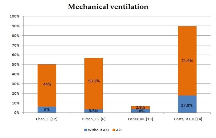
Int. J. Mol. Sci. 2021, 22, 8081                                                                                                       2 of 11

                                          2. Epidemiology
                                               With 84,230,049 cases of SARS-CoV-2 infection globally, at least one third is asymp-
                                          tomatic [8]. Among those COVID-19 patients who experience symptoms, about 80%
                                          develop mild to moderate symptoms, while 20% of cases present with severe symptoms
                                          over the course of the disease, of whom 6% become critically ill [9]. The overall mortality
                                          rate of COVID-19 patients is around 3% but, in the critically ill group, it can reach 50% [10].
                                               The incidence of acute kidney injury in COVID-19 varies in different case reports.
                                          Studies in China [5,10,11] have shown that AKI occurred in 5% to 29% within a median of
                                          7–14 days after admission, whereas reports from the United States [6,12,13] have shown
                                          greater rates reaching from 37% to 57% in COVID-19 positive patients. However, the onset
                                          of AKI in the US was observed much earlier—either upon admission or within 24 h of
                                          admission. Another study from Brazil also showed a high occurrence of AKI in 56% of
                                          COVID-19 patients, of which 67% developed stage 3 AKI [14].
                                               Fisher et al. [13] presented a comparison report between 3345 patients with COVID-
                                          19 and 1265 patients without COVID-19 during the same hospitalization period. AKI
                                          development in the COVID-19 (+) group was higher than in the COVID-19 (−) group (57%
                                          and 37%, respectively), and a significant number of patients positive for COVID-19 had
                                          stage 3 AKI compared with patients negative for COVID-19 (17.2% vs. 7.3%). Moreover,
                                          4.9% of the patients positive for COVID-19 required renal replacement therapy (RRT)
                                          compared with 1.6% of those negative for COVID-19. Other US studies reported RRT
                                          necessity in up to 19% of patients with COVID-19, while in Brazil it was up to 47% of
                                          patients.
                                               The results of incidence of AKI in COVID-19 patients are presented in Table 1.

                                                  Table 1. Incidence of AKI in COVID-19 patients.

                                                                                                                       RRT in Patients
                                                                                   Stages of AKI
                              All Patients           AKI                                                                 with AKI
                                                                       AKI 1            AKI 2            AKI 3
      Yang, X. [10]                52              15 (29%)
        Pei, G. [5]                333             22 (6.6%)         4 (18.2%)         7 (31.8%)        11 (50%)
     Cheng, Y. [11]                701             36 (5.1%)         13 (1.9%)         9 (1.3%)         14 (2%)
      Chan, L. [12]                3993          1835 (45.9%)        387 (21%)        199 (10.8%)     491 (26.7%)          347 (19%)
     Hirsch, J.S. [6]              5449          1993 (36.6%)       927 (46.5%)       447 (22.4%)     619 (31.1%)           14.3%
     Fisher, M. [13]               3345          1903 (56.9%)       942 (49.5%)       387 (20.3%)     574 (30.2%)         164 (4.9%)
   Costa, R.L.D. [14]              102            57 (55.9%)         10 (17.5%)        9 (15.8%)       38 (66.7%)         27 (47.4%)
    Ferlicot, S. [15]               47             1 (2.2%)           3 (6.4%)         2 (4.3%)        41 (87.2%)         30 (63.8%)

                                          3. Mechanism of SARS-CoV-2 Cellular Kidney Infection
                                               It is now a well-known fact that the main target of SARS-CoV-2 is the lungs; more
                                          precisely, type II pneumocytes. More and more studies published to date have proved that
                                          not only the lungs are exposed to infection, but so are the heart, liver, gastrointestinal tract,
                                          bone marrow and kidneys [3,16] Multiorgan tropism is due to the fact that SARS-CoV-2
                                          gains access to the cells through an endogenous viral receptor angiotensin converting
                                          enzyme 2 (ACE2) [17].
                                               In order for SARS-CoV-2 to enter the host, cells are required to bind its transmembrane
                                          spike (S) glycoprotein to cellular receptor ACE2. S consists of two subunits, each with
                                          a different function. S1 is responsible for binding to the host cell receptor, while S2 is
                                          used to fuse the viral membrane with the membrane of the infected cell [18,19]. Spike
                                          then requires proteolytic priming to be activated, which is granted by serine protease
                                          TMPRSS2. Therefore, ACE2 and TMPRSS co-expression is a key determinant for the
                                          entry of SARS-CoV-2 into host cells [20,21]. Once SARS-CoV-2 is in the cytosol of the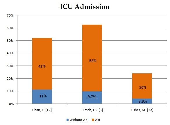
Int. J. Mol. Sci. 2021, 22, 8081                                                                                          3 of 11

                                   infected cell, the translation of its RNA and virion synthesis begins. It has been proven
                                   that genomic replication and virion assembly occur within the double vesicles of the
                                   endoplasmic reticulum (ER) and the Golgi complex [22].
                                         We can therefore conclude that susceptible kidney cells are those that express ACE2.
                                   Using RNA-Seq sequencing techniques, scientists were able to determine which kidney
                                   cell types comprised the ACE2 gene. The data show that ACE2 mRNA is mostly expressed
                                   in proximal tubular epithelial cells and podocytes [20,21]. This would concur with a report
                                   by Braun F. et al., who managed to isolate SARS-CoV-2 from epithelial cells of an autopsied
                                   kidney [23].

                                   4. Pathophysiology
                                        The best understood mechanism of kidney damage induced by SARS-CoV-2 is direct
                                   cellular infection. However, there are also few possible reasons for acute renal failure,
                                   such as inflammatory damage caused by cytokine storm, AKI related to acute respiratory
                                   distress syndrome (ARDS), kidney–lung crosstalk theory, hypovolemia and collapsing
                                   glomerulopathy [24–26].

                                   4.1. Direct Cellular Infection
                                         As mentioned before, SARS-CoV-2 penetrates through angiotensin converting enzyme
                                   2. The highest concentration of ACE2 in the kidneys was proven to be located in proximal
                                   tubular epithelial cells and podocytes [20,21,27]. Therefore, the direct infection of kidney
                                   cells by SARS-CoV-2 virus is the most likely mechanism for the development of acute
                                   kidney injury. Autopsy data also speak to this mechanism of AKI because many researchers
                                   found virus-like particles in epithelial cells of kidneys [24].

                                   4.2. Cytokine Storm and AKI Related to ARDS
                                        The abnormal immune response associated with SARS-CoV-2 is also a likely mecha-
                                   nism for the development of acute renal failure. At the root of these irregularities lies the
                                   cytokine storm and leukopenia [24]. Sepsis, a hemophagocytic syndrome, can lead to a
                                   so-called “cytokine storm”, which is a cytokine release syndrome (CRS). The most crucial
                                   cytokine responsible for this pathology is IL-6 [25]. IL-6 also occurs in ARDS complications
                                   of COVID-19. AKI in CRS is on intrarenal inflammation, increased vascular permeability,
                                   volume depletion and cardiomyopathy grounds. Cardiomyopathy can cause stasis in the
                                   renal veins, resulting in renal hypotension and hypoperfusion, leading to a reduction in
                                   the glomerular filtration rate. This phenomenon is called the syndrome type 1, which is
                                   manifested by endothelial damage, pleural effusions, edema, intra-abdominal hyperten-
                                   sion, third-space fluid loss, intravascular fluid depletion and hypotension [28]. Moreover,
                                   other complications of COVID-19, such as right and left ventricular failure, can cause AKI.
                                   The first one leads to blood stagnation in the kidneys, whereas the second one to reduced
                                   cardiac output and then to renal hypoperfusion [24]. There are five causes of AKI in ARDS:
                                   hemodynamic instability, hypoxemia/hypercapnia, acid-base dysregulation, inflammation
                                   and neurohormonal effects [25].

                                   4.3. Lung–Kidney Crosstalk Theory
                                        The occurrence of kidney dysfunctions in COVID-19 patients might be explained by
                                   the kidney–lung crosstalk theory. This is due to the increased concentration of cytokines in
                                   the blood, the release of which is promoted by lung injury. Elevated levels of cytokines,
                                   especially IL-6, cause an increase in alveolar capillary permeability and pulmonary hem-
                                   orrhage, and may even lead to distant-end organ dysfunction as damage to vascular
                                   endothelium in the kidneys. As a consequence, it leads to secondary hypoxia of the kidney
                                   and further damage to its structures. In patients who did not have chronic kidney disease
                                   or AKI, the research has shown that most of them manifested ARDS and/or AKI after
                                   developing pneumonia, which also testifies to the presence of lung–kidney crosstalk [28].
Int. J. Mol. Sci. 2021, 22, 8081 4 of 11
4.4. Hypovolemia
An incorrect distribution of fluids, especially hypovolemia (a consequence of fever
and tachypnea), may affect the kidneys. This condition causes renal hypoperfusion and,
consequently, renal failure. Endothelial damage, loss of fluid into the third space and
hypotension provoke renal hypoperfusion. Virus cells and cytokines produced by the
organism destroy the endothelium, which causes edema, ascites and hydrothorax, which in
turn leads to hypotension and a loss of fluids to the third space. The amount of circulating
fluid is reduced and this damages the kidneys in the prerenal mechanism. [28] Many
patients infected with SARS-CoV2 have gastrointestinal symptoms that greatly increase the
loss of fluid and further dehydration of the patient, mainly leading to pre-renal AKI [25].
In hemodynamically unstable patients, the venous flow deteriorates. Rhabdomyolysis (a
condition involving the rapid dissolution of damaged or injured skeletal muscle), metabolic
acidosis (increased level of hydrogen ions in the systemic circulation, which results in a
reduction in the level of serum bicarbonate) and hyperkalemia (serum or plasma potassium
level greater than 5.0 mEq/L to 5.5 mEq/L) are also associated with this condition. This has
a significant impact on the degradation of kidney function and, later on, on the occurrence
of AKI. [28] Therefore, optimizing hemodynamic is crucial to kidneys health.
4.5. Collapsing Glomerulopathy
Collapsing glomerulopathy (CG) is a histological term for focal segmental glomeru-
losclerosis defined by segmental or global glomerular collapse correlated with podocyte
proliferation, whose typical feature is proteinuria. CG has been associated with various
factors, but the essential one is the presence of Apolipoprotein 1 (APOL1) genotype, namely
alleles G1 and G2 [29]. Since the COVID-19 outbreak, there have been several case reports
presenting COVID-19 patients with CG, in which authors speculated about the possible
mechanisms linking it to SARS-CoV-2 infection. Kissling et al. [30] suggested direct vi-
ral toxicity on tubular cells as a pathomechanism of acute tubular necrosis in COVID-19
patients with G1 variant of APOL1 gene, based on post-mortem kidney examination. In
another report, Peleg et al. [31] failed to detect viral particles in kidney tissue; thus, cytokine
storm was presumed as a cause of collapsing glomerulopathy in COVID-19 patients. Both
theories are possible, especially with high-risk alleles of the APOL1 gene.
5. Histopathology
Many autopsies have been carried out since the beginning of the pandemic on COVID-
19 patients, especially in the search of complications the SARS-CoV-2 virus carries to
the human cells. Histopathological examinations used, among others, light microscopy,
electron microscopy and immunofluorescence.
COVID-19 patients with AKI rarely showed clinical symptoms. The kidneys were
often atypical in macroscopic examination, sometimes enlarged. However, microscopic
examination of the subjects showed acute tubular injury (ATI), tubulointerstitial injury
and glomerular injury, but nevertheless mild in relation to the degree of AKI and blood
creatinine concentration [15,32]. Other notable observations were: fibrosis, congestion
of the glomeruli and periurethral capillaries and the presence of glomerular fibrin. In
addition, changes in the kidneys included atherosclerosis, foci of ischemia, benign chronic
glomerular and tubulointerstitial lesions. Moreover, microscopy revealed the presence
of isometric vacuolization in the renal tubules as an important diagnostic point. These
changes in COVID-19 patients confirm direct viral infection into cells as a mechanism of
AKI. Moreover, they correlate with the presence of double-membrane-covered vesicles that
contain vacuoles and may be an indicator of active SARS-CoV-2 infection [32,33].
Acute tubular injury (ATI) is the constant condition found in COVID-19 patients
with AKI. ATI is characterized by the loss of the brush rim, degeneration of the vacuole,
flattening of the lumen of the tubule, cellular inclusions as well as necrosis and detachment
of the epithelium. These changes can be well observed in samples subjected to light
microscopy. The presence of hemosiderin and fibrin in the renal tubules has been rarelyInt. J. Mol. Sci. 2021, 22, 8081 5 of 11
observed [15,23,32]. However, no aggregated platelets were observed at all. Electron
microscopy revealed corona-like virus-like cells in the proximal part of the renal tubules
and in podocytes. Erythrocytes were present in the lumen of the periurethral vessels [23].
The footprint of interferon in capillaries in electron microscopy indicates a cytokine storm
as a mechanism for kidney damage. The visible swelling of the endothelial cells is also
evidence of damage to the endothelium [15]. The most frequent histological observations
are presented in Table 2.
Table 2. The most common histopathological observations in COVID-19.
Morphological Data Nephron Segments Pathophysiological Mechanism
Epithelial necrosis Proximal tubules Direct viral infections, hemodynamic
Cellular debris Lumen of tubules disorders, rhabdomyolysis
Myoglobin Tubules Rhabdomyolysis
Corona-like viruses Podocytes, tubules
Direct viral infections
Isometric vacuolization Tubules
Loss of brush border,
Tubules
flattening, damage Direct viral infections, cytokine storm
Swelling of endothelial cells Glomerulus
6. Clinical Features
As mentioned before, COVID-19 can be manifested as a mild or moderate infection or a
severe or critical illness. Mild or moderate symptoms include cough, fever, fatigue, dyspnea
and smell and taste loss. Severe COVID-19 cases additionally present with hypoxia and
>50% lung infiltration on imaging. The critical course of the disease includes respiratory
failure, SIRS and/or multiple organ failure [9].
Although the main feature of COVID-19 is pneumonia, we focused our research on
renal dysfunction. The most frequently reported disorder of the kidney was acute kidney
injury (AKI). In researched studies [5,6,11–13,16], the end point of AKI was defined by the
“Kidney Disease: Improving Global Outcomes (KDIGO)” criteria explained above. These
findings indicate that increase in serum creatinine in COVID-19 patients at admission can
be a negative prognostic factor in AKI development. In addition, majority of patients
who developed AKI presented with hematuria and proteinuria, although these were more
frequent in severe or critically ill patients (Table 3).
Table 3. Laboratory data in AKI patients with COVID-19.
Pei, G. [5] Cheng, Y. [11] Chan, L. [12] Hirsch, J.S. [6] Li, Z. [34]
n = 35 n = 53 n = 656 n = 1993 n = 147
Proteinuria 84% 26.0% 88/147 (60%)
negative 4/35 (11.4%) 16/53 (30.2%) 168 (26.0%) 31 (21%)
+ 24/35 (68.6%) 21/53 (39.6%) 206 (31.9%) 39 (27%)
++/+++ 7/35 (20.0%) 16/53 (30.2%) 194 (30.0%) 15 (10%)
+++ 78 (12.1%) 3 (2%)
p 0.002Int. J. Mol. Sci. 2021, 22, 8081 6 of 11
Table 3. Cont.
Pei, G. [5] Cheng, Y. [11] Chan, L. [12] Hirsch, J.S. [6] Li, Z. [34]
n = 35 n = 53 n = 656 n = 1993 n = 147
++/+++ 8/35 (22.9%) 12/53 (22.6%) 148 (27.3%) 16 (11%)
+++ 102 (18.8%) 13 (9%)
p 0.007Int. J. Mol. Sci. 2021, 22, 8081 7 of 11
Table 4. The correlation between age, sex, comorbid conditions and AKI development.
Pei, G. [5] n = 19 Chan, L. [12] n = 1835 Hirsch, J.S. [6] n = 1993 Fisher, M. [13] n = 1903
Age (years) 64.0 ± 8.1 71 (61–81) 69.0 (58.0, 78.0) 67.1 (15.3)
Sex (%)
Men 14 (73.7) 1270 (63.7) 1091 (57.3)
Women 734 (40) 812 (42.7)
Hypertension (%) 9 (47.4) 820 (45) 1292 (64.8)
Diabetes mellitus (%) 8 (42.1) 568 (31) 830 (41.6) 569 (29.9)
Chronic kidney disease (%) 339 (18) 287 (15.1)
ACEI/ARB treatment history (%) 4(21.1) 655 (32.9) 195 (10.2)
Coronary artery disease (%) 289 (14.5)
Congestive heart failure (%) 244 (13) 208 (10.4) 87 (4.6)
Liver disease (%) 99 (5) 42 (2.1)
Peripheral vascular disease (%) 194(11) 61 (3.1)
Cancer (%) 133 (6.7) 38 (2.0)
Obesity (%) 739 (37.1)
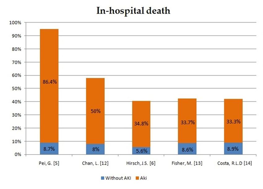
8. Impact of AKI Development on Hospitalization and Mortality Rate
In order to assess the impact of AKI development in patients with COVID-19 on
the course of hospitalization and mortality rate, we have compiled the data from five
different case reports and compared AKI patients with patients without AKI (Figures 1–3).
The occurrence of AKI in COVID-19-positive patients resulted in a significantly increased
number of admissions to the intensive care unit (ICU) in comparison to COVID-19 patients
without AKI. In the same group of patients, the need for mechanical ventilation was also
more noticeable. What stands out the most in our sheet is the fatality rate, which ranges
from 33.3% up to 86.4% in COVID-19 patients with AKI in comparison to COVID-19
patients without AKI, which varies from 5.6% to 9.3%. Therefore, patients with COVID-19
who develop AKI are at significantly higher risk of severe or critical course of the disease,
Int. J. Mol. Sci. 2021, 22, x FOR PEER REVIEW 8 of 12
respiratory failure and consequent mechanical ventilation. Moreover, AKI incidence
undeniably increases the mortality rate in COVID-19 patients.
Figure
Figure 1. Intensive
1. Intensive careadmission
care unit unit admission in patients
in patients with AKIwith
and AKI and
no AKI no AKI [6,12,13].
[6,12,13].Int. J. Mol. Sci. 2021, 22, 8081 8 of 11
Figure 1. Intensive care unit admission in patients with AKI and no AKI [6,12,13].
Int. J. Mol. Sci. 2021, 22, x FOR PEER REVIEW 9 of 12
Figure 2. The need for mechanical ventilation in patients with AKI and no AKI [6,12–14].
Figure 2. The need for mechanical ventilation in patients with AKI and no AKI [6,12–14].
Figure 3. In-hospital death in patients with AKI and no AKI [5,6,12–14].
Figure 3. In-hospital death in patients with AKI and no AKI [5,6,12–14].
9. Clinical Handling
9. Clinical Handling
There is no specific treatment for AKI caused by COVID-19, and it has to be based
onThere
KDIGOis noand
specific
othertreatment for AKI
guidelines. caused by COVID-19,
Prevention and itconsists
of AKI mainly has to be of
based on
individual fluid
KDIGO and other guidelines. Prevention of AKI mainly consists of individual fluid ther-
therapies (balanced crystalloids), discontinuation or reduction in nephrotoxic drugs and,
apies (balanced crystalloids), discontinuation or reduction in nephrotoxic drugs and, in
in the case of hypovolemia, the use of vasopressors. It is important to monitor kidney
the case of hypovolemia, the use of vasopressors. It is important to monitor kidney func-
function
tion with laboratory
with laboratory tests suchtests suchcreatinine
as serum as serumandcreatinine and urea levels.
urea levels.
Treatment should be systemic. We use antiviral drugs,
Treatment should be systemic. We use antiviral drugs, antibiotics, antibiotics, corticosteroids,
corticosteroids,
renin–angiotensin inhibitors, statins and anticoagulants. Drug therapy reduces the risk of the risk of
renin–angiotensin inhibitors, statins and anticoagulants. Drug therapy reduces
developing
developing AKIAKI
and and
causescauses a milder
a milder diseasedisease
course course
[39]. [39].
10.10. Renal
Renal Replacement
Replacement Therapy
Therapy
Renal
Renal replacement
replacement therapy
therapy (RRT)(RRT) is needed
is needed in up toin64%
upoftocritically
64% of critically
ill COVID-19ill COVID-19
patients who
patients develop
who developAKI AKI
(Table(Table
1) [6,12–15]. It is dueIttoisabnormal
1) [6,12–15]. concentrations
due to abnormal of elec-
concentrations of elec-
trolytes andand
trolytes volume
volume overload resistance
overload to pharmacological
resistance treatment.
to pharmacological The earliest
treatment. Thestud-
earliest studies
iessuggested
suggested that,
that, in
in hemodynamically
hemodynamically unstable
unstablepatients
patients with
withCOVID-19,
COVID-19, thethe
recom-
recommended
mended approach was continuous RRT (CRRT) [28]. However, clinicians observed a sig-
nificantly increased incidence of circuit clotting in COVID-19 patients, leading to pro-
longed time of treatment, as well as the unnecessary use of resources [24,40]. Therefore,
we must take caution in choosing the most safe and effective strategy.
Ramirez-Sandoval, J.C. et al. [41] reported the viability and safety of prolonged inter-Int. J. Mol. Sci. 2021, 22, 8081 9 of 11
approach was continuous RRT (CRRT) [28]. However, clinicians observed a significantly
increased incidence of circuit clotting in COVID-19 patients, leading to prolonged time
of treatment, as well as the unnecessary use of resources [24,40]. Therefore, we must take
caution in choosing the most safe and effective strategy.
Ramirez-Sandoval, J.C. et al. [41] reported the viability and safety of prolonged inter-
mittent renal replacement therapy (PIRRT) as a treatment option in COVID-19 patients
with AKI. The use of low-molecular-weight heparin (LMWH) in systemic anticoagulation
and unfractionated heparin (UFH) in regional anticoagulation was adapted and shown
to reduce the incidence of the circuit clotting observed in up to 13% of ICU patients with
COVID-19 infection. These findings were later confirmed in a report by Di Mario, F.
et al. [42], who tested the effectiveness of sustained low-efficiency dialysis (SLED), which
is a modality of PIRRT. However, in this study, the applied anticoagulation was regional
citrate anticoagulation (RCA) protocol, and it significantly lowered dialysis interruption by
circuit clotting to 6.1%. Moreover, the advantages of RCA over systemic heparin anticoagu-
lation protocols have been demonstrated in a comparative study by Arnold, F. et al. [43]
Taking into account these reports, we can assume that SLED with RCA protocol is the
safest and most efficient way of AKI management in hemodynamically unstable COVID-19
patients.
11. Conclusions
Acute kidney injury is common amongst patients with SARS-CoV-2 infection, espe-
cially critically ill ones, and is without a doubt associated with higher mortality. There are
numerous possible pathomechanisms which are still being investigated, but the most prob-
able ones are direct cellular invasion, ARDS, cytokine storm and hypovolemia. Histopatho-
logical reports showed that most COVID-19 patients with AKI presented acute tubular
damage, sometimes with necrosis and collapsing glomerulopathy. The most important
steps that should be taken in AKI prevention are the following: minimizing the risk of
hypovolemia and monitoring serum creatinine levels in the early stages of COVID-19
infection, especially in regard to the high-risk patients at an older age with diabetes melli-
tus, hypertension and cardiovascular diseases. However, when, despite our best efforts,
COVID-19 patients are hemodynamically unstable, the safest and most efficient way of AKI
management is SLED with RCA protocol. The evidence related to COVID-19 is changing
dynamically and we are still in need of more research for establishing the optimal treatment
of AKI in COVID-19 infection. Furthermore, physicians must be aware that patients who
recover from AKI induced by SARS-CoV-2 require monitoring of their kidneys on follow-
up, as there is rising evidence showing eGFR decreases among patients with a history of
COVID-19-associated AKI [44].
Author Contributions: All authors (M.G., S.L., E.M., B.F., J.R.) were involved in the preparation of
this article and J.R. revised the final version. All authors have read and agreed to the published
version of the manuscript.
Funding: This research received no external funding.
Institutional Review Board Statement: Not applicable.
Informed Consent Statement: Not applicable.
Data Availability Statement: The data used in this article are sourced from materials mentioned in
the References section.
Conflicts of Interest: The authors declare no conflict of interest.Int. J. Mol. Sci. 2021, 22, 8081 10 of 11
References
1. World Health Organisation (WHO). Available online: https://www.who.int/emergencies/diseases/novel-coronavirus-2019
(accessed on 4 March 2021).
2. Phan, L.T.; Nguyen, T.V.; Luong, Q.C.; Nguyen, T.V.; Nguyen, H.T.; Le, H.Q.; Nguyen, T.T.; Cao, T.M.; Pham, Q.D. Importation
and Human-to-Human Transmission of a Novel Coronavirus in Vietnam. N. Engl. J. Med. 2020, 382, 872–874. [CrossRef]
[PubMed]
3. Gupta, A.; Madhavan, M.V.; Sehgal, K.; Nair, N.; Mahajan, S.; Sehrawat, T.S.; Bikdeli, B.; Ahluwalia, N.; Ausiello, J.C.; Wan, E.Y.
Extrapulmonary manifestations of COVID-19. Nat. Med. 2020, 26, 1017–1032. [CrossRef] [PubMed]
4. Kellum, J.A.; Lameire, N.; Aspelin, P.; Barsoum, R.S.; Burdmann, E.A.; Goldstein, S.L.; Herzog, C.A.; Joannidis, M.; Kribben, A.;
Levey, A.S.; et al. Kidney disease: Improving global outcomes (KDIGO) acute kidney injury work group. KDIGO clinical practice
guideline for acute kidney injury. Kidney Int. Suppl. 2012, 1–138. [CrossRef]
5. Pei, G.; Zhang, Z.; Peng, J.; Liu, L.; Zhang, C.; Yu, C.; Ma, Z.; Huang, Y.; Liu, W.; Yao, Y.; et al. Renal Involvement and Early
Prognosis in Patients with COVID-19 Pneumonia. J. Am. Soc. Nephrol. 2020, 31, 1157–1165. [CrossRef] [PubMed]
6. Hirsch, J.S.; Ng, J.H.; Ross, D.W.; Sharma, P.; Shah, H.H.; Barnett, R.L. Acute kidney injury in patients hospitalized with COVID-19.
Kidney Int. 2020, 98, 209–218. [CrossRef]
7. Cummings, M.J.; Baldwin, M.R.; Abrams, D.; Jacobson, S.D.; Meyer, B.J.; Balough, E.M.; Aaron, J.G.; Jan, C.; Rabbani, L.E.; Hastie,
J.; et al. Epidemiology, clinical course, and outcomes of critically ill adults with COVID-19 in New York City: A prospective
cohort study. Lancet 2020, 395, 1763–1770. [CrossRef]
8. Oran, D.P.; Topol, E.J. The Proportion of SARS-CoV-2 Infections That Are Asymptomatic: A Systematic Review. Ann. Intern. Med.
2021, 174, 655–662. [CrossRef]
9. Wu, Z.; Mcgoogan, J.M. Characteristics of and Important Lessons From the Coronavirus Disease 2019 (COVID-19) Outbreak in
China. JAMA 2020, 323, 1239. [CrossRef] [PubMed]
10. Yang, X.; Yu, Y.; Xu, J.; Shu, H.; Xia, J.; Liu, H.; Wu, Y.; Zhang, L.; Yu, Z.; Fang, M.; et al. Clinical course and outcomes of critically
ill patients with SARS-CoV-2 pneumonia in Wuhan, China: A single-centered, retrospective, observational study. Lancet Respir.
Med. 2020, 8, 475–481. [CrossRef]
11. Cheng, Y.; Luo, R.; Wang, K.; Zhang, M.; Wang, Z.; Dong, L.; Li, J.; Yao, Y.; Ge, S.; Xu, G.; et al. Kidney disease is associated with
in-hospital death of patients with COVID-19. Kidney Int. 2020, 97, 829–838. [CrossRef] [PubMed]
12. Chan, L.; Chaudhary, K.; Saha, A.; Chauhan, K.; Vaid, A.; Zhao, S.; Paranjpe, I.; Somani, S.; Richter, F.; Miotto, R.; et al. AKI in
Hospitalized Patients with COVID-19. J. Am. Soc. Nephrol. 2021, 32, 151–160. [CrossRef] [PubMed]
13. Fisher, M.; Neugarten, J.; Bellin, E.; Yunes, M.; Stahl, L.; Johns, T.S.; Abramowitz, M.K.; Levy, R.; Kumar, N.; Mokrzycki, M.H.;
et al. AKI in Hospitalized Patients with and without COVID-19: A Comparison Study. J. Am. Soc. Nephrol. 2020, 31, 2145–2157.
[CrossRef]
14. Costa, R.L.D.; Sória, T.C.; Salles, E.F.; Gerecht, A.V.; Corvisier, M.F.; Menezes, M.A.D.M.; Ávila, C.D.S.; Silva, E.C.D.F.; Pereira,
S.R.N.; Simvoulidis, L.F.N. Acute kidney injury in patients with Covid-19 in a Brazilian ICU: Incidence, predictors and in-hospital
mortality. Braz. J. Nephrol. 2021. [CrossRef]
15. Ferlicot, S.; Jamme, M.; Gaillard, F.; Oniszczuk, J.; Couturier, A.; May, O.; Grünenwald, A.; Sannier, A.; Moktefi, A.; Le Monnier,
O.; et al. The spectrum of kidney biopsies in hospitalized patients with COVID-19, acute kidney injury, and/or proteinuria.
Nephrol. Dial. Transplant. 2021. [CrossRef]
16. Behzad, S.; Aghaghazvini, L.; Radmard, A.R.; Gholamrezanezhad, A. Extrapulmonary manifestations of COVID-19: Radiologic
and clinical overview. Clin. Imaging 2020, 66, 35–41. [CrossRef]
17. Zhou, P.; Yang, X.L.; Wang, X.G.; Hu, B.; Zhang, L.; Zhang, W.; Si, H.R.; Zhu, Y.; Li, B.; Huang, C.L.; et al. A pneumonia outbreak
associated with a new coronavirus of probable bat origin. Nature 2020, 579, 270–273. [CrossRef]
18. Hoffmann, M.; Kleine-Weber, H.; Schroeder, S.; Krüger, N.; Herrler, T.; Erichsen, S.; Schiergens, T.S.; Herrler, G.; Wu, N.-H.;
Nitsche, A.; et al. SARS-CoV-2 Cell Entry Depends on ACE2 and TMPRSS2 and Is Blocked by a Clinically Proven Protease
Inhibitor. Cell 2020, 181, 271–280. [CrossRef]
19. Walls, A.C.; Park, Y.-J.; Tortorici, M.A.; Wall, A.; Mcguire, A.T.; Veesler, D. Structure, Function, and Antigenicity of the SARS-CoV-2
Spike Glycoprotein. Cell 2020, 181, 281–292.e6. [CrossRef]
20. Khan, S.; Chen, L.; Yang, C.-R.; Raghuram, V.; Khundmiri, S.J.; Knepper, M.A. Does SARS-CoV-2 Infect the Kidney? J. Am. Soc.
Nephrol. 2020, 31, 2746–2748. [CrossRef]
21. Rahman, N.; Basharat, Z.; Yousuf, M.; Castaldo, G.; Rastrelli, L.; Khan, H. Virtual Screening of Natural Products against Type
II Transmembrane Serine Protease (TMPRSS2), the Priming Agent of Coronavirus 2 (SARS-CoV-2). Molecules 2020, 25, 2271.
[CrossRef]
22. Perlman, S.; Netland, J. Coronaviruses post-SARS: Update on replication and pathogenesis. Nat. Rev. Microbiol. 2009, 7, 439–450.
[CrossRef]
23. Braun, F.; Lütgehetmann, M.; Pfefferle, S.; Wong, N.M.; Carsten, A.; Lindenmeyer, T.M.; Nörz, D.; Heinrich, F.; Meißner, K.;
Wichmann, D.; et al. SARS-CoV-2 renal tropism associates with acute kidney injury. Lancet 2020, 396, 597–598. [CrossRef]
24. Ronco, C.; Reis, T.; Husain-Syed, F. Management of acute kidney injury in patients with COVID-19. Lancet Respir. Med. 2020, 8,
738–742. [CrossRef]Int. J. Mol. Sci. 2021, 22, 8081 11 of 11
25. Ahmed, A.R.; Ebad, C.A.; Stoneman, S.; Satti, M.M.; Conlon, P.J. Kidney injury in COVID-19. World J. Nephrol. 2020, 9, 18–32.
[CrossRef]
26. Sharma, P.; Uppal, N.N.; Wanchoo, R.; Shah, H.H.; Yang, Y.; Parikh, R.; Khanin, Y.; Madireddy, V.; Larsen, C.P.; Jhaveri, K.D.;
et al. COVID-19–Associated Kidney Injury: A Case Series of Kidney Biopsy Findings. J. Am. Soc. Nephrol. 2020, 31, 1948–1958.
[CrossRef]
27. Pan, X.-W.; Xu, D.; Zhang, H.; Zhou, W.; Wang, L.-H.; Cui, X.-G. Identification of a potential mechanism of acute kidney injury
during the COVID-19 outbreak: A study based on single-cell transcriptome analysis. Intensive Care Med. 2020, 46, 1114–1116.
[CrossRef]
28. Ronco, C.; Reis, T. Kidney involvement in COVID-19 and rationale for extracorporeal therapies. Nat. Rev. Nephrol. 2020, 16,
308–310. [CrossRef]
29. D’Agati, V.D.; Kaskel, F.J.; Falk, R.J. Focal Segmental Glomerulosclerosis. N. Engl. J. Med. 2011, 365, 2398–2411. [CrossRef]
30. Kissling, S.; Rotman, S.; Gerber, C.; Halfon, M.; Lamoth, F.; Comte, D.; Lhopitallier, L.; Sadallah, S.; Fakhouri, F. Collapsing
glomerulopathy in a COVID-19 patient. Kidney Int. 2020, 98, 228–231. [CrossRef] [PubMed]
31. Peleg, Y.; Kudose, S.; D’Agati, V.; Siddall, E.; Ahmad, S.; Nickolas, T.; Kisselev, S.; Gharavi, A.; Canetta, P. Acute Kidney Injury
Due to Collapsing Glomerulopathy Following COVID-19 Infection. Kidney Int. Rep. 2020, 5, 940–945. [CrossRef]
32. Santoriello, D.; Khairallah, P.; Bomback, A.S.; Xu, K.; Kudose, S.; Batal, I.; Barasch, J.; Radhakrishnan, J.; D’Agati, V.; Markowitz,
G. Postmortem Kidney Pathology Findings in Patients with COVID-19. J. Am. Soc. Nephrol. 2020, 31, 2158–2167. [CrossRef]
[PubMed]
33. Farkash, E.A.; Wilson, A.M.; Jentzen, J.M. Ultrastructural Evidence for Direct Renal Infection with SARS-CoV-2. J. Am. Soc.
Nephrol. 2020, 31, 1683–1687. [CrossRef] [PubMed]
34. Li, Z.; Wu, M.; Yao, J.; Guo, J.; Liao, X.; Song, S.; Yan, J. Caution on Kidney Dysfunctions of COVID-19 Patients. SSRN Electron. J.
2020. [CrossRef]
35. Richardson, S.; Hirsch, J.S.; Narasimhan, M.; Crawford, J.M.; Mcginn, T.; Davidson, K.W.; Barnaby, D.P.; Becker, L.B.; Chelico, J.D.;
Cohen, S.L.; et al. Presenting Characteristics, Comorbidities, and Outcomes Among 5700 Patients Hospitalized With COVID-19
in the New York City Area. JAMA 2020, 323, 2052. [CrossRef]
36. Henry, B.M.; Lippi, G. Chronic kidney disease is associated with severe coronavirus disease 2019 (COVID-19) infection. Int. Urol.
Nephrol. 2020, 52, 1193–1194. [CrossRef]
37. Morales, D.R.; Conover, M.M.; You, S.C.; Pratt, N.; Kostka, K.; Duarte-Salles, T.; Fernández-Bertolín, S.; Aragón, M.; Duvall,
S.L.; Lynch, K.; et al. Renin–angiotensin system blockers and susceptibility to COVID-19: An international, open science, cohort
analysis. Lancet Digit. Health 2021, 3, e98–e114. [CrossRef]
38. European Society of Cardiology. Position Statement of the ESC Council on Hypertension on ACEI-Inhibitors and Angiotensin
Receptor Blockers. 2020. Available online: https://www.escardio.org/Councils/Council-on-Hypertension-(CHT)/News/
position-statement-of-the-esc-council-on-hypertension-on-ace-inhibitors-and-ang (accessed on 1 June 2020).
39. Nadim, M.K.; Forni, L.G.; Mehta, R.L.; Connor, M.J.; Liu, K.D.; Ostermann, M.; Rimmelé, T.; Zarbock, A.; Bell, S.; Bihorac, A.; et al.
COVID-19-associated acute kidney injury: Consensus report of the 25th Acute Disease Quality Initiative (ADQI) Workgroup. Nat.
Rev. Nephrol. 2020. [CrossRef]
40. Helms, J.; Tacquard, C.; Severac, F.; Leonard-Lorant, I.; Ohana, M.; Delabranche, X.; Merdji, H.; Clere-Jehl, R.; Schenck, M.; Fagot
Gandet, F.; et al. High risk of thrombosis in patients with severe SARS-CoV-2 infection: A multicenter prospective cohort study.
Intensive Care Med. 2020, 46, 1089–1098. [CrossRef]
41. Ramirez-Sandoval, J.C.; Gaytan-Arocha, J.E.; Xolalpa-Chávez, P.; Mejia-Vilet, J.M.; Arvizu-Hernandez, M.; Rivero-Sigarroa, E.;
Torruco-Sotelo, C.; Correa-Rotter, R.; Vega-Vega, O. Prolonged Intermittent Renal Replacement Therapy for Acute Kidney Injury
in COVID-19 Patients with Acute Respiratory Distress Syndrome. Blood Purif. 2020, 1–9. [CrossRef]
42. Di Mario, F.; Regolisti, G.; Di Maria, A.; Parmigiani, A.; Benigno, G.D.; Picetti, E.; Barbagallo, M.; Greco, P.; Maccari, C.; Fiaccadori,
E. Sustained low-efficiency dialysis with regional citrate anticoagulation in critically ill patients with COVID-19 associated AKI:
A pilot study. J. Crit. Care 2021, 63, 22–25. [CrossRef]
43. Arnold, F.; Westermann, L.; Rieg, S.; Neumann-Haefelin, E.; Biever, P.M.; Walz, G.; Kalbhenn, J.; Tanriver, Y. Comparison of
different anticoagulation strategies for renal replacement therapy in critically ill patients with COVID-19: A cohort study. BMC
Nephrol. 2020, 21. [CrossRef]
44. Nugent, J.; Aklilu, A.; Yamamoto, Y.; Simonov, M.; Li, F.; Biswas, A.; Ghazi, L.; Greenberg, J.H.; Mansour, S.G.; Moledina, D.G.;
et al. Assessment of Acute Kidney Injury and Longitudinal Kidney Function after Hospital Discharge among Patients with and
without COVID-19. JAMA Netw. Open 2021, 4, e211095. [CrossRef] [PubMed]You can also read