Hippocampal CCR5/RANTES Elevations in a Rodent Model of Post-Traumatic Stress Disorder: Maraviroc (a CCR5 Antagonist) Increases Corticosterone ...
←
→
Page content transcription
If your browser does not render page correctly, please read the page content below
biomolecules

Article
Hippocampal CCR5/RANTES Elevations in a Rodent
Model of Post-Traumatic Stress Disorder: Maraviroc
(a CCR5 Antagonist) Increases Corticosterone Levels
and Enhances Fear Memory Consolidation
José Joaquín Merino 1, *, Vilma Muñetón-Gomez 2 , César Muñetón-Gómez 2 ,
María Ángeles Pérez-Izquierdo 3 , María Loscertales 4 and Adolfo Toledano Gasca 5
 1   Dpto. Farmacología, Farmacognosia y Botánica, Facultad de Farmacia, Universidad Complutense de
     Madrid (U.C.M). c/ Plaza Ramón y Cajal s/n, 28040 Madrid, Spain
 2   Universidad de La Salle Center, Facultad de Ciencias Agropecuarias, Av. Carrera 7. # 179-03 (sede norte),
     Bogotá, Colombia; hvcmg01@hotmail.com (V.M.-G.); cemuneton@unisalle.edu.co (C.M.-G.)
 3   Psychobiology Dept. Universidad Nacional de Educación a Distancia, UNED, 28040 Madrid, Spain;
     maperez@psi.uned.es
 4   Harvard Medical School, MGH, Massachussets General Hospital, 185 Cambridge St, Boston, MA 02114,
     USA; mloscertales@mgh.harvard.edu
 5   Department of Neuroanatomy, Instituto Cajal (CSIC), c/ Dr. Arce, 28.002 Madrid, Spain;
     atoledano@cajal.csic.es
 *   Correspondence: info@jjmerino.com; Tel.: 034-615-010216

 Received: 29 December 2019; Accepted: 27 January 2020; Published: 1 February 2020

 Abstract: Background: Contextual fear conditioning (CFC) is a rodent model that induces a high
 and long-lasting level of conditioning associated with traumatic memory formation; this behavioral
 paradigm resembles many characteristics of posttraumatic stress disorder (PSTD). Chemokines
 (chemotactic cytokines) play a known role in neuronal migration and neurodegeneration but their role
 in cognition is not totally elucidated. Aim: We ascertain whether CCR5/RANTES beta chemokines
 (hippocampus/prefrontal cortex) could play a role in fear memory consolidation (CFC paradigm).
 We also evaluated whether chronic stress restraint (21 days of restraint, 6-h/day) could regulate levels
 of these beta chemokines in CFC-trained rats; fear memory retention was determined taking the
 level of freezing (context and tone) by the animals as an index of fear memory consolidation 24 h
 after CFC training session; these chemokines (CCR5/RANTES) and IL-6 levels were measured in
 the hippocampus and prefrontal cortex of chronically stressed rats, 24 h after CFC post-training,
 and compared with undisturbed CFC-trained rats (Experiment 1). In Experiment 2, rats received
 1 mA of footshock during the CFC training session and fear memory consolidation was evaluated at
 12 and 24 h after CFC training sessions. We evaluated whether RANTES levels could be differentially
 regulated at 12 and 24 h after CFC training; in Experiment 3, maraviroc was administered to rats (i.m:
 100 mg/Kg, a CCR5 antagonist) before CFC training. These rats were not subjected to chronic stress
 restraint. We evaluated whether CCR5 blockade before CFC training could increase corticosterone,
 RANTES, or IL-6 levels and affects fear memory consolidation in the rats 24-h post-testing compared
 with vehicle CFC-trained rats. Results: Elevations of CCR5/RANTES chemokine levels in the
 hippocampus could have contributed to fear memory consolidation (24 h post-training) and chronic
 stress restraint did not affect these chemokines in the hippocampus; there were no significant
 differences in CCR5/RANTES levels between stressed and control rats in the prefrontal cortex
 (Experiment 1). In Experiment 2, hippocampal CCR5/RANTES levels increased and enhanced fear
 memory consolidation was observed 12 and 24 h after CFC training sessions with 1 mA of footshock.
 Increased corticosterone and CCR5/RANTES levels, as well as a higher freezing percentage to the
 context, were found at 24 h CFC post-testing in maraviroc-treated rats as compared to vehicle-treated
 animals (experiment-3). Conversely, IL-6 is not affected by maraviroc treatment in CFC training.

Biomolecules 2020, 10, 212; doi:10.3390/biom10020212                         www.mdpi.com/journal/biomolecules
Biomolecules 2020, 10, 212                                                                        2 of 19

    Conclusion. Our findings suggest a role for a hippocampal CCR5/RANTES axis in contextual fear
    memory consolidation; in fact, RANTES levels increased at 12 and 24 h after CFC training. When CCR5
    was blocked by maraviroc before CFC training, RANTES (hippocampus), corticosterone levels, and
    fear memory consolidation were greater than in vehicle CFC-trained rats 24 h after the CFC session.

    Keywords: post traumatic stress disorder; CCR5/RANTES chemokines; neural plasticity; chronic
    stress restraint; fear learning; neuro repair; neuroinmunology; neuroimmunomodulation

1. Introduction
     Immune responses can affect neural plasticity in brain areas (hippocampus and cortex); acute
stress enhances immune responses and chronic stress restraint provokes immunosuppression [1–7].
Chronic stress restraint provokes behavioral deficits in hippocampal-dependent tasks in rats [8–10]
and corticosterone, the stress hormone, affects synaptic terminal structure in the hippocampus [11,12]
and alters dendritic spine morphology in the rat medial prefrontal cortex [6].
     Cognition can be interfered with by blocking immune receptors in the central nervous system [5].
The association of immunomodulatory mechanisms with PTSD-like behavior alteration and immune
responses is not well understood. Chemokines (chemotactic cytokines) are G-coupled proinflammatory
cytokines involved in neuromodulation, neuronal migration [13,14]. RANTES (regulated on activation,
normal T-cell) is a small, secreted 8–10 kD chemokine involved in chemoattraction in eosinophils,
monocytes, and certain T leukocyte subsets. The ligand (RANTES: Regulated on Activation, Normal T
Cell Expressed and Secreted) binds to CCR5 [15] and also recognizes CCR1 chemokine receptors [16].
The CCR5 antagonist called maraviroc specifically blocks CCR5 while Met-RANTES blocks CCR1 and
CCR5 [17]. The CCR5/RANTES axis also contributes to neurodegeneration in the hippocampus [18–20]
as well as protecting neurons against insults in vitro [21]. Certain chemokines (CXCR4/SDF1 alpha)
regulate neuronal excitability [22,23]. These chemokines are detected by molecular techniques in the
hippocampus [24,25], which plays a role in contextual fear conditioning (CFC) [26].
     Recent evidence supports a role for immune dysfunction in psychiatric conditions such as post
traumatic stress disorder (PTSD) [8]. The contextual fear conditioning paradigm (CFC) is a PSTD
rodent model that induces a high and long-lasting level of conditioning associated with traumatic
memory formation [27,28]. This is a typical Pavlovian conditioning behavior [26]; the neutral stimulus
that did not elicit emotional responses is followed by an aversive stimulus (footshocks) in CFC-trained
rats using 1 mA footshocks; the re-exposure to the context (neutral stimulus) associates the neutral
stimulus with an aversive condition (footshock). These freezing levels correlated with corticosterone
levels in CFC-trained rats at 1 mA footshock [28]; the freezing is considered an index of fear memory
(context and tone) in this CFC behavioral paradigm; the freezing is evaluated at 12 and 24 h after
CFC training as an index of fear memory consolidation [28]. This CFC paradigm reproduces many
characteristics of PSTD, such as the persistence of traumatic memory [29]. In fact, PSTD patients
re-experience the traumatic event with distressing recollections, flashbacks, and psychologic distress
after encountering a stimulus that is reminiscent of the trauma [30,31]. The involvement of the CCR5
chemokine receptor in cognition and fear learning is unconfirmed. We try to resolve this question: are
CCR5/RANTES chemokines involved in traumatic memory consolidation in chronically stressed rats?

Aim
-      We studied whether chronic stress restraint could increase CCR5/RANTES chemokine as well as
       IL-6 levels in the hippocampus/prefrontal cortex (PFC) of rats subjected to 21 days of restraint;
       we evaluate if these chemokines play a role in fear memory consolidation in this CFC paradigm
       (1 mA, Experiment 1). These beta chemokines were measured 24 h after CFC training.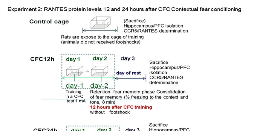
Biomolecules 2020, 10, 212                                                                           3 of 19

-     We evaluated whether CCR5/RANTES chemokine levels are differentially regulated at 12 and
      24 h post-training in CFC-trained rats with a footshock intensity of 1 mA (Experiment 2).
-     Once we confirmed elevated CCR5/RANTES levels at 12 and 24 h after CFC training (1 mA),
      we evaluated whether maraviroc (a CCR5 blocker, i.m injection: 100 mg/Kg) treatment administered
      for 3 consecutive days before CFC fear training could affect RANTES/IL-6 and corticosterone levels
      and also impairs fear memory consolidation 24 h after the CFC session (Experiment 3).

2. Material and Methods

2.1. Animals
      Male Wistar rats from Harlan Iberica (Barcelona, Spain), weighing from 165 to 185 g, were caged
in groups of three animals with free access to food and water under controlled temperature (22 ±
2 ◦ C) and light conditions (12 h:12 h light-dark cycle, light on at 8 A.M). All animals were handled
daily. According to their weight, animals were distributed into several experimental groups, including
undisturbed control rats (without stress or non-CFC trained rats).

2.2. Behavioral Methods

2.2.1. Chronic Stress Restraint Procedure
     All stress sessions took place in a room close to the colonies’ cages in the stabulary room. The daily
stress session was done by restraining rats for 21 consecutive days (6-h/day) in plastic restrainers
secured to the head and tail end with clip (from 09:00 a.m. to 15:00 p.m.) [27,28].

2.2.2. Contextual Fear Learning Paradigm: A Behavioral Paradigm of PSTD
      Rats were trained in a rodent box (30 × 37 × 25 cm) floored with 30 sheets of stainless steel,
through which animals received a footshock of 1 mA from a shock generator (LEICA I.C, Model
L-I100-26, Spain) connected to the floor during the training session. Briefly, rats were trained for
5 min in the contextual fear conditioning model. During this training session, animals were allowed
to explore the environment for 180 s and, after this time, received three consecutive footshocks of
1 mA of intensity every 60 s, following our previous protocols [27,28]. During the training sessions,
all undisturbed rats remained in their cages without receiving footshocks or other treatments. Thus,
this training session consisted of three footshocks of 1 mA intensity in the conditioning chamber,
producing effects that resemble many characteristics of PSTD [28]. Memory formation was evaluated
by freezing percentage to the context and tone as an index of fear memory in these CFC-trained rats
with 1 mA footshocks; the fear memory consolidation was evaluated at 12 or 24 h after the CFC training
session (depending on the experiment); freezing percentage at 24 h CFC post-training is considered as
an index of conditioned fear memory consolidation, and it was evaluated for 8 min. However, during
this fear memory retention phase evaluated 12/24 h after the CFC session, rats did not receive any
footshock [27,28]. This followed behavioral protocol was previously published by us.
      Last, CCR5/RANTES protein levels were measured by ELISA/Western blot in crude synaptosomes
(hippocampus/cortex), following our own protocols [27,28]. All rats were sacrificed one day after the
induction of chronic stress and also 1 day after the end of the CFC training; the hippocampus was dissected
(hippocampus and prefrontal cortex) and stored at −80 ◦C for further biochemical evaluation of chemokines.

2.3. Biochemical Methods

2.3.1. Synaptosomes Isolation (Hippocampus and Prefrontal Cortex)
     Synaptosomes were obtained following a protocol modified from Lynch and Voss (1991). Briefly,
the hippocampus was dissected and homogenized in 1 mL of lysis buffer (10 volumes), containing 0.32 M
sucrose, HEPES 5 mM, 1 µg/µL aprotinin, 1 µg/µL leucopeptin, 1 µg/µL peptastine, and 1 mM DTT.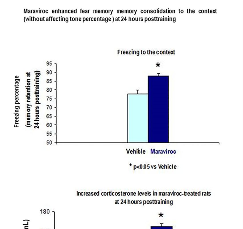
Biomolecules 2020, 10, 212                                                                         4 of 19

These homogenates were centrifuged for 5 min at 1000× g, 4 ◦ C using a JA 20.21 rotor. The supernatant
was centrifuged for 15 min at 15000× r.p.m, 4 ◦ C. After removing this supernatant, the final pellet
contains synaptosomes, which were resuspended in PBS 1 X buffer plus HEPES 5 mM, 1 µg/µL
aprotinin, 1 µg/µL leucopeptin, 1 µg/µL peptastine, and 1 mM DTT. The total protein estimation was
quantified by Bradford; finally, absorbance was read at 595 nm in a DIGYSCAN spectrophotometer
(UNED, Madrid).

2.3.2. ELISA CCR5/RANTES Protein Levels (Crude Synaptosomes)
     The CCR5/RANTES delta chemokines were measured by ELISA in crude synaptosomes following
the manufacturer’s instructions and own protocols [28]. The used kits for ELISA were RANTES
(#MMR00 R&D system, MN, USA); CCR5 (#ABIN626788 antibodies-online company, Germany,
detection range: 0.312 ng/mL—20 ng/mL), IL-6 kit (# R6000B, R&D, MN, USA). The detection limit for
RANTES is 2 pg/mL. Briefly, 10 mg of samples were loaded in their respective plate in order to detect
these markers; the capture antibody (concentration 10 µg/µL) was incubated overnight (RANTES: o/n)
at 4 ◦ C in PBS (phosphate buffer saline). After four washes with PBS plus 0.05% Tween 20, plates
were incubated with a biotinylated secondary antibody (1:500 dilution) for 2 h at room temperature
(RT). After 3 washes, the signal was enhanced by adding streptavidin-HRP (1:5000, R&D System, MN,
USA) to the plate (30 min). The reaction was allowed to develop in dark conditions by adding OPD
1 µg/µL (ortophenylenediamine, Sigma, Madrid, Spain) in citrate buffer for 10 min and 30% H2 O2 .
After stopping the reaction with H2 SO4 , the absorbance was read at 492 nm wavelength (DIGYSCAN
spectrophotometer reader) following our own protocol [28].

2.3.3. Western Blot for CCR5 Detection in Crude Synaptosomes
      Briefly, CCR5 was quantified in synaptosomes from the hippocampus by Westerm blot. Samples
were homogenized in 500 µL of buffer (PBS/0.1% Nonidet P-4/0.1% SDS/0.5% deoxycholic acid)
containing aprotinine (5.7 µg/mL), sodium vanadate (1 mmol/l); and phenylmethylsulfonyl fluoride
(100 µg/µL); all these reagents were added immediately after the homogenization procedure.
After centrifugation (15000 × r.p.m, 20 min), aliquotes from the supernatants were collected and
stored at −20 ◦ C until further biochemical analysis.
      Synaptosomes from the hippocampus were boiled for 3 min at 90 ◦ C in 30 mM Tris-HCl buffer at pH
7.4, containing 0.05% lauryl dodecyl sulphate sodium (SDS) and beta-mercaptoethanol. Equal amounts
of proteins were loaded (40 µg of total protein) and transferred to PVDF membranes at 1 mA/cm2 (1A,
200 v during 1 h in a TE 22 Transfer system, Amersham Pharmacy, Madrid, Spain). After blocking
nonspecific binding, samples were overnight incubated (o/n) with 5% of milk powered milk free
of saturated fat 1:1 in TBS-Tween 20 in 50 mM Tris-HCl, pH = 8, 138 mM NaCl, 0.05% Tween 20
(TBST); the CCR5 antibody was incubated o/n at 12 µg/µL (AbCAM, UK; # ab65850). This CCR5
polyclonal antibody recognizes the N-terminal region of CCR5) and does not cross-react with other
chemokine receptors. Beta actine was included as a loading control (stripping) at 1:5000 in TBS-Tween.
After blocking membranes with TBS-T in the NAP blocker (Genotech), the primary CCR5 antibody
was added at 1:500. The secondary anti rabbit HRP horseradish-peroxidase-conjugated antibody
was used at 1:20000 for 1 h at room temperature. Finally, blots were developed with the ECL+
chemoluminescence system (Amersham Pharmacia, Spain) and densitometry of bands expressed as a
percentage of controls.

2.3.4. Corticosterone Levels
     Trunk blood was collected by centrifugation and pooled in heparinized vials for centrifugation at
2780 rpm for 10 min at 4 ◦ C. Plasma was collected out and stored at −20 ◦ C until assays were performed.
Plasma B was assayed in duplicate with the RIA kit (ICN Biomedical Inc. Costa Mesa, California,
USA). This intra-assay coefficient of variation was 4.4%.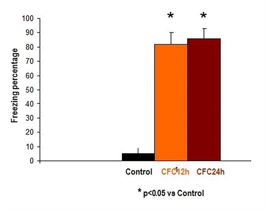
Biomolecules 2020, 10, 212                                                                                                  5 of 19

3. Results

3.1. Experiment 1: Effect of Chronic Stress Restraint and Fear Learning in CCR5/RANTES Levels
(Hippocampus/Prefrontal Cortex)
     In Experiment 1, we evaluate whether chronic stress restraint and/or contextual fear conditioning
training (CFC) increase hippocampal/PFC CCR5/RANTES protein levels 24 h post-testing. We quantified
freezing levels like an index for memory consolidation at 24 h after the CFC session, following our
own protocols [28]. Animals were subjected to 21 days of chronic stress restraint in plastic restrainers.
One day later (day 23), a subgroup of animals from the “stress condition” (n = 16) or “undisturbed”
(n = 16) groups were trained in a contextual fear conditioning paradigm (CFC) paradigm with 1 mA
footshocks. Rats were re-exposed to the context at 24 h after training in order to evaluate fear memory
consolidation but without receiving footshocks. The bifactorial ANOVA analyze a possible interactive
effect between stress and/or fear learning (PSTD model) in delta chemokine levels; for this purpose,
“stress factor” and/or “fear learning (CFC)” effect/s were evaluated by including four groups (UND =
control, ST, CFC24, ST + CFC24).
     These CCR5/RANTES chemokines were measured by ELISA (hippocampus/prefrontal cortex)
and Western blot in crude synaptosomes. The experimental design included these groups: (i) control
(UND: undisturbed rats, n = 8) that did not receive treatment/s, (ii) animals subjected to 21 days
of chronic stress restraint in plastic restrainers (ST: 6-h/day, n = 8); (iii) contextual fear conditioning
(CFC)-trained rats received the footshocks (1 mA); chemokines were measured by ELISA (crude
synaptosomes from hippocampus/PFFC) in rats re-exposed to the context at 24 h post-testing (CFC24,
n = 8). The levels of freezing response to the context and tone were evaluated as an index of fear
memory consolidation at 24 h after the CFC session (n = 8). (iv) Rats subjected to 21 days of chronic
stress restraint (6-h/day) and subsequently, 1 day after the last restraint session, these animals were
CFC trained with 1 mA of footshocks (ST + CFC24, n = 8); rats were re-exposed to the training context
at 24 h after the CFC session (CFC24) without receiving footshocks, following own protocols [28].
After the last stress session, the rats were returned to their respective cages. The undisturbed rats
(UND: controls) remained in their home cages without behavioral manipulation (see design in Figure 1).
All rats were sacrificed 24 h after their last experimental condition. Undisturbed controls (UND) were
sacrificed at the same time as the rest of the groups. Chemokines were evaluated by ELISA in crude
synaptosomes (hippocampus/prefrontal
                   Biomolecules 2020, 10, 212 cortex) at 24 h after the CFC session.       6 of 22

                Figure 1. Behavioral protocol for chronic stress and/or contextual fear conditioning.
                                  Figure 1. Behavioral protocol for chronic stress and/or contextual fear conditioning.

                           Since structural alterations have been demonstrated in the hippocampus [12] and prefrontal
                      cortex of rats subjected to 21 days of chronic stress restraint [6], we evaluated whether levels of
                      these chemokines could be affected by chronic stress and/or fear memory consolidation at 24 h after
                      a CFC session.

                      3.1.1. Hippocampus: increased hippocampal CCR5 Levels in Chronically Stressed Rats 24 Hours
                      after Contextual Fear Conditioning Training (24 Hours Post-testing)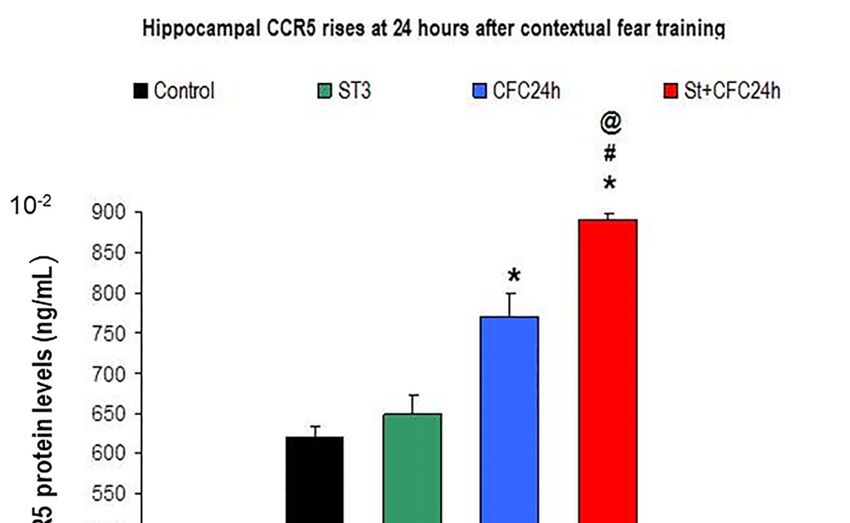
Biomolecules 2020, 10, 212                                                                                      6 of 19

     Since structural alterations have been demonstrated in the hippocampus [12] and prefrontal
cortex of rats subjected to 21 days of chronic stress restraint [6], we evaluated whether levels of these
chemokines could be affected by chronic stress and/or fear memory consolidation at 24 h after a
CFC session.

3.1.1. Hippocampus: Increased Hippocampal CCR5 Levels in Chronically Stressed Rats 24 Hours after
Contextual Fear Conditioning Training (24 Hours Post-testing)
     Bifactorial ANOVA revealed higher hippocampal CCR5 levels at 24 h after the CFC training
session (F (1, 31) = 0.3 p < 0.05) as well as a significant effect for the “stress factor” (F (1, 31) = 0.3;
p < 0.05); the interaction between the two factors also increased CCR5 protein levels “(F (1, 31) = 0.014;
p = 0.049); the post hoc Bonferroni also revealed elevated CCR5 in stressed animals that were trained
in a CFC paradigm compared to CFC-trained rats (without stress, p < 0.05) as well as compared to
undisturbed         10, 212rats (p < 0.05, Figure 2).
                 control
 Biomolecules 2020,                                                                                   7 of 22

      Figure 2. CCR5 protein levels in crude synaptosomes from the hippocampus of chronic stress and/or
      CFC-trained rats (24 h after the CFC session).
       Figure 2. CCR5 protein levels in crude synaptosomes from the hippocampus of chronic stress
3.1.2. and/or
       RANTES CFC-trained  rats (24 h after the CFC session).
                 (Hippocampus)

 3.1.2.The ANOVA
        RANTES        revealed a significant effect for fear memory consolidation (F (1, 31) = 23.74; p = 0.000)
                    (Hippocampus)
as well as for the “stress factor” (F (1, 31) = 0.7, p < 0.05); however, there was no interactive effect between
these The   ANOVA
       factors (F (1, 31)revealed
                          = 8.01; p a= significant  effect
                                       0.97; n.s). The     for fearpost
                                                       Bonferroni    memory     consolidation
                                                                         hoc test               (F (1.53)
                                                                                  revealed higher  RANTES = 23.74;
                                                                                                             levelspat=
 0.000)  as well   as for  the “stress    factor”  (F (1.53) = 0.7, p <  0.05); however,   there  was  no  interactive
24 h post-CFC session than in control animals (without stress, p < 0.05, Figure 3). In addition, chronically
 effect between
stressed  rats that these   factors (F (1.53)
                     were CFC-trained            = 8.01;RANTES
                                            had higher    p = 0.97;levels
                                                                     n.s). than
                                                                           The controls
                                                                                Bonferroni
                                                                                         (p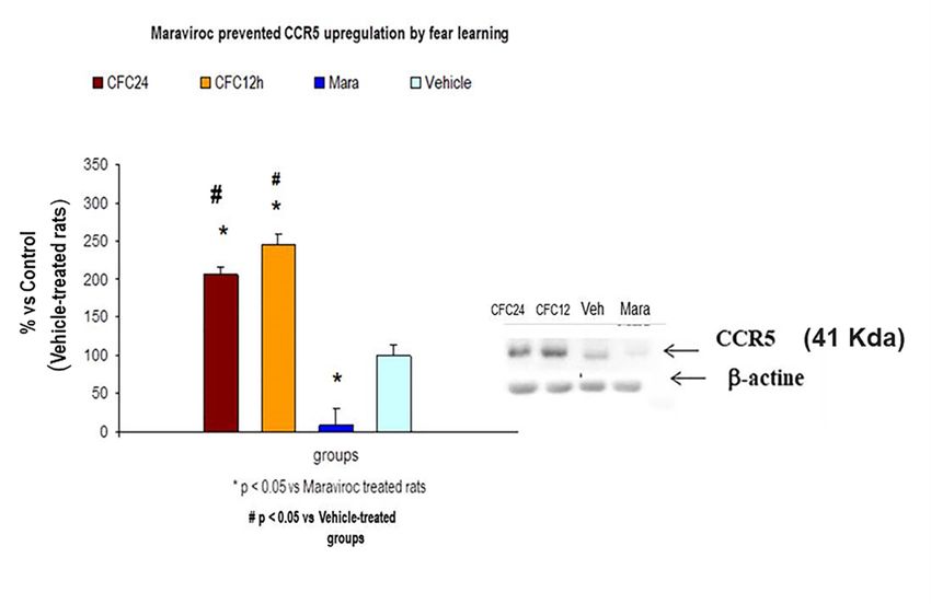
Biomolecules 2020, 10, 212
              Biomolecules 2020, 10, 212                                                                        8 of 22
                                                                                                                           7 of 19

                            Figure 3. Increased RANTES protein levels 24 after CFC post-testing.
                                      Figure 3. Increased RANTES protein levels 24 after CFC post-testing.
3.1.3. Increased IL-6 Levels by Fear Learning in the Hippocampus of Chronically Stressed Rats 24
            3.1.3. Increased IL-6 Levels by Fear Learning in the Hippocampus of Chronically Stressed Rats 24
Hours after CFC Training with 1 mA
                Hours after CFC Training with 1 mA
     The Mann–Whitney
                 The Mann–Whitney   analyses    revealed
                                           analyses   revealedaugmented      IL-6protein
                                                                augmented IL-6     protein    levels
                                                                                         levels       in homogenates
                                                                                                in homogenates  from the from the
hippocampus by chronic stress; in fact, IL-6 protein level elevations were observed in chronically
            hippocampus       by   chronic  stress; in fact, IL-6 protein level elevations  were  observed in chronically
            stressed
stressed rats   (p
Biomolecules 2020, 10, 212                                                                                         8 of 19

     The Figure 4 shows hippocampal mean ± S.E.M (standard error of mean) of IL-6 protein levels
(Figure 4); The groups are also indicated in CCR5 levels (Figure 2), RANTES levels (Figure 3) in the
hippocampus of chronically stressed animals and/or fear memory consolidation at 24 h after the CFC
session (n = 8 rats/group).
     UND: Undisturbed control rats did not receive treatment/s (n = 8).
     ST: Rats subjected to 21 days of chronic stress restraint in plastic restrainers (6-h/day, n = 8).
     CFC24: Rats trained in a contextual fear conditioning paradigm CFC (1 mA of footshocks) and
re-exposed to the conditioning chamber 24 h after CFC (without receiving footshocks).
     ST + CFC24: Rats subjected to 21 days of chronic stress restraint were trained in a contextual fear
conditioning paradigm (CFC, 1 mA). These animals were re-exposed to the conditioning chamber
24 h after the CFC session (n = 8); these beta chemokines were evaluated by ELISA 24 h after the CFC
session in crude synaptosomes (hippocampus).

3.1.4. Prefrontal Cortex: Chronic Stress and/or Contextual Fear Conditioning (CFC) did not Affect
CCR5/RANTES Protein Levels in the Prefrontal Cortex 24 h after the CFC Session
      With regard to CCR5 levels in the prefrontal cortex, the ANOVA did not shown an effect for “stress
factor” (F (1, 31) = 0.3; p = 0.58; n.s) and there was a lack of effect from the “conditioning factor” (F (1,
31) = 0.18, p = 0.66; n.s). We found a slight tendency for interaction between both factors F (1, 31) = 2.565,
p = 0.115; n.s). Similar results were observed for RANTES levels in a bifactorial ANOVA in the PFC (data
not  shown). The post hoc Tukey test showed a tendency toward lower levels in the cortex of chronically
 Biomolecules 2020, 10, 212                                                                            10 of 22
stressed rats 24 h after the CFC session as compared to chronically stressed rats (p = 0.1; n.s; Figure 5).

      Figure 5. CCR5 protein levels in the prefrontal cortex are not regulated by chronic stress restraint or
       Figure
      fear     5. CCR5
           learning     protein levels in the prefrontal cortex are not regulated by chronic stress restraint or
                    in rats.
       fear learning in rats.
      Figure 5 shows no effect for CCR5 protein levels in the prefrontal cortex of rats subjected to chronic
stressFigure
        restraint5 shows
                   as well no
                            as aeffect  foreffect
                                  lack of    CCR5 forprotein   levels in
                                                      fear learning.      the was
                                                                       CCR5     prefrontal
                                                                                    evaluatedcortex  of rats
                                                                                                 in crude     subjected to
                                                                                                           synaptosomes
 chronic
(PFC,   n =stress   restraint as well
             8 animals/group).      The as   a lack
                                         graph       of effect
                                                  indicates  meanfor values
                                                                     fear learning.
                                                                              ± S.E.M.CCR5 was evaluated in crude
 synaptosomes       (PFC, n = (control
      UND: Undisturbed          8 animals/group).
                                          rats) did notThereceive
                                                            graphtreatment/s
                                                                    indicates mean(n = values
                                                                                        8).     ± S.E.M.
      ST: Rats subjected to 21 days of chronic stress restraint in plastic restrainers (6-h/day, n = 8).
      UND:      Undisturbed     (control   rats) did  not  receive  treatment/s    (n =  8).
      ST: RatsRats
      CFC24:      subjected
                       trainedtoin21adays   of chronic
                                      contextual    fearstress   restraintparadigm
                                                          conditioning      in plastic CFC
                                                                                        restrainers
                                                                                              (1 mA(6-h/day,    n = 8).and
                                                                                                      of footshocks)
      CFC24:toRats
re-exposed             trained in a contextual
                  the conditioning     chamber 24fear     conditioning
                                                       h after CFC (withoutparadigm     CFCfootshocks).
                                                                                 receiving    (1 mA of footshocks) and
      ST + CFC24:
 re-exposed     to the Rats
                       conditioning
                             subjectedchamber
                                         to 21 days 24of
                                                       h after  CFC
                                                          chronic     (without
                                                                   stress         receiving
                                                                           restraint          footshocks).
                                                                                      were trained   in a contextual fear
      ST + CFC24:
conditioning            Rats subjected
                  paradigm     (CFC, 1 mA).to 21These
                                                   days animals
                                                         of chronic    stress
                                                                    were       restraint to
                                                                           re-exposed     were
                                                                                             the trained  in a contextual
                                                                                                 conditioning    chamber
 fear
24     conditioning
    h after              paradigm
             the CFC session     (n = (CFC,    1 beta
                                       8); these   mA).chemokines
                                                          These animals       were re-exposed
                                                                       were evaluated      by ELISA to24the  conditioning
                                                                                                          h after the CFC
 chamber
session   in 24  h after
              crude       the CFC session
                     synaptosomes               (n = 8); these beta chemokines were evaluated by ELISA 24 h
                                       (hippocampus/PFC).
 after the CFC session in crude synaptosomes (hippocampus/PFC).

3.2. Experiment 2: Effect of Fear Learning in RANTES Levels (12 and 24 h Post-testing)
   In Experiment 2, rats were not subjected to chronic stress restraint. We evaluate whether
RANTES could be differentially regulated at two time windows after the CFC session (12 and 24
Biomolecules 2020, 10, 212                                                                               9 of 19

3.2. Experiment 2: Effect of Fear Learning in RANTES Levels (12 and 24 h Post-testing)
     In Experiment 2, rats were not subjected to chronic stress restraint. We evaluate whether RANTES
could be differentially regulated at two time windows after the CFC session (12 and 24 post-testing).
For this aim, rats were trained with 1 mA of footshocks in a CFC paradigm. Rats trained with 1 mA
footshocks were re-exposed to the context at 12 and 24 h after the CFC session (without receiving further
footshocks). Fear memory consolidation was measured as an index of fear memory consolidation (12
and 24 h after the CFC session) and RANTES levels were measured at 12 and 24 h after the CFC training
session without receiving any further footshocks (n = 7 rats/group). The undisturbed control cage (UND)
 Biomolecules 2020, 10, 212                                                                         11 of 22
included rats exposed to the cage conditioning chamber but without receiving footshocks (Figure 6).

              Figure 6. Behavioral protocol for beta chemokine detection in contextual fear learning.
               Figure 6. Behavioral protocol for beta chemokine detection in contextual fear learning.
3.2.1. Increased Corticosterone and Enhanced Freezing Percentage to the Context at 12 and 24 h after
the CFC Session with 1 mA Footshocks
 3.2.1. Increased Corticosterone and Enhanced Freezing Percentage to the Context at 12 and 24 h
 afterWetheobserved
            CFC Session with 1plasma
                    increased   mA Footshocks
                                      corticosterone levels and enhanced freezing percentage to the
context at 12 and 24 h after the CFC session (p < 0.05, Figure 7) as compared to controls, in agreement
       We observed increased plasma corticosterone levels and enhanced freezing percentage to the
with own previous findings [27,28].
 context at 12 and 24 h after the CFC session (p < 0.05, Figure 7) as compared to controls, in
      Figure 7 shows the mean ± S.E.M freezing percentage to the context at 12 h (orange) as well
 agreement with own previous findings [27,28].
as 24 h (brown) after the CFC session with 1 mA footshocks (Figure 7). Freezing is an index of
fear memory consolidation (n = 8 rats/group). Controls (black) did not receive footshocks and are
undisturbed controls; the rats were only expose to the cage without receiving electric footshocks.
Freezing percentages to the context were measured at 12 and 24 h after CFC training. The results are
shown as mean ± S.E.M.
      Control: Control rats exposed to the cage conditioning chamber (n = 5).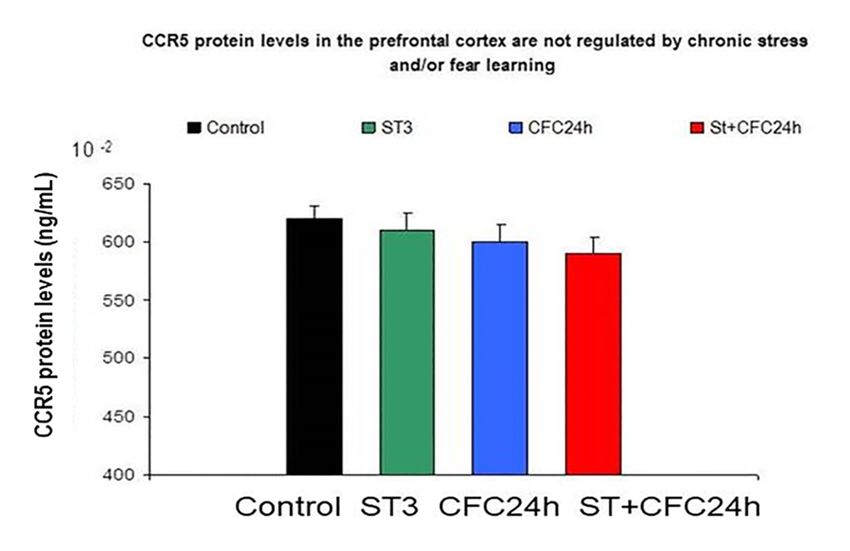
Biomolecules 2020, 10, 212                                                                                                     10 of 19

     CFC12: Rats trained in a contextual fear conditioning paradigm (1 mA of footshocks) and
re-exposed to the conditioning chamber at 12 h (CFC12, n = 7) or 24 h (CFC24, n = 7) after the CFC
session but without     receiving footshocks (n = 7).
                Biomolecules 2020, 10, 212                                     12 of 22

      Figure 7. Increased freezing levels at 12 and 24 h after the CFC session. * p < 0.05 vs. undisturbed rats
                      Figure 7. Increased freezing levels at 12 and 24 h after the CFC session. * p < 0.05 vs. undisturbed
      (exposed to the rats
                      cage).
                           (exposed to the cage).

3.2.2. Increased RANTES
                    Figure 7 Protein    Levels
                             shows the mean      in the
                                             ± S.E.M     Hippocampus
                                                     freezing                 12context
                                                              percentage to the   and at2412Hours    after
                                                                                             h (orange)     CFC
                                                                                                        as well as Training with
1 mA of Electric24Footshocks
                   h (brown) after the CFC session with 1 mA footshocks (Figure 7). Freezing is an index of fear
                     memory consolidation (n = 8 rats/group). Controls (black) did not receive footshocks and are
      The Bonferrroni   analysis
                undisturbed          revealed
                              controls;           CCR5/RANTES
                                        the rats were only expose to the elevations     in the hippocampus
                                                                          cage without receiving electric footshocks. at 12 and 24 h
                Freezing percentages to the context were measured at 12 and 24 h after CFC training. The results
after the CFC session   asascompared
                are shown     mean ± S.E.M. with control rats exposed to the cage (without receiving footshocks,
p < 0.05 in both cases).
                     Control:Interestingly,      RANTES
                              Control rats exposed  to the cage levels  were
                                                                conditioning    significantly
                                                                             chamber (n = 5).     higher at 24 h after CFC as
                     CFC12: Rats trained in a contextual fear conditioning paradigm (1 mA of footshocks) and re-
compared with exposed
                CFC atto12      h post-training values (Figure 8).
                           the conditioning chamber at 12 h (CFC12, n = 7) or 24 h (CFC24, n = 7) after the CFC
                   Biomolecules 2020, 10, 212                                                                       13 of 22
                     session but without receiving footshocks (n = 7).

                     3.2.2. Increased RANTES Protein Levels in the Hippocampus 12 and 24 Hours after CFC Training
                     with 1 mA of Electric Footshocks
                          The Bonferrroni analysis revealed CCR5/RANTES elevations in the hippocampus at 12 and 24
                     h after the CFC session as compared with control rats exposed to the cage (without receiving
                     footshocks, p < 0.05 in both cases). Interestingly, RANTES levels were significantly higher at 24 h
                     after CFC as compared with CFC at 12 h post-training values (Figure 8).

                         Figure 8. Increased RANTES levels at 12 and 24 h after the CFC session.
                                          Figure 8. Increased RANTES levels at 12 and 24 h after the CFC session.

                        Control: Control rats exposed to the cage conditioning chamber (n = 5) without footshocks.
                        CFC12: Rats trained in a contextual fear conditioning paradigm (1 mA of footshocks) and re-
                   exposed to the conditioning chamber at 12 h (CFC12, n = 7) or 24 h (CFC24, n = 7) after the CFC
                   session but without receiving footshocks (n = 7).
                        RANTES levels were measured at 12 and 24 h after the CFC session. Figure 7 shows the mean
                   (RANTES) ± S.E.M values at 12 and 24 h CFC post-testing (n = 8 animals/group).
Biomolecules 2020, 10, 212 11 of 19
Control: Control rats exposed to the cage conditioning chamber (n = 5) without footshocks.
CFC12: Rats trained in a contextual fear conditioning paradigm (1 mA of footshocks) and
re-exposed to the conditioning chamber at 12 h (CFC12, n = 7) or 24 h (CFC24, n = 7) after the CFC
session but without receiving footshocks (n = 7).
RANTES levels were measured at 12 and 24 h after the CFC session. Figure 7 shows the mean
(RANTES) ± S.E.M values at 12 and 24 h CFC post-testing (n = 8 animals/group).
3.3. Experiment 3: Effect of CCR5 Blockade by Maraviroc in Corticosterone and Freezing Levels (24 Hours after
CFC Training Session)
As the RANTES rises seem to be associated with fear memory consolidation at 24 h after the
CFC session, we evaluated whether CCR5 blockade by maraviroc affects corticosterone levels and
alters freezing levels (context and tone) as compared to vehicle CFC-trained rats. For this purpose,
rats received 1 mA of footshocks during CFC training following our own protocols [28]. The maraviroc
was i.m administered (n = 7, a CCR5 blocker, i.m: 100 mg/Kg) to rats for 3 consecutive days before CFC
fear training with 1 mA of footshock intensity. The vehicle-treated rats received vehicle for 3 consecutive
days (saline, i.m) before CFC training (n = 7). These rats were trained in a CFC with 1 mA of footshock
intensity; both experimental groups were re-exposed to the context in the conditioned chamber box 24 h
after the CFC session without receiving any further footshocks. Corticosterone and freezing percentage
(context and tone) were compared between maraviroc and vehicle CFC-trained rats at 24 h after the CFC
session. Freezing percentage (context and tone) were evaluated during 8 min as an index of fear memory
consolidation (n =10,
Biomolecules 2020, 7/group)
212 but these animals did not receive footshocks (Figure 9). 14 of 22
Figure 9. Maraviroc increased freezing percentage to the context and elevated corticosterone levels in
CFC-trained rats.
Figure 9. Maraviroc increased freezing percentage to the context and elevated corticosterone levels
in CFC-trained rats.
3.3.1. Maraviroc i.m Injections for 3 Consecutive Days before CFC Training (1 mA Footshocks)
Increased Corticosterone Levels and Enhanced Freezing to the Context at 24 Hours after the CFC
Session as Compared to Vehicle CFC-Treated rats (1 mA)
These maraviroc-trained rats (100 mg/Kg, i.m) had increased corticosterone levels as well as
enhanced freezing percentage to the context at 24 h after the CFC session as compared to vehicle
CFC-trained rats (p < 0.05, Figure 10).Biomolecules 2020, 10, 212 12 of 19
3.3.1. Maraviroc i.m Injections for 3 Consecutive Days before CFC Training (1 mA Footshocks)
Increased Corticosterone Levels and Enhanced Freezing to the Context at 24 Hours after the CFC
Session as Compared to Vehicle CFC-Treated rats (1 mA)
These maraviroc-trained rats (100 mg/Kg, i.m) had increased corticosterone levels as well as
enhanced freezing percentage to the context at 24 h after the CFC session as compared to vehicle
Biomolecules 2020, 10, 212 15 of 22
CFC-trained rats (p < 0.05, Figure 10).
Figure 10. Enhanced freezing to the context in maraviroc-trained rats together increased plasma
corticosterone levels, 24 h after CFC training session as compare to vehicle CFC-trained rats with 1 mA
Figure 10. (p
footshocks < 0.05, n freezing
Enhanced = 7 animals/group, p < 0.05).
to the context in maraviroc-trained rats i.m:
Maraviroc (3 days, together increased
100 mg/Kg) plasma
treatment
corticosterone
before levels,enhanced
CFC training 24 h afterfreezing
CFC training session
percentage to as
thecompare to vehicle
context when CFC-trained
rats were rats to
re-exposed with
the1
mA footshocks(p < 0.05, n = 7 animals/group, p < 0.05). Maraviroc (3 days, i.m: 100 mg/Kg)
conditioning chamber at 24 h after the CFC sessions compared to vehicle CFC-trained rats (1 mA of treatment
before CFC training enhanced freezing percentage to the context when rats were re-exposed to the
footshocks).
conditioning chamber at 24 h after the CFC sessions compared to vehicle CFC-trained rats (1 mA of
Figure 10 showed increased corticosterone levels and higher freezing percentage to the context at
footshocks).
24 h after the CFC session in maraviroc-treated rats.
3.3.2. Maraviroc i.m Injections for 3 Consecutive Days before CFC Training (1 mA Footshocks) did
not Prevented RANTES Overexpression as Compared to Vehicle CFC-Treated Rats (1 mA)
RANTES levels were still higher in the hippocampus of maraviroc-trained rats, 24 h after CFC
training session, as compared to vehicle CFC-trained rats with 1 mA footshocks (p < 0.05, n = 7Biomolecules 2020, 10, 212 13 of 19
3.3.2. Maraviroc i.m Injections for 3 Consecutive Days before CFC Training (1 mA Footshocks) did not
Prevented RANTES Overexpression as Compared to Vehicle CFC-Treated Rats (1 mA)
RANTES levels were still higher in the hippocampus of maraviroc-trained rats, 24 h after CFC
training session, as compared to vehicle CFC-trained rats with 1 mA footshocks (p < 0.05, n = 7
animals/group, p < 0.05). The Western blot analysis showed increased CCR5 protein levels 12 and
24 h after CFC session. This 41 Kda band for CCR5 was almost completely abolished by maraviroc
treatment although
Biomolecules 2020, 10,there
212 was a slight signal as compared to vehicle-treated rats. The Western 16 of 22 blot
indicated all densitometry (n = 5/group). CCR5 increased 12 and 24 h after the CFC session and
maraviroc
maraviroc prevented
prevented this upregulation (n = 5–7
this upregulation (n =rats/group).
5–7 rats/group).
All dataAll were
data expressed
were expressed as a
as a percentage
of thepercentage of the vehicle-control
vehicle-control group, which group,
is 100%which is 100%
(Figure 11). (Figure 11).
Figure 11. Maraviroc induced an almost complete CCR5 blockade in maraviroc CFC-trained rats as
compared to vehicle-treated animals (n = 5 rats/group, p < 0.05). Vehicle-treated rats are expressed as
Figure 11. Maraviroc induced an almost complete CCR5 blockade in maraviroc CFC-trained rats
100% of band signal (densitometry).
as compared to vehicle-treatedAllanimals
data are(nexpressed as percentage
= 5 rats/group, p < 0.05).control (vehicle percentage).
Vehicle-treated rats are
CCR5 is upregulated by fear memory (12 and 24 h after CFC session) and maraviroc
expressed as 100% of band signal (densitometry). All data are expressed as percentage prevented
controlthese
upregulations (see
(vehicle Western blot
percentage). CCR5(41 Kda band for
is upregulated by CCR5) and quantification).
fear memory (12 and 24 h afterBands for CCR5
CFC session) andwere
maraviroc prevented these upregulations (see Western blot (41 Kda band for CCR5)
normalized with beta actine. Mara: Maraviroc CFC-treated rats (100 mg/Kg, i.m); Vehicle: Rats received and
quantification). Bands for CCR5 were normalized with beta actine. Mara: Maraviroc CFC-treated
vehicle and these animals were trained in a CFC paradigm (1 mA); CFC12h: Fear memory consolidation
rats (100 mg/Kg, i.m); Vehicle: Rats received vehicle and these animals were trained in a CFC
at 12 h after CFC session; CFC24h: Fear memory consolidation at 24 h after CFC session.
paradigm (1 mA); CFC12h: Fear memory consolidation at 12 h after CFC session; CFC24h: Fear
memory consolidation at 24 h after CFC session.
3.3.3. Maraviroc i.m Injections for 3 Consecutive Days before CFC Training (1 mA Footshocks) did not
Affect IL-6 (24 Hours Post-testing) Protein Levels as Compared to Vehicle-CFC Treated Rats (1 mA)
3.3.3. Maraviroc i.m Injections for 3 Consecutive Days before CFC Training (1 mA Footshocks) did
not Affect IL-6
Maraviroc (24 Hours
treatment Post-testing)
augmented Protein Levels
RANTES levels as
at Compared to Vehicle-CFC
24 h after the CFC training Treated Ratsalthough
session (1
mA) did not differ in maraviroc-treated rats as compared to vehicle CFC-trained animals (p > 0.05,
IL-6 levels
n.s, n = 7/group,
MaravirocFigure 12). augmented RANTES levels at 24 h after the CFC training session
treatment
In order toIL-6
although evaluate
levelswhether the CCR5
did not differ blockade affects rats
in maraviroc-treated corticosterone
as compared levels under CFC-trained
to vehicle basal conditions
(in theanimals (p of
absence > 0.05, n.s,orn learning),
stress = 7/group, Figure 12).
we compared this stress hormone in maraviroc (100 mg/Kg, i.m.,
n = 5)-treated
In order to evaluate
controls whether the
and unstressed CCR5(without
controls blockade CFC
affects n = 5). levels
corticosterone
training, under indicated
Our results basal
conditions (in the absence of stress or learning), we compared this stress hormone
that corticosterone levels did not differ between both groups (145 ± 18 (control undisturbed) vs. in maraviroc (100156 ±
mg/Kg, i.m., n = 5)-treated controls and unstressed controls (without CFC training, n = 5). Our
19 (maraviroc) ng/mL, p > 0.05, n.s, Figure 12).
results indicated that corticosterone levels did not differ between both groups (145 ± 18 (control
undisturbed) vs. 156 ± 19 (maraviroc) ng/mL, p > 0.05, n.s, Figure 12).Biomolecules 2020,10,
Biomolecules2020, 10,212
212 14
17 of
of 19
22
Figure 12. Maraviroc did not prevent RANTES elevations (mean ± S.E.M) in CFC-trained rats as
compared to vehicle-treated animals (n = 7 rats/group, p < 0.05). In addition, IL-6 levels did not differ
Figure 12.
between Maraviroc
both did not
experimental prevent
groups RANTES
(p > 0.05. n.s, nelevations (mean ± S.E.M) in CFC-trained rats as
= 7 rats/group).
compared to vehicle-treated animals (n = 7 rats/group, p < 0.05). In addition, IL-6 levels did not
4. Discussion
differ between both experimental groups (p > 0.05. n.s, n = 7 rats/group).
The specificity of RANTES elevations in the rat hippocampus but not in the prefrontal cortex
4. Discussion
could contribute to fear memory consolidation at 12 and 24 h after a CFC session; however,
The specificity of RANTES
hippocampal/prefrontal CCR5/RANTES elevations in the
levels did rat
nothippocampus
differ betweenbut ratsnot in the prefrontal
subjected to chronic cortex
stress
could contribute to fear memory consolidation at 12 and 24 h after
restraint and controls (without stress). The Pavlovian principle associates an innocuous cue (context a CFC session; however,
hippocampal/prefrontal
or tone) with an aversiveCCR5/RANTES
stimulus (electricallevelsfootshock).
did not differ When between
rats were ratsre-exposed
subjected to 24 chronic
h later
stress
to the restraint and controls
context (without (without
electrical stress). The
footshocks), the Pavlovian
IL-6 levelsprinciple
were higher associates
in thean innocuous cue
hippocampus of
(context orstressed
chronically tone) withrats,an aversive that
suggesting stimulus (electrical
CFC training footshock).
contributed When ratsIL-6
to increased were (ST + CFC24)
re-exposed
levels 24 h
later
in to the context
chronically stressed(without
rats aselectrical
compared footshocks), the IL-6 levels
to non-footshocked stressedwere higher
rats. These in hippocampal
the hippocampus Il-6
of chronically
elevations stressed rats, suggesting
in footshock-trained rats could that CFC
reflect trainingconditioning
Pavlovian contributed(fear to increased IL-6 levels (STat+
memory consolidation)
CFC24)
24 in CFC
h after the chronically
training stressed
session [30].ratsThisas elevations
comparedin proinflammatory
to non-footshocked stressed
cytokine, like Il-6rats.
levelThese
[31],
hippocampal
contribute Il-6 elevations
to aversive in footshock-trained
conditioned behavior; these findingsrats could reflect
in fear Pavlovian
learning are in conditioning
concordance with (fear
memory
the increasedconsolidation)
production ofat Il-624 h after the
by peripheral blood CFC training cells
mononuclear session [30]. inThis
observed PSTDelevations in
patients [30].
proinflammatory
In addition, exposure cytokine, like IL-6 level
to psychological [31],elevates
stressors contribute to aversive
plasma IL-6 levels conditioned
[31]; although behavior;
CCR5 these
glial
findings inactivation
chemokine fear learning are in injury
after axonal concordance
associated withwiththe increased
entorhinal production
cortex of Il-6leukocytes
lesions directs by peripheral
[32],
blood mononuclear cells observed in PSTD patients [30]. In addition,
these beta chemokines (hippocampal/cortex) levels did not differ between stressed and control rats. exposure to psychological
stressors elevatesregulate
Chemokines plasma neuronal
IL-6 levels [31]; although
migration [33] andCCR5are alsoglial
playchemokine activation after axonal
a role in neuroinflammation and
injury associated with
neurodegeneration [34,35].entorhinal
CCR5/RANTES cortex chemokines
lesions directs wereleukocytes
measured [32], in crudethese beta chemokines
synaptosomes since
(hippocampal/cortex)
chemokine ligands (likelevels
SDF did not differ
1 alpha) can bebetween
rapidly stressed
secreted and control and
by neurons rats.transported to synapses
Chemokines
after their synthesisregulate
[23]. Theneuronal migration
CCR5/RANTES [33] andcould
elevations are also
induceplayneuroplastic
a role in neuroinflammation
changes involved
and neurodegeneration [34,35]. CCR5/RANTES chemokines were
in fear memory consolidation at 24 h after the CFC session. ELISA data showed increased measured in crude synaptosomes
CCR5
since chemokine
protein levels after ligands
the CFC (like SDF 1(12
session alpha)
and 24 canh be rapidly secreted
post-testing); by neurons
maraviroc treatment andbefore
transported
the CFC to
synapsesabolished
learning after their
this synthesis
upregulation. [23].Since
Thecertain
CCR5/RANTES
chemokines,elevations
i.e., stromal could
derivate induce
factorneuroplastic
1 (CXCL12
changes involved in fear memory consolidation at 24 h after the CFC session. ELISA data showed
increased CCR5 protein levels after the CFC session (12 and 24 h post-testing); maraviroc treatmentBiomolecules 2020, 10, 212 15 of 19 = SDF1 Alpha, the ligand for CXCR4) can guide neuronal migration by interacting with specific chemokine receptors in the hippocampus [36,37] and prefrontal cortex [38], we suspect that these hippocampal elevations contribute to fear learning consolidation. In fact, the addition of RANTES recombinant to an NT-2 cell line in vitro induced synaptogenesis and neurite outgrowth [39]. In addition, chemokine SDF-1 = (CXCL12) differentially regulates axonal elongation and branching in hippocampal neurons [40]. The CCR5 rise enhanced long-term memory consolidation in the hippocampus of trained rats in passive avoidance (another aversive learning paradigm) [41]. These data agree with CCR5 elevations in the hippocampus of CFC-trained rats 24 h after the CFC session. Chemokines contribute neural-activity-dependent changes and PSA-NCAM (polysialylated-cell adhesion molecule) also contributes to traumatic memory consolidation in rats [26,28,29,42]. As dendrite-selective redistribution of chemokine receptors (i.e, CXCR4) has been observed following agonist stimulation [43], these RANTES elevations could promote neuroplastic changes via CCR5 during traumatic memory (fear memory) consolidation. Maraviroc (a CCR5 antagonist) treatment before fear learning increased corticosterone levels and also enhanced fear memory consolidation (to the context) at 24 h after the CFC session as compared to vehicle CFC-trained rats. The findings concur with the enhanced freezing responses to the context reported in CFC-trained mice lacking CX3CR1 (transgenic mice lacking this delta chemokine receptor) [44]. Moreover, the enhanced fear memory in maraviroc CFC-trained rats agrees with the improved cognitive responses of viral-suppressed chronic HIV seropositive patients (suppressed viral load) following CCR5 antagonism by maraviroc [43]. Maraviroc crosses the blood–brain barrier and it specifically blocks CCR5 but not CCR1 [45]. Thus, enhancement of freezing responses at 24 h after the CFC session produced by maraviroc do not exclude the possibility that RANTES could bind to CCR1 in the hippocampus. As RANTES levels were still higher in maraviroc-treated rats as compared to vehicle-treated animals, we should not exclude that RANTES levels could have provoked desensitization on other chemokine receptors (i.e, CCR1). For example, Met-RANTES (a CCR5 antagonist) internalized CCR5 in a slower, less potent manner than the agonists CCL3 and RANTES = CCL5, [46]. Met-RANTES (a CCR5 antagonist) is also capable of partial agonist activity regarding receptor signaling and internalization. As receptor trafficking impacts on cell surface expression and the ability of the receptor to respond to more ligands, this information may indicate an alternative regulation of CCR5 by Met-RANTES that allows the modified ligand to reduce inflammation through stimulation of a pro-inflammatory receptor. In addition, chemokine receptor deficiency is associated with increased levels of ligands (fractalkine = CX3CL1) in circulation and tissues in transgenic lacking CX3CR1 -/- [47]. In our study, Il-6 levels did not differ between maraviroc CFC-trained rats and vehicle-treated animals. There is controversy about the involvement of CX3CR1 (a delta chemokine receptor) in contextual fear learning; two studies have analyzed the role of CX3CR1 in the hippocampus of transgenic mice lacking CX3CR1. Schubert I et al. (2019) reported increased post-shock freezing in CX3CR1-/- transgenic mice, which expressed significantly higher post-reminder shock freezing as compared with CX3CR1+/+ wild type mice [8]. These findings are in concordance with higher freezing responses (to the context) found in maraviroc-treated rats when CCR5 was blocked. In fact, when rats were exposed to the context at 24 h after CFC training, stronger fear memory consolidation and higher RANTES and corticosterone levels were observed in maraviroc CFC-trained rats than in vehicle CFC-trained animals. The indirect evidence leads to the suspicion that CCR5 blockade enhances fear memory consolidation, which concurs with Schubert’s findings in CX3CR1 transgenic mice [8]. Conversely, our findings are in contraposition to Rogers and coworkers’ evidences [48]. The latter reported reduced freezing to the context in transgenic mice lacking CX3CR1 as compared to wild-type mice by CFC fear learning [48]. When exposed to the shock context at 24 h post-training, CX3CR1 transgenic mice showed defective synaptic plasticity and reduced fear memory consolidation [48]. Moreover, CX3CR1-deficient mice have impaired long-term potentiation, an electrophysiological model of learning and memory [48]. The indirect evidence suggest that CCR5 blockade increases fear memory consolidation as well as corticosterone levels. In fact, RANTES levels
Biomolecules 2020, 10, 212                                                                                     16 of 19

are still higher in maraviroc-treated rats as compared to vehicle CFC-trained rats. This CCR5 blockade
by maraviroc did not affect Il-6 proinflammatory cytokine levels.
     Neurobiological studies support the altered function of limbic brain areas regulating stress and
emotional responses, such as the hippocampus and the amygdala, in PTSD [49]. PTSD patients have
impaired processing of fear, leading to re-experiencing, avoidance, and symptoms of hyperarousal
to trauma reminders. The inability to extinguish intense fear memories is a clinical problem in
patients with psychiatric disorders involving dysregulation of fear, phobias, and panic disorder [49–51].
Cognitive therapy and psychostimulants (nootropic, stabilizing mood drugs, or antipsychotics) could
prevent anxiety-related disorders in PSTD [50]. The CCR5/RANTES axis emerges as a possible
pharmacological target in neuropsychiatry.

5. Conclusions
     The CCR5/RANTES chemokine elevations in the hippocampus, 12 and 24 h after CFC training,
could contribute to fear memory consolidation. When CCR5 was blocked by maraviroc before CFC
training, plasma corticosterone levels and fear memory consolidation were greater than in vehicle
CFC-trained rats at 24 h after the CFC session. In addition, RANTES levels were still higher by CCR5
blockade in maraviroc CFC-trained rats as compared to vehicle CFC-trained animals. Conversely, IL-6
levels were not affected by maraviroc treatment.

Author Contributions: J.J.M.: wrote the manuscript, maraviroc experiment, experiment design, ELISA and
Western blot analysis for CCR5/RANTES. V.M.-G., C.M.-G.: statistical analysis, and graph elaboration. M.Á.P.-I.:
behavioral studies. M.L.: corticosterone and experimental design, supervision and antibody provision. A.T.G.:
supervision. All authors have read and agreed to the published version of the manuscript.
Funding: UNED-research projects for young researchers (IP: JJ Merino) and research projects about chemokines,
neurodegeneration and neural plasticity to JJ Merino (IP, 2003–2007, included, UNED); Maria Loscertales (USA)
supported part of this study.
Acknowledgments: We thank María Pilar González for her critical reading of this study. The Article Processing
Charge) APC has been supported by SEMERETEC (Sociedad Española de Medicina Regenerativa y Terapia
Celular, Madrid) and FEMEL (Fundación Española de Medicina Estética y Longevidad, Madrid, Spain).
Conflicts of Interest: All authors declare no conflict of interest. The funders had no role in the design of the study;
in the collection, analyses, or interpretation of data; in the writing of the manuscript; or in the decision to publish
the results.

References
1.    Black, P.H. Central nervous system-immune system interactions: Psychoneuroendocrinology of stress and
      its immune consequences. Antimicrob. Agents Chemother. 1994, 38, 1–6. [CrossRef] [PubMed]
2.    Dhabhar, F.S.; McEwen, B.S. Acute stress enhances while chronic stress suppresses cell-mediated immunity
      in vivo: A potential role for leukocyte trafficking. Brain Behav. Immun. 1997, 11, 286–306.
3.    Flügge, G. Stress, glucocorticoids and structural plasticity of the hippocampus. Neurosci. Biobehav. Rev. 1998,
      23, 295–300.
4.    Glaser, R.; Kiecolt-Glaser, J.K. Stress-induced immune dysfunction: Implications for health. Nat. Rev. Immunol.
      2005, 5, 243–251. [CrossRef] [PubMed]
5.    Cohen, H.; Zohar, J.; Gidron, Y.; Matar, M.A.; Belkin, D.; Loewenthal, U.; Kozlovsky, N.; Kaplan, Z. Blunted
      HPA axis response to stress influences susceptibility to posttraumatic stress response in rats. Biol. Psychiatry
      2006, 15, 1208–1218. [CrossRef] [PubMed]
6.    Radley, J.J.; Rocher, A.B.; Rodriguez, A.; Ehlenberger, D.B.; Dammann, M.; McEwen, B.S.; Morrison, J.H.;
      Wearne, S.L.; Hof, P.R. Repeated stress alters dendritic spine morphology in the rat medial prefrontal cortex.
      J. Comp. Neurol. 2008, 1, 1141–1150. [CrossRef] [PubMed]
7.    Ziv, Y.; Ron, N.; Butovsky, O.; Landa, G.; Sudai, E.; Greenberg, N.; Cohen, H.; Kipnis, J.; Schwartz, M. Immune
      cells contribute to the maintenance of neurogenesis and spatial learning abilities in adulthood. Nat. Neurosci.
      2006, 9, 268–275. [CrossRef]
Biomolecules 2020, 10, 212 17 of 19
8. Schubert, I.; Ahlbrand, R.; Winter, A.; Vollmer, L.; Lewkowich, I.; Sah, R. Enhanced fear and altered neuronal
activation in forebrain limbic regions of CX3CR1- deficient mice. Brain Behav Immun 2018, 68, 34–43, Epub
2017. [CrossRef]
9. Kleen, J.K.; Sitomer, M.T.; Killeen, P.R.; Conrad, C.D. Chronic stress impairs spatial memory and motivation
for reward without disrupting motor ability and motivation to explore. Behav. Neurosci. 2006, 120, 842–851.
[CrossRef]
10. McEwen, B.S. Glucocorticoids, depression, and mood disorders: Structural remodeling in the brain.
Metabolism 2005, 54 (Suppl. 1), 20–23. [CrossRef]
11. Magariños, A.M.; Verdugo, J.M.; McEwen, B.S. Chronic stress alters synaptic terminal structure in
hippocampus. PNAS 1997, 9, 14002–14008.
12. Rostène, W.; Kitabgi, P.; Parsadaniantz, S.M. Chemokines: A new class of neuromodulator? Nat. Rev. Neurosci.
2007, 8, 895–903.
13. Guyon, A.; Nahon, J.L. Multiple actions of the chemokine stromal cell-derived factor-1 alpha on neuronal
activity. J. Mol. Endocrinol. 2007, 38, 365–376. [CrossRef] [PubMed]
14. Lin, Y.L.; Mettling, C.; Portalès, P.; Rouzier, R.; Clot, J.; Reynes, J.; Corbeau, P. The chemokine CCL5 regulates
the in vivo cell surface expression of its receptor, CCR5. AIDS 2008, 30, 430–432. [CrossRef]
15. Anders, H.J.; Vielhauer, V.; Frink, M.; Linde, Y.; Cohen, C.D.; Blattner, S.M.; Kretzler, M.; Strutz, F.; Mack, M.;
Gröne, H.J.; et al. Chemokine receptor CCR-1 antagonist reduces renal fibrosis after unilateral ureter ligation.
J. Clin. Investig. 2002, 109, 251–259. [CrossRef]
16. Wells, T.N.; Power, C.A.; Shaw, J.P.; Proudfoot, A.E. Chemokine blockers—Therapeutics in the making?
Trends Pharmacol. Sci. 2006, 27, 41–47. [CrossRef]
17. Galasso, J.M.; Harrison, J.K.; Silverstein, F.S. Excitotoxic brain injury stimulates expression of the chemokine
receptor CCR5 in neonatal rats. Am. J. Pathol. 1998, 153, 1631–1640. [CrossRef]
18. Maung, R.; Hoefer, M.M.; Sánchez, A.B.; Sejbuk, N.E.; Medders, K.E.; Desai, M.K.; Catalan, I.C.;
Donwling, C.C.; de Rozieres, C.M.; Garden, G.A.; et al. CCR5 knockout mice prevents neuronal injury
and behavioural impairment induced in a transgenic mice model by a CXCR4-using HIV-1 glycoprotein.
J. Inmunol. 2004, 15, 1895–1910.
19. Glass, W.G.; Hickey, M.J.; Hardison, J.L.; Liu, M.T.; Manning, J.E.; Lane, T.E. Antibody targeting of the CC
chemokine ligand 5 results in diminished leukocyte infiltration into the central nervous system and reduced
neurologic disease in a viral model of multiple sclerosis. J. Immunol. 2004, 1, 4018–4025. [CrossRef]
20. Bruno, V.; Copani, A.; Besong, G.; Scoto, G.; Nicoletti, F. Neuroprotective activity of chemokines against
N-methyl-D-aspartate or beta-amyloid-induced toxicity in cult ure. Eur. J. Pharmacol. 2000, 7, 117–121. [CrossRef]
21. Di Filippo, M.; Sarchielli, P.; Picconi, B.; Calabresi, P. Neuroinflammation and synaptic plasticity: Theoretical
basis for a novel, immune-centred, therapeutic approach to neurological disorders. Trends Pharmacol. Sci.
2008, 29, 402–412. [CrossRef] [PubMed]
22. Kasiyanov, A.; Fujii, N.; Tamamura, H.; Xiong, H. Modulation of network-driven, GABA-mediated giant
depolarizing potentials by SDF-1 alpha in the developing hippocampus. Dev. Neurosci. 2008, 30, 285–292.
[CrossRef]
23. Ahmed, F.; Tessarollo, L.; Thiele, C.; Mocchetti, I. Brain-derived neurotrophic factor modulates expression of
chemokine receptors in the brain. Brain Res. 2008, 28, 1–11. [CrossRef] [PubMed]
24. Geppert, A.M. Constitutive patterns of RANTES, MCP-1 and MIP-1 alpha expression at the mRNA and
protein level during postnatal development of the rat brain. Folia Neuropathol. 2003, 41, 79–88. [PubMed]
25. Fanselow, M.S. Contextual fear, gestalt memories, and the hippocampus. Behav. Brain Res. 2000, 1, 73–81.
[CrossRef]
26. Sandi, C.; Merino, J.J.; Cordero, M.I.; Kruyt, N.D.; Murphy, K.J.; Regan, C.M. Modulation of hippocampal
NCAM polysialylation and spatial memory consolidation by fear conditioning. Biol. Psychiatry 2003, 15,
599–607. [CrossRef]
27. Merino, J.J.; Cordero, M.I.; Sandi, C. Regulation of hippocampal cell adhesion molecules NCAM and L1
by contextual fear conditioning is dependent upon time and stressor intensity. Eur. J. Neurosci. 2000, 12,
3283–3290. [CrossRef]
28. Vieweg, W.V.; Julius, D.A.; Fernandez, A.; Beatty-Brooks, M.; Hettema, J.M.; Pandurangi, A.K. Posttraumatic
stress disorder: Clinical features, pathophysiology, and treatment. Am. J. Med. 2006, 19, 383–390. [CrossRef]You can also read