Scientific update on COVID-19 - CLINIC & PHYSIOPATHOLOGY - Updated on April 19th 2021 - Infectiologie
←
→
Page content transcription
If your browser does not render page correctly, please read the page content below
CLINIC &
                                        PHYSIOPATHOLOGY
https://www.coreb.infectiologie.com/                                  https://reacting.inserm.fr/

                                       Scientific update
                                         on COVID-19

                                         Updated on April 19th 2021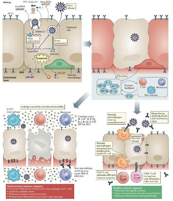
2
Redaction committee
Boris Lacarra – AP-HP Robert Debré
F-Xavier Lescure – Inserm, AP-HP Bichat, COREB
Guillaume Mellon – AP-HP Bichat, COREB
Inmaculada Ortega Perez – ANRS|Maladies infectieuses émergentes
Eric D’Ortenzio – ANRS|Maladies infectieuses émergentes, Inserm, AP-HP
Erica Telford – Inserm

Reviewing committee
Jean-Marc Chapplain – CHU Rennes, COREB                     Jean-Christophe Lucet – Inserm, AP-HP Bichat
Flavie Chatel – COREB                                       Claire Madelaine – ANRS|Maladies infectieuses émergentes
Hélène Coignard – HCL, COREB                                Matthieu Mahevas – Inserm, AP-HP Henri-Mondor
Dominique Costagliola – Inserm                              Emmanuelle Vidal Petiot – Inserm, AP-HP Bichat
Marie-Paule Kieny – Inserm                                  Benoit Visseaux – Inserm, AP-HP Bichat
Quentin Le Hingrat – Inserm, AP-HP Bichat
3

CLINICAL

 Questions:
 - What are the mechanisms of action of SARS-CoV-2?
 - What are the cellular and humoral host responses against SARS-CoV-2 infection?
 - What are the clinical features of COVID-19 in adults and children?
 - Is there multiple-organ damages associated to COVID-19?
 - What are the long term effects of Covid-19?
4
Physiopathology
• Binding to host cell through ACE2 receptor by spike (S) protein
o Lung, Kidney, Heart, Brain …
• Fusion of the viral envelope with cellular membrane (TMPRSS2)
• Virus hijacks the cell machinery
• Host cell pyroptosis and release damage-associated molecular
o ATP, nucleic acid, ASC oligomer …
• Inflammatory response
o Pro-inflammatory cytokines & chemokines: IL-6, IP-10, MCP1 …
• Attract other cells (monocytes, macrophage, T cells …)
o Pro-inflammatory feedback loop
o Eliminates the infected cells before the virus spreads
BUT sometimes (10 to 15 days after symptom onset)
• Accumulation of immune cells
o Hyper-inflammatory response
o Lung damage and multi-organ damage
Tay MZ, et al. Nat Rev Immunol. Apr 2020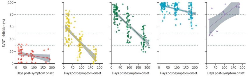
5
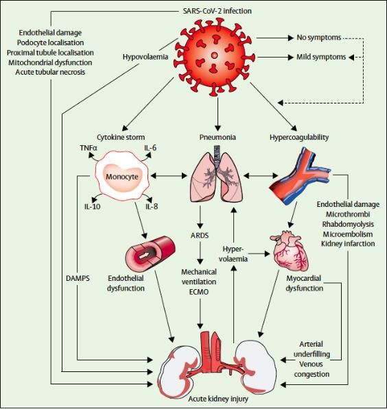
Physiopathology
• SARS-CoV-2 targets ACE2 receptor and infected cells via
« priming »
o Renin- Angiotensin system dysregulation
o Activation of innate and adaptative immune pathways
o Cytokine storm
o coagulation pathway hypercoagulation
• Multi-organ damage
o Kidney, heart, lungs, vessel, immune system ….
Battle D, et al. JASN. May 2020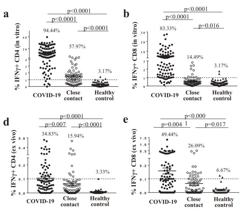
6

Auto-antibodies & type I IFN & COVID-19
                Neutralizing auto-Abs against type I IFN could lead to life-threatening COVID-19 pneumoniae?

987 patients hospitalized for life-threatening COVID-19
663 patients asymptomatic or mildly symptomatic (COVID-19)
1227 healthy controls
Auto-antibodies against IFN-α2 and/or IFN-ω?
• 135 of 987 critically ill patients had IgG auto-Abs against at least
  one type I IFN.
Auto-Abs neutralize IFN-α2 and/or IFN-ω in vitro?
• 101 of 987 life-threatening COVID-19 had neutralizing IgG auto-
  Abs against at least one type I IFN:
    • 51% against IFN-α2 and IFN-ω,                                          FACS plots depicting IFN-α2- or IFN-ω-induced pSTAT1 in the
    • 36% against IFN-α2 only,                                               presence of 10% healthy control or anti-IFN-α2/ω- auto-Abs-
                                                                             containing patient plasma (top panel) or an IgG-depleted
    • 13% against IFN-ω only.                                                plasma fraction (bottom panel).
• Auto-Abs detected in only 4 of 1227 controls and none of 663
  asymptomatic or mild-symptomatic patients.
                                                             IgG depletion from patients with auto-Abs restored normal pSTAT1
                                                             induction after IFN-α2 and IFN-ω stimulation.

                                                                                                   Bastard P, et al. Science. Sep 2020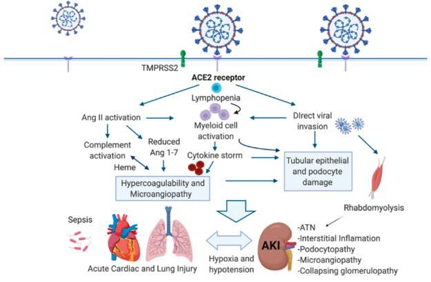
7
Auto-antibodies & type I IFN & COVID-19
Auto-Abs against all IFN-α subtypes?
• All patients (22) with neutralizing auto-Abs against IFN-α2 IFN-α levels in the plasma or
had auto-Abs against all 13 IFN-α subtypes serum of patients.
• Early treatment with IFN-α is unlikely to be beneficial
Auto-Abs against IFN-β?
• 1,9% of the patients had auto-Abs against IFN-β
• All were severe COVID-19
• Treatment with injected or nebulized IFN-β may have
beneficial effects
In vitro and in vivo?
• In patients with neutralizing auto-Abs against IFN-α2, the
baseline levels of type I IFN-dependent transcripts were
low, Auto-Abs against type I IFNs are a cause of severe SARS-
• Neutralizing in vitro & in vivo CoV-2 infection.
• Suggesting a pre-existing or concomitant biological impact Provides an explanation for the major sex bias in severe
in vivo COVID-19 and the increase in risk with age
Clinical and therapeutic implications
Bastard P, et al. Science. Sep 2020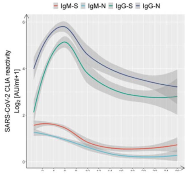
8

    C5a-C5aR1 axis & COVID-19
C5a anaphylatoxin and its receptor C5aR1 play a key role in the initiation and maintenance of inflammatory response
     • Recruiting and activating neutrophils and monocytes
82 individuals: 10 healthy control, 10 paucisymptomatic COVID-19, 34 with pneumonia & 28 with ARDS due to SARS-CoV-2.

           Concentration of C5a desArg in plasma                          C5a is detected in lung sample from COVID-19 patients

                        An increase in plasma C5a levels
                        proportional to COVID-19 severity.

                        Increased systemic and local
                        complement pathway activities on
                        the peripheral blood.

                               Saliva specimens could be effective in the diagnosis of COVID-19

                                                                                                     Carvelli J, et al. Nature. Jul 2020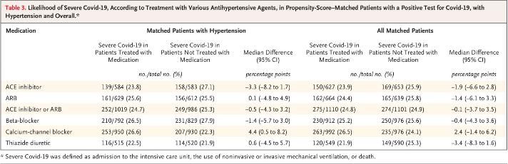
9
C5a-C5aR1 axis & COVID-19
C5a production leads to the chemo-attraction and Potential therapeutic strategy C5a-C5aR1 axis blockade.
activation of myeloid cells in the lung release of Avdoralimab = mAb against C5aR1.
inflammatory cytokines. In vitro:
• inhibited C5a-induced neutrophil activation,
Possible that the vasculitis associated with severe • Inhibited the C5a-induced migration of neutrophils.
COVID-19 is linked to the production of C5a.
CD45+ immune cell infiltration in BALF In mice:
C5a-R1 expression (red) • Mice received an intranasal instillation of recombinant
human C5a developed ALI.
• Avdoralimab prevented albumin release in BALF
• Avdoralimab inhibited the increase in IL-6, TNF and CCL2.
• Avdoralimab inhibited ALI in mice
CR5a-C5aR1 axis blockade might be used to prevent the excessive lung
Neutrophils and monocytes in BALF expressed C5aR1. inflammation and endothelialitis associated with ARDS in COVID-19
patients
Carvelli J, et al. Nature. Jul 2020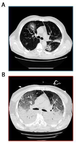
10

SARS-CoV-2 specific T cell immunity
 SARS-CoV2 specific T cells in patients with COVID-19
 • 36 individuals after recovery from mild to severe COVID-19.
 • T cell response against selected structural (N) and non-structural
   proteins (NSP7, NSP13 & ORF1).
 • Use of an unbiased method with overlapping peptides.
 • Peripherical blood mononuclear cell (PBMC) of the 36 patients were
   stimulated for 18h with the different peptides pools.

 • In 36 out of 36 individuals, found specific T cell that recognized
   multiple regions of the N-protein (IFNγ spot)

                                                                        Le Bert N, et al. Nature. Jul 2020
11
SARS-CoV-2 specific T cell immunity
SARS-CoV2 specific T cells in unexposed donors
• 37 donors: not exposed to SARS-CoV and SARS-CoV-2 The percentage of individuals with N-specific responses
• Detection of SARS-CoV-2-specific IFNγ responses in 19 out of 37
unexposed donor.
• The unexposed group showed a mixed response to the N protein or to
NSP7 and NSP13.
• These SARS-CoV-2-reactive cells from unexposed donors had the
capacity to expand after stimulation with SARS-CoV-2-specific
peptides.
Infection with betacoronaviruses induces multi-specific
and long lasting T cell immunity against the structural N
protein.
Le Bert N, et al. Nature. Jul 2020NEW
12
SARS-CoV-2 specific T cell immunity
Samples: peripheral blood mononuclear cells (PBMCs) from:
- 90 COVID-19 patients, collected 48-86 days after disease onset
- 69 close contacts (NAT-neg, SARS-CoV-2 IgG/IgM-neg), collected 48-86 days after contact
with COVID-19 patient
- 63 healthy donors, collected in September 2019
in vitro: PBMC expansion and 10 day-stimulation with peptide pool targeting spike,
membrane and envelope glycoproteins, nucleocapsid, RNA polymerase ORF1ab
ex vivo: PBMCs stimulated overnight with peptide pool
94.44% CD4+ and 83.33% CD8+ SARS-CoV-2 specific T cells of COVID-19 patients, and
57.97% CD4+ and 14.49 CD8+ of close contacts underwent in vitro expansion.
Healthy donors showed minimal cross-reactive T cells from other coronaviruses, but at a
significantly lower frequency than T cell immunity of close contacts.
ex vivo data corroborated these results and showed significant differences between T cell
memory pools and INFγ production of patients and close contacts.
Memory T cell immunity is detectable in both symptomatic and asymptomatic COVID-19
patients, with no significant difference in T cell pool size and qualities. INFγ expressing T cell exantion upon in vitro and ex vivo PBMC
stimulation with peptide pools encompassing viral epitopes
Following in vitro expansion, virus-specific memory CD4+ T cell pool correlated with titers
of IgG against S RBD and N protein.
SARS-CoV-2-specific memory T-cell immunity was observed in COVID-19 patients and close contacts at D48-86
Wang Z, et al. Nature Commun. Mar 2021NEW
13
SARS-CoV-2 specific B cell immunity
21 Severe (S)-Cov vs 18 Mild (M)-Cov patients assessed at 3 and 6 months
S-specific IgG are stable with time in both cohorts, but appear significantly higher in
S-CoV patients
At 6 months, B cells mostly resided in the memory B cell (MBC) compartment in
both cohorts, while S-specific antigen secreting cells were marginally detectable. S-
specific MBCs were at higher frequencies S-CoV patients, but present also in M-CoVs.
In both S-CoVs and M-CoVs, the proportion of S-specific activated B cells (ABCs)
steadily decreased over time, along with an increase of S-specific classical, resting
MBCs.
A robust and stable S-specific MBC population is induced in both M- and S-CoV patients
Sokal A, et al. Cell. Mar 2021NEW
14
SARS-CoV-2 specific B cell immunity
87 participants assessed at 1.3 and 6.2 months after infection
Antibody response:
• Anti-RBD and ELISA anti-N antibodies in plasma decreased significantly between
Relative change in
1.3 and 6.2 months. plasma antibody levels
• IgM showed the greatest decrease in anti-RBD reactivity (53%), followed by IgG
(32%); anti-RBD IgA decreased by only 15% and anti-N IgG levels by 22%.
• Individuals with persistent post-acute symptoms had significantly higher levels of
anti-RBD IgG and anti-N total antibody.
• Neutralising activity: NT50 was 401 and 78 at 1.3 and 6.2 months, respectively
5-fold decrease. Neutralizing activity was directly correlated with IgG anti-RBD.
B cell response:
• The % of RBD-binding memory B cells increased marginally between 1.3 and 6.2
months (n=21).
Percentage of antigen-
• although the magnitude of the RBD-specific memory B cell compartment is specific B cells
conserved between 1.3 and 6.2 months after infection, there is extensive clonal
turnover and antibody sequence evolution, consistent with prolonged germinal
centre reactions.
Gaebler C. et al. Nature. Jan 202115
Immunological assessment
Cohort study of 178 confirmed SARS-CoV-2 infection
Asymptomatic infection = 20,8% (37/178 patients)
37 asymptomatic matched with 37 mild symptomatic patients
Viral shedding:
• Initial Ct value were similar in the two groups
• Asymptomatic group had a significantly longer duration of viral
shedding (19 days versus 14 days; p=0.028)
IgG and IgM, 3 to 4 weeks after exposure (acute phase):
• IgG positivity rates similar between the two groups (81 and 84% of
asymptomatic and symptomatic, respectively)
• IgG levels in the asymptomatic group (median S/CO, 3.4; IQR, 1.6–
10.7) were lower than the symptomatic group (median S/CO, 20.5;
IQR, 5.8–38.2; p = 0.005)
• IgM levels were similar in the two groups (62 and 78% of positivity
of asymptomatic and symptomatic, respectively)
Long QX, et al. Nat Med. Jun 202016
Immunological assessment
IgG and IgM, 8 weeks after exposure (convalescent phase)
• A decline of IgG is observed among >90% of
patients
• 40% and 13% of asymptomatic individuals IgG+ at
the acute phase became seronegative
Similar observations were made for neutralizing antibodies
Asymptomatic patients had a reduced inflammatory
response with lower concentration of circulating cytokines
and chemokines
The relatively low seroprevalence and its decrease within
2-3 months after infection highlights the potential limits of
serology for diagnostic and the need of timely serosurvey
Limits
Viral RNA shedding does not equate viral infectivity
(not assessed in this study)
Serological observations may depend in part on the
commercial assay used
Long QX, et al. Nat Med. Jun 202017
Antibody response to SARS-CoV-2
The distribution of antibody sequences from six individuals
The number in the inner circle indicates the number of
Cohort of 149 cases and contacts: 111 with SAR-CoV-2 PCR positive + 46 close sequences analyzed for the individual denoted above the
contacts. circle. White indicates sequences isolated only once, and
Free of symptoms at least 14 days at the time of sample collection. grey or colored pie slices are proportional to the number of
clonally related sequences.
Convalescent plasma samples
• Binding to SARS-CoV-2 RBD and trimetric S protein?
IgG response: 78% showed anti-RBD and 70% anti-S
IgM response: 15% showed anti-RBD and 34% anti-S
Anti-RBD IgG levels moderately correlated with age and severity
• Neutralizing activities? the half-maximal neutralizing titer (NT 50)
Generally low: NT5018
Antibody response to SARS-CoV-2
• Do monoclonal antibodies have neutralizing activity? The normalized relative luminescence values for cell
lysates of 293TACE2 cells 48 h after infection with SARS-
Among 89 RBD-binding antibodies tested, we found 52 that neutralized CoV-2 pseudovirus in the presence of increasing
SARS-CoV-2 pseudovirus with IC50 values ranging from 3 to 709 ng/ml. concentrations of monoclonal antibodies.
Potent neutralizing antibodies found irrespective of the NT 50 values.
Even individuals with modest plasma neutralizing activity have rare
IgG memory B cells that produce potent SARS-CoV-2-neutralizing
antibodies.
Plasma neutralizing activity is low in most convalescent individuals
Recurrent anti-SARS-CoV-2 RBD antibodies with potent neutralizing
activity can be found in all individuals.
A vaccine designed to elicit such antibodies could be broadly effective.
Robbiani DF, et al. Nature. Aug 202019
Neutralizing antibodies to SARS-CoV-2 infection
Understanding the protective effects of the immune response ⇔ neutralizing effects of SARS-CoV-2 antibodies
Neutralizing activity of serum samples in relation to ELISA titers.
Mont Sinai Health System screen individuals for antibodies to SARS-CoV-2
• 72,401 individuals screening : 30,082 positive & 42,319 negative
• Vast majority of positive individuals have moderate-to-high titer of anti-
spike antibodies.
• Seroconverters = titer of 1:320 or higher
Neutralizing effects quantitative microneutralization assay
- 120 samples of known ELISA titers ranging from negative to ≥1:2880
- Neutralization titers significantly correlated with spike-binding titers
- 90% of seroconverters make detectible neutralizing antibody responses
Wajnberg A, et al. Science. Oct 202020
Neutralizing antibodies to SARS-CoV-2 infection
Antibody titer stability over time
Longevity of the antibody response:
- Slow decline in titer over time
- Initial increase in individuals with a initial titer of 1:320 or lower
- Titer remains relatively stable for several months after infection (∼ 5)
- Good correlation between neutralization and ELISA titers on day 148
Correlation between specific level of antibody and risk of (re)infection?
- Still unclear for infection with SARS-CoV-2 in humans
Individuals who have recovered from mild COVID-19 experience
relatively robust antibody response to the spike
Correlation between spike-binding titers and neutralization titers
Stable antibody titers over 3 months and modest declines at the
5-month time point
Cannot provide conclusive evidence : do this antibody responses protect from reinfection?
Wajnberg A, et al. Science. Oct 2020NEW
21
Long term humoral response against SARS-CoV-2
Anti-SARS-CoV-2 antibody persistance in COVID-19 patients after 6 months (1/3)
532/9542 individuals tested positive for pan-immunoglobulins Seroconversion rates of neutralising Ab at baseline and
(Wuhan). second follow-up:
- Seroprevalence adjusted for sex, age group and district: 6.92%
- 1st follow-up at 2 months, 2nd at 6 months
Baseline 2nd follow-up P value
IgG IgA IgM Neutralising Ab Confirmed
18 (66·7%) 16 (59·3%) 0·54
(n=27)
Baseline
532 (100%) 84 (15.8%) 69 (13.0%) 212 (39.8%) Symptomatic
(n=532) 35 (63·6%) 35 (63·6%) 1·000
(n=55)
1st follow-up
354 (97.5%) 36 (9.9%) 14 (3.9%) 162 (44.6%) Asymptomatic
(n=363) 88 (34·8%) 103 (40·7%) 0·38
(n=253)
2nd follow-up
413 (91.0%) 16 (3.5%) 7 (1.5%) 187 (41.2%) Total
(n=454) 141 (42·1%) 154 (46·0%) 0·55
(n=335)
Titers of pan-Ig, IgG , IgM and IgA continuosly decreased
significantly accross the study period The proportion of patients positive for IgM, IgA and IgG
decreased in all three subgroubs
He Z. et al. Lancet. Mar 2021NEW
22
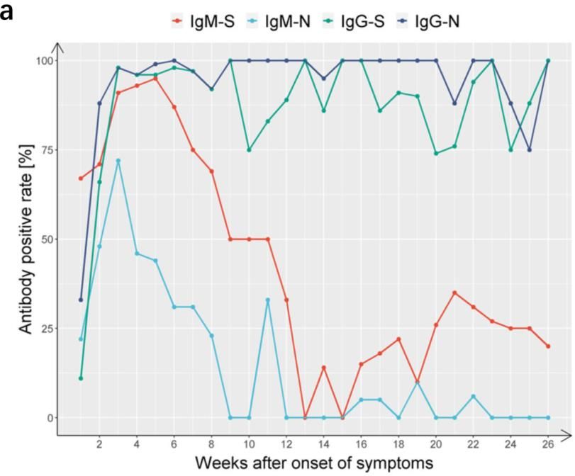
Long term humoral response against SARS-CoV-2
Anti-SARS-CoV-2 antibody persistance in COVID-19 patients after 6 months (2/3)
IgM and IgG responses against RBD of S and N proteins over 26 weeks (W) in 349 symptomatic COVID-19 patients, Wuhan
Antibody positivity rate: Antibody titers:
• W1: IgM-S (67%) > IgG-N (33%) > IgM-N (22%) > IgG-S (11%) • IgM-N and IgM-S peaked at W3 and 4, and fell below
• IgM-S peaked (95%) at W5, then decreased below 35% after W13 cutoff value at W9 and 12
• IgG-N and IgG-S peaked at W4 and 5, respectively,
• IgM-N reached 72% at W3, then became undetectable at W10-12
underwent a contraction phase (W6-14) and then
• IgG-N and IgG-S reached high positivity rate at W2 and 3 stabilised and high level over the study period
respectively, and remained high over the study period
IgG-RBD-S titer was highly positively correlated with
neutralising activity
Wu J, et al. Nature Commun. Mar 2021NEW
23
Long term humoral response against SARS-CoV-2
Anti-SARS-CoV-2 antibody persistance in COVID-19 patients after 6 months (3/3)
Neutralising antibodies (nAB) in 164 COVID-19 patients, Singapore, 180 days post symptom onset
5 patterns of nAb dynamics observed: Persistent group showed higher levels of pro-inflammatory
• Negative – did not reach 30% inhibition level): 12% cytokines (IFN-γ, IL-12p70, and IL-17A) and chemokine (IP-10),
• Rapid waning – positive early on but seroverting: 27% and growth factors as compared with other groups at 180 days
• Slow waning – remain nAb-positive over study period: 29% All patients maintained specific (NP, M, S) T-cell response at 180
• Persistent – minimal nAb decay 32% days
• Delayed response – increase of nAb ≥90 days post- Disease severity independently was associated with persistent
symptom onset: 2% antibody levels
Linear regression model of each
grouping for neutralising
antibody level. Dashed lines
represents 30%, 50%, and 80% of
sVNT percentage inhibition.
Chia WN, et al. Lancet Microbe. Mar 202124
Clinical features
Median time (41 patients admitted to hospital)
• From onset of symptoms to first hospital
admission
o 7 days [4,0–8,0]
• From illness onset to dyspnea
o 8 days [5,0–13,0]
• To ARDS
o 9 days [8,0–14,0]
• To ICU admission
o 10,5 days
• To mechanical ventilation
o 10,5 days [7,0–14,0]
Huang C, et al. Lancet. Feb 2020 Berlin DA, et al. NEJM. May 202025
Clinical features
China, 1 590 hospitalized patients (13,4% of all cases reported in China)
Age (median): 48,9 ± 16,3 years
Male: 904 (57,3 %)
Comorbidities Symptoms Outcomes
• Hypertension: 16,9 % • Fever: 88 % • Pharyngalgia: 14,7 % • Critical illness: 131 (8,24 %)
• Cough: >70 % • Headache: 15,4 % • ICU admission: 99 (6,23 %)
• Diabetes: 8,2 %
• Fatigue: 42,8 % • Chill: 12,2 % • Mechanical ventilation: 50 (3,1 %)
• CHD: 3,7 % • Shortness of breath: 20,8 % • Nausea/vomiting: 5,8 %
• Cerebrovascular disease: 1,9 % • Myalgia/arthralgia: 17,5 % • Diarrhea: 4,2 %
• COPD: 1,5 %
Case fatality rate: 50 (3,1 %)
• Chronic kidney disease: 1,3 % Abnormal chest CT: 1130 (71,1 %)
• Malignancy: 1,1 %
Guan W, et al. Eur Respi J. Jun 202026
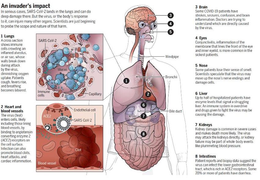
Organ damage
Wadman M, et al. Science. Apr 202027
Radiology
Monocentric – from 16 January to 17 February
90 patients - Median follow up: 18 days [5 – 43]
CT interpretation (366 CT scan)
Each lung divided into 3 zones
Overall CT score (max = 24)
Results
• Increase median values of CT score with time
• Peak levels of lung involvement: 6-11d from symptom
onset
• Ground glass opacity (GGO) is the most common finding
• More diverse manifestations around 6-11d and after
• Sensitivity of CT for SARS-CoV-2 increase over time
• At discharge: 64% still had abnormalities
Limitations : No subgroup analysis (mild and severe)
Bilateral GGO is the most common manifestation
Rapid extension and specific pattern of evolution
Wang Y, et al. Radiology. Mar 202028
Radiology
Ground glass opacity in a 35-year-old woman with COVID-19 pneumonia
J1 J5 J11 J15
TIME
Wang Y, et al. Radiology. Mar 202029
Heart & COVID-19
Acute myocarditis
• 7 – 17% of hospitalized patients ECG and echocardiographic abnormalities
• 22 – 31% patients admitted in ICU • Correlated with worse outcomes
• 7% of COVID-19 related deaths
Acute myocardial infarction
• Viral illness increase the risk
• Inflammation + hypercoagulability increased risk
Acute heart failure
• 20-25% of patients in their initial presentation
• Increased risk of mortality
• New cardiomyopathy or exacerbation?
Dysrhythmias
• 17% of hospitalized and 44% of ICU patients
• Hypoxia, inflammatory, abnormal metabolism
Venous thromboembolic event
• Increased risk
• Inflammation, organ dysfunction, abnormal coagulation
• 16-17% of pulmonary embolism
Long B, et al. Am J Emerg Med. Apr 202030
Kidney & COVID-19
Introduction
• > 40% cases of COVID-19 have abnormal proteinuria at hospital
admission ACE2
pathways
• Patients admitted to ICU with COVID-19:
• 20 to 40% have an AKI
• 20% require renal replacement therapy (RRT)
Pathophysiology multifactorial with predisposing factors
Management
• Implementation of KDIGO guidelines
• Restore normal volume status
• Reduce the risk of
• Pulmonary oedema
• Right ventricular overload
• Congestion
• Application of lung-protective ventilation
• RRT
• Volume overload ± refractory hypoxemia
• Right jugular vein
• Anticoagulation protocols: LMWH or UFH
Ronco C, et al. Lancet Respir Med. May 202031 Kidney & COVID-19 Prospective cohort – 1 hospital in China – 701 patients Cumulative incidence of AKI subgrouped by baseline serum creatine • Prevalence of acute kidney injury (AKI)? • Association between markers of kidney injury and death? Age (median): 63 years with 52,4% male Illness onset to admission: 10 days Kidney injury (at admission) • Elevated serum creatinine (SC) at admission 14,4% • Elevated BUN at admission 13,1% • GFR
32
Kidney & COVID-19 After adjusting
Kidney abnormalities ↑ in hospital death
Cumulative incidence for in-hospital death
High prevalence of kidney disease among hospitalized patients with COVID-19
Association between kidney involvement and poor outcome
Early detection and effective intervention of kidney involvement
Impact on long-term outcomes?
Cheng Y, et al. Kidney Int. May 202033
Neuropsychiatric disorders & COVID-19
Temporal distribution for cases notified to the CoroNerve Study group
Online network of secure rapid-response case report notification portals
(CoroNerve platforms)
From April 2 to April 26, 2020 in the UK
153 unique cases (correlated with the national case identification data)
• 114 = confirmed SARS-CoV-2 infection
• 6 = probable SARS-CoV-2 infection
• 5 = possible SARS-CoV-2 infection
• 28 excluded because missing data
4 clinical syndromes associated with COVID-19
• Cerebrovascular event = 77 cases
o Ischemic stroke / intracerebral hemorrhage
• Altered mental status = 39 cases
o Encephalopathy /encephalitis / primary psychiatric
diagnoses / …
• Peripheral neurology = 6 cases Age distribution of
patients –
• Other neurological disorders = 3 cases
case definitions for
cerebrovascular and
Acute alteration in mental status were overrepresented in young patients neuropsychiatric
events
Cerebrovascular events in COVID-19 vasculopathy
Viral neurotropism? Host immune responses? Genetic factors?
Varatharaj A, et al. Lancet Psychiatry. June 202034
Severity of depressive symptoms & COVID-19
Who is most at risk and how their experiences are evolving as the pandemic continues?
Explore the severity levels of depressive symptoms among individuals at high
Characteristics of study participants (extract)
risk.
• Cohort study (COVID-19 Social Study in the UK)
• Depressive symptoms were measured on 7 occasions: the 9-item Patient
Health Questionnaire (PHQ-9)
• Exposures self-reported during the interview
Group-based trajectories of depressive symptoms were estimated using latent
growth mixture (LGM) modeling.
51 417 participants:
• Oldest age group > 60 y 32,1% (higher proportion)
• Higher proportion of participants in the low and medium-income groups
• 22,1% were essential worker
• 19,9% had mental heakth condition
• 11,3% had psychological or physical abuse
Group-Based Trajectories of Depressive Symptoms
Severe depressive symptoms decreased following the Class 1: low depressive symptom trajectory
Class 2: moderate depressive symptom trajectory
start of the lockdown but began to increase again Class 3: severe depressive symptom trajectory
Iob E, et al. JAMA Netw Open. Oct 202035
Severity of depressive symptoms & COVID-19
Associations of Sociodemographic, Psychosocial, and Health-Related Risk
The risk of severe depressive symptoms was higher among people: Factors With the Severe Depressive Symptom Trajectories
- Experiencing abuse or low social support
- With low SEP
- With preexisting mental or physical health condition
Preexisting mental health condition versus no preexisting:
- Mean PHQ-9 score more than 2-fold higher
Psychological distress experienced during this pandemic may result
in an increased incidence of various adverse physical health
outcomes.
Model 1: adjusting for age, sex and COVID-19 symptoms
Limits: Model 2: adjusting for other risk factors
- Not random sample & not nationally representative
- Self-reported measures bias (underreported The odds of severe depressive symptoms were more than 5-fold higher in
sensitive information) those facing socioeconomic disadvantage
- Causality cannot be assumed
- Lack data on individuals prior to lockdown Importance of developping strategies to identify at-risk person
Iob E, et al. JAMA Netw Open. Oct 20202 types of phenotypes 36
ARDS & COVID-19 Type «L»: Low elastance
• Gas volume nearly normal
• Vt 7-8 ml/kg DV37
Antihypertensive drugs & COVID-19
• Observational study
• Lombardy Region in Italy - data extracted from the registry
• February 21 to March 11
• Patient older than 40 years
• 6272 cases matched to 30759 controls (on age, sex & municipality
residence)
• Use of antihypertensive drugs
o ARBs 22,2% among cases and 19,2% among controls
o ACE inhibitors 23,9% among cases and 21,4% among controls
Limits
• Neither ARBs nor ACE inhibitors had a significant association with risk • Change in strategy to test for coronavirus during
of COVID-19 study
• Information on drug use is limited to prescription
o Risk similar for women and men
• Exposure to antihypertensive drug not available after
o Not modified by age – severity of clinical manifestation – course of December 2019
COVID-19
• Control group included persons with COVID-19
o No evidence of an independent relationship between RAAS • Unmeasured confounders
blockers and the susceptibility to COVID-19
Mancia G, et al. NEJM. May 202038
Antihypertensive drugs & COVID-19
• Observational study
• New-York University - Use of the NYU Langone Health
• March 1 to April 15, 2020
• All patients with Covid-19 test results recorded
• Extracted from the chart (preceding 18 months)
o Medical history
o Medication data
• For a given medication, used a propensity-score Limits
models that adjusted for multiple variable • Variation in the diagnostic characteristic for the
• 12594 patients COVID-19 testing method
o 5894 COVID-19+ • Multiple tests for some patients
• Some patients may have been tested at other heath
o 4357 history of hypertension 2573 COVID-19+
systems
• No association with any medication studied of • May not reflect actual drug exposure
o Risk of severe COVID-19 • Not account for socioeconomic status, insurance, …
o Increased likelihood of a positive test • Additional unmeasured confounders
Rule out that the risk was higher among treated
patients than among untreated patients
Reynolds HR, et al. NEJM. May 202039
Risk factors of mortality The proportion of hospitalized and fatal SARS-CoV-2 cases per 100 000
individuals relative to the total Danish population within each age group
Nationwide cohort of all Danish individuals tested for SARS-CoV-2
The study cohort was linked to the Danish administrative and health registrie
11 122 cases with PCR positive: 80% were community-managed & 20% were hospitalized
(whereas 2,8% in an ICU)
30 days all cause of mortality = 5,2%
Risk factors of death:
Sex:
• adjusted for age and number of co-morbidities, ORs = 2,1;CI95% [1.7–2.6] for men
Age:
• 70 – 79 years: OR= 15; CI95% [9– 26]
• 80-89 years: OR= 30; CI95% [17–52]
• >90 years: OR= 90; CI95% [50–162]
Proportion of patients
Number of co-morbidities: dying among SARS-CoV-
• OR=5.2; CI95% [3.4–8.0], for cases with at least four co-morbidities 2 PCR-positive cases
• 79% of deaths had at least two co-morbidities within different
Chronic diseases: subgroups of age and
• Ischemic heart disease & hypertension ORs 1,1 to 1,3 number of comorbidities
• Organ transplantation OR 3,4
Reilev M, et al. Int J Epidemiol. Sep 202040
2549 children in USA
• Age (median): 11 years [0 – 17] • Symptoms (on 291 cases) • Outcomes (on 745 cases)
• Fever: 56% • Hospitalized: 147
• Male: 57 %
• Cough: 54%
• ICU admission: 15
• Exposure to a COVID-19 patients: • Dyspnea: 13%
91% (household / community) • Diarrhea: 13% • Case fatality rate: 0,1%
• Nausea/vomiting: 11%
• Abdominal pain: 5,8%
• …
Children aged41
Pediatric inflammatory multisystem syndrome
Observation of a large number of children hospitalized for cardiogenic shock potentially associated with
SARS-CoV-2
• Retrospective cohort – 2 countries (France & Switzerland) – 14 centers
• 35 children - Age (median): 10 years [2 – 16] – 51% were male
• 88% were positive for SARS-CoV-2 (nasopharyngeal swabs or serology)
Evolution
• 71% had total recovery left ventricular ejection fraction at day 7
• Time to full recovery = 2 days [2 – 5]
Treatment (no recommendation for the moment)
• 62% had invasive respiratory support
• 28% needed VA-ECMO
Differences with Kawasaki disease
New disease related to SARS-CoV-2? No precise arguments - Older (median age: 8 to 10y)
Shares some similarities with KD - Incomplete forms of KD
- Limited number of coronary
Understanding the immune mechanisms of this disease is a priority artery dilatation
Belhadjer Z, et al. Circulation. May 202042 Pediatric inflammatory multisystem syndrome Cohort of patients with KD in Paris region associated with SARS-CoV-2 ( 16 patients) Compared with a historical cohort of «classical KD» ( 220 patients) Cohort of Kawa-COVID-19 • Median age = 10 y IQR [4,7 – 12,5] • Median time from the onset of KD to hospitalization was 5 days • RT PCR all site positive: 69% (11 cases) • Cardiac ultrasound was abnormal in 11 patients • No death – all are in remission Kawa-COVID-19 versus historical cohort • Older 10 vs 2 years (p
NEW
       Long Covid in hospitalized patients                                                                                      43

Timeline of post-acute COVID-19. Acute COVID-19 usually lasts until 4 weeks from symptom onset, beyond which
replication-competent SARS-CoV-2 has not been isolated. Post-acute COVID-19 is defined as persistent symptoms
and/or delayed or long-term complications beyond 4 weeks from the onset of symptoms. The common symptoms
observed in post-acute COVID-19 are summarized.
                                                                                    Nalbandian A, et al. Nature Med. Mar 2021
NEW
Long Covid in hospitalized patients 44
Cohort of adult Covid-19 patients hospitalized between Jan and May 2020,
Wuhan (China), 1733 patients enrolled – 6 month follow-up
Temporal changes of seropositivity of anti-SARS-CoV-2 antibodies
Total Scale 3 Scale 4 Scale 5-6
(no supplemental (supplemental (HFNC, NIV or
(94 patients)
oxygen) oxygen) IMV )
At least one symptom 1265/1655 (76%) 344/424 (81%) 820/1114 (74%) 101/117 (86%)
mMRC score 1196/1615 (74%) 323/425 (76%) 802/1079 (74%) 71/111 (64%)
Pain or discomfort
431/1616 (27%) 111/422 (26%) 274/1082 (25%) 46/112 (41%)
(EQ-5D-5L questionnaire)
Anxiety or depression
367/1617 (23%) 98/425 (23%) 233/1081 (22%) 36/111 (32%)
(EQ-5D-5L questionnaire)
Quality of life (0-100) 367/1617 (23%) 98/425 (23%) 233/1081 (22%) 36/111 (32%)
Distance walked in 6 min –
392/1692 (23%) 103/423 (24%) 255/1153 (22%) 34/116 (29%)
lower than normal range
eGFRNEW
Long Covid in hospitalized patients 45
Cohort of adult Covid-19 patients hospitalized between Mar and Jun 2020, Italy, 238 patients enrolled – 4 month follow-up
(27.7% no oxygen required; 20.6% noninvasive ventilation; 8.8% mechanical ventilation; 11.8% ICU)
Covid-19 symptoms Pulmonary Function Testing Physical Performance Evaluation
Acute phase At follow-up DLCONEW
Long Covid in hospitalized patients 46
47 780 individuals (mean age 65, 55% men) hospitalised with covid-19 and discharged
alive by 31 August 2020, exactly matched to controls from a pool of ̴50 million people
in England for personal and clinical characteristics.
Admission to hospital for covid-19 was associated with an increased risk of
readmission (3.5 times greater) and death (7.7 times greater) after discharge,
compared to matched control.
Rates of multiorgan dysfunction after discharge were higher in the Covid-19
cohort as compared to controls (Respiratory diseases 29% of individuals [27.3
times greater for new onset diagnoses], diabetes 4.0% [3.0], MAjor
Cardiovascular Events 4.8% [2.8], chronic kidney disease 1.5% [1.9], chronic
liver disease 0.3% [1.5]).
Absolute risk of death, readmission, multiorgan dysfunction after discharge
was greater in individuals ≥70 and of white ethnic background.
Secondary analysis showed that individuals discharged from ICU after covid-19
experienced greater rates of death and readmission than those not admitted
to ICUs.
Ayoubkhani D, et al. BMJ. Mar 202147 CLINIC & PHYSIOPATHOLOGY (April 2021) 1. What are the mechanisms of action of SARS-CoV-2? - It uses ACE2 receptor to enter the cell and can produce a hyper-inflammatory response - Activation of innate and adaptative immune pathways - Auto-Abs against type I IFNs are a cause of severe SARS-CoV-2 infection 1. What are the cellular and humoral host responses against SARS-CoV-2 infection? - Induces long lasting T and B cell immunity against the Spike protein and the structural N protein - Recovered from mild COVID-19 robust antibody response to spike protein - Most symptomatic and asymptomatic patients present strong IgM and IgG responses, the latter lasting up to 6 months - Anti-spike protein antibody titers appear to correlate with viral neutralization for several months 2. What is the clinical presentation of COVID-19 in adults and children? - Most persons are asymptomatic or mildly symptomatic - Independent risk factors of mortality: age – obesity – chronic disease - Children are less represented than adults and have less severe or critical forms of the disease 3. Is there multiple-organ damage? - Predominantly lung damage prognostic of the disease - Several cases of heart & kidney damage 4. What are the long term effects of Covid-19 (Long Covid)? - Long term effects include fatigue, pulmonary function impairment and psychological sequelae up to 6 months after infection - ICU admission for Covid-19 is associated to increased risks of readmission and death
References                                                                                                                               48
1. Tay MT, et al. The trinity of COVID-19: immunity, inflammation and intervention. Nat Rev Immunol. 2020 Jun;20(6):363-374. doi:
    10.1038/s41577-020-0311-8.
2. Batlle D, et al. Acute Kidney Injury in COVID-19: Emerging Evidence of a Distinct Pathophysiology. J Am Soc Nephrol. 2020 Jul;31(7):1380-
    1383. doi: 10.1681/ASN.2020040419.
3. Le Bert N, et al. SARS-CoV-2-specific T cell immunity in cases of COVID-19 and SARS, and uninfected controls. Nature. 2020
    Aug;584(7821):457-462. doi: 10.1038/s41586-020-2550-z.
4. Wang Z, et al. Exposure to SARS-CoV-2 generates T-cell memory in the absence of a detectable viral infection. Nat Commun. 2021 Mar
    19;12(1):1724. doi: 10.1038/s41467-021-22036-z.
5. Sokal A, et al. Maturation and persistence of the anti-SARS-CoV-2 memory B cell response. Cell. 2021 Mar 4;184(5):1201-1213.e14. doi:
    10.1016/j.cell.2021.01.050.
6. Gaebler C, et al. Evolution of antibody immunity to SARS-CoV-2. Nature. 2021 Mar;591(7851):639-644. doi: 10.1038/s41586-021-03207-w.
7. Long QX, et al. Antibody responses to SARS-CoV-2 in patients with COVID-19. Nat Med. 2020 Jun;26(6):845-848. doi: 10.1038/s41591-020-
    0897-1.
8. Robbiani DF, et al. Convergent antibody responses to SARS-CoV-2 in convalescent individuals. Nature. 2020 Aug;584(7821):437-442. doi:
    10.1038/s41586-020-2456-9.
9. Bastard P, et al. Autoantibodies against type I IFNs in patients with life-threatening COVID-19. Science. 2020 Oct 23;370(6515):eabd4585.
    doi: 10.1126/science.abd4585.
10. Wajnberg A, et al. Robust neutralizing antibodies to SARS-CoV-2 infection persist for months. Science. 2020 Dec 4;370(6521):1227-1230.
    doi: 10.1126/science.abd7728.
11. He Z, et al. Seroprevalence and humoral immune durability of anti-SARS-CoV-2 antibodies in Wuhan, China: a longitudinal, population-level,
    cross-sectional study. Lancet. 2021 Mar 20;397(10279):1075-1084. doi: 10.1016/S0140-6736(21)00238-5.
References 49
12. Wu J, et al. SARS-CoV-2 infection induces sustained humoral immune responses in convalescent patients following symptomatic COVID-19.
Nat Commun. 2021 Mar 22;12(1):1813. doi: 10.1038/s41467-021-22034-1.
13. Chia WN, et al. Dynamics of SARS-CoV-2 neutralising antibody responses and duration of immunity: a longitudinal study. Lancet Microbe.
2021 Mar 23. doi: 10.1016/S2666-5247(21)00025-2. Online ahead of print.
14. Carvelli J, et al. Association of COVID-19 inflammation with activation of the C5a-C5aR1 axis. Nature. 2020 Dec;588(7836):146-150. doi:
10.1038/s41586-020-2600-6.
15. Reilev M, et al. Characteristics and predictors of hospitalization and death in the first 11 122 cases with a positive RT-PCR test for SARS-CoV-
2 in Denmark: a nationwide cohort. Int J Epidemiol. 2020 Oct 1;49(5):1468-1481. doi: 10.1093/ije/dyaa140.
16. Mancia G, et al. Renin-Angiotensin-Aldosterone System Blockers and the Risk of Covid-19. N Engl J Med. 2020 Jun 18;382(25):2431-2440.
doi: 10.1056/NEJMoa2006923.
17. Reynolds HR, et al. Renin-Angiotensin-Aldosterone System Inhibitors and Risk of Covid-19. N Engl J Med. 2020 Jun 18;382(25):2441-2448.
doi: 10.1056/NEJMoa2008975.
18. Huang C, et al. Clinical features of patients infected with 2019 novel coronavirus in Wuhan, China. Lancet. 2020 Feb 15;395(10223):497-506.
doi: 10.1016/S0140-6736(20)30183-5.
19. Berlin DA, et al. Severe Covid-19. N Engl J Med. 2020 Dec 17;383(25):2451-2460. doi: 10.1056/NEJMcp2009575.
20. Guan W, et al. Comorbidity and its impact on 1590 patients with COVID-19 in China: a nationwide analysis. Eur Respir J. 2020
May;55(5):2000547. doi: 10.1183/13993003.00547-2020.
21. Wadman M, et al. A rampage through the body. Science. 2020 Apr 24;368(6489):356-360. doi: 10.1126/science.368.6489.356.
22. Wang Y, et al. Temporal Changes of CT Findings in 90 Patients with COVID-19 Pneumonia: A Longitudinal Study. Radiology. 2020
Aug;296(2):E55-E64. doi: 10.1148/radiol.2020200843.References 50
23. Long B, et al. Cardiovascular complications in COVID-19. Am J Emerg Med. 2020 Jul;38(7):1504-1507. doi: 10.1016/j.ajem.2020.04.048.
24. Ronco C, et al. Management of acute kidney injury in patients with COVID-19. Lancet Respir Med. 2020 Jul;8(7):738-742. doi:
10.1016/S2213-2600(20)30229-0.
25. Cheng Y, et al. Kidney disease is associated with in-hospital death of patients with COVID-19. Kidney Int. 2020 May;97(5):829-838. doi:
10.1016/j.kint.2020.03.005.
26. Varatharaj A, et al. Neurological and neuropsychiatric complications of COVID-19 in 153 patients: a UK-wide surveillance study. Lancet
Psychiatry. 2020 Oct;7(10):875-882. doi: 10.1016/S2215-0366(20)30287-X.
27. Iob E, et al. Levels of Severity of Depressive Symptoms Among At-Risk Groups in the UK During the COVID-19 Pandemic. JAMA Netw Open.
2020 Oct; 3(10): e2026064. doi: 10.1001/jamanetworkopen.2020.26064.
28. Gattinoni L, et al. COVID-19 Does Not Lead to a "Typical" Acute Respiratory Distress Syndrome. Am J Respir Crit Care Med. 2020 May
15;201(10):1299-1300. doi: 10.1164/rccm.202003-0817LE.
29. Gattinoni L, et al. COVID-19 pneumonia: different respiratory treatments for different phenotypes? Intensive Care Med. 2020
Jun;46(6):1099-1102. doi: 10.1007/s00134-020-06033-2.
30. CDC COVID-19 Response Team. Coronavirus Disease 2019 in Children - United States, February 12-April 2, 2020. MMWR Morb Mortal Wkly
Rep. 2020 Apr 10;69(14):422-426. doi: 10.15585/mmwr.mm6914e4.
31. Belhadjer Z, et al. Acute Heart Failure in Multisystem Inflammatory Syndrome in Children in the Context of Global SARS-CoV-2 Pandemic.
Circulation. 2020 Aug 4;142(5):429-436. doi: 10.1161/CIRCULATIONAHA.120.048360.
32. Pouletty M, et al. Paediatric multisystem inflammatory syndrome temporally associated with SARS-CoV-2 mimicking Kawasaki disease
(Kawa-COVID-19): a multicentre cohort. Ann Rheum Dis. 2020 Aug;79(8):999-1006. doi: 10.1136/annrheumdis-2020-217960.
33. Nalbandian A, et al. Post-acute COVID-19 syndrome. Nat Med. 2021 Mar 22. doi: 10.1038/s41591-021-01283-z. Online ahead of print.References 51
34. Huang C, et al. 6-month consequences of COVID-19 in patients discharged from hospital: a cohort study. Lancet. 2021 Jan 6;397(10270):220-
232. doi: 10.1016/S0140-6736(20)32656-8.
35. Bellan M, et al. Respiratory and Psychophysical Sequelae Among Patients With COVID-19 Four Months After Hospital Discharge. JAMA Netw
Open. 2021 Jan 4;4(1):e2036142. doi: 10.1001/jamanetworkopen.2020.36142.
36. Ayoubkhani D, et al. Post-covid syndrome in individuals admitted to hospital with covid-19: retrospective cohort study. BMJ. 2021 Mar
31;372:n693. doi: 10.1136/bmj.n693.Contacts Dr. Guillaume Mellon Dr Eric D’Ortenzio guillaume.mellon@aphp.fr eric.dortenzio@inserm.fr
You can also read