SARS-COV-2 ON OCULAR SURFACES IN A COHORT OF PATIENTS WITH COVID-19 FROM THE LOMBARDY REGION, ITALY
←
→
Page content transcription
If your browser does not render page correctly, please read the page content below
Research

                JAMA Ophthalmology | Original Investigation

                SARS-CoV-2 on Ocular Surfaces in a Cohort of Patients
                With COVID-19 From the Lombardy Region, Italy
                Claudio Azzolini, MD; Simone Donati, MD; Elias Premi, MD; Andreina Baj, MD; Claudia Siracusa, BMedBiol;
                Angelo Genoni, BMedSci; Paolo A. Grossi, MD; Lorenzo Azzi, MD; Fausto Sessa, MD; Francesco Dentali, MD;
                Paolo Severgnini, MD; Giulio Minoja, MD; Luca Cabrini, MD; Maurizio Chiaravalli, MD; Giovanni Veronesi, MD;
                Giulio Carcano, MD; Lorenzo S. Maffioli, MD; Angelo Tagliabue, MD

                                                                                                                                   Supplemental content
                   IMPORTANCE Since February 2020, coronavirus disease 2019 (COVID-19) has spread rapidly
                   all over the world, with an epidemiological cluster in Lombardy, Italy. The viral
                   communicability may be mediated by various body fluids, but insufficient information is
                   available on the presence of the virus in human tears.
                   OBJECTIVES To investigate the rate of severe acute respiratory syndrome coronavirus 2
                   (SARS-CoV-2) in tears collected from patients with COVID-19 by means of real-time reverse
                   transcriptase–polymerase chain reaction (rRT-PCR) assay and to assess the association of
                   virus presence with concomitant clinical conditions.

                   DESIGN, SETTING, AND PARTICIPANTS Cross-sectional study conducted between April 9 and
                   May 5, 2020. The setting was intensive care units at Azienda Socio-Sanitaria Territoriale
                   (ASST) Sette-Laghi Hospital, University of Insubria, in Varese, Lombardy, Italy. A conjunctival
                   swab was performed in 91 patients hospitalized for COVID-19, which was clinically diagnosed
                   by rRT-PCR assay on nasopharyngeal swabs and by radiological imaging. Conjunctival swabs
                   from 17 additional healthy volunteer participants with no symptoms of COVID-19 were
                   examined to evaluate the availability and applicability of the conjunctival swab test.

                   EXPOSURE SARS-CoV-2 detection by means of rRT-PCR assay performed on the collected
                   samples obtained by conjunctival swabs.

                   MAIN OUTCOMES AND MEASURES Conjunctival swab and nasopharyngeal swab results are
                   reported, as well as demographic and clinical data.

                   RESULTS A total of 108 participants (mean [SD] age, 58.7 [14.2] years; 55 female and 53 male)
                   were tested for SARS-CoV-2 using rRT-PCR assay, including 91 patients hospitalized with
                   COVID-19 and 17 were healthy volunteers. SARS-CoV-2 was found on the ocular surface in 52
                   of 91 patients with COVID-19 (57.1%; 95% CI, 46.3%-67.5%), with a wide variability in the
                   mean viral load from both eyes. Among a subset of 41 patients, concordance of 63.0% (95%
                   CI, 41.0%-81.0%) was found between positive conjunctival and nasopharyngeal swab test
                   results when performed within 2 days of each other. In 17 of these patients, nasopharyngeal
                   swab results were negative for SARS-CoV-2. In 10 of these 17 patients, conjunctival swab
                   results were positive for the virus.

                   CONCLUSIONS AND RELEVANCE In this study, SARS-CoV-2 RNA was found on the ocular
                   surface in a large part of this cohort of patients with COVID-19, although the infectivity of this
                   material could not be determined. Because patients may have positive test results with a
                   conjunctival swab and negative results with a nasopharyngeal swab, use of the slightly
                   invasive conjunctival swab may be considered as a supplementary diagnostic test.

                                                                                                                              Author Affiliations: Author
                                                                                                                              affiliations are listed at the end of this
                                                                                                                              article.
                                                                                                                              Corresponding Author: Claudio
                                                                                                                              Azzolini, MD, Unit of Ophthalmology,
                                                                                                                              Azienda Socio-Sanitaria Territoriale
                                                                                                                              (ASST) dei Sette Laghi, Department
                                                                                                                              of Medicine and Surgery, University
                                                                                                                              of Insubria, Via Guicciardini 9, 21100
                   JAMA Ophthalmol. doi:10.1001/jamaophthalmol.2020.5464                                                      Varese, Italy (claudio.azzolini@
                   Published online March 4, 2021.                                                                            uninsubria.it).

                                                                                                                                                                (Reprinted) E1
                                                         © 2021 American Medical Association. All rights reserved.

Downloaded From: https://jamanetwork.com/ by Hamlaoui Mohamed on 03/07/2021
Research Original Investigation SARS-CoV-2 on Ocular Surfaces in a Cohort of Patients With COVID-19 From the Lombardy Region, Italy
I
n February 2020, coronavirus disease 2019 (COVID-19) as-
sociated with severe acute respiratory syndrome ap- Key Points
peared in China1-12 and rapidly spread all over the world,
Question What is the qualitative and quantitative presence of
with an epidemiological cluster in Northern Italy.9,13-17 Sev- severe acute respiratory syndrome coronavirus 2 (SARS-CoV-2) on
eral reasons explain this rapid diffusion in the Lombardy re- the ocular surface in patients with coronavirus disease 2019
gion of Italy. The high population density with increased pos- (COVID-19) hospitalized in intensive care units at a university
sibility of interpersonal contact, the prevalence of respiratory hospital in Lombardy, Italy?
pathologies because of pollution, contact with the Chinese Findings Using reverse transcription–polymerase chain reaction
population for travel and business, and the nonwindy, tem- assay, this study found that SARS-CoV-2 was present on the ocular
perate climate conditions promote the persistence of a virus surface in 52 of 91 patients with COVID-19 (57.1%). The virus may
in the environment. More than 90 000 citizens from the Lom- also be detected on ocular surfaces in patients with COVID-19
bardy region have officially been affected by the disease, with when the nasopharyngeal swab is negative.
a high death toll reported.18 The actual numbers are certainly Meaning These results suggest that SARS-CoV-2 may diffuse from
much higher. ocular surfaces to the body.
The etiological factor responsible for the disease has been
identified in a new betacoronavirus named severe acute re- ratory devices, as recommended by anesthesiologists for the
spiratory syndrome coronavirus 2 (SARS-CoV-2). This virus has safety of patients. We also collected specimens from 34 eyes
high human transmission via airways but has a medium of 17 healthy volunteer participants (10 women and 7 men) to
virulence.19 Therefore, many people test positive for the pres- evaluate the availability and applicability of the conjunctival
ence of the virus without any signs of disease. SARS-CoV-2 RNA swab test.
has been found in the nasopharyngeal tract and bronchial Before the conjunctival swab procedure, an ophthalmolo-
drainage,1 in saliva,20 in tears,21-24 in urine,25 and in feces26 but gist (E.P.) examined the status of eyelids, conjunctiva, and cor-
not in seminal fluids.27 nea. Eye examinations were done at the bedside without a slit-
The objective of the present study was to use real-time re- lamp. Clinical information about hospitalization timing, results
verse transcriptase–polymerase chain reaction (rRT-PCR) analy- of diagnostic and serological examinations, and type of respi-
sis to investigate the presence of SARS-CoV-2 in tears col- ratory device was recorded using a smartphone during the pro-
lected from patients with COVID-19. We also aimed to assess cedure and later transcribed. We also documented results of
the association of virus presence with concomitant systemic the last nasopharyngeal swab for each patient.
and local clinical conditions. The sampling procedure (Figure 1) was performed at the
bedside by the same ophthalmologist (E.P.) in both eyes. The
samples were obtained without topical anesthesia and after
sufficient time had elapsed since lacrimal substitutes had been
Methods
used. The conjunctival swabs were performed in the right eye
Study Design first, and paired (right and left) conjunctival swabs were kept
This cross-sectional study was conducted between April 9 and separate. A sample was available from only 1 eye in 6 patients
May 5, 2020, in intensive care units at Azienda Socio- (right eye in 4 patients and left eye in 2 patients) because of
Sanitaria Territoriale (ASST) Sette-Laghi Hospital, University difficulties during the sampling procedure (lack of coopera-
of Insubria, in Varese, Lombardy, Italy. A conjunctival swab was tion or technical problems). The conjunctival samples were ab-
performed in 2 cohorts (patients hospitalized for COVID-19 and sorbed by a dedicated swab with short fiber strands opti-
healthy participants) and was examined by rRT-PCR assay to mized for virus samples, with minimum patient discomfort
detect the presence of SARS-CoV-2. The study was carried out (FLOQSwabs; COPAN, Brescia, Italy). Swabs for sampling of
in accordance with the guidelines of the Declaration of tears are shown in the eFigure in Supplement 1.
Helsinki28 and subsequent revisions and with the authoriza- The conjunctival swab was then inserted into a dedicated
tion of the Ethics Committee and Institutional Advisory Board vial with a transport fluid (UTM-RT–Hanks balanced salt so-
of ATS (Agenzia per la Tutela della Salute) Insubria in Varese, lution enriched with proteins and sugars and with a neutral
Italy. Oral informed consent was obtained from study partici- pH; COPAN). The use of this fluid ensures that the samples are
pants, who did not receive a stipend. All collected data were preserved in ambient conditions for up to 48 hours. The vials
deidentified and protected by privacy safeguards. The study were delivered to the laboratory within 45 minutes and stored
is registered on ClinicalTrials.gov (Identifier: NCT04402853). at −80 °C after virus inactivation for 1 minute at 90 °C. The labo-
We collected specimens and clinical data from 176 eyes of ratory researcher (A.B.) then processed the samples within
91 patients hospitalized for COVID-19 in 3 different intensive 2 days.
care units (ICUs). Clinical diagnosis of COVID-19 disease was From the vial, 140 μL of each sample was subjected to RNA
confirmed by nasopharyngeal swab positivity, symptoms of se- extraction (QIAmp viral RNA mini kit; QIAGEN) and eluted in
vere respiratory distress, characteristic chest imaging (radio- 60 μL. One-step real-time polymerase chain reaction (PCR) was
graph and computed tomographic scan with ground glass performed (Luna universal qPCR master mix; New England Bio-
opacifications), and lymphopenia.5,8 All patients were from the Labs) from 5 μL of extracted RNA. Forward (5′-ACCTTCCCAGG-
central-northern area of Lombardy. We excluded patients with TAACAAACCA-3′) and reverse (5′-TTACCTTTCGGTCACACCCG-
continuous positive airway pressure helmets or similar respi- 3′) primers targeting the 5′ untranslated region (5′UTR) of
E2 JAMA Ophthalmology Published online March 4, 2021 (Reprinted) jamaophthalmology.com
© 2021 American Medical Association. All rights reserved.
Downloaded From: https://jamanetwork.com/ by Hamlaoui Mohamed on 03/07/2021SARS-CoV-2 on Ocular Surfaces in a Cohort of Patients With COVID-19 From the Lombardy Region, Italy Original Investigation Research
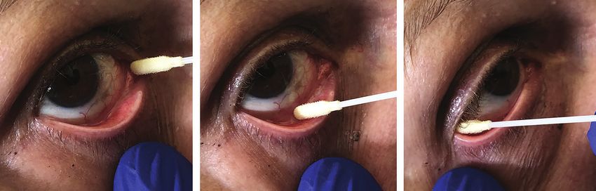
Figure 1. Procedure for Sampling of Tears
A Beginning B Middle C End
A, First, the swab is leaned on the
caruncula for 5 seconds. B, It is then
slowly moved across the exposed
inferior fornix conjunctiva to the
external fornix (C) within 5 seconds.
The samples were obtained without
topical anesthesia and after sufficient
time had elapsed since lacrimal
substitutes had been used. In all
patients, the conjunctival swabs were
performed in the right eye first.
SARS-CoV-2 were used. Primers were designed using soft- crete variables, respectively. The same statistics were reported
ware (CLC Genomics Workbench; QIAGEN), and their speci- in patients with positive vs negative conjunctival swab re-
ficity was checked using the BLAST database.29 sults and were compared using t tests or χ2 tests for continu-
Samples were run in 4 replicates together with a quanti- ous and discrete or dichotomous variables, respectively. Time
fied positive control (SARS-CoV-2 RNA control; Twist Biosci- between conjunctival swab performance and COVID-19 diag-
ence) on a PCR system (QuantStudio 5 real-time PCR; Thermo nosis was reported in weeks (range, 1-4 weeks).
Fisher Scientific), with an annealing temperature of 60 °C. Pro- We estimated the rate of patients with positive conjunc-
vided at a concentration of 106 copies/μL, serial dilutions of tival swab results in the overall sample since COVID-19 diag-
RNA control from 104 to 10 copies/μL were used to construct nosis using the exact binomial distribution for 95% CIs. The
a standard curve to perform an absolute quantification. Re- same analyses were replicated for the latest available naso-
sults were expressed as copies/μL (a positive viral load was de- pharyngeal swab. In addition, we reported the prevalence of
fined as >50 copies/μL in at least 1 eye) according to the de- concordant-positive conjunctival swab results in both eyes. The
tection and amplification ability of the rRT-PCR instrument. viral load distribution variable was defined as the average be-
In case of an uncertain result, an end point rRT-PCR and sub- tween eyes in patients with concordant-positive results and
sequent sequencing of the 5′UTR region were performed on as the viral load in the positive eye for individuals with posi-
the same RNA. tive conjunctival swab results in 1 eye only. We estimated the
Retrotranscription, amplification, and sequence reaction rate of both positive conjunctival and nasopharyngeal swabs,
were performed with a thermal cycler (Veriti; Perkin Elmer), as well as both negative conjunctival and nasopharyngeal
and 251–base pair amplicon detection was performed with a swabs, and the 95% CIs from the exact binomial distribution.
chip (LabChipGx Touch24; Perkin Elmer). Obtained ampli- The analyses were performed using SAS, 9.4 release (SAS In-
cons were sequenced by the Sanger method (SeqStudio ge- stitute Inc), and graphics were drawn with R (The R Project for
netic analyzer; Thermo Fisher Scientific). All collected speci- Statistical Computing). All P values were 2-sided but P values
mens have been preserved in vials at −80 °C for future research. were not adjusted for multiple analyses.
Electronic medical records (eTable in Supplement 2) were
uploaded and stored in a dedicated database. The system was
provided by a medical platform in a data warehouse in Milan,
Italy (Eumeda platform hosted by Aruba Business srl) to en-
Results
sure data security and uninterrupted availability. The plat- A total of 108 participants (mean [SD] age, 58.7 [14.2] years;
form enabled efficient and immediate data visibility and rapid 55 female and 53 male) were tested for SARS-CoV-2 using rRT-
data extraction. PCR assay. Ninety-one were hospitalized patients with COVID-
19, and 17 were healthy participants. The mean (SD) age of the
Statistical Analysis healthy participants was 49.5 (5.2) years. All conjunctival swabs
Because of a lack of data regarding the prevalence of positive were negative for SARS-CoV-2 among the healthy partici-
conjunctival swab results in the Italian COVID-19 population, pants. The evaluation of the availability and applicability of
there was no formal sample size calculation. Given the size of the conjunctival swab test in these 17 showed that the proce-
the reference population for the hospital and the epidemic dure is annoying for the participant and easily repeatable by
trend curve at the start of the study, we expected to accrue ap- the operator.
proximately 100 patients. This size would enable estimation Characteristics of the 91 hospitalized patients are shown
of the proportion of positive tests with a precision of about in Table 1. Of these, 58 patients (63.7%) had positive nasopha-
10%.30 ryngeal swab results and 33 (36.3%) had negative nasopha-
We summarized demographic and clinical features of eli- ryngeal swab results.
gible participants. Mean and standard proportions or abso- SARS-CoV-2 was found on the ocular surface in 52 of the
lute and relative frequencies were used for continuous and dis- 91 patients hospitalized with COVID-19 (57.1%; 95% CI, 46.3%-
jamaophthalmology.com (Reprinted) JAMA Ophthalmology Published online March 4, 2021 E3
© 2021 American Medical Association. All rights reserved.
Downloaded From: https://jamanetwork.com/ by Hamlaoui Mohamed on 03/07/2021Research Original Investigation SARS-CoV-2 on Ocular Surfaces in a Cohort of Patients With COVID-19 From the Lombardy Region, Italy
Table 1. Demographic and Clinical Characteristics of 91 Patients Overall and by Conjunctival Swab Positivity
or Negativity for SARS-CoV-2
No. (%)
Conjunctival
All patients swab positive Conjunctival swab
Variable (N = 91) (n = 52)a negative (n = 39)
Age, mean (SD), y 67.9 (13.2) 68.3 (13.5) 67.3 (12.9)
Sex
Female 45 (50.5) 27 (51.9) 18 (46.2)
Male 46 (49.5) 25 (48.1) 21 (53.8)
Nasopharyngeal swab test
Positive 58 (63.7) NA NA
Negative 33 (36.3) NA NA
Comorbidities at COVID-19 diagnosis
Cardiovascular diseases 53 (58.2) 31 (59.6) 22 (56.4)
Respiratory diseases 13 (14.3) 9 (17.3) 4 (10.3)
Autoimmune diseases 22 (24.2) 15 (28.8) 7 (17.9)
Neurological diseases 32 (35.2) 17 (32.7) 15 (38.5)
Endocrine diseases 65 (71.4) 38 (73.1) 27 (69.2)
Ocular diseases 5 (5.5) 2 (3.8) 3 (7.7)
Previous ocular surgery 3 (3.3) 2 (3.8) 1 (2.6)
Ocular signsb
Hyperemia 3 (3.3) 3 (5.8) 0
Secretions 3 (3.3) 3 (5.8) 0
Abbreviations: COVID-19, coronavirus
Blepharitis 5 (5.5) 4 (7.7) 1 (2.6) disease 2019; NA, not applicable;
Other signs 8 (8.8) 7 (13.5) 1 (2.6) SARS-CoV-2, severe acute respiratory
Hospitalization department syndrome coronavirus 2.
a
Infectious diseases department 31 (34.1) 19 (36.5) 12 (30.8) A positive viral load was defined as
greater than 50 copies/μL in at least
High intensity medicine department 42 (46.2) 24 (46.2) 18 (46.2) 1 eye.
Intensive care department 18 (19.8) 9 (17.3) 9 (23.1) b
Presence of signs or symptoms in at
Respiratory devices at time of conjunctival swabc least 1 eye.
c
Ambient air 21 (23.1) 9 (17.3) 12 (30.8) Listed from least to greatest
intensity (excluding patients with
Cannulas 16 (17.6) 10 (19.2) 6 (15.4)
continuous positive airway pressure
Venturi mask 30 (33.0) 19 (36.5) 11 (28.2) helmets or similar respiratory
Reservoir mask 13 (14.3) 8 (15.4) 5 (12.8) devices, as recommended by
anesthesiologists for the safety of
Intubation 11 (12.1) 6 (11.5) 5 (12.8)
patients).
67.5%). There was a wide variability in the average viral load junctival swab results. Neither the hospital department type
from both eyes (median [range], 284 copies/μL [29-45 000 nor the respiratory device at the time of conjunctival swab was
copies/μL]). associated with the presence of the virus on the ocular sur-
The virus was present in both eyes in 31 of 52 patients. face (Table 1).
Several patients (22 of 31 [71%]) had a slight difference in Fifty-eight (63.7%; 95% CI, 53.0%-73.6%) of the hospital-
viral load values between the 2 eyes. A discrepancy in con- ized patients had positive nasopharyngeal swab results (Table 2).
junctival swab results within the same patient (ie, 1 eye No differences in the number of positive conjunctival swabs were
positive and 1 eye negative) was observed in 21 of 91 found among the 91 hospitalized patients according to time since
patients (23.1%); a viral load greater than 50 copies/μL was COVID-19 diagnosis and clinical signs.
detected in 1 eye. The highest viral load of a single eye (up Forty-one patients had both nasopharyngeal and con-
to 90 000 copies/μL) was found in patients with the virus junctival swabs performed on the same day or within
detected in both eyes. The lowest value of viral load found 2 days. A positive concordance of 63.0% (95% CI, 41.0%-
in patients considered positive for the conjunctival swab (58 81.0%) was found between conjunctival and nasopharyn-
copies/μL) was found in patients in whom the virus was geal swabs when performed within 2 days of each other.
detected in 1 eye only. Among these 41 patients, only 7 of 17 patients (41.2%; 95%
No association was found between virus detection and co- CI, 18.0%-67.0%) tested had negative conjunctival and
morbidities at COVID-19 diagnosis. A slightly higher preva- nasopharyngeal swab results (Table 3). Again, 10 of these 17
lence of ocular signs or symptoms of surface inflammation in patients had negative nasopharyngeal swab results but posi-
at least 1 eye was present among patients with positive con- tive conjunctival swab results, with a mean viral load value
junctival swab results than among those with negative con- of 881.7 copies/μL (range, 29-6900 copies/μL).
E4 JAMA Ophthalmology Published online March 4, 2021 (Reprinted) jamaophthalmology.com
© 2021 American Medical Association. All rights reserved.
Downloaded From: https://jamanetwork.com/ by Hamlaoui Mohamed on 03/07/2021SARS-CoV-2 on Ocular Surfaces in a Cohort of Patients With COVID-19 From the Lombardy Region, Italy Original Investigation Research
Table 2. Comparison of Patients With Positive Conjunctival Swab Results
and Those With Positive Nasopharyngeal Swab Resultsa
Abbreviations: COVID-19, coronavirus
Conjunctival swab positive Nasopharyngeal swab positive disease 2019; NA, not applicable.
No.b/total No.e/total a
A positive viral load was defined as
Variable No.c % (95% CI)d No.c % (95% CI)d greater than 50 copies/μL in at least
All patients 52/91 57.1 (46.3-67.5) 58/91 63.7 (53.0-73.6) 1 eye.
b
Time since COVID-19 Number of patients with positive
diagnosis, wk conjunctival swab results.
1 14/26 53.9 (33.3-73.4) NA 73.3 (58.1-85.4) c
Number of patients with a COVID-19
2 15/27 55.6 (35.3-74.5) NA 82.4 (56.6-96.2) diagnosis.
d
3 10/14 71.4 (41.9-91.6) NA 33.3 (0.1-70.1) The 95% CIs are from the exact
binomial distribution.
≥4 13/24 54.2 (32.8-74.5) NA 40.0 (19.1-64.0)
e
Number of patients with positive
χ2 Test P value NA .71 NA .005
nasopharyngeal swab results.
Table 3. Results of Conjunctival and Nasopharyngeal Swab Tests Among 41 Patients With Both Tests Performed Within 2 Days
Nasopharyngeal swab positive Nasopharyngeal swab negative
No.a/total No.d/total
Variable No.b Prevalence (95% CI)c No.e Prevalence (95% CI)c
All patients 15/24 0.63 (0.41-0.81) 7/17 0.41 (0.18-0.67)
Time since COVID-19 diagnosis, wk
1 7/8 0.88 (0.47-0.99) 2/6 0.33 (0.04-0.78)
≥2 8/16 0.50 (0.25-0.75) 5/11 0.45 (0.17-0.77)
c
Abbreviation: COVID-19, coronavirus disease 2019. The 95% CIs are from the exact binomial distribution.
a d
Number of patients with positive conjunctival swab results. Number of patients with negative conjunctival swab results.
b e
Number of patients in whom the results of the last nasopharyngeal swab were Number of patients in whom the results of the last nasopharyngeal swab were
positive. negative.
Figure 2 shows the median values and 25th to 75th per- samples were analyzed in various laboratories using differ-
centiles of viral load in 52 patients with positive conjunctival ent rRT-PCR procedures within the same study. Third, the
swab results. The median viral load was 1120 copies/μL for tear samples may have been incubated with various fluids.
week 1, 303 copies/μL for week 2, 424 copies/μL for week 3, Fourth, the interval from acquisition to processing was not
and 295 copies/μL for week 4. All 34 eyes of the 17 healthy con- stated, which may have altered the results if high. Fifth,
trol participants tested negative for SARS-CoV-2. knowledge about virus behavior and characteristics has
increased in the last few months, with growing availability
of detection methods.
There was a wide variability in the mean viral load from
Discussion both eyes in the studied cohort. Several patients had a slight
In this study, SARS-CoV-2 was present on the ocular surface difference between the 2 eyes. A discrepancy in conjunctival
in patients with COVID-19 and was quantitatively and quali- swab results (ie, 1 eye positive and 1 eye negative) was ob-
tatively detectable, but the infectivity of this material and served in 21 of 91 patients (23.1%). The variability in viral load
thus the definitive clinical relevance could not be deter- among the patients and the discrepancy between eyes might
mined from this study. The high rates reported in this study have different explanations. The presence of the virus on the
(Tables 1 and 2) may be attributable to different reasons. eye surface could be variable (sometimes low or undetect-
The sample collections were performed by the same oph- able). In addition, the sampling procedure may be uncomfort-
thalmologist following a precisely defined procedure and able and poorly done, especially when performed in the
using a dedicated swab for molecular testing. Tear samples second eye.
were processed in the same laboratory, which has extensive In comparison of conjunctival swab and nasopharyngeal
experience in processing thousands of body fluid samples a swab results in 41 patients with COVID-19 when tests were
day from patients with COVID-19. Time between sample performed within 2 days of each other, we observed a posi-
incubation and processing was minimized, as described in tive concordance of 63.0% (95% CI, 41.0%-81.0%) between
the Methods section, and a real-time PCR primer targeted to the results of the tests (Table 3, left column). In the same
the 5′UTR region of SARS-CoV-2 was used in all cases.20 subgroup, we observed that among the 17 patients with
The few positive coronavirus conjunctival swab results COVID-19 that had a negative nasopharyngeal swab test
reported in the literature21-24,31-35 may be because of several result, 10 patients had a positive conjunctival swab (Table 3,
critical issues in sampling and laboratory processes. First, right column). These patients demonstrated a high viral
the specimen collection procedures were not well explained load (approximate mean, 1000 copies/μL) in tear samples,
in the articles and may be not reproducible. Second, pointing out that the virus was present in the body despite
jamaophthalmology.com (Reprinted) JAMA Ophthalmology Published online March 4, 2021 E5
© 2021 American Medical Association. All rights reserved.
Downloaded From: https://jamanetwork.com/ by Hamlaoui Mohamed on 03/07/2021Research Original Investigation SARS-CoV-2 on Ocular Surfaces in a Cohort of Patients With COVID-19 From the Lombardy Region, Italy
into the body through the nasolacrimal duct, which is a
Figure 2. Viral Load in 52 Conjunctival Swab–Positive Patients
According to Time Since COVID-19 Diagnosis
pathway to the pharynx.46-48 This contagion occurs despite
the use of protective masks for the mouth and nose. The
50 000 clinical case of the deceased ophthalmologist Dr Li Wen-
liang of Wuhan, China, described in the literature may be an
example of such COVID-19 spread.49 Although the infectiv-
ity of the viral material detected in the present study is
Viral load, copies/μL
5000
unknown, these results support the use of eye protection
for people working in environments where infection
through the ocular route is feasible. 50-58 Eye protection
500 probably should be considered if viral material might
exceed certain limits, especially in the absence of wind or
indoor systems designed to clear the air.
0 We evaluated the association between positive conjunc-
1 2 3 4
tival swab results and the use of different respiratory
Time since COVID-19 diagnosis, wk
devices9 in patients with COVID-19, as considered in the eli-
Shown are the median values and 25th to 75th percentiles of viral load.
gibility criteria stated in the Methods section of our study. It
COVID-19 indicates coronavirus disease 2019. is speculated that invasive maneuvers and respiratory
devices like continuous positive airway pressure masks or
helmets may increase the risk of viral diffusion by creating a
being undetectable in the nasopharyngeal tract. Studies26,36 closed environment around the head. 59 No associations
have reported the presence of SARS-CoV-2 in various body were found between respiratory device use and conjuncti-
fluids, but it was not always detectable in the nasopharyn- val swab results. This finding indicates that respiratory
geal tract in those cases. devices may not be associated with viral diffusion of SARS-
There could be many different explanations for the CoV-2 into tears (Table 1).
presence of the virus on the ocular surface, but these rea- We observed a low rate of ocular signs or symptoms in pa-
sons are all speculative. For example, direct contagion from tients positive for the conjunctival swab (Table 1). A persis-
airborne droplets by infected people is possible, as well as tent palpebral edema could be secondary to the prone posi-
particles diffused in the atmosphere.1,9 Atmospheric par- tion of a patient for respiratory reasons, as we find in patients
ticulates are known to function as carriers for many chemi- a few days after ocular surgery when a prone position is
cal and biological contaminants, including viruses.37 By pig- necessary.60 However, data in the current literature are dis-
gybacking, viruses adhere to atmospheric fine powders38 cordant about eye surface inflammatory involvement be-
consisting of solid or liquid particles that are able to remain cause many patients show different signs and symptoms, with
in the atmosphere for hours, days, or longer, especially in a difficult grading and evaluation.61
nonwindy and polluted climate like the Po Valley in In respiratory airways, the infection and cellular entry
Lombardy.39 Increasing numbers of patients with COVID-19 of SARS-CoV-2 are mediated by the spike glycoprotein of
have been diagnosed in this region since March 3, 2020, coronavirus and the host cellular SARS-CoV receptor for
corresponding to excessive atmospheric particulate levels40 angiotensin-converting enzyme 2 (ACE2).62 Type II trans-
recorded from February 10 to 29, 2020. This interval is rec- membrane serine protease (TMPRSS2) is required to pro-
ognized as the incubation period before clinical manifesta- mote SARS-CoV-2 entry by ACE2 cleavage to promote viral
tion of the virus. uptake. 63 However, the expression of ACE2 receptors in
Regarding other means of viral diffusion into the eye, anterior ocular tissues, such as the conjunctiva or cornea,
the literature reports direct contact with infected surfaces has yet to be established. More research exploring the
by the hands and transport to the mouth, nose, or other hypothesis of SARS-CoV-2 ocular infection through ACE2
mucous membranes, such as the conjunctiva.1,9,41,42 SARS- may be warranted. Various clinical signs of SARS-CoV-2
CoV-2 remains viable in aerosol form for hours even with a have been described in the literature,64 but they appear to
decreased infectious titer.43 It may stay on various surfaces be nonspecific and different from viral conjunctivitis and
longer. For example, it can be detected on plastic for up to keratitislike adenovirus. Knowing that SARS-CoV-2 is pre-
72 hours, although with a great reduction in virus titer (ie, sent in the conjunctiva may represent future scenarios for
the virus could be viable on dry surfaces until 72 hours even immunological and body reaction therapy in these patients.
if a reduction ofSARS-CoV-2 on Ocular Surfaces in a Cohort of Patients With COVID-19 From the Lombardy Region, Italy Original Investigation Research
Limitations
This study has some limitations. We could not determine the in- Conclusions
fectivity of the viral material detected and thus the definitive
clinical relevance. Other limitations include the cross-sectional This cross-sectional study found that SARS-CoV-2 RNA was
design of the study, which lacks long-term prospective evalua- present on ocular surfaces in a large portion of the study
tion of the patients. The limited number of negative conjuncti- cohort, although the infectivity of this material could not be
val swab in the presence of a positive nasopharyngeal test in pa- determined from the study. Findings suggest that individu-
tients with COVID-19 may be due to difficulties in patients’ tears als with COVID-19 may test positive with a conjunctival
sampling and current overall limited knowledge. To evaluate the swab and test negative with a nasopharyngeal swab. The
availability and applicability of the conjunctival swab test, we slightly invasive conjunctival swab may be considered as a
studied a group of 17 healthy volunteers with no symptoms or supplementary diagnostic test for COVID-19. Ongoing devel-
signs of COVID-19. All conjunctival swabs were negative for SARS- opment of procedures and laboratory testing tools may
CoV-2 among these participants. improve the ability to investigate this use in the future.
ARTICLE INFORMATION Drafting of the manuscript: Azzolini, Donati, Premi, 4. Lu R, Zhao X, Li J, et al. Genomic
Baj, Azzi, Dentali, Severgnini, Tagliabue. characterisation and epidemiology of 2019 novel
Accepted for Publication: October 10, 2020.
Critical revision of the manuscript for important coronavirus: implications for virus origins and
Published Online: March 4, 2021. intellectual content: Azzolini, Donati, Premi, receptor binding. Lancet. 2020;395(10224):565-574.
doi:10.1001/jamaophthalmol.2020.5464 Siracusa, Genoni, Grossi, Sessa, Minoja, Cabrini, doi:10.1016/S0140-6736(20)30251-8
Author Affiliations: Unit of Ophthalmology, Chiaravalli, Veronesi, Carcano, Maffioli. 5. Huang C, Wang Y, Li X, et al. Clinical features of
Azienda Socio-Sanitaria Territoriale (ASST) dei Sette Statistical analysis: Veronesi. patients infected with 2019 novel coronavirus in
Laghi, Department of Medicine and Surgery, Obtained funding: Azzolini. Wuhan, China. Lancet. 2020;395(10223):497-506.
University of Insubria, Varese, Italy (Azzolini, Administrative, technical, or material support: doi:10.1016/S0140-6736(20)30183-5
Donati, Premi); Laboratory of Microbiology, ASST Azzolini, Premi, Baj, Siracusa, Genoni, Grossi, Azzi, 6. Zhu N, Zhang D, Wang W, et al; China Novel
dei Sette Laghi, Department of Medicine and Dentali, Minoja, Tagliabue. Coronavirus Investigating and Research Team.
Surgery, University of Insubria, Varese, Italy (Baj); Supervision: Azzolini, Donati, Sessa, Severgnini, A novel coronavirus from patients with pneumonia
Laboratory of Medicine, Service of Cytogenetics Cabrini, Chiaravalli, Carcano, Maffioli. in China, 2019. N Engl J Med. 2020;382(8):727-733.
and Medical Genetics, ASST dei Sette Laghi, Varese, Conflict of Interest Disclosures: Dr Azzolini reported doi:10.1056/NEJMoa2001017
Italy (Siracusa); Department of Biotechnology and that his university received grants from Bayer and 7. Chen N, Zhou M, Dong X, et al. Epidemiological
Life Sciences, University of Insubria, Varese, Italy NovartisforophthalmicprojectsandthatBayer,Novartis, and clinical characteristics of 99 cases of 2019
(Genoni); Unit of Infectious and Tropical Diseases, Alcon, Allergan, Santen, Topcon, Thea, Dompé, and novel coronavirus pneumonia in Wuhan, China:
ASST dei Sette Laghi, Department of Medicine and Bausch & Lomb provided grant funds to a nonprofit a descriptive study. Lancet. 2020;395(10223):507-
Surgery, University of Insubria, Varese, Italy Italian ophthalmic association he chaired from 2016 to 513. doi:10.1016/S0140-6736(20)30211-7
(Grossi); Unit of Oral Medicine and Pathology, ASST 2019. No other disclosures were reported. 8. Wang D, Hu B, Hu C, et al. Clinical characteristics of
dei Sette Laghi, Department of Medicine and 138 hospitalized patients with 2019 novel coronavirus–
Surgery, University of Insubria, Varese, Italy (Azzi, Funding/Support: This study received financial
infected pneumonia in Wuhan, China. JAMA. 2020;323
Tagliabue); Unit of Pathology, ASST dei Sette Laghi, support from the research resources of the
(11):1061-1069. doi:10.1001/jama.2020.1585
Department of Medicine and Surgery, University of Department of Medicine and Surgery, University of
Insubria, Varese, Italy. 9. Giwa AL, Desai A, Duca A. Novel 2019
Insubria, Varese, Italy (Sessa); Unit of High Intensity coronavirus SARS-CoV-2 (COVID-19): an updated
Medicine, ASST dei Sette Laghi, Department of Role of the Funder/Sponsor: The funding source overview for emergency clinicians. Emerg Med Pract.
Medicine and Surgery, University of Insubria, had no role in the design and conduct of the study; 2020;22(5):1-28.
Varese, Italy (Dentali); Cardiosurgery Intensive Care collection, management, analysis, and
10. Corman VM, Landt O, Kaiser M, et al. Detection
Unit, ASST dei Sette Laghi, Department of interpretation of the data; preparation, review, or
of 2019 novel coronavirus (2019-nCoV) by
Biotechnology and Life Sciences, University of approval of the manuscript; and decision to submit
real-time RT-PCR. Euro Surveill. 2020;25(3):23-30.
Insubria, Varese, Italy (Severgnini); Transplants the manuscript for publication. doi:10.2807/1560-7917.ES.2020.25.3.2000045
Intensive Care Unit, ASST dei Sette Laghi, Varese, Additional Contributions: We gratefully
Italy (Minoja); Intensive Care Unit, ASST dei Sette 11. Habibzadeh P, Stoneman EK. The novel
acknowledge the nursing staff of the various coronavirus: a bird’s eye view. Int J Occup Environ
Laghi, Department of Biotechnology and Life departments involved in the care of patients with Med. 2020;11(2):65-71. doi:10.15171/ijoem.2020.1921
Sciences, University of Insubria, Varese, Italy COVID-19 and the laboratory personnel involved in
(Cabrini); Unit of Ophthalmology, ASST dei Sette 12. Zhou F, Yu T, Du R, et al. Clinical course and risk
this study. We thank Andrea Falco, BEng, for
Laghi, Varese, Italy (Chiaravalli); Research Center in factors for mortality of adult inpatients with
construction and management of the dedicated COVID-19 in Wuhan, China: a retrospective cohort
Epidemiology and Preventive Medicine (EPIMED), database for the electronic medical records
Department of Medicine and Surgery, University of study. Lancet. 2020;395(10229):1054-1062. doi:
(Eumeda platform hosted by Aruba Business srl). 10.1016/S0140-6736(20)30566-3
Insubria, Varese, Italy (Veronesi); Unit of General, He was not compensated for his contributions.
Emergency and Transplant Surgery, ASST dei Sette 13. Sebastiani G, Massa M, Riboli E. Covid-19
Laghi, Department of Medicine and Surgery, REFERENCES epidemic in Italy: evolution, projections and impact
University of Insubria, Varese, Italy (Carcano); Chief of government measures. Eur J Epidemiol. 2020;
1. Guan WJ, Ni ZY, Hu Y, et al; China Medical
Medical Officer, ASST dei Sette Laghi, Varese, Italy 35(4):341-345. doi:10.1007/s10654-020-00631-6
Treatment Expert Group for Covid-19. Clinical
(Maffioli); Chancellor, University of Insubria, Varese, characteristics of coronavirus disease 2019 in 14. Rizzi M, Castelli F, Latronico N, Focá E.
Italy (Tagliabue). China. N Engl J Med. 2020;382(18):1708-1720. SARS-CoV-2 invades the West: how to face a
doi:10.1056/NEJMoa2002032 COVID-19 epidemic in Lombardy, Northern Italy?
Author Contributions: Dr Azzolini had full access to all
Infez Med. 2020;28(2):133-134.
of the data in the study and takes responsibility for the 2. She J, Jiang J, Ye L, Hu L, Bai C, Song Y. 2019 Novel
integrityofthedataandtheaccuracyofthedataanalysis. coronavirus of pneumonia in Wuhan, China: emerging 15. Porcheddu R, Serra C, Kelvin D, Kelvin N, Rubino
Concept and design: Azzolini, Dentali, Cabrini, attack and management strategies. Clin Transl Med. S. Similarity in case fatality rates (CFR) of COVID-19/
Carcano, Maffioli. 2020;9(1):19. doi:10.1186/s40169-020-00271-z SARS-COV-2 in Italy and China. J Infect Dev Ctries.
2020;14(2):125-128. doi:10.3855/jidc.12600
Acquisition, analysis, or interpretation of data: 3. Sommer A. Humans, viruses, and the eye: early
Azzolini, Donati, Premi, Baj, Siracusa, Genoni, report from the COVID-19 front line. JAMA 16. Asperges E, Novati S, Muzzi A, et al; COVID-19
Grossi, Azzi, Sessa, Dentali, Severgnini, Minoja, Ophthalmol. 2020;138(5):578-579. doi:10.1001/ IRCCS San Matteo Pavia Task Force. Rapid response to
Cabrini, Chiaravalli, Veronesi, Tagliabue. jamaophthalmol.2020.1294 COVID-19 outbreak in Northern Italy: how to convert a
jamaophthalmology.com (Reprinted) JAMA Ophthalmology Published online March 4, 2021 E7
© 2021 American Medical Association. All rights reserved.
Downloaded From: https://jamanetwork.com/ by Hamlaoui Mohamed on 03/07/2021Research Original Investigation SARS-CoV-2 on Ocular Surfaces in a Cohort of Patients With COVID-19 From the Lombardy Region, Italy
classic infectious disease ward into a COVID-19 conjunctiva. J Med Virol. 2020;92(10):1757-1758. dental practice. Int J Oral Sci. 2020;12(1):9. doi:10.
response centre. J Hosp Infect. 2020;S0195-6701(20) doi:10.1002/jmv.25856 1038/s41368-020-0075-9
30119-5. doi:10.1016/j.jhin.2020.03.020 34. Peng Y, Zhou YH. Is novel coronavirus disease 51. Lai THT, Tang EWH, Chau SKY, Li KKW. Reply to:
17. Gagliano A, Villani PG, Co’ FM, et al. COVID-19 (COVID-19) transmitted through conjunctiva? J Med Ocular manifestation, eye protection, and
epidemic in the middle province of Northern Italy: Virol. 2020. Published online March 16, 2020. doi: COVID-19. Graefes Arch Clin Exp Ophthalmol. 2020;
impact, logistics, and strategy in the first line 10.1002/jmv.25753 258(6):1341. doi:10.1007/s00417-020-04663-2
hospital. Disaster Med Public Health Prep. 2020:1-5. 35. Zhang X, Chen X, Chen L, et al. The evidence of 52. Mungmungpuntipantip R, Wiwanitkit V. Ocular
18. Odone A, Delmonte D, Scognamiglio T, SARS-CoV-2 infection on ocular surface. Ocul Surf. manifestation, eye protection, and COVID-19.
Signorelli C. COVID-19 deaths in Lombardy, Italy: 2020;18(3):360-362. doi:10.1016/j.jtos.2020.03.010 Graefes Arch Clin Exp Ophthalmol. 2020;258(6):1339.
data in context. Lancet Public Health. 2020;5(6):e310. 36. Azzi L, Carcano G, Dalla Gasperina D, Sessa F, doi:10.1007/s00417-020-04662-3
doi:10.1016/S2468-2667(20)30099-2 Maurino V, Baj A. Two cases of COVID-19 with 53. Wan KH, Huang SS, Young AL, Lam DSC.
19. Gomez LM, Meszaros VA, Turner WC, positive salivary and negative pharyngeal or Precautionary measures needed for ophthalmologists
Ogbunugafor CB. The epidemiological signature of respiratory swabs at hospital discharge: a rising during pandemic of the coronavirus disease 2019
pathogen populations that vary in the relationship concern. Oral Dis. 2020. Published online April 25, (COVID-19). Acta Ophthalmol. 2020;98(3):221-222.
between free-living parasite survival and virulence. 2020. doi:10.1111/odi.13368 doi:10.1111/aos.14438
Viruses. 2020;12(9):E1055. doi:10.3390/v12091055 37. Peng L, Zhao X, Tao Y, Mi S, Huang J, Zhang Q. 54. Zeri F, Naroo SA. Contact lens practice in the
20. Azzi L, Carcano G, Gianfagna F, et al. Saliva is a The effects of air pollution and meteorological time of COVID-19. Cont Lens Anterior Eye. 2020;43
reliable tool to detect SARS-CoV-2. J Infect. 2020;81 factors on measles cases in Lanzhou, China. Environ (3):193-195. doi:10.1016/j.clae.2020.03.007
(1):e45-e50. doi:10.1016/j.jinf.2020.04.005 Sci Pollut Res Int. 2020;27(12):13524-13533. doi:10. 55. Lai THT, Tang EWH, Chau SKY, Fung KSC, Li
21. Seah IYJ, Anderson DE, Kang AEZ, et al. 1007/s11356-020-07903-4 KKW. Stepping up infection control measures in
Assessing viral shedding and infectivity of tears in 38. Setti L, Passarini F, De Gennaro G, et al. ophthalmology during the novel coronavirus
coronavirus disease 2019 (COVID-19) patients. SARS-CoV-2RNA found on particulate matter of outbreak: an experience from Hong Kong. Graefes
Ophthalmology. 2020;127(7):977-979. doi:10.1016/j. Bergamo in Northern Italy: first evidence. Environ Res. Arch Clin Exp Ophthalmol. 2020;258(5):1049-1055.
ophtha.2020.03.026 2020;188:109754. doi:10.1016/j.envres.2020.109754 doi:10.1007/s00417-020-04641-8
22. Zhou Y, Duan C, Zeng Y, et al. Ocular findings 39. Conticini E, Frediani B, Caro D. Can 56. Li JO, Lam DSC, Chen Y, Ting DSW. Novel
and proportion with conjunctival SARS-COV-2 in atmospheric pollution be considered a co-factor in coronavirus disease 2019 (COVID-19): the
COVID-19 patients. Ophthalmology. 2020;127(7): extremely high level of SARS-CoV-2 lethality in importance of recognising possible early ocular
982-983. doi:10.1016/j.ophtha.2020.04.028 Northern Italy? Environ Pollut. 2020;261:114465. manifestation and using protective eyewear. Br J
23. Willcox MD, Walsh K, Nichols JJ, Morgan PB, doi:10.1016/j.envpol.2020.114465 Ophthalmol. 2020;104(3):297-298. doi:10.1136/
Jones LW. The ocular surface, coronaviruses and 40. Lombardia ARPA. Agenzia Regionale bjophthalmol-2020-315994
COVID-19. Clin Exp Optom. 2020;103(4):418-424. Protezione Ambiente: air data, March 2020. 57. Siedlecki J, Brantl V, Schworm B, et al. COVID-19:
doi:10.1111/cxo.13088 Accessed March 15, 2020. https://www. ophthalmological aspects of the SARS-CoV 2 global
24. Sun CB, Wang YY, Liu GH, Liu Z. Role of the eye arpalombardia.it/Pages/ARPA_Home_Page.aspx pandemic. Klin Monbl Augenheilkd. 2020;237(5):675-
in transmitting human coronavirus: what we know 41. Chan JF, Yuan S, Kok KH, et al. A familial cluster of 680. doi:10.1055/a-1164-9381
and what we do not know. Front Public Health. pneumonia associated with the 2019 novel coronavi- 58. Yan Y, Chen H, Chen L, et al. Consensus of
2020;8:155. doi:10.3389/fpubh.2020.00155 rus indicating person-to-person transmission: a study Chinese experts on protection of skin and mucous
25. Sun J, Zhu A, Li H, et al. Isolation of infectious of a family cluster. Lancet. 2020;395(10223):514-523. membrane barrier for health-care workers fighting
SARS-CoV-2 from urine of a COVID-19 patient. doi:10.1016/S0140-6736(20)30154-9 against coronavirus disease 2019. Dermatol Ther.
Emerg Microbes Infect. 2020;9(1):991-993. doi:10. 42. Phan LT, Nguyen TV, Luong QC, et al. Published online March 13, 2020.
1080/22221751.2020.1760144 Importation and human-to-human transmission of 59. Ralli M, Candelori F, Cambria F, et al. Impact of
26. Jiang X, Luo M, Zou Z, Wang X, Chen C, Qiu J. a novel coronavirus in Vietnam. N Engl J Med. 2020; COVID-19 pandemic on otolaryngology,
Asymptomatic SARS-CoV-2 infected case with viral 382(9):872-874. doi:10.1056/NEJMc2001272 ophthalmology and dental clinical activity and future
detection positive in stool but negative in 43. van Doremalen N, Bushmaker T, Morris DH, perspectives. Eur Rev Med Pharmacol Sci. 2020;24
nasopharyngeal samples lasts for 42 days. J Med Virol. et al. Aerosol and surface stability of SARS-CoV-2 as (18):9705-9711. doi:10.26355/eurrev_202009_23062
2020;92(10):1807-1809. doi:10.1002/jmv.25941 compared with SARS-CoV-1. N Engl J Med. 2020; 60. Donati S, Caprani SM, Airaghi G, et al. Vitreous
27. Song C, Wang Y, Li W, et al. Absence of 2019 382(16):1564-1567. doi:10.1056/NEJMc2004973 substitutes: the present and the future. Biomed Res
novel coronavirus in semen and testes of COVID-19 44. Otter JA, Donskey C, Yezli S, Douthwaite S, Int. 2014;2014:351804. doi:10.1155/2014/351804
patients. Biol Reprod. 2020;103(1):4-6. doi:10.1093/ Goldenberg SD, Weber DJ. Transmission of SARS 61. Wu P, Duan F, Luo C, et al. Characteristics of
biolre/ioaa050 and MERS coronaviruses and influenza virus in ocular findings of patients with coronavirus disease
28. World Medical Association. World Medical healthcare settings: the possible role of dry surface 2019 (COVID-19) in Hubei Province, China. JAMA
Association Declaration of Helsinki: ethical contamination. J Hosp Infect. 2016;92(3):235-250. Ophthalmol. 2020;138(5):575-578. doi:10.1001/
principles for medical research involving human doi:10.1016/j.jhin.2015.08.027 jamaophthalmol.2020.1291
subjects. JAMA. 2013;310(20):2191-2194. doi:10. 45. Han Y, Wu N, Zhu W, et al. Detection of HIV-1 62. Lange C, Wolf J, Auw-Haedrich C, et al.
1001/jama.2013.281053 viruses in tears of patients even under long-term Expression of the COVID-19 receptor ACE2 in the
29. National Center for Biotechnology Information. HAART. AIDS. 2011;25(15):1925-1927. doi:10.1097/ human conjunctiva. J Med Virol. 2020. Published
BLAST. Accessed October 31, 2020. https://blast. QAD.0b013e32834b3578 online May 6, 2020. doi:10.1002/jmv.25981
ncbi.nlm.nih.gov/Blast.cgi 46. Qing H, Li Z, Yang Z, et al. The possibility of 63. Hoffmann M, Kleine-Weber H, Schroeder S,
30. Hajian-Tilaki K. Sample size estimation in COVID-19 transmission from eye to nose. Acta et al. SARS-CoV-2 cell entry depends on ACE2 and
epidemiologic studies. Caspian J Intern Med. 2011;2 Ophthalmol. 2020;98(3):e388. doi:10.1111/aos.14412 TMPRSS2 and is blocked by a clinically proven
(4):289-298. 47. Lu CW, Liu XF, Jia ZF. 2019-nCoV transmission protease inhibitor. Cell. 2020;181(2):271-280.e8.
through the ocular surface must not be ignored. doi:10.1016/j.cell.2020.02.052
31. Xia J, Tong J, Liu M, Shen Y, Guo D. Evaluation of
coronavirus in tears and conjunctival secretions of Lancet. 2020;395(10224):e39. doi:10.1016/S0140- 64. Aiello F, Gallo Afflitto G, Mancino R, et al.
patients with SARS-CoV-2 infection. J Med Virol. 6736(20)30313-5 Coronavirus disease 2019 (SARS-CoV-2) and
2020;92(6):589-594. doi:10.1002/jmv.25725 48. Kuo ICA. Rashomon moment? ocular colonization of ocular tissues and secretions:
involvement and COVID-19. Ophthalmology. 2020; a systematic review. Eye (Lond). 2020;34(7):1206-
32. Liu Z, Sun CB. Conjunctiva is not a preferred 1211. doi:10.1038/s41433-020-0926-9
gateway of entry for SARS-CoV-2 to infect 127(7):984-985. doi:10.1016/j.ophtha.2020.04.027
respiratory tract. J Med Virol. 2020;92(9):1410-1412. 49. Parrish RK II, Stewart MW, Duncan Powers SL. 65. Xie C, Lu J, Wu D, et al. False negative rate of
doi:10.1002/jmv.25859 Ophthalmologists are more than eye doctors: in COVID-19 is eliminated by using nasal swab test.
memoriam Li Wenliang. Am J Ophthalmol. 2020; Travel Med Infect Dis. 2020;37:101668. doi:10.1016/
33. Guo D, Xia J, Shen Y, Tong J. SARS-CoV-2 may j.tmaid.2020.101668
be related to conjunctivitis but not necessarily 213:A1-A2. doi:10.1016/j.ajo.2020.02.014
spread through the conjunctiva SARS-CoV-2 and 50. Peng X, Xu X, Li Y, Cheng L, Zhou X, Ren B.
Transmission routes of 2019-nCoV and controls in
E8 JAMA Ophthalmology Published online March 4, 2021 (Reprinted) jamaophthalmology.com
© 2021 American Medical Association. All rights reserved.
Downloaded From: https://jamanetwork.com/ by Hamlaoui Mohamed on 03/07/2021You can also read