EARLIER INTERVENTION FOR AMD - Retina Today
←
→
Page content transcription
If your browser does not render page correctly, please read the page content below

Insert to      January/February 2019                                                          Sponsored by Ellex

                                                       As multimodal imaging redefines
                                                  diagnosis, a new laser technology may
                                                     make early interventional treatment
                                                        possible for some types of AMD.

2RT:
                                                      EARLIER
                                                      INTERV ENTION
                                                      FOR AM D
NETAN CHOUDHRY, MD, FRCS(C) | DAVID CHOW, MD | ROBYN GUYMER, AM, MBBS, PHD | FRANK HOLZ, MD | RISHI P. SINGH, MD
PARTICIPANTS
                 NETAN CHOUDHRY, MD, FRCS(C)                                             FRANK HOLZ, MD
                 n
                   C o-Founder and Medical Director, Vitreous Retina Macula              n Professor and Chairman, Department of Ophthalmology,
                   Specialists of Toronto, Toronto, Canada                                  University of Bonn, Germany, Bonn, Germany
                 n
                   Lecturer, Department of Ophthalmology & Visual Sciences,              n frank.holz@ukb.uni-bonn.de
                   University of Toronto, Toronto, Canada                                n Financial disclosures: Consultant (Acucela, Apellis, Bayer,
                 n
                   netan.choudhry@gmail.com                                                 Boehringer-Ingelheim, Bioeq/Formycon, Galimedix, Genentech,
                 n
                   F inancial disclosures: Consultant and/or Speaker (Allergan, Bayer,      Geuder, Grayburg Vision, Heidelberg Engineering, LinBioscience,
                   Carl Zeiss Meditec, Ellex, Johnson & Johnson, Optos, Topcon);            Novartis, Pixium Vision, Oxurion, Stealth BioTherapeutics); Grant
                   Research Equipment (Carl Zeiss Meditec, Optos, Topcon)                   Support or Sponsored Research (Acucela, Allergan, Apellis,
                                                                                            Bayer, Bioeq/Formycon, Carl Zeiss Meditec, CenterVue, Ellex,
                 DAVID CHOW, MD                                                             Galimedix, Genentech, Grayburg Vision, Heidelberg Engineering,
                 n
                   R etina Specialist and Director, Toronto Retina Institute,               LinBioscience, NightStarX, Novartis, Optos, Oxurion, Pixium
                   Toronto, Canada                                                          Vision, Stealth BioTherapeutics)
                 n
                   Assistant Professor, St. Michael’s Hospital, University of Toronto,
                   Toronto, Canada                                                       RISHI P. SINGH, MD
                 n
                   davidrchow@me.com                                                     n Retina Specialist, Cleveland Clinic, Cleveland, Ohio
                 n
                   F inancial disclosures: Consultant (Alcon, Allergan,                  n Associate Professor of Ophthalmology, Case Western Reserve
                   Bausch + Lomb, Bayer, Katalyst, Novartis, Optovue)                       University, Cleveland, Ohio
                                                                                         n
                                                                                            President, Retina World Congress, San Diego, California
                 ROBYN GUYMER, AM, MBBS, PHD                                              n
                                                                                            drrishisingh@gmail.com
                 n
                   Professor of Ophthalmology, Melbourne University,                      n
                                                                                            Financial disclosures: Consultant (Alcon, Carl Zeiss Meditec,
                   Melbourne, Australia                                                     Genentech, Novartis, Optos, Regeneron Pharmaceuticals, Inc.);
                 n
                   Deputy Director, Centre for Eye Research, Australia,                     Sponsored Research (Apellis)
                   Melbourne, Australia
                 n rh.guymer@unimelb.edu.au

                 n
                   F inancial disclosures: Advisory Board (Apellis, Bayer,
                   Genentech, Novartis)

2 INSERT TO RETINA TODAY | JANUARY/FEBRUARY 2019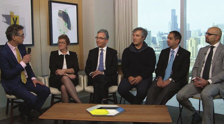
2RT:
                                                                                        2 R T: E AR LI E R I N T E R V E N T I O N F O R A M D

                                                                   EARL I ER
                                                                   I NTERVENTI ON
                                                                   FOR AM D
As multimodal imaging redefines diagnosis, a new laser technology may make early interventional treatment possible
for some types of AMD.

       As the ability to detect and
    diagnose age-related macular
    degeneration (AMD) has evolved
    to encompass earlier stages of the
    disease, the effort to develop earlier
    treatments has intensified. Recent
    research efforts have yielded some
    interesting results. Retina Today
    hosted a roundtable discussion
    during AAO Chicago 2018, chaired
    by Ellex Chief Medical Officer,
    David Lubeck, MD. The roundtable
    brought together a group of leading
    international experts in AMD to
    discuss changing treatments for the
    disease and how they envision the          Figure 1. Leading experts gather during AAO 2018 in Chicago. Left to right: David Lubeck, MD (moderator); Robyn
    future (Figure 1).                         Guymer, AM, MBBS, PhD; Frank Holz, MD; David Chow, MD; Rishi P. Singh, MD; and Netan Choudhry, MD, FRCS(C).

THE UNMET NEED FOR EARLY AMD TREATMENT
   We have seen a recent focus on the social and human
s

    costs of advanced AMD (Figure 2),1,2 which highlights
    the need for interventions to diagnose and treat the
    condition earlier in the disease process. How has this
    new focus changed the way you treat AMD?

   Frank Holz, MD: It is a huge unmet need. As we have come
                                                                            Figure 2. AMD has a significant effect on the global population.1,2
to understand the enormous burden that patients shoulder
in the late stages of this disease, it has become obvious that
an intervention that prevents the late-stage disease from                   efficacious treatment for late-stage dry AMD with geographic
developing is needed. Today, we can treat the neovascular                   atrophy (GA). An early intervention to slow progression of the
phenotype quite successfully, but we do not have any                        disease would help enormously.

                                                                                                             JANUARY/FEBRUARY 2019 | INSERT TO RETINA TODAY 3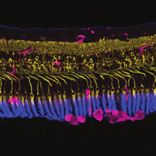
2 R T: E AR LI E R I N T E R V E N T I O N F O R A M D

                                                                        treatments that depend on the disease stage. Practitioners
           Patients and physicians are desperate…I am                   have tended to lump all AMD that is not wet into the dry cat-
                                                                        egory. Perhaps this was acceptable when there were no specific
                      pleased that we now can                           treatments for AMD at different stages of progression, but as
                                                                        we now start to discuss intervening early, we need patients and
         offer a treatment to patients with early AMD that              other doctors to understand what stage they have.
         may slow down progress of the disease, thereby                    Prof. Holz and I were part of the Ryan Beckman Initiative, so
                                                                        we would like people to use the Beckman Classification’s five
                 lowering their risk of losing vision.                  subgroups (Table 1).3
                                                   —David Chow, MD         Early AMD is defined based on the size of drusen, which is medi-
                                                                        um. Intermediate AMD involves either large drusen or medium
                                                                        drusen plus pigment. Finally, in late AMD, there may be atrophy
      David Chow, MD: One of the things we know about                   or neovascular disease.
   patients who do well on AMD therapies is how important                  With multimodal imaging, we are now able to detect atrophy
   their baseline VA is. If we can preserve their vision at a much      earlier and earlier, which is why the Classification of Atrophy
   earlier stage, when their vision is quite good, then their long-     Meeting focuses in part on trying to standardize how we define
   term outcome can be quite good as well. The benefit of early         the changes we see on OCT. Prof. Holz and I both contributed
   intervention is a consistent factor we have been able to extract     to a paper earlier this year that classifies complete atrophy and
   from all the studies we have ever done on anti-VEGF therapy.         incomplete atrophy based on which layers of the OCT are miss-
                                                                        ing.4 Complete atrophy includes photoreceptor and retinal pig-
     Robyn Guymer, AM, MBBS, PhD: The treatments for wet                ment epithelium (RPE) loss, and incomplete atrophy has a lesser
   AMD are quite efficacious, but they are also quite expensive and     degree of RPE damage and outer retinal atrophy. We are trying
   are a huge burden in terms of patients’ quality of life. Patients    to get everyone to use these terms so that we can all contribute
   would fare much better if we could intervene early, so we would      to trials that determine the risks associated with these stages.
   not reach a point where we need to rely on anti-VEGF treatments.
                                                                               Diagnostically, how would you approach a patient with
                                                                        s

      Dr. Chow: Prof. Holz hit it on the head. Patients and physi-              early or intermediate AMD?
   cians are desperate for something to treat patients with dry
   AMD and resulting visual loss. We know many patients who               Netan Choudhry, MD, FRCS(C): Traditionally, prior to
   suffer significant visual loss from this disease are patients with   the LEAD trial,5 our options for counseling patients about
   dry AMD who go on to develop GA. So many patients in our             the available therapies were limited, and these patients were
   practices are suffering. They are always walking in my door say-     somewhat neglected. We know that the AREDS vitamins are
   ing, “Why is it that you can give the other patient a needle, but    helpful for a certain subclass of patients with AMD, but now
   you cannot do anything for me?” I am pleased that we now             we are evaluating patients in the context of a new treatment
   can offer a treatment to patients with early AMD that may            modality, namely 2RT. We look at multimodal imaging, OCT,
   slow down progression of the disease, thereby lowering their         the clinical picture, the size of the drusen, and the presence
   risk of losing vision.                                               or absence of GA. Even nascent GA will be important as we
                                                                        evaluate these patients, as Prof. Guymer described.
   DIAGNOSING AND STAGING AMD
       As we discuss wet and dry AMD, and early,                          Rishi P. Singh, MD: It might be worth pointing out that all
   s

        intermediate, and late-stage disease, we should                 of these stages have two really important features that clini-
        ensure we are clear about these terms. Please                   cians use frequently. First, they help us to determine the level
        describe the different stages of AMD. How do new
        diagnostic technologies allow you to detect these                            TABLE 1. AMD: BECKMAN CLASSIFICATION 3
        stages, and how does this impact your choice of
        treatment?                                                       1.   No AMD (normal aging changes)
                                                                         2.   Early AMD
      Prof. Guymer: It makes matters confusing that many physi-          3.   Intermediate AMD
   cians use the term “dry AMD” when referring to late-stage GA
   or any disease that is not “wet” AMD. It is critical that we all      4.   Late AMD, neovascular (wet AMD)
   use the same terminology, particularly when we are developing         5.   Late AMD, GA (dry AMD)

4 INSERT TO RETINA TODAY | JANUARY/FEBRUARY 2019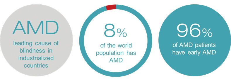
2 R T: E AR LI E R I N T E R V E N T I O N F O R A M D
of visual detriment, so we can say, for example, that a patient we know that patients with late-stage AMD will progress at a
has intermediate AMD and visual decline as a result. Second, different rate than those who have earlier disease. That allows
the stages allow us to determine the disease progression, and us to give our patients a prognosis.
LEAD TRIAL OF 2RT RETINAL REJUVENATION
Figure 3. 2RT
Courtesy of Professor Erica L. Fletcher MScOptom, PhD (Department of Anatomy and
Neuroscience, the University of Melbourne, Australia).
2RT is a nanosecond pulsed laser (Table 2). The technology induces a
was developed by Ellex with the goal of eliminating thermal mononuclear
damage to the neural retina, thus sparing the photoreceptors cell response,
from apoptosis during retinal laser treatment. It induces a including the
mononuclear cell response, including the release of microglia release of
(Figure 3). The 2RT laser’s mode of action is the combination microglia (pink).
of nanosecond pulses and engineered Nanopix Technology In this image, the
(Ellex), which produces a proprietary pixelated beam to a very retinal microglia
are shown
specific proportion of RPE cells, subjecting only targeted indi-
extending their
vidual cells to undergo apoptosis and allowing for a natural processes
healing response to restore retinal function. This ensures tar- through the outer
geted treatment of individual RPE cells. No other retinal laser nuclear layer
delivers such a short pulse, nor produces a pixelated beam. towards the laser
treatment site.
TABLE 2. THE KEY PARAMETERS OF 2RT
3 nanosecond pulse duration
400 µm spot size
Nanopix Technology
Pixelated beam profile
In 2018, the LEAD randomized clinical trial of 2RT
s
technology showed that it may slow progression to
late AMD in patients without reticular pseudodrusen 3 6-month, multicenter, randomized, sham-controlled trial.
s s s s
(RPD) (Figure 4).5 What do you consider the role of 2RT 2 92 patients enrolled had bilateral large drusen without signs of atrophy on OCT.
in your clinical practice? P articipants were randomized to 2RT or sham treatment at 6-month intervals.
M ultimodal imaging showed that overall, there was a positive trend towards slow-
Prof. Guymer: It was important to do a properly conducted ing disease progression to late AMD, but this did not reach statistical significance.
randomized clinical trial, which has been lacking for subthresh- T he benefit of 2RT was shown in a post hoc analysis, where progression to late
s
old lasers in general. This was the first robust clinical trial for AMD was significantly slowed for 222 participants without coexistent RPD.
2RT, and results showed that, overall, there was no benefit
Figure 4. The 2RT LEAD study results.5
of the laser treatment in slowing progression to late AMD.
However, in a post hoc analysis, there appeared to be a differ-
ential treatment effect depending upon the presence of RPD, validate the post hoc analyses. Imaging technologies now allow
with those without RPD having less progression and those with us to clearly detect RPD, which sit atop the RPE differently
RPD having a greater risk of progression to late AMD in the from sub-RPE drusen.
treatment group. It is very important to remember that we
need to validate the results with a second trial. We must have a Prof. Guymer: The results look very promising. Subthreshold
replication study showing the same results. nanosecond laser treatment may help slow progression in
patients without RPD, and the basic science certainly is very
Prof. Holz: We would very much welcome an opportunity solid. The LEAD results were derived from a post hoc analysis,
to slow progression. Since we learned about the encourag- and we uncovered an interesting finding once we drilled down
ing results of the LEAD trial of the 2RT system led by Prof. to look at treatment effect modification analysis. In this post
Guymer,5 we have been reflecting on the design of a study to hoc analysis, patients were randomized on the basis of the
JANUARY/FEBRUARY 2019 | INSERT TO RETINA TODAY 5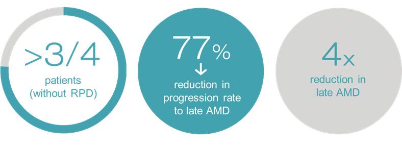
2 R T: E AR LI E R I N T E R V E N T I O N F O R A M D

   RPD. Until we do that and find the same results, we have a        If you look at the literature on RPD, it goes from those num-
   promising result, but not a proven intervention at this stage.    bers up to 92% in a few papers.7 Based on your results, it seems
                                                                     as though the way we classify RPD for 2RT will become criti-
      Dr. Chow: The work that Prof. Guymer has done is ground-       cally important.
   breaking. It is essential that someone has actually done a
   proper prospective trial with subthreshold laser technology,         Prof. Guymer: Correct. According to the work of Christine
   because many other technologies with subthreshold lasers          A. Curcio, PhD, from the United States,8 we can find RPD in
   have suffered as a consequence of not having a properly run       almost anyone. It will be interesting to learn in the future if
   prospective trial.                                                patients can afford to have a small amount of RPD and still
      In the post hoc analysis, Prof. Guymer’s trial highlights a    benefit from 2RT. Some subjects were predominately RPD,
   clinical finding about RPD that goes beyond the average retina    but they had to have at least one standard drusen to get into
   specialist’s diagnostic experience. Although we all know RPD,     the trial. Going forward, we have to ask whether we should
   we have never used it as an important piece of diagnostic         include patients with RPD in 2RT studies. I do not think we
   information that could strongly influence our treatment deci-     should automatically exclude them, because it would be
   sions. I think Prof. Guymer’s work highlighting RPD is such       a shame to deny any chance to slow down progression to
   an incredibly important factor in determining whether this        patients who are at risk. Just as one trial requires verification
   technology may or may not be effective. In fact, I would love     before we can say treatment has value for one group, we also
   to understand how you defined RPD. Was that purely photos?        need to see verification in a second trial of whether it lacks
   Was it OCT? Was it scanning laser ophthalmoscopy?                 value for another population.

      Prof. Guymer: Right from the beginning of the design           s  Dr. Singh, as a physician who has not used 2RT
   phase, we wanted to look for RPD as a variable. What we did           yet, how are you presently managing early AMD?
   not think about was whether the effect of 2RT would differ            How do you think you might incorporate 2RT into
   depending on the presence or absence of RPD. We followed              your practice?
   a very strict definition of RPD that occupies about a page of
   the paper. Basically, we wanted patients to have at least five       Dr. Singh: Right now, it is about waiting and watching the
   RPD spread over more than one OCT scan and validated on           patient. We do not have many options for our patients with
   another en face OCT image. We needed to see RPD definitively      this condition. We do give them AREDS vitamin therapy and
   on two en face images because an OCT scan shows the central       the ForeseeHome monitoring device (Notal Vision), which has
   cube, and RPD can occur outside the cube.                         been shown to improve the ability to detect early conversion
      We classified RPD as definitely, possibly, or definitely not   to exudative AMD at home, so we can get them in with earlier
   present, and analyzed the results, comparing definitely versus    VA changes.9 Beyond that, we do nothing more. If we had a
   the rest. We determined that 2RT’s greatest potential efficacy    good treatment option for these patients that was proven in
   lay with patients without RPD.                                    a large trial, we would be more than willing to adopt it and
                                                                     probably use it for many of our patients.
     Dr. Chow: You have written another paper that elegantly
   looked at the rate of RPD and its risk of causing GA, which was   INITIATING 2RT FOR INTERMEDIATE AMD
   about 12% to 14%.6 That was based on fundus photography.             Let us discuss the clinical scenario of patients
                                                                     s

                                                                         with intermediate AMD. How would you describe
                                                                         these patients?

          It is essential that someone has actually done a              Prof. Holz: Although they can have perfect 20/20 vision,
                                                                     often they have functional deficits. For example, they take
          proper prospective trial with subthreshold laser           longer to adapt from the sunny outdoors to a dark room.
        technology, because many other technologies with             Dim light is terrible for them. It would be a second aim of any
                                                                     treatment to not only halt progression to late stages of AMD,
       subthreshold lasers have suffered as a consequence            but also perhaps to improve vision in the intermediate stage of
           of not having a properly run prospective trial.           the disease. We have not yet learned whether AREDS vitamin
                                                                     supplements for intermediate AMD will produce any improve-
                                                   —David Chow, MD   ment. The hypothesis is that if a treatment can get rid of exces-
                                                                     sive extracellular material, perhaps photoreceptors and the RPE

6 INSERT TO RETINA TODAY | JANUARY/FEBRUARY 2019
2 R T: E AR LI E R I N T E R V E N T I O N F O R A M D
for me. Prof. Guymer, I would love to hear your comments.
I know that GA was excluded in the trial criteria, but do
When there is nascent GA hiding and about to we know if, for example, early GA is an absolute “no” with
become full-thrust, that would not be an ideal this technology?
situation to offer 2RT. Ideally, we would like to offer it Prof. Guymer: We do not know because they were not in
earlier in the disease progression, particularly to our the trial. We have tried to look back at the data to see if there
is any indication that a small patch of nascent GA could be
patients with intermediate AMD. reversed by causing the RPE to sort of bridge the gap. Looking
closely at the trial’s results, there may be a few instances where
—Netan Choudhry, MD, FRCS(C) the nascent GA seemed to improve for whatever reason, so
it could be possible to use 2RT when there are very small
amounts of nascent GA. However, if you want an early-stage
will function better, but this needs to be tested. endpoint, then what are we going to measure if patients are
starting with atrophy?
Dr. Choudhry: That is one of the things we see in our prac- It will be interesting to see what the US FDA wants us to do.
tice as well. Patients with intermediate AMD start to complain Will they want us to treat patients and observe all the way to
of visual issues that generally did not exist in the disease’s early GA, in which case we could potentially include some nascent
stages because the drusen burden or RPE changes were low. atrophy and determine whether we can slow it down? As we
At the intermediate stage, they have decreased retinal sensitiv- continue to follow patients from the LEAD trial, we may find
ity, slowing dark adaptation, and metamorphopsia from larger that we have slowed the progression from nascent to full-
drusen that are starting to develop. We get patients involved blown GA. It is possible that treatment may be of help to the
in vision rehabilitation therapy or using magnifiers, and they rest of the eye.
experience benefits from that process very quickly. It is a criti- The other interesting thing, of course, is the bilateral effect.
cal stage within the AMD spectrum when intervention would In the animal studies, there seemed to be some benefit to the
really be ideal. other eye,10 and our very early pilot study showed this as well.11
It may be possible to treat one eye and see some benefit to the
Looking at the clinical and diagnostic spectrum, what other eye in slowing down progression. We have a lot to learn
s
are your treatment criteria to initiate 2RT? about this from the preclinical data because there are mea-
surable indicators in both eyes that appear to suggest we are
Dr. Choudhry: Currently, we look to the LEAD trial for guid- improving the removal of extracellular debris.
ance in terms of which types of patients should be offered
treatment with 2RT. As we have just discussed, RPD is one fac- Prof. Holz: Perhaps to the point, we should create the
tor for exclusion. It is not indicated for patients with GA. When same experience and offer the same options to patients who
there is nascent GA hiding and about to become full-thrust, present with symptoms and show a precursor to atrophy
that would not be an ideal situation to offer 2RT. Ideally, we on their B-scans. It would be a shame to exclude them from
would like to offer it earlier in the disease progression, particu- future treatment, particularly because atrophy is present in
larly to our patients with intermediate AMD. such a high proportion of patients. Is incipient atrophy the
point of no return or not? We do not know. In any case, we
Prof. Holz: In Europe we do not offer the treatment at this should continue to develop treatments that show efficacy in
stage. Results from the LEAD trial are promising,5 but the the presence of GA in terms of slowing lesion enlargement.
next steps of another clinical trial informed by the LEAD trial
need to take place before it gets approval from the regulatory Dr. Chow: I agree with you, but I was discouraged
authorities. We look forward to the commencement of by the strong connection between RPD and GA in the
that trial. post hoc analysis.
Dr. Chow: One practical issue is that since we received Prof. Guymer: It is the patients with RPD who are most
the 2RT laser in our office, our referrals have not met the affected by the dark adaptation problems. They potentially
inclusion criteria: intermediate AMD with no visual loss. The progress to atrophy more quickly. They are the very patients
more common patient has already started to suffer some you really want to treat.
overt visual loss and has early GA. That has been the issue In fact, drusen by itself may be less of a risk than we have
JANUARY/FEBRUARY 2019 | INSERT TO RETINA TODAY 72 R T: E AR LI E R I N T E R V E N T I O N F O R A M D
previously understood. In the AREDS study, they were not
able to look at RPD in everyone.12 They did not have all the
multimodal imaging we are using. As a result, we do not know One of the things that has come out of this trial,
the risk of progression for a patient who has drusen without where a paradigm shift is upon us and we are about
RPD. The two things together are clearly not good news.
to embark on a possible new treatment for AMD, is
There have been previous studies on laser treatment of the responsibility to educate peers more about the
s
AMD. How is the LEAD trial different?
nonexudative form of AMD.
Prof. Guymer: The first studies go back many decades. Don —Netan Choudhry, MD, FRCS(C)
Gass actually noticed serendipitously that when he used a
thermal laser on diabetic patients, their drusen resolved.13 In
the 1990s, researchers in both America and Europe tried using
a thermal laser to reduce drusen, and some of them were referred patients with drusen plus pigment, and we found
stopped due to concerns about neovascularization as a com- that about 40% of these people had nascent GA. The
plication. Subsequently, Cochrane’s analysis showed that was referring clinician would think they had early AMD, but when
not the case.14 we looked more closely, they already had the beginnings of
Previous studies focused on the old thermal lasers. In con- atrophy. Forming a patient cohort with drusen plus some
trast, the nonthermal 2RT laser, has a very different mechanism pigment and no nascent GA took quite some time, but we
of action. We cannot extrapolate LEAD findings to other types could reduce that time by teaching optometrists to identify
of lasers, even subthreshold lasers. As far as I know, LEAD is the the necessary characteristics on OCT. Eventually, we hope
only randomized clinical trial of a subthreshold nanosecond that we can build a registry that enables us to recruit much
laser for AMD. more quickly.
TEACHING PEERS TO CLASSIFY AND REFER AMD Prof. Holz: As the classification develops, it will become
We have discussed the use of advanced imaging more complex. We have simplified this classification over the
s
to perform a more detailed analysis of AMD, past decades, and now high-resolution imaging modalities
and then tailoring therapy accordingly. What does this give us a new level of granularity. Yes, we need to address the
mean for primary care doctors referring role of optometrists as the first contact, but I am convinced
patients to you? that artificial intelligence and machine learning will permit the
imaging modalities themselves to make assessments and clas-
Dr. Choudhry: One of the things that has come out of sifications right away. For example, we will not have to search
this trial, where a paradigm shift is upon us and we are about for RPD in each and every B-scan in the future. Artificial intel-
to embark on a possible new treatment for AMD, is the ligence will help identify these patients.
responsibility to educate peers more about the nonexudative
form of AMD. Most primary eye care providers can identify a Dr. Choudhry: As Prof. Guymer mentioned, the term “dry
subretinal hemorrhage, pigment epithelial detachment, and so AMD” should be much more specific because the treatments
forth. But now they may need to recognize all the subtleties are likely going to be aimed at subtypes within the spectrum of
of the different types of drusen through a GA classification nonexudative AMD.
system. A great deal of new nomenclature is being developed
to describe the anatomy of nonexudative AMD. All of this will Prof. Guymer: Yes, it is very important because when
inform the development of inclusion criteria for the next trial patients come and tell us they have been told they have dry
or actual treatment that we are rolling out, like 2RT. AMD, physicians say, “Well, it is too late.” But when we take a
closer look, they have actually got intermediate AMD that is
Prof. Guymer: In Australia, optometrists see many of these not wet, so their doctor has called it dry. If we all use the same
patients and refer them to ophthalmologists or for studies. terms to describe each condition more clearly, at least the right
Many of our optometry practices have OCT, but they need patients will be referred for trials, and no one will be missed.
to know what they are seeing, so now we are actively teach-
ing optometrists what to look for on OCT. Prof. Holz: Even for late dry AMD with GA, there is huge vari-
It took us about 3 years to recruit for the LEAD trial, which ability. We now have predictive factors that explain some 40% of
is a long time given the prevalence of AMD. Optometrists morphological factors in future progression rates. In order to
8 INSERT TO RETINA TODAY | JANUARY/FEBRUARY 20192 R T: E AR LI E R I N T E R V E N T I O N F O R A M D
architecture to discuss the structures of the eye and levels of
AMD. I have many figures in my office to demonstrate what
When patients come and tell us they have been told AMD looks like in various disease states. It is very effective
they have dry AMD, physicians say, “Well, it is too to compare normal eyes to their own abnormal images side
by side.
late.” But when we take a closer look, they have
actually got intermediate AMD that is not wet, so Dr. Choudhry: I also try to give patients some context about
why they have this condition, because “Why do I have this?” is
their doctor has called it dry. If we all use the same often one of their first questions. We talk about the influence
terms to describe each condition more clearly, at of family history, genetic factors, such as light-colored skin or
eyes, and choices, like smoking. The ways people acquire AMD
least the right patients will be referred for trials, and are so multifaceted that the origin can be a difficult concept
no one will be missed. for people to grasp.
—Robyn Guymer, AM, MBBS, PhD Prof. Holz: We talked about the genesis of this term “dry
AMD” and how it is still being used in the community. We all
have to get better about what we say, because if we tell patients
select the right patients that may be helped by a specific interven- they have dry AMD the relatives look it up, they see a major
tion in GA trials, we need more complex, detailed classification. cause of severe visual loss linked to GA. It is a miseducation for
many people who will never lose vision from earlier stages. We
Let us use this forum as an educational tool. Is there have to be extremely careful about how we use the term.
s
a tailored version of AMD diagnosis that optometrists
and comprehensive ophthalmologists could use in Prof. Guymer: I tell patients that it is sort of like finding high
their daily practices to help them master the disease’s blood pressure. You feel fine, but you are at risk of a stroke or
complexities? a heart attack, and that could happen in the future. I explain
that early or intermediate AMD is not late AMD, but they are
Prof. Guymer: In Australia, we are trying to get optometry at risk of progressing to late AMD and experiencing vision loss
to classify AMD using the Beckman Classification as the first as a result. I am also careful when I am injecting patients with
step.3 Next, we look for RPD, which requires more advanced anti-VEGF therapy to not say something like, “Great, you have
imaging and a color photo, as well as the ability to read the a dry retina,” which they could confuse with dry AMD.
signs, for accurate diagnosis. Beckman plus RPD analysis go a
long way. Do you get into the detailed types and the levels of
s
AMD based on the classification system?
Dr. Singh: Beckman is the first of the new classifications
in the OCT era, and it has been important to learn as a lay Dr. Choudhry: I usually do not, particularly at the initial
clinician who is a part of many studies. One of the interesting consultation. Some people are more curious than others, but
things about this approach is it shows that the presence of this is a lot to take in, and it is often over their heads. When
drusen alone does not constitute AMD. That was probably patients are diagnosed, I think it is enough to describe the
the biggest of many takeaways from the LEAD trial for me. I nature of exudative and nonexudative AMD to patients and
tell my patients about this all the time when I receive referrals alert them to the warning signs they need to watch for—
for suspected AMD. Hopefully our optometry colleagues are namely, visual changes that could indicate progression. Those
receiving this message as well. details, along with the context of their own lifestyle risk factors,
make them feel confident they know what is going on and feel
ENRICHING PATIENT EDUCATION empowered to make decisions about their lifestyles and when
We have laid out some of the more detailed ways you to seek help.
s
diagnose and approach AMD. Will this affect your
conversations with patients? Prof. Holz: General knowledge of the eye tends to be low.
We are all working in our countries to improve awareness of
Dr. Singh: To just say the word “retina” is deceiving to the the disease through different measures. As Dr. Singh pointed
patient, who does not have that level of understanding. I start out, even the term “retina” is often unknown, as is “macula.”
by explaining AMD in terms of the eye as a camera, using its I have heard many patients say, “I have macula.” They think it
JANUARY/FEBRUARY 2019 | INSERT TO RETINA TODAY 92 R T: E AR LI E R I N T E R V E N T I O N F O R A M D
is a disease. We are working hard with patient organizations
to improve the level of knowledge and awareness, as has been
done in other fields of medicine. The most important thing we
can do is to teach patients to see the doctor about any vision
changes because early detection depends on how people inter-
pret their vision deficits.
Prof. Guymer: Once my patients have been diagnosed and
understand AMD, I begin showing them their images from
visit to visit, to both teach and reassure them. When they
come in for their yearly visit, they are quite keen to look at
the drusen on their OCT. I explain that drusen come and go
and that does not matter. I am looking for nascent GA, and
if they do not have it, then they are stable. I also show them
Courtesy of Robyn Guymer AM, MBBS, PhD.
autofluorescence. I explain that I am looking for a mottled
pattern that would indicate “RPD,” and if I do not see it, that
is a good sign. It means that they do not seem to have certain
high-risk factors.
Do any of you mention the possibility of a laser
s
treatment for AMD?
Figure 5. RPE cells treated with the 2RT laser shown to be migrating and dividing in the eye.
Dr. Chow: When I discuss laser treatment with patients
referred to me for the 2RT, I use a garbage truck analogy. works, then maybe we do not need to do it very often. We have
The RPE cells are like garbage trucks, and these little spots made a 70-year-old RPE into a 35-year-old RPE, and that might
I am showing them on the picture are building up because be enough. If it is not as effective as we want, do we have to
the garbage trucks are breaking down. With the 2RT laser, repeat it? Should we change the dose? In the LEAD study, we
we try to reinvigorate those garbage trucks to get rid of the chose to retreat patients, but we did not change the dose.
garbage again. My patients all smile at the garbage truck story,
and it makes laser treatment for this complex disease easy Dr. Chow: In the LEAD study, you delivered 400 μm sub-
to understand. threshold laser spots at 12 locations on the retina—six each
in the arcs above and below the superior and inferior vascular
Dr. Choudhry: We are still early in the adoption phase. We arcades. Retreatment was based on 6-month evaluations up to
have read that the treatment is cutting edge, and we are work- 30 months. How did you arrive at the number and location of
ing towards educating our primary eye care colleagues about those spots? Did you play around with doubling or tripling them?
which types of patients should be considered for 2RT laser
treatment. We generally are not discussing it with patients Prof. Guymer: We chose to do 12 spots because that is what
until we put that process in place. was done in thermal laser studies. I was also involved in the
pilot study, and when we treated 50 people at 1,000 μm from
POTENTIAL FOR FUTURE STUDIES OF 2RT the fovea, it became quite clear that we did not need to be
Are you currently treating patients with 2RT or that close. We moved out a bit to be safer.
s
enrolling patients in studies within your practice? By doing 12 spots, repeated if necessary, based on 6-month
follow-up visits, we could scatter the spots for retreatment,
Dr. Chow: There are many variables to be studied and a rather than going over the same area again. John Marshall,
great deal of technique-related study opportunities, but I think PhD, FRCPath(Hon), from the United Kingdom, who came
before we get there, we want to ensure we are doing no harm. up with the concept that causing the RPE to reinvigorate
would have a beneficial effect, would like us to do more
Prof. Guymer: That is good point. For example, we do not spots. We are trying 100 spots now in a small pilot study.
really know whether we have to treat patients with the laser Again, we are looking at dark adaption as the safety signal,
more than once. We think it works by causing RPE to divide hoping to do some good by improving dark adaptation, but
(Figure 5), as shown in the results of preclinical studies. If that certainly making sure we are not making it worse.
10 INSERT TO RETINA TODAY | JANUARY/FEBRUARY 20192 R T: E AR LI E R I N T E R V E N T I O N F O R A M D
Remember that benefits of 2RT treatment come from Dr. Choudhry: My thoughts are not that different from the
removing a few RPE cells in a disease where the RPE is not rest of our group. I would want to see more data first. We all
functioning properly. When RPD are present, the RPE cells are practice to do no harm, which is why we do studies to deter-
being overly stressed, and the RPE is failing to deal with the mine where there is harm and where there is safety for the
debris accumulating at both sides. patient. I think thus far, we have seen some markers for safety
with the LEAD study, and the mounting data ultimately seem
WOULD YOU RECOMMEND 2RT FOR YOURSELF OR A promising for offering 2RT treatment. n
LOVED ONE?
If you had intermediate AMD, or a loved one had it,
s
1. Wong WL, Su X, Li X, et al. Global prevalence of age-related macular degeneration and disease burden projection for 2020
would you want 2RT treatment to be done? and 2040: a systematic review and meta-analysis. Lancet Glob Health. 2014;2:e106-116.
2. World Health Organization (WHO). Global Initiative for the Elimination of Avoidable Blindness: action plan 2006-2011. WHO.
2007.
Prof. Guymer: I would involve them in the next study. 3. Ferris FL 3rd, Wilkinson CP, Bird A, et al. Clinical classification of age-related macular degeneration. Ophthalmology.
2013;120(4):844-851.
4. Sadda SR, Guymer R, Holz FG, et al. Consensus definition for atrophy associated with age-related macular degeneration on
Prof. Holz: Agreed. I would enroll them in the next study OCT: classification of atrophy report 3. Ophthalmology. 2018;125(4):537-548.
and explore the many ideas we have discussed for studying fur- 5. Guymer RH, Wu Z, Hodgson LAB, et al. Subthreshold nanosecond laser intervention in age-related macular degeneration: the
ther aspects of this treatment. LEAD randomized controlled clinical trial. Ophthalmology. 2018;pii:S0161-6420(18):32135-3.
6. Finger RP, Wu Z, Luu CD, et al. Reticular pseudodrusen: a risk factor for geographic atrophy in fellow eyes of individuals with
unilateral choroidal neovascularization. Ophthalmology. 2014;121(6):1252-1256.
Dr. Chow: I am going to sound overly aggressive, but I would 7. Sivaprasad S, Bird A, Nitiahpapand R, et al. Perspectives on reticular pseudodrusen in age-related macular degeneration. Surv
actually treat them. The major caveat would be if my family Ophthalmol. 2016;61(5):521-537.
member had RPD all over the place, in which case I would say, 8. Curcio CA, Messinger JD, Sloan KR, McGwin G, Medeiros NE, Spaide RF. Subretinal drusenoid deposits in non-neovascular
age-related macular degeneration: morphology, prevalence, topography, and biogenesis model. Retina. 2013;33(2):265-276.
“Not right now.” But if my mother did not have RPD, and she
9. Chew EY, Clemons TE, Bressler SB, et al. Randomized trial of a home monitoring system for early detection of choroidal
were starting to notice a loss of vision, I would treat with 2RT. neovascularization home monitoring of the Eye (HOME) study. Ophthalmology. 2014;121(2):535-544.
We see patients all the time who are losing vision, and they 10. Jobling AI, Guymer RH, Vessey KA, et al. Nanosecond laser therapy reverses pathologic and molecular changes in age-
know it. They are begging us to do something. Now instead of related macular degeneration without retinal damage. FASEB J. 2015;29(2):696-710.
11. Guymer RH, Brassington KH, Dimitrov P, et al. Nanosecond-laser application in intermediate AMD: 12-month results of
telling those patients, “Sorry, I do not have anything,” I would fundus appearance and macular function. Clin Exp Ophthalmol. 2014;42(5):466-479.
treat with 2RT in the absence of RPD. 12. Chew EY, Clemons T, SanGiovanni JP, et al. The Age-Related Eye Disease Study 2 (AREDS2): Study Design and Baseline
Characteristics (AREDS2 Report Number 1). Ophthalmology. 2012;119(11): 2282–2289.
Dr. Singh: The data are mounting. My choice would depend 13. Gass JD. Photocoagulation of macular lesions. Trans Am Acad Ophthalmol Otolaryngol. 1971;75:580e608.
14. Virgili G, Michelessi M, Parodi MB, et al. Laser treatment of drusen to prevent progression to advanced age-related macular
on the long-term results of the LEAD study, so we can see if degeneration. Cochrane Database Syst Rev. 2015;10:CD006537.
patients progress to neovascular AMD or GA over time.
To watch videos of this roundtable, go to bit.ly/2MdnShs
2RT® has a CE Mark (Conformité Européene) for treatment in patients with early AMD where it can produce bilateral improvements in macular appearance and function. 2RT® has a CE Mark (Confor-
mité Européene) and US Food and Drug Administration (FDA) (510k) market release for the treatment of Clinically Significant Macular Edema (CSME). 2RT® is not approved for sale in the US for the
indication of early AMD.
JANUARY/FEBRUARY 2019 | INSERT TO RETINA TODAY 11You can also read