Lentivirus-mediated gene therapy for Fabry disease - Nature
←
→
Page content transcription
If your browser does not render page correctly, please read the page content below
ARTICLE
                  https://doi.org/10.1038/s41467-021-21371-5                 OPEN

                  Lentivirus-mediated gene therapy for Fabry disease
                  Aneal Khan1, Dwayne L. Barber 2,3, Ju Huang2, C. Anthony Rupar4,5,6, Jack W. Rip4, Christiane Auray-Blais7,
                  Michel Boutin7, Pamela O’Hoski8, Kristy Gargulak9, William M. McKillop9, Graeme Fraser10, Syed Wasim11,
                  Kaye LeMoine12, Shelly Jelinski13,14, Ahsan Chaudhry15, Nicole Prokopishyn16, Chantal F. Morel17,
                  Stephen Couban18,24, Peter R. Duggan 19, Daniel H. Fowler20, Armand Keating2,21, Michael L. West22,
                  Ronan Foley8 & Jeffrey A. Medin 2,9,23 ✉
1234567890():,;

                  Enzyme and chaperone therapies are used to treat Fabry disease. Such treatments are
                  expensive and require intrusive biweekly infusions; they are also not particularly efficacious. In
                  this pilot, single-arm study (NCT02800070), five adult males with Type 1 (classical) phe-
                  notype Fabry disease were infused with autologous lentivirus-transduced, CD34+-selected,
                  hematopoietic stem/progenitor cells engineered to express alpha-galactosidase A (α-gal A).
                  Safety and toxicity are the primary endpoints. The non-myeloablative preparative regimen
                  consisted of intravenous melphalan. No serious adverse events (AEs) are attributable to the
                  investigational product. All patients produced α-gal A to near normal levels within one week.
                  Vector is detected in peripheral blood and bone marrow cells, plasma and leukocytes
                  demonstrate α-gal A activity within or above the reference range, and reductions in plasma
                  and urine globotriaosylceramide (Gb3) and globotriaosylsphingosine (lyso-Gb3) are seen.
                  While the study and evaluations are still ongoing, the first patient is nearly three years post-
                  infusion. Three patients have elected to discontinue enzyme therapy.

                  1 Department   of Medical Genetics, Metabolics and Pediatrics, Alberta Children’s Hospital, Cumming School of Medicine, Research Institute, University of
                  Calgary, Calgary, AB, Canada. 2 University Health Network, Toronto, ON, Canada. 3 Department of Laboratory Medicine and Pathobiology, University of
                  Toronto, Toronto, ON, Canada. 4 Department of Pathology and Laboratory Medicine, Western University, London, ON, Canada. 5 Department of Pediatrics,
                  Western University, London, ON, Canada. 6 Children’s Health Research Institute, London, ON, Canada. 7 Division of Medical Genetics, Department of
                  Pediatrics, CIUSSS de l’Estrie-CHUS Hospital Fleurimont, Université de Sherbrooke, Sherbrooke, QC, Canada. 8 Department of Pathology and Molecular
                  Medicine, McMaster University and Juravinski Hospital and Cancer Centre, Hamilton, ON, Canada. 9 Department of Pediatrics, Medical College of
                  Wisconsin, Milwaukee, WI, USA. 10 Department of Oncology, McMaster University and Juravinski Hospital and Cancer Centre, Hamilton, ON, Canada.
                  11 Cancer Clinical Research Unit, Princess Margaret Cancer Centre, Toronto, ON, Canada. 12 Nova Scotia Health Authority, QEII Health Sciences Centre,

                  Canadian Fabry Disease Initiative, Nova Scotia Fabry Disease Program, Halifax, NS, Canada. 13 Alberta Children’s Hospital and Foothills Medical Centre,
                  Calgary, AB, Canada. 14 Tom Baker Cancer Centre, Alberta Health Services, Calgary, AB, Canada. 15 Departments of Oncology and Medicine, Alberta Blood
                  and Marrow Transplant Program, University of Calgary, Calgary, AB, Canada. 16 Department of Pathology and Laboratory Medicine, Cumming School of
                  Medicine, University of Calgary, Calgary, AB, Canada. 17 Fred A. Litwin Family Centre in Genetic Medicine, Department of Medicine, University Health
                  Network, Toronto, ON, Canada. 18 Division of Hematology, Department of Medicine, Dalhousie University, Halifax, NS, Canada. 19 Cumming School of
                  Medicine, University of Calgary, Calgary, AB, Canada. 20 Rapa Therapeutics, Rockville, MD, USA. 21 University of Toronto, Princess Margaret Cancer Centre,
                  Toronto, ON, Canada. 22 Division of Nephrology, Department of Medicine, Dalhousie University, Halifax, NS, Canada. 23 Department of Biochemistry, Medical
                  College of Wisconsin, Milwaukee, WI, USA. 24Deceased: Stephen Couban. ✉email: jmedin@mcw.edu

                  NATURE COMMUNICATIONS | (2021)12:1178 | https://doi.org/10.1038/s41467-021-21371-5 | www.nature.com/naturecommunications                                1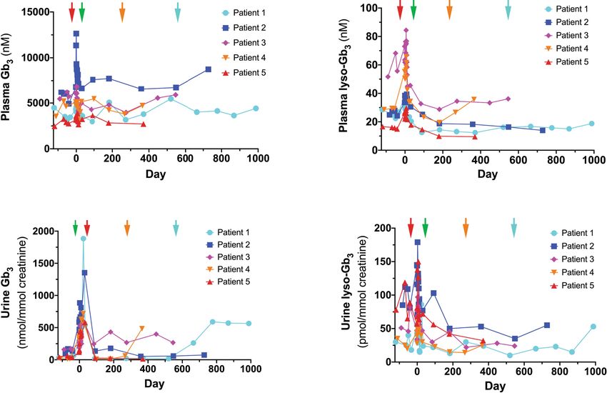
ARTICLE                                                                    NATURE COMMUNICATIONS | https://doi.org/10.1038/s41467-021-21371-5

I
    n Fabry disease, mutations of the X-linked GLA gene lead to                tolerance. We employed a recombinant LV with a self-
    accumulation of glycosphingolipids including globo-                        inactivating LTR design and an optimized Kozak start sequence
    triaosylceramide (Gb3)1,2 and globotriaosylsphingosine                     to deliver a human codon-optimized α-gal A transgene20. A
(lyso-Gb3)3,4. This results in end-organ damage to the kidneys,                single ex vivo LV transduction allowed for controlled dosing of
heart, and brain leading to a decreased life expectancy5,6. Current            the vector with a relatively low multiplicity of infection that
approved treatments for Fabry disease include enzyme therapy                   should minimize insertional mutagenesis events20. We also uti-
(ET) and an oral pharmacologic chaperone (migalastat)7–9.                      lized reduced-intensity melphalan conditioning, enabling out-
Biweekly ET can reduce Gb3 levels in urine, plasma, and tissues                patient management, fewer Grade 3 or 4 adverse events, and
but is intrusive, not curative, and progressive disease continues to           reduced cost. We report safety and outcome measures of the first
cause clinical symptoms and a decreased lifespan10,11. Moreover,               gene therapy trial for Fabry disease.
antibody formation directed against the recombinant enzyme
occurs, which may affect therapy outcome10. The short plasma
half-life12 requires biweekly infusions at considerable cost.                  Results
Despite these issues, ET is recommended for treatment of Fabry                 Patient enrollment. Male patients with known Fabry disease from
patients to prevent progression in conjunction with nonspecific                 the Canadian Fabry Disease Initiative study of ET were approa-
adjunctive therapies13–15. Migalastat is protein-variant specific,              ched for possible enrollment. Seven male Fabry disease patients
and therefore only available to a subset of Fabry patients with                previously treated with ET, ages 29–48 years, were enrolled
amenable mutations8.                                                           (Table 1, Supplementary Table 1); two patients failed screening
   Gene therapy, in theory, would enable Fabry patients to receive             tests associated with inclusion and exclusion trial criteria. Patients
a single treatment that could be more effective than current                   were followed from January 2017 to February 2020 for this study.
options and free them from ET. Transduced cell populations that                Ongoing follow-up will extend until February 2024.
continuously produce α-gal A may be more effective clinically. As
well, cross-correction may make Fabry disease particularly                     Study objectives. The primary objectives of this study were to
amenable to gene therapy; enabling systemic correction with a                  determine the safety and toxicity of autologous stem cell trans-
lower number of vector transduced cells16,17. Transgenic mice,                 plantation with mobilized CD34+ hematopoietic cells transduced
with tissue α-gal A activity >10,000 times endogenous levels, were             with a lentiviral vector containing human codon-optimized α-gal
healthy and did not have altered cyto-architecture;18,19 thus high             A cDNA in adult male Fabry disease patients. Several secondary
levels of α-gal A may not be deleterious.                                      objectives were analyzed including monitoring levels of α-gal A
   In a pilot safety study, we have targeted enriched CD34+                    activity in plasma, peripheral blood leukocytes and bone-marrow-
hematopoietic stem/progenitor cells (HSPCs) for lentivirus (LV)-               derived mononuclear cells, and measuring levels of Gb3 and lyso-
mediated gene therapy in patients with Fabry disease                           Gb3 in plasma and urine. In addition, the transduction efficiency
(NCT02800070, Fig. 1). Transduced HSPCs may deliver the                        of CD34+ hematopoietic cells was assessed as well as the pre-
functional enzyme to sites not accessible to ET. HSPC progeny                  sence and persistence of marked cells expressing α-gal A in
may be a major source of Gb3 and lyso-Gb3; this may correct                    peripheral blood. An exploratory objective was also incorporated
substrate accumulation at the point of origin. Use of autologous               to track the levels of anti-α-gal A antibodies in recipients. The
primitive HSPCs with conditioning may facilitate immune                        primary endpoint of this study was toxicity as assessed using the

       Phase 1:           Phase 2:                                         Phase 3:                                                        Phase 4:
      Screening         Pre-treatment                                  Treatment Phase                                                  Post-Treatment
        Phase               Phase                                                                                                      Follow-up Phase

                                                       Mobilization                                               Transplant         Follow-up at regional
                                               Mobilize patient cells at                                                               stem cell center
                                                                                     Approval by Health Canada

      Adult male             Cease             regional stem cell center
                                                                                       Certificate of Analysis

                                                                                                                 Patient receives    After 1 month of
                              ET                                                                                                     scheduled follow-up
                                               Apheresis to isolate                                              melphalan at
         Fabry                                                                                                                       post-transplant, safety
                                               nucleated blood cells at                                          the regional stem
        disease            Establish           regional stem cell center                                                             review of patient data
                                                                                                                 cell center
        patient            baseline
         on ET              α-gal A            Ship sample immediately                                           Thaw and infusion   Resume ET at 1 month
                            activity           for stem cell isolation                                           of autologous       post-transplant
                                                                                                                 transduced cells
                                               Ship CD34+ selected cells.                                                            Pause ET at six
                                               Transduction with lentiviral-                                                         months post-transplant
                                               α-gal A, cryopreservation                                                             if certain clinical
                                                                                                                                     endpoints are reached.

        21 days             30 days
                                                                               Approximately
         before              before                      1-7 days                                                  Day -2 to +12         Up to 5 years
                                                                                 30 days
        Phase 2             Phase 3

Fig. 1 Study schema. Five men with Type I Fabry disease were infused with autologous transduced CD34+-selected hematopoietic stem/progenitor cells
engineered to express α-galactosidase A (α-gal A) following mild ablation. ET enzyme therapy.

2                  NATURE COMMUNICATIONS | (2021)12:1178 | https://doi.org/10.1038/s41467-021-21371-5 | www.nature.com/naturecommunications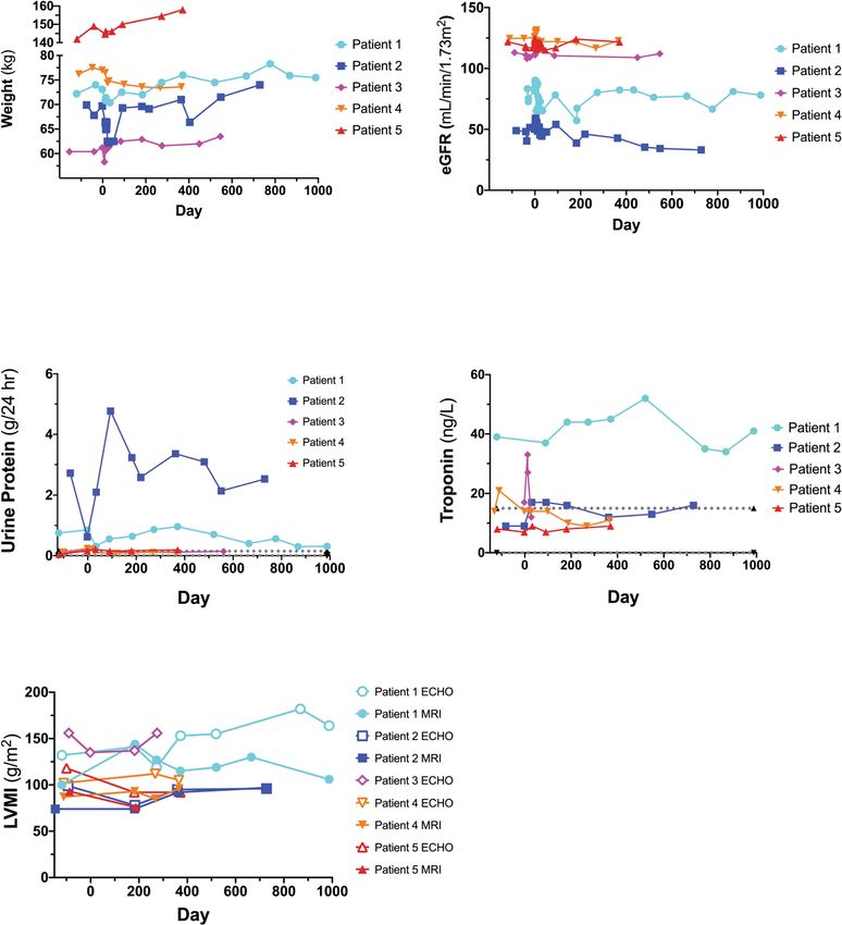
NATURE COMMUNICATIONS | https://doi.org/10.1038/s41467-021-21371-5                                                                                          ARTICLE

 Table 1 Baseline patient demographics and treatment phase outcomes.

 Parameter                                                  Patient 1              Patient 2                  Patient 3            Patient 4            Patient 5
 Age (years)                                                48                     39                         39                   37                   29
 GLA mutation                                               p.Gln321Arg            p.Ser345Pro                p.Ala143Pro          p.Ala143Pro          p.Tyr134Ser
 Age at diagnosis (years)                                   36                     29                         0                    4                    14
 Age started ET (years)                                     36                     33                         36                   26                   29
 Fabry symptoms at baseline
 Acroparesthesia                                                                                              x
 Angiokeratoma                                              x                      x                                               x                    x
 Cardiomyopathy                                             x                                                 x                    x
 Chronic kidney disease                                                            x
 Cold intolerance                                                                  x
 Corneal verticillata                                                              x                          x                    x
 Gastrointestinal                                           x                      x                                               x                    x
 Heat intolerance                                                                  x
 Hypertension                                                                                                                      x                    x
 Hypohidrosis                                                                      x                                               x                    x
 Headaches and migraines                                                                                                                                x
 Pain                                                                                                                              x                    x
 Peripheral sensory neuropathy                                                     x
 Proteinuria                                                x                      x
 Tinnitus or hearing loss                                                                                     x                    x                    x
 Mobilization agents                                        G-CSF                  G-CSF + plerixafor         G-CSF + plerixafor   G-CSF + plerixafor   G-CSF
 Apheresis yield CD34+ cells ×106/kg                        8.4                    9.2                        18.1                 9.0                  5.1
 Drug product VCN (copies/genome)                           0.68                   1.43                       0.81                 1.37                 1.13
 Drug product infused: CD34+ cells ×106/kg                  4.9                    6.4                        13.8                 6.2                  3.1
 Colony PCRa
    Mock                                                    0%                     0%                         0%                   ND                   ND
    LV-AGA (Drug Product)                                   52%                    62%                        54%                  ND                   ND
    Colony PCR on Day −2 BM samples                         ND                     0/86 (0%)                  0/88 (0%)            0/92 (0%)            0/88 (0%)
    Colony PCR on Day 28 BM samples                         10/28 (35.7%)          70/92 (76.1%)              63/90 (70.0%)        52/92 (56.5%)        56/88 (63.6%)
    Actual study day for colony PCR                         Day 33                 Day 28                     Day 28               Day 28               Day 32
 Day −2 BM α-gal A nmoles/hr/mg                             0.3                    1.0                        1.6                  1.0                  0.8
 Day 28 BM α-gal A nmoles/hr/mg                             220                    350                        169                  320                  569
 Time to engraftment (absolute neutrophil                   Day 12                 Day 13                     Day 11               Day 11               Day 12
 count >0.5 x 10⁹ cells per L)b
 Time to engraftment                                        Day 12                 Day 12                     Day 12               Day 11               Day 12
 (platelet count >20 x 109 per L)
 Day −2 BM VCN copies/genome                                0.00                   0.00                       0.00                 0.00                 0.00
 Day 28 BM VCNc copies/genome                               0.47                   0.89                       0.33                 1.10                 1.21
 Maximum PB VCN copies/genome                               0.55                   1.10                       0.78                 0.65                 1.30
 First Day Plasma α-Gal A activity observed                 Day 8d                 Day 7                      Day 6                Day 6                Day 7
 Days from Cell Infusion to ET withdrawal                   +548                   ND                         −41e                 +214                 ND

 BM bone marrow, ET enzyme therapy, ND not done, PB peripheral blood, PCR polymerase chain reaction, VCN vector copy number.
 aTransduction efficiency—colony PCR assays.
 bRef. 35.
 cDay 28–33.
 dDefined by activity >2 times the average of pretransplant values.
 ePatient 3 discontinued ET prior to infusion and chose not to resume.

National Cancer Institute of Canada (NCIC) Common Termi-                                       patients were administered a single dose of melphalan IV at
nology Criteria for Adverse Events (CTCAE), Version 4.03.                                      100 mg/m2 on Day −1. On Day 0, autologous CD34+ transduced
Because of the small sample size of this clinical study, there was                             cells were infused. Filgrastim (5 μg/kg) was administered sub-
insufficient power for comprehensive statistical analysis.                                      cutaneously daily from Day 5 until neutrophil count reached
                                                                                               ≥1.5 x 10⁹ cells/L. All patients (except Patient 3) restarted ET
Mobilization and leukapheresis. During the pretreatment phase,                                 30 days after infusion. According to a Health Canada-approved
ET was stopped for a minimum of 30 days prior to transplant. ET                                protocol amendment, all patients are eligible to discontinue
was stopped so that we could obtain a consistent cohort of                                     ET. Two patients subsequently discontinued ET (Patient 1 at day
baseline measurements. After cessation of ET, peripheral blood                                 548, Patient 4 at day 214 after transplantation, respectively). A
(PB) CD34+ HSPCs were mobilized using filgrastim in Patients 1                                  third patient (Patient 3) chose not to resume ET after
and 5, and filgrastim and plerixafor in Patients 2–4. Leukapher-                                transplantation.
esis yielded 5.1–18.1 x 10⁶ CD34+ cells/kg, with a final total
number of 3.65–8.35 x 10⁸ CD34+ cells (Table 1). The drug                                      α-gal A enzyme activity. Circulating α-gal A activity was
product yield was 3.1–13.8 x 10⁶ CD34+ cells/kg (Table 1).                                     first detected in all patients between Days 6 and 8 following
                                                                                               infusion and attained reference range levels in all patients
Treatment phase. Enzyme activity, colony PCR, and VCN assays                                   (Fig. 2a). Leukocyte α-gal A reached specific activity levels
were performed on the drug product (Table 1). Prior to infusion,                               above the reference range (Fig. 2b). Both plasma α-gal A

NATURE COMMUNICATIONS | (2021)12:1178 | https://doi.org/10.1038/s41467-021-21371-5 | www.nature.com/naturecommunications                                                3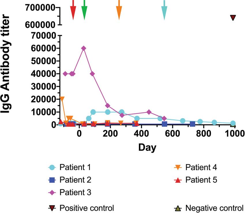
ARTICLE NATURE COMMUNICATIONS | https://doi.org/10.1038/s41467-021-21371-5
a
b
c
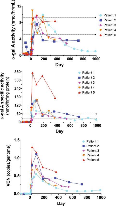
Fig. 2 α-galactosidase A (α-gal A) enzyme activity and vector copy number (VCN). a Plasma α-gal A activity attained reference range levels in all
patients; although decreased over time, the plasma α-gal A enzyme activity levels are above what is observed in Fabry disease patients and have not
returned to original baseline levels. The reference ranges (dotted lines) were defined by Dr. Rupar’s laboratory based on 150 specimens referred for
diagnostic testing. Males with classic Fabry disease have plasma levels around 1 nmol/h/ml. b Leukocyte α-gal A attained supranormal specific activity
levels for each patient. Although decreased over time, leukocyte α-gal A-specific enzyme activity levels are above what is seen in Fabry disease patients
and have not returned to original baseline levels. The reference ranges (dotted lines) were defined by Dr. Rupar’s laboratory based on 150 specimens
referred for diagnostic testing. c VCN in peripheral blood reached between 0.55 and 1.10 copies/genome in all patients, and although decreased over time,
has remained above 0.05 copies/genome in all patients to date (almost 3 years in Patient 1).
enzyme activity levels and leukocyte α-gal A specific enzyme Vector copy number. The infused drug product vector copy
activity levels decreased over time; yet the activity levels are number (VCN) ranged from 0.68 to 1.43 (copies/genome)
above Fabry disease patients21 and have not returned to ori- (Table 1). The VCN ranged from 0.33 to 1.21 (copies/genome) in
ginal baseline levels (Fig. 2a, b)22. Plasma and leukocyte α-gal bone marrow (BM) aspirates obtained from each patient at one
A-specific activities mirrored each other (Supplementary month (Table 1). PB VCN reached a range of 0.55 to 1.10 copies/
Fig. 1). genome in all patients and has decreased over time but has
4 NATURE COMMUNICATIONS | (2021)12:1178 | https://doi.org/10.1038/s41467-021-21371-5 | www.nature.com/naturecommunicationsNATURE COMMUNICATIONS | https://doi.org/10.1038/s41467-021-21371-5 ARTICLE
a b
c d
Fig. 3 Total plasma and urine globotriaosylceramide (Gb3) and lyso-Gb3 levels. Plasma Gb3 (a), plasma lyso-Gb3 (b), urine Gb3 (c), and urine lyso-Gb3
(d) levels are illustrated for each patient. Red arrow is at Day −30 when ET was stopped prior to mobilization. Green arrow is at Day 30 when ET was
restarted for Patients 1, 2, 4, and 5. The orange arrow demarks when Patient 4 stopped ET at Day 214 and the blue arrow is when Patient 1 stopped ET at
Day 548. Patient 3 chose not to restart ET.
remained above 0.05 copies/genome in all patients (almost 3 years disease (Fig. 4d). Left ventricular mass index (LVMI) monitoring
in Patient 1) (Table 1, Fig. 2c). VCN mirrored leukocyte α-gal A- by MRI and ECHO revealed that cardiac hypertrophy observed in
specific activity (Supplementary Fig. 2). Patient 1 was relatively stable for nearly three years post-infusion
(Fig. 4e). LVMI was also stable for Patients 2, 4, and 5 during the
Plasma and urine Gb3/lyso-Gb3 levels. Total plasma and urine study period.
Gb3 levels were variable, especially during the Treatment Phase.
Plasma and urine lyso-Gb3 levels were reduced over time in most Anti-α-gal A antibody titer. Anti-α-gal A antibody (IgG) levels
patients (Fig. 3). This reduction was sustained after dis- increased for Patient 1 within 6 months post-infusion, before
continuation of ET in Patient 3, with the exception of urine Gb3. gradually declining (Fig. 5). Patient 3 had the highest titer at
Increases in urine Gb3 levels were also observed when Patients 1 screening, which was maintained through mobilization and the
and 4 stopped ET treatment. Urine lyso-Gb3 also increased after Treatment Phase but decreased after 6 months. Patients 4 and 5
Patient 1 stopped ET treatment. Plasma Gb3 levels in Patient 3 had low titers at screening, which dropped by mobilization
fluctuated after infusion, but rose at later time points. Plasma Gb3 (Patient 5) or after the Treatment Phase (Patient 4). Patient 2 had
and plasma lyso-Gb3 have increased in Patient 4 after this patient no detectable anti-α-gal A antibody.
chose to stop ET. Data for the Treatment Phase for all parameters
shown in Figs. 2 and 3 are illustrated in Supplementary Fig. 3. Safety monitoring. No unexpected trends or safety events have
been identified. The safety profile is consistent for patients
Clinical parameters. Patients initially experienced a drop in undergoing melphalan conditioning for autologous hematopoie-
weight during the Treatment Phase. In Patients 1–3 and 5, tic cell transplantation (Supplementary Table 2). Two AEs
weights increased over time following transplantation (Fig. 4a). (nausea [Grade 1] and cough [Grade 2]) were possibly related to
eGFR increased during the Treatment Phase in all patients the investigational product. There were 20 AEs reported of Grade
(Fig. 4b). Following gene therapy, eGFR returned to baseline 3 or 4 (Supplementary Table 2). Patient 2 experienced anorexia
levels and was relatively stable in all patients, except for Patient 2. over 51 days corresponding to a lack of appetite (Supplementary
This patient displayed progressive chronic kidney disease with Table 2) and this was assessed to be related to study procedures.
significant proteinuria (Table 1) during screening. Linear All AEs Grade 3 or higher were unrelated to protocol treatment,
regression demonstrated that Patient 2 had the steepest eGFR but were related to the study procedures. Two SAEs were related
slope, whereas the eGFR slope was near zero for the remaining to the study procedures (febrile neutropenia in Patient 4 and
patients (Supplementary Fig. 4). Proteinuria was also described in peripherally inserted central catheter line infection/thrombosis of
Patient 1 during screening (Table 1); 24-h urinary protein mea- the right arm in Patient 5).
surements confirmed this observation in Patients 1 and 2
(Fig. 4c). Patient 1 was described with left ventricular hyper- Discussion
trophy during screening (Table 1). Monitoring of troponin levels In this pilot clinical trial of LV-mediated gene therapy in 5 men
suggests that Patient 1 is displaying cardiac features of Fabry with Type 1 (classical) Fabry disease, all patients demonstrated a
NATURE COMMUNICATIONS | (2021)12:1178 | https://doi.org/10.1038/s41467-021-21371-5 | www.nature.com/naturecommunications 5ARTICLE NATURE COMMUNICATIONS | https://doi.org/10.1038/s41467-021-21371-5
a b
c d
e
Fig. 4 Clinical parameters. Weight (a), estimated Glomerular Filtration Rate (eGFR) (b), urinary protein secretion (c), troponin (d), and Left Ventricular
Mass Index (LVMI) (e) was monitored during the course of the trial for all patients. eGFR was calculated using the Chronic Kidney Disease-Epidemiology
Collaboration formula. Values from Magnetic Resonance Imaging (MRI) and Echocardiography (ECHO) are shown for LVMI (e). Patient 3 did not attend all
cardiac assessments and has been omitted from Fig. 4e.
sustained safety profile. One patient is now out nearly 3 years throughout the study period (ranging from 12-33 months post-
from his infusion date. We chose a reduced-intensity condition- infusion), with the exception of Patient 2 who is showing
ing regimen for drug product engraftment, based on many years symptoms of chronic kidney disease associated with his Fabry
of transplant experience with melphalan in Toronto. Since there disease. Plasma lyso-Gb3 levels decreased in all patients with the
is limited experience with transplant conditioning procedures in exception of Patient 4 where an increase was observed. Plasma
Fabry patients, and Fabry disease patients have considerable Gb3 levels were generally stable in all patients, except for increases
comorbidities, we also wanted to establish whether mobilization at later time points in Patients 2 and 3. Urine lyso-Gb3 values
and engraftment could be tolerated in this population. All were increased in Patient 1, stable in Patient 4, and decreased in
patients received a low melphalan dose; 3 men (Patients 1, 4, and Patients 2, 3 and 5. Finally, urine Gb3 levels were stable in
5) received the treatment as an outpatient and returned home the Patients 2 and 5 who continued ET after gene therapy, but were
same day. Full ablation regimens may require lengthy hospital observed to increase in Patients 1, 3, and 4 who elected to
stays and result in additional AEs23–27. withdraw from their ET infusions. Interestingly, Patient 3 who
We demonstrated efficient LV-mediated gene transfer into chose to stop ET prior to mobilization had sustained low levels of
enriched Fabry patient CD34+ cells. VCN levels were modest, plasma lyso-Gb3 and urine lyso-Gb3; a result of the gene therapy
minimizing the chance of genotoxicity. Our regimen led to product alone.
increased circulating and intracellular α-gal A activity. All It is possible that a more myelo-suppressive conditioning
patients reached reference range levels. Values declined over time, regimen would result in better engraftment of LV/AGA trans-
possibly due to an exhaustion of transduced short-term repopu- duced cells—although this hypothesis remains to be tested. In
lating HSPCs, but remained above what is typically observed in selection of a partial myeloablative conditioning regimen, we
Fabry patients and well above pretreatment levels in all five cases. considered that our prospective patients were relatively healthy
Whether the enzyme values reach an asymptote reflecting males with Fabry disease who were receiving ET prior to
engraftment of transduced long-term HSPCs remains to be recruitment to our trial. Unlike most other acute disorders that
determined. VCN and α-gal A activities were well correlated, have been treated by gene therapy, Fabry disease is a chronic
underscoring the fidelity of our therapeutic LV construct. Patient disease. As such, our clinical team and our regulatory body were
weight, eGFR, proteinuria and LVMI were stable for all patients reluctant to implement a full myeloablative conditioning regimen.
6 NATURE COMMUNICATIONS | (2021)12:1178 | https://doi.org/10.1038/s41467-021-21371-5 | www.nature.com/naturecommunicationsNATURE COMMUNICATIONS | https://doi.org/10.1038/s41467-021-21371-5 ARTICLE
All data presented within this manuscript have deidentified each patient and
regional stem cell center. Recruitment (from September 2016 to October 2018) was
restricted to men (age 18–50) with confirmed Type 1 (classical) phenotype Fabry
disease (with GLA genotyping) who had received ET for at least 6 months prior to
study enrollment. Inclusion criteria comprised an estimated glomerular filtration
rate (eGFR), > 45 mL/min/1.73 m2 (chronic kidney disease-epidemiology colla-
boration equation [CKD-EPI]) and left ventricular ejection fraction >45%. Patients
with advanced Fabry disease were excluded. Additional eligibility criteria can be
found in Supplementary Table 3. Criteria for ET withdrawal is indicated in Sup-
plementary Table 4. The current version of the Health Canada-approved clinical
protocol can be accessed in the Supplementary Data.
Lentiviral vector. The LV-AGA vector has been described previously20. Large-
scale, high-titer clinical-grade, recombinant lentivector was manufactured, purified,
and qualified by the Indiana University Vector Production Facility, Indianapolis,
IN. A total of five patients were treated in this Phase I safety trial due to the
quantity of the lentiviral vector produced for this study.
Study design. Patients were recruited to three regional hematopoietic stem cell
centers (Calgary, AB; Toronto, ON; and Halifax, NS). All steps of mobilization,
apheresis, conditioning, and transplantation occurred at the relevant site for each
patient. Patients were initially mobilized with filgrastim (granulocyte-colony sti-
mulating factor [G-CSF] 16 µg/kg). If peripheral blood CD34+ counts were low
(i.e., if the predicted CD34+ count wasARTICLE NATURE COMMUNICATIONS | https://doi.org/10.1038/s41467-021-21371-5
have been no treatment-related serious adverse events. A Data Monitoring Safety 21. Maruyama, H. et al. Effectiveness of plasma lyso-Gb3 as a biomarker for
Committee (DSMC) reviewed clinical and safety data after the Treatment Phase for selecting high-risk patients with Fabry disease from multispecialty clinics for
Patients 1 and 3. Interim analysis and publication of this clinical trial was genetic analysis. Genet. Med. 21, 44–52 (2019).
authorised by the DSMC. As this is the first lentivirus-directed gene therapy trial to 22. Politei, J., Schenone, A. B., Cabrera, G., Heguilen, R. & Szlago, M. Fabry
focus on adults with a lysosomal storage disorder, the DSMC supported reporting disease and enzyme replacement therapy in classic patients with same
of safety data up to 1-year post-infusion of each patient. mutation: different formulations–different outcome? Clin. Genet. 89, 88–92
(2016).
Reporting summary. Further information on research design is available in the Nature 23. Biffi, A. et al. Lentiviral hematopoietic stem cell gene therapy benefits
Research Reporting Summary linked to this article. metachromatic leukodystrophy. Science 341, 1233158 (2013).
24. Boztug, K. et al. Stem-cell gene therapy for the Wiskott-Aldrich syndrome. N.
Engl. J. Med. 363, 1918–1927 (2010).
Data availability 25. Ribeil, J. A. et al. Gene therapy in a patient with sickle cell disease. N. Engl. J.
The datasets generated during and/or analyzed during the current study are available Med. 376, 848–855 (2017).
from the corresponding author on reasonable request. Source data are provided with 26. Thompson, A. A. et al. Gene therapy in patients with transfusion-dependent
this paper. beta-thalassemia. N. Engl. J. Med. 378, 1479–1493 (2018).
27. Ferrua, F. et al. Lentiviral haemopoietic stem/progenitor cell gene therapy for
Received: 5 May 2020; Accepted: 25 January 2021; treatment of Wiskott-Aldrich syndrome: interim results of a non-randomised,
open-label, phase 1/2 clinical study. Lancet Haematol. 6, e239–e253 (2019).
28. Yoshimitsu, M. et al. Correction of cardiac abnormalities in Fabry mice by
direct intraventricular injection of a recombinant lentiviral vector that
engineers expression of alpha-galactosidase A. Circ. J. 70, 1503–1508 (2006).
29. Skoog, W. A. & Beck, W. S. Studies on the fibrinogen, dextran and
phytohemagglutinin methods of isolating leukocytes. Blood 11, 436–454
References (1956).
1. Brady, R. O. et al. Enzymatic defect in Fabry’s disease. Ceramidetrihexosidase 30. Auray-Blais, C. et al. Urinary biomarker investigation in children with Fabry
deficiency. N. Engl. J. Med. 276, 1163–1167 (1967). disease using tandem mass spectrometry. Clin. Chim. Acta 438, 195–204
2. Platt, F. M., d’Azzo, A., Davidson, B. L., Neufeld, E. F. & Tifft, C. J. Lysosomal (2015).
storage diseases. Nat. Rev. Dis. Prim. 4, 27 (2018). 31. Boutin, M. & Auray-Blais, C. Multiplex tandem mass spectrometry analysis of
3. Aerts, J. M. et al. Elevated globotriaosylsphingosine is a hallmark of Fabry novel plasma lyso-Gb(3)-related analogues in Fabry disease. Anal. Chem. 86,
disease. Proc. Natl Acad. Sci. USA 105, 2812–2817 (2008). 3476–3483 (2014).
4. Auray-Blais, C. et al. Urinary globotriaosylsphingosine-related biomarkers for 32. Lavoie, P., Boutin, M. & Auray-Blais, C. Multiplex analysis of novel urinary
Fabry disease targeted by metabolomics. Anal. Chem. 84, 2745–2753 (2012). lyso-Gb3-related biomarkers for Fabry disease by tandem mass spectrometry.
5. Desnick, R. J. et al. Fabry disease, an under-recognized multisystemic disorder: Anal. Chem. 85, 1743–1752 (2013).
expert recommendations for diagnosis, management, and enzyme 33. Lee, C. J., Fan, X., Guo, X. & Medin, J. A. Promoter-specific lentivectors for
replacement therapy. Ann. Intern. Med. 138, 338–346 (2003). long-term, cardiac-directed therapy of Fabry disease. J. Cardiol. 57, 115–122
6. Waldek, S., Patel, M. R., Banikazemi, M., Lemay, R. & Lee, P. Life expectancy (2011).
and cause of death in males and females with Fabry disease: findings from the 34. Hazari, H. et al. Comparison of cardiac magnetic resonance imaging and
Fabry Registry. Genet. Med. 11, 790–796 (2009). echocardiography in assessment of left ventricular hypertrophy in Fabry
7. Eng, C. M. et al. Safety and efficacy of recombinant human alpha-galactosidase disease. Can. J. Cardiol. 34, 1041–1047 (2018).
A replacement therapy in Fabry’s disease. N. Engl. J. Med. 345, 9–16 (2001). 35. Kurtzberg J. et al. Placental blood as a source of hematopoietic stem cells for
8. Germain, D. P. et al. Treatment of Fabry’s disease with the pharmacologic transplantation into unrelated recipients. N. Engl. J. Med. 335(3), 157–166
chaperone migalastat. N. Engl. J. Med. 375, 545–555 (2016). (1996).
9. Schiffmann, R. et al. Enzyme replacement therapy in Fabry disease: a
randomized controlled trial. JAMA 285, 2743–2749 (2001).
10. El Dib, R. et al. Enzyme replacement therapy for Anderson-Fabry disease.
Cochrane Database Syst. Rev. 7, CD006663 (2016). Acknowledgements
11. Rombach, S. M. et al. Long term enzyme replacement therapy for Fabry We thank the patients for their commitment to the project. We thank Cindy Yau, Rupi
disease: effectiveness on kidney, heart and brain. Orphanet J. Rare Dis. 8, 47 Mangat, Sarah Young, and Pam Degendorfer from Ozmosis Research, Inc., for their
(2013). support of the trial. From the Cumming School of Medicine, University of Calgary, we
12. Eng, C. M. et al. A phase 1/2 clinical trial of enzyme replacement in Fabry acknowledge Andrew Daly and John Klassen for inpatient care to all stem cell transplant
disease: pharmacokinetic, substrate clearance, and safety studies. Am. J. Hum. recipients. We acknowledge Kathleen Estes who is managed by Donna Simcoe of Simcoe
Genet. 68, 711–722 (2001). Consultants, Inc. for help with medical writing. Funding for writing support was pro-
13. Biegstraaten, M. et al. Recommendations for initiation and cessation of vided by AVROBIO, Inc who did not contribute to the content and only provided an
enzyme replacement therapy in patients with Fabry disease: the European administrative review of the final manuscript. We made final decisions regarding all
Fabry Working Group consensus document. Orphanet J. Rare Dis. 10, 36 content. The following institutions provided funding for this study: Canadian Institutes
(2015). of Health Research (CIHR, grant number 119187), The Kidney Foundation of Canada,
14. Ortiz, A. et al. Fabry disease revisited: management and treatment and the MACC Fund. Financial support for the study was also provided by
recommendations for adult patients. Mol. Genet. Metab. 123, 416–427 (2018). AVROBIO, Inc.
15. Wanner, C. et al. European expert consensus statement on therapeutic goals in
Fabry disease. Mol. Genet. Metab. 124, 189–203 (2018).
16. Medin, J. A. et al. Correction in trans for Fabry disease: expression, secretion
and uptake of alpha-galactosidase A in patient-derived cells driven by a high- Author contributions
titer recombinant retroviral vector. Proc. Natl Acad. Sci. USA 93, 7917–7922 α-gal A enzymatic assays were performed by the London Health Sciences Centre Clinical
(1996). Biochemical Genetics Laboratory, London, ON (J.W.R. and C.A.R.). Gb3 and lyso-Gb3
17. Takenaka, T., Qin, G., Brady, R. O. & Medin, J. A. Circulating alpha- analyses were performed at the Université de Sherbrooke, Sherbrooke, QC (M.B. and
galactosidase A derived from transduced bone marrow cells: relevance for C.A.-B.). Vector copy number assays and ELISAs to measure anti-α-gal IgG antibodies
corrective gene transfer for Fabry disease. Hum. Gene Ther. 10, 1931–1939 were performed at the University Health Network, Toronto, ON (J.H., K.G., D.L.B., and
(1999). J.A.M.), and the Medical College of Wisconsin, Milwaukee, WI (K.G., W.M.M., and
18. Ashley, G. A., Desnick, R. J., Gordon, R. E. & Gordon, J. W. High J.A.M.). The FACTs team had confidential access to the data through the trial man-
overexpression of the human alpha-galactosidase A gene driven by its agement company. The sponsor (University Health Network) played no role in the study
promoter in transgenic mice: implications for the treatment of Fabry disease. design, data collection and analyses, or drafting of the manuscript. A.K., D.L.B., J.H.,
J. Invest. Med. 50, 185–192 (2002). C.A.R., C.A.-.B., W.M.M., D.H.F., A.K., M.L.W., R.F., and J.A.M. participated in research
19. Kase, R. et al. Immunohistochemical characterization of transgenic mice design. Research was performed by A.K., D.L.B., J.H., C.A.R., J.W.R., C.A.-B., M.B., P.O.,
highly expressing human lysosomal alpha-galactosidase. Biochim. Biophys. K.G., W.M.M., G.F., S.W., K.L., S.J., A.C., N.P., C.F.M., S.C., P.R.D., D.H.F., A.K.,
Acta 1406, 260–266 (1998). M.L.W., R.F., and J.A.M. Data were analyzed by A.K., D.L.B., J.H., C.A.R., J.W.R.,
20. Huang, J. et al. Lentivector iterations and pre-clinical scale-up/toxicity testing: C.A.-B., M.B., K.G., W.M.M., D.H.F., A.K., M.L.W., R.F., and J.A.M. J.A.M. and D.L.B.
targeting mobilized CD34(+) cells for correction of Fabry disease. Mol. Ther. wrote the manuscript and it was reviewed by A.K., C.A.R., C.A.-B., D.H.F., A.K., M.L.W.,
Methods Clin. Dev. 5, 241–258 (2017). and R.F. All authors approved the final manuscript before publication.
8 NATURE COMMUNICATIONS | (2021)12:1178 | https://doi.org/10.1038/s41467-021-21371-5 | www.nature.com/naturecommunicationsNATURE COMMUNICATIONS | https://doi.org/10.1038/s41467-021-21371-5 ARTICLE
Competing interests Additional information
A.K. received grants, consulting fees, revenue distribution agreement, speaker fees and Supplementary information The online version contains supplementary material
travel support with AVROBIO, Inc. as well as revenue distribution agreement with available at https://doi.org/10.1038/s41467-021-21371-5.
University Health Network regarding gene therapy using technology from this work.
D.L.B. and J.H. were partially paid from a Sponsored Research Agreement—AVRO- Correspondence and requests for materials should be addressed to J.A.M.
BIO, Inc. C. A. Rupar has the following financial relationships to disclose: the Bio-
chemical Genetics clinical diagnostic laboratory at his home institution is contracted Peer review information Nature Communications thanks Christina Yap, Juan Bueren
by AVROBIO, Inc. to assay enzymes on a fee for service basis. He is the laboratory and the other anonymous reviewer(s) for their contribution to the peer review of this
director but receives no personal compensation. C.A.-B. has received a service contract work. Peer reviewer reports are available.
and honoraria for biomarker analysis with AVROBIO, Inc., grant from CIHR. K.G.
had travel paid for by AVROBIO, Inc. S.W. has received nonfinancial support from Reprints and permission information is available at http://www.nature.com/reprints
Sanofi-Genzyme, nonfinancial support from Takeda Pharmaceuticals (formerly Shire
HGT), personal fees and nonfinancial support from Amicus Therapeutics. K.L. has Publisher’s note Springer Nature remains neutral with regard to jurisdictional claims in
received travel grant and honorarium from Amicus Therapies; travel grant and published maps and institutional affiliations.
speaker fees from Sanofi-Genzyme; travel grant, consulting fees and speaker fees from
Takeda Pharmaceuticals; medical advisor to the Canadian Fabry Disease Association.
C.F.M. has received grants, personal fees, and nonfinancial support from Takeda Open Access This article is licensed under a Creative Commons Attri-
Pharmaceuticals (previously Shire HGT), grants, personal fees and nonfinancial
bution 4.0 International License, which permits use, sharing, adaptation,
support from Sanofi-Genzyme, nonfinancial support from Amicus Therapeutics. A.K.
distribution and reproduction in any medium or format, as long as you give appropriate
has received consultancy fees from AVROBIO, Inc. unrelated to this study. M.L.W.
credit to the original author(s) and the source, provide a link to the Creative Commons
has received research grants, consulting fees, speaker fees and travel support with
license, and indicate if changes were made. The images or other third party material in
Amicus Therapeutics, Protalix, Sanofi-Genzyme and Takeda, revenue distribution
this article are included in the article’s Creative Commons license, unless indicated
agreement with University Health Network regarding gene therapy using technology
otherwise in a credit line to the material. If material is not included in the article’s Creative
from this work. J.A.M. has the following financial relationships to disclose: SAB—Rapa
Commons license and your intended use is not permitted by statutory regulation or
Therapeutics. Honoraria—Sanofi-Genzyme, Shire. Co-Founder—AVROBIO, Inc.
exceeds the permitted use, you will need to obtain permission directly from the copyright
Shareholder—AVROBIO, Inc. Grants from Canadian Institutes of Health Research
and Kidney Foundation of Canada and AVROBIO, Inc. M.B., A.C., P.R.D., R.F., D.H. holder. To view a copy of this license, visit http://creativecommons.org/licenses/by/4.0/.
F., G.F., S.J., W.M.M., P.O., N.P., and J.W.R. have no financial relationships to disclose
in relation to this trial. © The Author(s) 2021
NATURE COMMUNICATIONS | (2021)12:1178 | https://doi.org/10.1038/s41467-021-21371-5 | www.nature.com/naturecommunications 9You can also read