Exogenous sodium hydrosulfide protects against high glucose induced injury and inflammation in human umbilical vein endothelial cells by ...
←
→
Page content transcription
If your browser does not render page correctly, please read the page content below
MOLECULAR MEDICINE REPORTS 23: 67, 2021

            Exogenous sodium hydrosulfide protects against high
        glucose‑induced injury and inflammation in human umbilical
               vein endothelial cells by inhibiting necroptosis
                   via the p38 MAPK signaling pathway
                          JIAQIONG LIN1*, XIAOYONG LI2*, YAN LIN3*, ZENA HUANG4 and WEN WU1

        1
         Department of Endocrinology, Guangdong Geriatrics Institute, Guangdong Provincial People's Hospital, Guangdong
      Academy of Medical Sciences, Guangzhou, Guangdong 510080; Departments of 2Surgery and 3Nephrology, The Third
     Affiliated Hospital of Guangzhou Medical University, Guangzhou, Guangdong 510150; 4Department of General Medicine,
      Guangdong General Hospital, Guangdong Academy of Medical Sciences, Guangzhou, Guangdong 510080, P.R. China

                                        Received May 6, 2020; Accepted October 5, 2020

                                                 DOI: 10.3892/mmr.2020.11706

Abstract. In recent years hydrogen sulfide (H2S) has demon‑        immunofluorescence assay and ELISAs. The results demon‑
strated vasculoprotective effects against cell death, which        strated that necroptosis and the p38 MAPK signaling
suggests its promising therapeutic potential for numerous          pathway mediated HG‑induced injury and inflammation.
types of disease. Additionally, a protective effect of exogenous   Notably, NaHS was discovered to significantly ameliorate
H2S in HG‑induced injuries in HUVECs was demonstrated,             p38 MAPK/necroptosis‑mediated injury and inflammation in
suggesting a potential protective effect for diabetic vascular     response to HG, as evidenced by an increase in cell viability,
complications. The present study aimed to investigate the          a decrease in ROS generation and loss of MMP, as well as
mechanism accounting for the cytoprotective role of exoge‑         the reduction in the secretion of proinflammatory cytokines.
nous H2S against high glucose [HG (40 mM glucose)]‑induced         In addition, the upregulated expression of RIP3 induced by
injury and inflammation in human umbilical vein endothelial        HG was repressed by treatment with SB203580, while the
cells (HUVECs). HUVECs were exposed to HG for 24 h to              HG‑induced upregulation of p‑p38 expression levels were
establish an in vitro model of HG‑induced cytotoxicity. The        significantly downregulated following the treatment of Nec‑1
cells were pretreated with sodium hydrosulfide (NaHS), a           and RIP3‑siRNA. In conclusion, the findings of the present
donor of H 2S, or inhibitors of necroptosis and p38 MAPK           study indicated that NaHS may protect HUVECs against
prior to the exposure to HG. Cell viability, intracellular reac‑   HG‑induced injury and inflammation by inhibiting necroptosis
tive oxygen species (ROS), mitochondrial membrane potential        via the p38 MAPK signaling pathway, which may represent a
(MMP), IL‑1β, IL‑6, IL‑8, TNF‑ α, phosphorylated‑(p)38             promising drug for the therapy of diabetic vascular complica‑
and receptor‑interacting protein 3 (RIP3) expression levels        tions.
were detected using the indicated methods, including Cell
Counting Kit 8, fluorescence detection, western blotting,          Introduction

                                                                   Diabetes mellitus, which is characterized by hyperglycemia,
                                                                   affects >415 million people worldwide (1). The condition is a
Correspondence to: Dr Wen Wu, Department of Endocrinology,         significant economic burden and seriously affects the quality
Guangdong Geriatrics Institute, Guangdong Provincial People's      of life of diabetic patients (2). Owing to hyperglycemia, the
Hospital, Guangdong Academy of Medical Sciences, 106 Zhongshan     dysfunction of the vascular endothelium may lead to vascular
Second Road, Yuexiu, Guangzhou, Guangdong 510080, P.R. China       lesions and cause further severe implications, such as retinop‑
E‑mail: wuwen1964@163.com                                          athy (3), diabetic nephropathy (4), cardiovascular disease (5,6)
Dr Yan Lin, Department of Nephrology, The Third Affiliated         and neuropathy (7). Previous studies have reported that
Hospital of Guangzhou Medical University, 63 Duobao Road,          multiple factors, including mitochondrial dysfunction (8),
Liwan, Guangzhou, Guangdong 510150, P.R. China                     oxidative stress (9) and the activity of various signaling
E‑mail: linyan2013@gzhmu.edu.cn                                    molecules, such as sirtuin 1 (10) and p53 (11), were implicated
                                                                   in the high glucose [HG (40 mM glucose)]‑induced dysfunc‑
*
    Contributed equally                                            tion of the vascular endothelium. Accumulating evidence has
                                                                   suggested that p38 MAPK may be associated with diabetic
Key words: sodium hydrosulfide, hyperglycemia, necroptosis, p38
                                                                   complications; for example, Song et al (12) discovered that
MAPK, human umbilical vein endothelial cells
                                                                   the p38 signaling pathway mediated renal injury in diabetic
                                                                   nephropathy model mice. Furthermore, the activation of p38
2                                 LIN et al: INHIBITING NECROPTOSIS VIA THE p38 MAPK PATHWAY

MAPK was identified in HK‑2 cells stimulated with HG,                 expression levels of genes in 10 samples, including five healthy
while the inhibition of p38 MAPK exerted a protective role            subjects and five patients with diabetes with microvascular
over cell function (13). Notably, inflammation has also been          diseases, was conducted using R package (version 3.6.3;
considered as a crucial player in the development of diabetic         https://cran.r‑project.org/bin/windows/base/old/3.6.3/). Gene
mellitus, especially in vascular disorders (14‑16). In addi‑          set enrichment analysis (GSEA) of all genes was also performed
tion, accumulating studies have reported that p38 MAPK                using R package. P
MOLECULAR MEDICINE REPORTS 23: 67, 2021                                                 3

mixture. The mixture was maintained at room temperature for             lysed with RIPA lysis buffer for 30 min at 4˚C. Total protein
30 min to form complexes, and then the mixture was added to             was quantified using a BCA protein assay kit and 30 µg
12‑well‑plates with 500 µl OptiMEM and incubated at 37˚C in             protein/lane was separated via 12% SDS‑PAGE. The separated
a 5% CO2 incubator. The medium was replaced after 24 h with             proteins were transferred onto a PVDF membrane and blocked
DMEM with 10% FBS, which contained neither siRNA nor                    with 5% free‑fat milk for 90 min at room temperature. The
the transfection reagent.                                               membranes were then incubated with the following primary
                                                                        antibodies at 4˚C overnight with gentle agitation: Anti‑RIP3
Cell viability assay. HUVECs were cultured in 96‑well plates            (1:1,000), anti‑p38 (1:1,000) and anti‑p‑p38 (1:1,000) or
at a density of 1x10⁴ cells/ml. Following incubation at 37˚C            anti‑GAPDH (1:5,000). Following incubation with the primary
for 24 h, cells received different treatments as described in           antibody, the membranes were washed with 0.1% TBS
‘cell culture and treatment’ section and then washed with               Tween-20 and then incubated with the secondary antibody
PBS. Subsequently, according to the manufacturer's protocol,            (1:5,000) for 60 min at room temperature. The membranes
10 µl CCK‑8 solution was added to each well and incubated               were washed with 0.1% TBS Tween-20 and protein bands
at 37˚C for 2 h. The absorbance was measured at 450 nm                  were visualized using ECL and exposure to X‑ray films. To
with a microplate reader (Multiskan MK3 Microplate reader;              semi‑quantify protein expression levels, the X‑ray films were
Thermo Fisher Scientific, Inc.). The mean optical density (OD)          scanned and analyzed with ImageJ software. The experiment
of 3 wells in the indicated groups was used to calculate the            was repeated 3 times.
cell viability according to the following formula: Percentage
of cell viability (%)=(OD treatment group/OD control group) x100. The   Immunofluorescence assay. HUVECs at a density of
experiment was repeated 5 times.                                        1x10⁵ cells/ml were cultured at 37˚C in DMEM with 10% FBS
                                                                        on glass coverslips for 24 h. Cells were fixed with 4% para‑
Measurement of intracellular reactive oxygen species (ROS)              formaldehyde for 15 min at temperature, permeabilized with
generation. Intracellular ROS generation was measured                   PBS containing 0.2% Triton X‑100 and blocked with 1% BSA
by determining the oxidation of DCFH‑DA to fluorescent                  (Seebio; http://www.seebio.cn) for 20 min at room temperature.
dichlorofluorescein (DCF). Briefly, HUVECs at a density of              The slides were subsequently incubated with the anti‑p‑p38
1x10⁵ cells/ml were cultured at 37˚C in DMEM with 10% FBS               primary antibody (1:200) overnight at 4˚C. Following the
on 6‑well plates for 24 h. Following treatment as described             primary antibody incubation, the glass coverslips were
above, cells were collected for determination of ROS genera‑            washed with 0.1% PBS‑Tween 20 (PBST) and incubated with
tion. The cells were washed three times with PBS. Then,                 a FITC‑labelled anti‑rabbit secondary antibody (1:1,000) for
10 µM DCFH‑DA solution in serum‑free DMEM was added                     1 h at 37˚C, then washed by PBST and stained with DAPI for
to the slides and incubated at 37˚C for 30 min. The cells               5 min at temperature. Stained cells were visualized using a
were washed 5 times with PBS and DCF fluorescence was                   fluorescence microscope at x40 magnification (Axio Imager
measured over the entire field of vision using a fluorescence           Z1).
microscope at x40 magnification connected to an imaging
system (BX50‑FLA; Olympus Corporation). The mean fluo‑                  Measurement of the secretory levels of IL‑1β, IL‑6, IL‑8 and
rescence intensity (MFI) from five random fields, which is              TNF‑a using ELISAs. HUVECs were cultured in 96‑well
used an index for ROS production, was analyzed using ImageJ             growth‑medium plates at 37˚C for 24 h. Then, after receiving
(1.47i software; National Institutes of Health). The experiment         different treatments as described above, the secretory levels of
was repeated 5 times.                                                   IL‑1β, IL‑6, IL‑8 and TNF‑a in the culture supernatant, which
                                                                        were acquired via centrifuging at 500 x g for 5 min at 4˚C,
Examination of mitochondrial membrane potential (MMP).                  were analyzed using their respective ELISA kits, according to
The MMP was analyzed using a fluorescent dye named Rh123.               the manufacturers' protocols. The experiment was performed
The depolarization of the MMP results in a loss of MMP and              5 times.
a decrease in green fluorescence (39). Briefly, HUVECs at
a density of 1x10⁵ cells/ml were cultured at 37˚C in DMEM               Statistical analysis. All data are presented as the mean ± SEM.
with 10% FBS on 6‑well plates for 24 h. Following treatment             Statistical differences between groups were determined using
as described above, cells were collected for examination                a one‑way ANOVA followed by a LSD post‑hoc test for
of MMP. The cells were washed three times with PBS. The                 3 groups and by a Tukey's post‑hoc test for ≥3 groups using
cells were subsequently incubated with 1 µM Rh123 at 37˚C               SPSS 20.0 software (IBM Corp.). P
4 LIN et al: INHIBITING NECROPTOSIS VIA THE p38 MAPK PATHWAY Figure 1. Exogenous NaHS attenuates the HG‑induced upregulation of the expression levels of RIP3 and p‑p38 in HUVECs. (A) PCA of the GSE43950 dataset obtained from the Gene Expression Omnibus database. Samples including DMMVC and control were separated into two cluster. (B) Gene Set Enrichment Analysis for all genes in the GSE43950 dataset. Genes involved in the necroptosis pathway were enriched in diabetes with microvascular diseases. Expression levels of (C) RIP3 and (D) p‑p38/p38 ratio were analyzed and semi‑quantified using western blotting. HUVECs were pretreated with or without 400 µM NaHS for 30 min prior to exposure to 40 mM HG. (E) Representative micrographs of immunofluorescence staining of HUVECs with an anti‑p‑p38 antibody (green) and the fluorescent nuclear stain DAPI (blue) following the indicated treatments. Scale bar, 50 µm. Data are presented as the mean ± SEM (n=3). *P
MOLECULAR MEDICINE REPORTS 23: 67, 2021                                                               5

Figure 2. Positive feedback loop between p38 MAPK and necroptosis pathways in HUVECs. (A) Expression levels of RIP3 were semi‑quantified by western
blotting analysis. HUVECs were treated with 40 mM HG for 24 h with or without pretreatment with 3 µM SB203580 for 1 h. (B) Expression levels of
p‑p38/p38 were analyzed using western blotting. HUVECs were treated with 40 mM HG for 24 h with or without pretreatment with 100 µM Nec‑1 for 24 h.
(C) Transfection efficiency of RIP3‑siRNA in HUVECs was determined using western blotting. (D) Expression levels of p‑p38/p38 were determined using
western blotting with or without RIP3‑siRNA transfection. Data are presented as the mean ± SEM (n=3). *P
6                                     LIN et al: INHIBITING NECROPTOSIS VIA THE p38 MAPK PATHWAY

Figure 3. Role of necroptosis and p38 MAPK inhibition in the protective effects of H2S against HG‑induced cytotoxicity in HUVECs. (A) HUVECs were
treated with 40 mM HG for 24 h with or without the pretreatment with 400 µM NaHS for 30 min or 3 µM SB203580 (the inhibitor of p38) for 60 min and
the cell viability was analyzed using a CCK‑8 assay. HUVECs were treated with 40 mM HG for 24 h with or without pretreatment with (B) 100 µM Nec‑1 or
transfection with (C) RIP3‑siRNA for 24 h and the cell viability was analyzed using a CCK‑8 assay. Data are presented as the mean ± SEM (n=5). **P
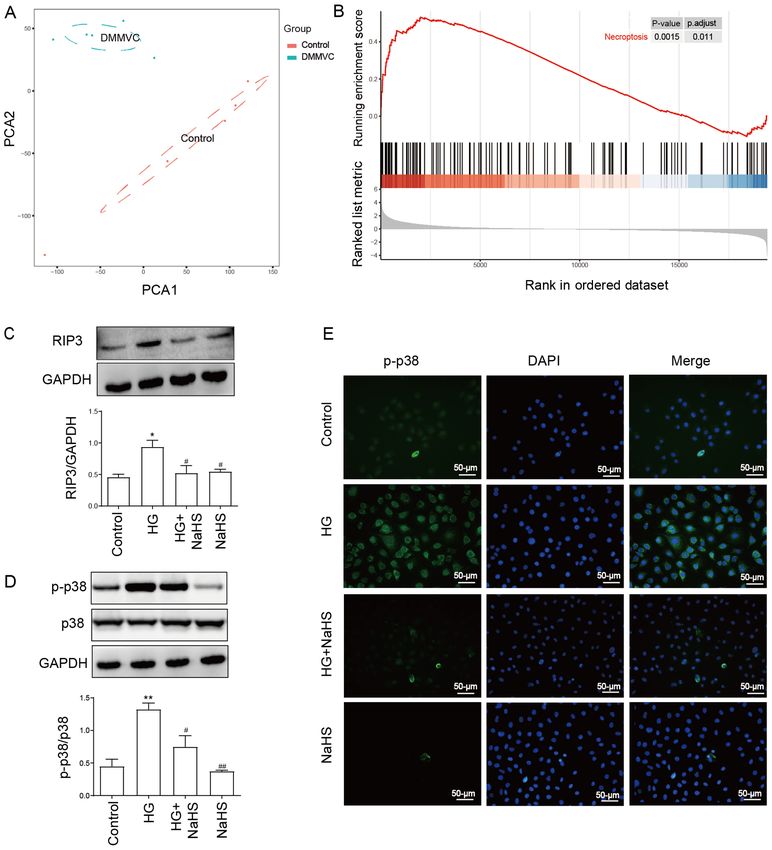
MOLECULAR MEDICINE REPORTS 23: 67, 2021                                                              7

Figure 5. Role of necroptosis and p38 MAPK inhibition on the protective effects of NaHS against the HG‑induced loss of MMP in HUVECs. After the
HUVECs were treated with the indicated treatments, the MMP was analyzed using the fluorescent dye, Rhodamine 123, followed by fluorescence microscopy.
Scale bar, 50 µm. Data are presented as the mean ± SEM (n=5). **P
8                                     LIN et al: INHIBITING NECROPTOSIS VIA THE p38 MAPK PATHWAY

Figure 6. Role of necroptosis and p38 MAPK inhibition on the protective effects of NaHS against HG‑induced inflammation in HUVECs. HUVECs were
treated with 40 mM HG for 24 h with or without pretreatment with 400 µM NaHS for 30 min and ELISAs were used to analyze the secretory levels of
(A) IL‑1β, (B) IL‑6, (C) IL‑8 and (D) TNF‑α. HUVECs were treated with 40 mM HG for 24 h with or without pretreatment with 100 µM Nec‑1 for 24 h and
ELISAs were used to analyze the secretory levels of (E) IL‑1β, (F) IL‑6, (G) IL‑8 and (H) TNF‑α. HUVECs were treated with 40 mM HG for 24 h with or
without transfection with RIP3‑siRNA for 24 h and ELISAs were used to analyze the secretory levels of (I) IL‑1β, (J) IL‑6, (K) IL‑8 and (L) TNF‑α. HUVECs
were treated with 40 mM HG for 24 h with or without pretreatment with 3 µM SB203580 for 1 h and ELISAs were used to analyze the secretory levels of
(M) IL‑1β, (N) IL‑6, (O) IL‑8 and (P) TNF‑α. Data are presented as the mean ± SEM (n=5). *P
MOLECULAR MEDICINE REPORTS 23: 67, 2021                                                9

or chemical hypoxia‑treated HaCaT cells (46,47). The current       reported that following the application of a p38 pathway
results identified that NaHS displayed strong anti‑inflamma‑       inhibitor, HG‑induced oxidative stress was attenuated in cere‑
tory properties, as evidenced by the reduced secretory levels      bral endothelial cells (18). Furthermore, another study also
of IL‑1β, IL‑6, IL‑8 and TNF‑α.                                    demonstrated that the p38 inhibitor improved HG‑induced
    Necroptosis, also named programmed necrosis, was               dysfunction of endothelial cells (56). Similar to these observa‑
identified as a new form of cell death (48). Previous evidence     tions, the present results illustrated that HG‑induced injury in
has indicated that necroptosis was involved in the develop‑        HUVECs was significantly inhibited following the treatment
ment of atherosclerosis (49), retinopathy (50) and myocardial      with SB203580, as evidenced by decreased cytotoxicity, MMP
damage (51). GSEA analysis of the GSE43950 dataset revealed        dissipation and ROS generation, as well as the severity of
a significant enrichment in the necroptosis pathway during         inflammation. These results demonstrated that the inhibition
diabetes with microvascular disease, suggesting the important      of the p38 MAPK signaling pathway attenuated the injury
role of the necroptosis pathway in diabetes with vasculopathy.     induced by HG, including cytotoxicity, ROS production,
Notably, in our previous study, it was discovered that necrop‑     mitochondrial injury and inflammation damage in HUVECs,
tosis contributed to the protective effect of NaHS (29). The       indicating that the activation of p38 MAPK may be a potential
present study also demonstrated that HG induced injuries,          mechanism accounting for HG‑induced injury.
including cytotoxicity, ROS production and mitochondrial                In our previous study, it was revealed that NaHS mediated
injury, were subsequently ameliorated following the transfec‑      a protective effect in HG‑induced apoptosis and necroptosis in
tion with RIP3‑siRNA or pretreatment with NaHS. Overall,           HUVECs (29). Consistent with this previous study, the present
these findings supported the hypothesis that NaHS may              results suggested that NaHS exerted significant cytoprotection
ameliorate HG‑induced damage, including cytotoxicity, ROS          in HUVECs, as evidenced by an increase in cell survival,
accumulation, MMP loss and inflammation, at least in part,         reduction in ROS production and loss of MMP. In addition,
via inhibiting necroptosis. However, the specific mechanism        RIP3‑siRNA was used to further determine the role of necrop‑
remains unclear.                                                   tosis pathway in HG‑induced injuries in the present study.
    Cao et al (52) reported that the p38 MAPK pathway was          Notably, both NaHS and RIP3‑siRNA effectively suppressed
activated in HG‑induced pancreatic cancer development.             the inflammatory response induced by HG. Our previous study
In H9c2 cells, which were exposed to 35 mM glucose, p38            reported the protective effect of NaHS (29), but the specific
MAPK was found to mediate HG‑induced damages, such                 signaling pathways via which it operates remained unknown.
as apoptosis, ROS overgeneration and the loss of MMP (53).         Therefore, the present study was conducted to further investi‑
Hence, the present study investigated the expression levels of     gate the specific mechanism. The results demonstrated that the
p38 MAPK in HUVECs treated with HG using western blot‑             p38 MAPK signaling pathway was implicated in protection of
ting and immunofluorescence. Similar to the previous findings,     NaHS. Furthermore, in the present study, a positive feedback
the results revealed a significant upregulation of p‑p38 expres‑   loop was identified between necroptosis and the p38 MAPK
sion levels following the treatment with 40 mM HG, indicating      signaling pathway. The inhibitor of p38 MAPK, SB203580,
that the activation of p38 may be induced by HG exposure.          significantly inhibited the HG‑induced expression levels of
A previous study demonstrated that the p38 MAPK pathway            RIP3. Combined with the aforementioned findings that both
was responsible for the protective effect of H2S in HG‑induced     p38 MAPK and necroptosis contributed to cell dysfunction, the
injury in H9c2 cells (53). Therefore, the current study further    present study indicated that necroptosis mediated HG‑induced
investigated whether the p38 MAPK pathway contributed              injury by activating p38 MAPK. Interestingly, the HG‑induced
to the protective effect of NaHS in HUVECs. The results            upregulated expression levels of p‑p38 were discovered to be
showed that the upregulated p‑p38 expression levels induced        diminished by Nec‑1 and RIP3‑siRNA. This positive feedback
by HG were reduced following the pretreatment with NaHS,           may play a crucial role in regulating HG‑induced injury and
implying the regulatory effect of NaHS on p‑p38 activation.        inflammation. For example, the positive feedback of necroptosis
Hence, these findings further suggested that p38 MAPK              and p38 MAPK may trigger the cascade amplification effect,
signaling pathway inhibition may contribute to NaHS protec‑        resulting in the aggravation of the inflammatory reaction and
tion in HG‑induced injury. These results are consistent with       tissue injury. Therefore, the present results provide two poten‑
previous studies. For example, it was previously reported that     tial therapeutic targets for diabetic vascular complications.
H2S exerted anti‑inflammatory effects repressing p38 phos‑              However, there are some limitations to the present study.
phorylation (54). These results suggested that the inhibition      Firstly, only in vitro investigations were performed; therefore,
of p38 MAPK may be the main mechanism of H 2S protec‑              further in vivo experiments using model mice, for example,
tion. Another study demonstrated that p38 was implicated in        are required to validate the results. Secondly, a constitutively
zVADfmk‑mediated necroptosis (55). Thus, the role of p38 in        activated p38 activator, such as hesperetin, should be used in
HG‑induced necroptosis was subsequently investigated. The          further experiments to further strengthen the conclusions.
upregulated expression levels of RIP3 induced by HG were                In conclusion, the findings of the present study elucidated
significantly reduced following the treatment with SB203580,       the mechanism underlying the protective effect of NaHS against
which indicated that p38 MAPK participated in HG‑mediated          HG‑induced injury and inflammation. The results suggested
necroptosis. Taken together, these above results suggested that    that NaHS H 2S may protect HUVECs against HG‑induced
NaHS may protect HUVECs against HG‑induced necroptosis             injury by inhibiting necroptosis, which may be mediated
by inhibiting the p38 MAPK pathway.                                through the p38 MAPK signaling pathway. Furthermore, to
    Further experiments were performed to clarify the role         the best of our knowledge, this was the first study to iden‑
of p38 in HG‑induced injury in HUVECs. A previous study            tify the positive feedback loop between necroptosis and the
10 LIN et al: INHIBITING NECROPTOSIS VIA THE p38 MAPK PATHWAY
p38 MAPK signaling pathway. These findings highlighted 7. Yerra VG, Areti A and Kumar A: Adenosine monophosphate‑acti‑
vated protein kinase abates hyperglycaemia‑induced neuronal
the vasculoprotective effects of NaHS and may provide an injury in experimental models of diabetic neuropathy: Effects
improved understanding for developing effective therapeutic on mitochondrial biogenesis, autophagy and neuroinflammation.
strategies for diabetic vascular complications. Mol Neurobiol 54: 2301‑2312, 2017.
8. Suzuki K, Olah G, Modis K, Coletta C, Kulp G, Gerö D,
Szoleczky P, Chang T, Zhou Z, Wu L, et al: Hydrogen sulfide
Acknowledgements replacement therapy protects the vascular endothelium in
hyperglycemia by preserving mitochondrial function. Proc Natl
Acad Sci USA 108: 13829‑13834, 2011.
Not applicable. 9. Ceriello A, Novials A, Ortega E, Canivell S, La Sala L,
Pujadas G, Esposito K, Giugliano D and Genovese S:
Funding Glucagon‑like peptide 1 reduces endothelial dysfunction,
inflammation, and oxidative stress induced by both hypergly‑
cemia and hypoglycemia in type 1 diabetes. Diabetes Care 36:
The present study was funded by a grant provided from 2346‑2350, 2013.
the National Natural Science Foundation of China (grant 10. Chen H, Wan Y, Zhou S, Lu Y, Zhang Z, Zhang R, Chen F,
Hao D, Zhao X, Guo Z, et al: Endothelium‑specific SIRT1
no. 81450062). overexpression inhibits hyperglycemia‑induced upregulation of
vascular cell senescence. Sci China Life Sci 55: 467‑473, 2012.
Availability of data and materials 11. Yokoyama M, Shimizu I, Nagasawa A, Yoshida Y, Katsuumi G,
Wakasugi T, Hayashi Y, Ikegami R, Suda M, Ota Y, et al: p53
plays a crucial role in endothelial dysfunction associated with
The datasets analyzed during the current study are available hyperglycemia and ischemia. J Mol Cell Cardiol 129: 105‑117,
in the Gene Expression Omnibus repository (http://www.ncbi. 2019.
12. Song W, Wei L, Du Y, Wang Y and Jiang S: Protective effect
nlm.nih.gov/geo). of ginsenoside metabolite compound K against diabetic
nephropathy by inhibiting NLRP3 inflammasome activation
Authors' contributions and NF‑ κ B/p38 signaling pathway in high‑fat diet/strepto‑
zotocin‑induced diabetic mice. Int Immunopharmacol 63:
227‑238, 2018.
All authors contributed to the study conception and design. 13. Chen P, Yuan Y, Zhang T, Xu B, Gao Q and Guan T: Pentosan
Experiments were performed by XL and YL. Data collection polysulfate ameliorates apoptosis and inf lammation by
suppressing activation of the p38 MAPK pathway in high
and analysis were performed by WW. The manuscript was glucose‑treated HK‑2 cells. Int J Mol Med 41: 908‑914, 2018.
written, drafted and designed by JL, who also performed the 14. Chen Y, Wang JJ, Li J, Hosoya KI, Ratan R, Townes T and
experiments. Bioinformatics analysis was performed by ZH. Zhang SX: Activating transcription factor 4 mediates hypergly‑
caemia‑induced endothelial inflammation and retinal vascular
All authors read and approved the final manuscript. leakage through activation of STAT3 in a mouse model of
type 1 diabetes. Diabetologia 55: 2533‑2545, 2012.
Ethics approval and consent to participate 15. Perkins JM, Joy NG, Tate DB and Davis SN: Acute effects of
hyperinsulinemia and hyperglycemia on vascular inflammatory
biomarkers and endothelial function in overweight and obese
Not applicable. humans. Am J Physiol Endocrinol Metab 309: E168‑E176, 2015.
16. Jung UJ and Choi MS: Obesity and its metabolic complications:
The role of adipokines and the relationship between obesity,
Patient consent for publication inflammation, insulin resistance, dyslipidemia and nonalco‑
holic fatty liver disease. Int J Mol Sci 15: 6184‑6223, 2014.
Not applicable. 17. Li J, Bao L, Zha D, Zhang L, Gao P, Zhang J and Wu X:
Oridonin protects against the inf lammatory response in
diabetic nephropathy by inhibiting the TLR4/p38‑MAPK and
Competing interests TLR4/NF‑κ B signaling pathways. Int Immunopharmacol 55:
9‑19, 2018.
18. Arcambal A, Taïlé J, Rondeau P, Viranaïcken W, Meilhac O and
The authors declare that they have no competing interests. Gonthier MP: Hyperglycemia modulates redox, inflammatory
and vasoactive markers through specific signaling pathways in
References cerebral endothelial cells: Insights on insulin protective action.
Free Radic Biol Med 130: 59‑70, 2019.
19. Shanmuganathan S and Angayarkanni N: Chebulagic acid
1. Jeffery N and Harries LW: β‑cell differentiation status in type 2 chebulinic acid and Gallic acid, the active principles of Triphala,
diabetes. Diabetes Obes Metab 18: 1167‑1175, 2016. inhibit TNFα induced pro‑angiogenic and pro‑inflammatory
2. da Rocha Fernandes J, Ogurtsova K, Linnenkamp U, activities in retinal capillary endothelial cells by inhibiting
Guariguata L, Seuring T, Zhang P, Cavan D and Makaroff LE: p38, ERK and NFkB phosphorylation. Vascul Pharmacol 108:
IDF diabetes atlas estimates of 2014 global health expenditures 23‑35, 2018.
on diabetes. Diabetes Res Clin Pract 117: 48‑54, 2016. 20. Dhuriya YK and Sharma D: Necroptosis: A regulated inflam‑
3. Sahajpal NS, Goel RK, Chaubey A, Aurora R and Jain SK: matory mode of cell death. J Neuroinflammation 15: 199, 2018.
Pat hologica l p er t u rbat ions i n d iabet ic ret i nopat hy: 21. Zhe‑Wei S, Li‑Sha G and Yue‑Chun L: The role of necroptosis
Hyperglycemia, AGEs, oxidative stress and inflammatory path‑ in cardiovascular disease. Front Pharmacol 9: 721, 2018.
ways. Curr Protein Pept Sci 20: 92‑110, 2019. 22. Chan FKM, Luz NF and Moriwaki K: Programmed necrosis
4. Liu H, Wang X, Liu S, Li H, Yuan X, Feng B, Bai H, Zhao B, in the cross talk of cell death and inflammation. Annu Rev
Chu Y and Li H: Effects and mechanism of miR‑23b on Immunol 33: 79‑106, 2015.
glucose‑mediated epithelial‑to‑mesenchymal transition in 23. He S, Wang L, Miao L, Wang T, Du F, Zhao L and Wang X:
diabetic nephropathy. Int J Biochem Cell Biol 70: 149‑160, 2016. Receptor interacting protein kinase‑3 determines cellular
5. Laakso M and Kuusisto J: Insulin resistance and hyperglycaemia necrotic response to TNF‑alpha. Cell 137: 1100‑1111, 2009.
in cardiovascular disease development. Nat Rev Endocrinol 10: 24. Cho YS, Challa S, Moquin D, Genga R, Ray TD, Guildford M and
293‑302, 2014. Chan FKM: Phosphorylation‑driven assembly of the RIP1‑RIP3
6. Li Y, Shelat H, Wu H, Zhu M, Xu J and Geng YJ: Low circu‑ complex regulates programmed necrosis and virus‑induced
lating level of IGF‑1 is a distinct indicator for the development of inflammation. Cell 137: 1112‑1123, 2009.
cardiovascular disease caused by combined hyperglycemia and 25. Newton K and Manning G: Necroptosis and inflammation. Annu
dyslipidemia. Int J Cardiol 171: 272‑273, 2014. Rev Biochem 85: 743‑763, 2016.MOLECULAR MEDICINE REPORTS 23: 67, 2021 11
26. Silke J, Rickard JA and Gerlic M: The diverse role of RIP kinases 43. Liu X, Ma D, Zheng S, Zha K, Feng J, Cai Y, Jiang F, Li J and
in necroptosis and inflammation. Nat Immunol 16: 689‑697, Fan Z: The roles of nitric oxide and hydrogen sulfide in the
2015. anti‑atherosclerotic effect of atorvastatin. J Cardiovasc Med
27. Negroni A, Colantoni E, Pierdomenico M, Palone F, Costanzo M, (Hagerstown) 16: 22‑28, 2015.
Oliva S, Tiberti A, Cucchiara S and Stronati L: RIP3 AND 44. Bełtowski J, Wójcicka G and Jamroz‑Wiśniewska A: Hydrogen
pMLKL promote necroptosis‑induced inflammation and alter sulfide in the regulation of insulin secretion and insulin sensi‑
membrane permeability in intestinal epithelial cells. Dig Liver tivity: Implications for the pathogenesis and treatment of diabetes
Dis 49: 1201‑1210, 2017. mellitus. Biochem Pharmacol 149: 60‑76, 2018.
28. Wang L, Wang T, Li H, Liu Q, Zhang Z, Xie W, Feng Y, 45. Wang Y, Zhao X, Jin H, Wei H, Li W, Bu D, Tang X, Ren Y,
Socorburam T, Wu G, Xia Z and Wu Q: Receptor interacting protein Tang C and Du J: Role of hydrogen sulfide in the development
3‑mediated necroptosis promotes lipopolysaccharide‑induced of atherosclerotic lesions in apolipoprotein E knockout mice.
inflammation and acute respiratory distress syndrome in mice. Arterioscler Thromb Vasc Biol 29: 173‑179, 2009.
PLoS One 11: e0155723, 2016. 46. Guo R, Wu K, Chen J, Mo L, Hua X, Zheng D, Chen P, Chen G,
29. Lin J, Chen M, Liu D, Guo R, Lin K, Deng H, Zhi X, Zhang W, Xu W and Feng J: Exogenous hydrogen sulfide protects against
Feng J and Wu W: Exogenous hydrogen sulfide protects human doxorubicin‑induced inflammation and cytotoxicity by inhibiting
umbilical vein endothelial cells against high glucoseinduced p38MAPK/NFκ B pathway in H9c2 cardiac cells. Cell Physiol
injury by inhibiting the necroptosis pathway. Int J Mol Med 41: Biochem 32: 1668‑1680, 2013.
1477‑1486, 2018. 47. Yang C, Yang Z, Zhang M, Dong Q, Wang X, Lan A, Zeng F,
30. Feng T, Chen W, Zhang C, Xiang J, Ding HM, Wu LL and Chen P, Wang C and Feng J: Hydrogen sulfide protects against
Geng D: The p38/CYLD pathway is involved in necroptosis chemical hypoxia‑induced cytotoxicity and inflammation in
induced by oxygen‑glucose deprivation combined with ZVAD in HaCaT cells through inhibition of ROS/NF‑κ B/COX‑2 pathway.
primary cortical neurons. Neurochem Res 42: 2294‑2304, 2017. PLoS One 6: e21971, 2011.
31. Qin S, Yang C, Huang W, Du S, Mai H, Xiao J and Lü T: 48. Degterev A, Huang Z, Boyce M, Li Y, Jagtap P, Mizushima N,
Sulforaphane attenuates microglia‑mediated neuronal necrop‑ Cuny GD, Mitchison TJ, Moskowitz MA and Yuan J: Chemical
tosis through down‑regulation of MAPK/NF‑ κ B signaling inhibitor of nonapoptotic cell death with therapeutic potential for
pathways in LPS‑activated BV‑2 microglia. Pharmacol Res 133: ischemic brain injury. Nat Chem Biol 1: 112‑119, 2005.
218‑235, 2018. 49. Lin J, Li H, Yang M, Ren J, Huang Z, Han F, Huang J, Ma J,
32. Wang D, Zhao M, Chen G, Cheng X, Han X, Lin S, Zhang X Zhang D, Zhang Z, et al: A role of RIP3‑mediated macrophage
and Yu X: The histone deacetylase inhibitor vorinostat prevents necrosis in atherosclerosis development. Cell Rep 3: 200‑210,
TNFα‑induced necroptosis by regulating multiple signaling 2013.
pathways. Apoptosis 18: 1348‑1362, 2013. 50. Murakami Y, Matsumoto H, Roh M, Suzuki J, Hisatomi T,
33. Łowicka E and Bełtowski J: Hydrogen sulfide (H2S)‑the third Ikeda Y, Miller JW and Vavvas DG: Receptor interacting protein
gas of interest for pharmacologists. Pharmacol Rep 59: 4‑24, kinase mediates necrotic cone but not rod cell death in a mouse
2007. model of inherited degeneration. Proc Natl Acad Sci USA 109:
34. Gemici B, Elsheikh W, Feitosa KB, Costa SK, Muscara MN and 14598‑14603, 2012.
Wallace JL: H2S‑releasing drugs: Anti‑inflammatory, cytopro‑ 51. Luedde M, Lutz M, Carter N, Sosna J, Jacoby C, Vucur M,
tective and chemopreventative potential. Nitric Oxide 46: 25‑31, Gautheron J, Roderburg C, Borg N, Reisinger F, et al: RIP3,
2015. a kinase promoting necroptotic cell death, mediates adverse
35. Citi V, Piragine E, Testai L, Breschi MC, Calderone V and remodelling after myocardial infarction. Cardiovasc Res 103:
Martelli A: The role of hydrogen sulfide and H2S‑donors in 206‑216, 2014.
myocardial protection against ischemia/reperfusion injury. Curr 52. Cao L, Chen X, Xiao X, Ma Q and Li W: Resveratrol inhibits
Med Chem 25: 4380‑4401, 2018. hyperglycemia‑driven ROS‑induced invasion and migration
36. Xu S, Liu Z and Liu P: Targeting hydrogen sulfide as a prom‑ of pancreatic cancer cells via suppression of the ERK and p38
ising therapeutic strategy for atherosclerosis. Int J Cardiol 172: MAPK signaling pathways. Int J Oncol 49: 735‑743, 2016.
313‑317, 2014. 53. Xu W, Wu W, Chen J, Guo R, Lin J, Liao X and Feng J:
37. Kumar M and Sandhir R: Hydrogen sulfide suppresses homocys‑ Exogenous hydrogen sulfide protects H9c2 cardiac cells against
teine‑induced glial activation and inflammatory response. Nitric high glucose‑induced injury by inhibiting the activities of the p38
Oxide 90: 15‑28, 2019. MAPK and ERK1/2 pathways. Int J Mol Med 32: 917‑925, 2013.
38. Huang Z, Dong X, Zhuang X, Hu X, Wang L and Liao X: Exogenous 54. Perry MM, Tildy B, Papi A, Casolari P, Caramori G, Rempel KL,
hydrogen sulfide protects against high glucose‑induced inflam‑ Halayko AJ, Adcock I and Chung KF: The anti‑proliferative and
mation and cytotoxicity in H9c2 cardiac cells. Mol Med Rep 14: anti‑inflammatory response of COPD airway smooth muscle
4911‑4917, 2016. cells to hydrogen sulfide. Respir Res 19: 85, 2018.
39. Zhang L, Jia YH, Zhao XS, Zhou FH, Pan YY, Wan Q, Cui XB, 55. Koike A, Hanatani M and Fujimori K: Pan‑caspase inhibitors
Sun XG, Chen YY, Zhang Y and Cheng SB: Trichosanatine induce necroptosis via ROS‑mediated activation of mixed
alleviates oxidized low‑density lipoprotein induced endothelial lineage kinase domain‑like protein and p38 in classically acti‑
cells injury via inhibiting the LOX‑1/p38 MAPK pathway. Am vated macrophages. Exp Cell Res 380: 171‑179, 2019.
J Transl Res 8: 5455‑5464, 2016. 56. Mazrouei S, Sharifpanah F, Caldwell RW, Franz M, Shatanawi A,
40. Daniele G, Guardado Mendoza R, Winnier D, Fiorentino TV, Muessig J, Fritzenwanger M, Schulze PC and Jung C: Regulation
Pengou Z, Cornell J, Andreozzi F, Jenkinson C, Cersosimo E, of MAP kinase‑mediated endothelial dysfunction in hypergly‑
Federici M, et al: The inflammatory status score including IL‑6, cemia via arginase I and eNOS dysregulation. Biochim Biophys
TNF‑α, osteopontin, fractalkine, MCP‑1 and adiponectin under‑ Acta Mol Cell Res 1866: 1398‑1411, 2019.
lies whole‑body insulin resistance and hyperglycemia in type 2
diabetes mellitus. Acta Diabetol 51: 123‑131, 2014.
41. Smith RP and Gosselin RE: Hydrogen sulfide poisoning. J Occup This work is licensed under a Creative Commons
Med 21: 93‑97, 1979. Attribution-NonCommercial-NoDerivatives 4.0
42. Chatzianastasiou A, Bibli SI, Andreadou I, Efentakis P, International (CC BY-NC-ND 4.0) License.
Kaludercic N, Wood ME, Whiteman M, Di Lisa F, Daiber A,
Manolopoulos VG, et al: Cardioprotection by H2S donors: Nitric
oxide‑dependent and ‑independent mechanisms. J Pharmacol
Exp Ther 358: 431‑440, 2016.You can also read