Supplementation with psyllium seed husk reduces myocardial damage in a rat model of ischemia/reperfusion - NRP
←
→
Page content transcription
If your browser does not render page correctly, please read the page content below
Nutrition Research and Practice. 2019
                                                                               Published online 2019 May 7
                                                                               ⓒ2019 The Korean Nutrition Society and the Korean Society of Community Nutrition
                                                                               http://e-nrp.org

Supplementation with psyllium seed husk reduces myocardial
damage in a rat model of ischemia/reperfusion
Sun Ha Lim and Jongwon Lee§
Department of Biochemistry, School of Medicine, Catholic University of Daegu, 33 Duryugongwon-ro 17-gil, Nam-gu, Daegu 42472, Republic of Korea

  BACKGROUND/OBJECTIVES: Myocardial infarction (MI) is caused by extensive myocardial damage attributed to the occlusion
  of coronary arteries. Our previous study in a rat model of ischemia/reperfusion (I/R) demonstrated that administration of arabinoxylan
  (AX), comprising arabinose and xylose, protects against myocardial injury. In this study, we undertook to investigate whether
  psyllium seed husk (PSH), a safe dietary fiber containing a high level of AX (> 50%), also imparts protection against myocardial
  injury in the same rat model.
  MATERIALS/METHODS: Rats were fed diets supplemented with PSH (1, 10, or 100 mg/kg/d) for 3 d. The rats were then subjected
  to 30 min ischemia through ligation of the left anterior descending coronary artery, followed by 3 h reperfusion through
  release of the ligation. The hearts were harvested and cut into four slices. To assess infarct size (IS), an index representing
  heart damage, the slices were stained with 2,3,5-triphenyltetrazolium chloride (TTC). To elucidate underlying mechanisms, Western
  blotting was performed for the slices.
  RESULTS: Supplementation with 10 or 100 mg/kg/d of PSH significantly reduces the IS. PSH supplementation (100 mg/kg/d)
  tends to reduce caspase-3 generation and increase BCL-2/BAX ratio. PSH supplementation also upregulates the expression
  of nuclear factor erythroid 2-related factor 2 (NRF2), and its target genes including antioxidant enzymes such as glutathione
  S-transferase mu 2 (GSTM2) and superoxide dismutase 2 (SOD2). PSH supplementation upregulates some sirtuins (NAD+-dependent
  deacetylases) including SIRT5 (a mitochondrial sirtuin) and SIRT6 and SIRT7 (nuclear sirtuins). Finally, PSH supplementation upregulates
  the expression of protein kinase A (PKA), and increases phosphorylated cAMP response element-binding protein (CREB) (pCREB),
  a target protein of PKA.
  CONCLUSIONS: The results from this study indicate that PSH consumption reduces myocardial I/R injury in rats by inhibiting
  the apoptotic cascades through modulation of gene expression of several genes located upstream of apoptosis. Therefore,
  we believe that PSH can be developed as a functional food that would be beneficial in the prevention of MI.
  Nutrition Research and Practice 2019 May 7; pISSN 1976-1457 eISSN 2005-6168

  Keywords: Infarction, apoptosis, sirtuin, functional food

INTRODUCTION*                                                                         resulting in endothelial dysfunction, which permits low-density
                                                                                      lipoprotein (LDL) to enter the intima [7-9]. The LDL absorbed
   A subset of coronary heart disease (CHD), myocardial                               is oxidized and then engulfed by macrophages, which transform
infarction (MI) is a consequence of irreversible heart damage.                        into foam cells. Proliferation of the foam cells leads to plaque
The damage results from the extensive death of myocardial                             formation, resulting in narrowing of the arteries. Overall, this
cells, and is attributed to prolonged ischemia with or without                        is a slow process and usually takes several decades [10]. Previous
reperfusion, generated by occlusion of the coronary arteries.                         studies have demonstrated the efficacy of psyllium seed husk
Progression to MI can be divided into the preocclusion and                            (PSH) consumption in reducing risk factors such as hyperlipi-
postocclusion steps [1-4] (Fig. 1).                                                   demia [11-13], diabetes [14], and hypertension [15,16], thereby
   In the preocclusion steps, atherosclerosis results in narrowing                    inhibiting the preocclusion steps (Fig. 1).
of the arteries, which, in turn, is triggered by major risk factors                      In the postocclusion steps, abrupt rupture of the plaques
such as hypertension, hypercholesterolemia, and diabetes [5,                          results in clot formation which, in turn, occludes the artery and
6]. These risk factors exert oxidative stress on the endothelium                      results in myocardial ischemia. The resultant ischemia greatly

This work was supported by a grant of Comprehensive and Integrative Medicine R&D project through Comprehensive and Integrative Medicine Institute (CIMI),
funded by the Ministry of Health & Welfare (MOHW), Republic of Korea (Grant Number : CIMI-15-02-08), the Basic Science Research Program through the National
Research Foundation of Korea (NRF) funded by the Ministry of Education (No. 2017R1D1A1B03034255) to J. L., and supported by the National Research Foundation
of Korea (NRF) grant funded by the Korea government(MSIT) (No. NRF-2017R1C1B2010220) to S. H. L.
§
  Corresponding Author: Jongwon Lee, Tel. 82-53-650-4471, Fax. 82-53-621-4106, Email. leejw@cu.ac.kr
Received: December 17, 2018, Revised: March 13, 2019, Accepted: March 23, 2019
This is an Open Access article distributed under the terms of the Creative Commons Attribution Non-Commercial License (http://creativecommons.org/licenses/by-nc/3.0/)
which permits unrestricted non-commercial use, distribution, and reproduction in any medium, provided the original work is properly cited.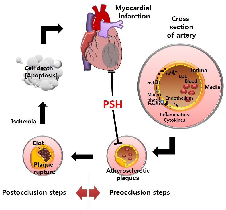
2                                                               Myocardial protection through psyllium intake

                                                                                              consisting of arabinose and xylose), arabinose itself, as well as
                                                                                              hot water extract of ground wheat, reduces myocardial injury
                                                                                              at the postocclusion steps in a rat MI model [26]. Our findings
                                                                                              implicate that any foods containing high levels of AX may be
                                                                                              efficacious in preventing MI through the action of arabinose
                                                                                              during the postocclusion steps. To test the hypothesis, we
                                                                                              conducted a literature search and chose PSH (soluble dietary
                                                                                              fiber) as a candidate since PSH is rich in AX (approximately 60
                                                                                              wt% of PSH) and yields approximately 20 and 46 wt% of
                                                                                              arabinose and xylose, respectively, when hydrolyzed [27]. Based
                                                                                              on these findings, we undertook to investigate whether PSH
                                                                                              supplementation for 3 d reduces the myocardial injury in a rat
                                                                                              model of MI where the left anterior descending coronary artery
                                                                                              (LAD) was occluded for 30 min through ligation, and then
                                                                                              reperfused for 3 h through release of ligation. We further inves-
                                                                                              tigated whether PSH supplementation reduces the myocardial
                                                                                              injury by inhibiting the apoptotic cascades, including PKA to
                                                                                              caspase-3 (CASP3). This study provides some evidence
                                                                                              indicating that PSH is able to prevent MI by inhibiting only the
                                                                                              postocclusion steps, since the 3 d period chosen to supplement
                                                                                              PSH is too short to affect the preocclusion steps which usually
Fig. 1. Summary of development of myocardial infarction (Modified from [4]).
In the preocclusion steps, LDL enters the intima due to endothelial dysfunction. LDL          takes several decades [10] (Fig. 1).
absorbed is oxidized to oxLDL, and oxLDL is engulfed by the macrophages. The
macrophages are then transformed to foam cells which subsequently proliferate, resulting
in the formation of atherosclerotic plaques and narrowing of the arteries. In the             MATERIALS AND METHODS
post-occlusion steps, abrupt rupture of the plaques leads to clot formation in the lesion.
This event can occlude the artery and subsequently result in myocardial ischemia. As a        Materials
result, ATP generation is greatly reduced due to interruption of oxidative phosphorylation,
and myocardial cells die through apoptosis and necrosis. As regions of cell death become        PSH was purchased from Pharmatech Korea (Yongin, Gyeonggi-
extensive, myocardial infarction ensues. PSH, psyllium seed husk; LDL, low-density            do, Republic of Korea). Other reagents were purchased from
lipoprotein; oxLDL, oxidized LDL.
                                                                                              Sigma-Aldrich (St. Louis, MO, USA), unless stated otherwise. PSH
                                                                                              was analyzed to contain approximately 66% AX and 21%
reduces ATP generation and produces excessive reactive oxygen                                 arabinose (wt/wt) [28].
species (ROS) from the cytosol and mitochondria when coupled
with reperfusion, in particular. Consequently, myocardial cells                               Animals
get damaged and die through necrosis and apoptosis, including                                    Eight-week-old male Sprague Dawley (SD) rats were purchased
generation of caspase 3 from procaspase 3, subsequently                                       from Samtaco Inc. (Osan, Gyeonggi-do, Republic of Korea).
                                                  -
leading to MI. ROS, including superoxide anion (O2 ) and hydrogen                             Experiments were carried out according to the protocols for
peroxide (H2O2), is mainly generated in the mitochondria [17].                                animal care and use of laboratory animals, approved by the
ROS reacts with polyunsaturated fatty acids (PUFA) such as                                    Institutional Animal Care and Research Advisory Committee of
cardiolipin (a mitochondrial lipid), resulting in the production                              Catholic University, Daegu, South Korea (No. DCIAFCR-151230-
of toxic byproducts such as 4-hydroxy-2-nonenal (HNE) and                                     20-Y). Animals were housed with food and water available ad
malondialdehyde (MDA) [17]. ROS levels are reduced by the                                     libitum under diurnal lighting conditions and in a temperature-
                       -
transformation of O2 to H2O2 by superoxide dismutase (SOD)                                    controlled environment until the start of the experiment.
[18], followed by the conversion of H2O2 into innocuous water
by a catalase reaction [18]. On the other hand, 4-hydroxynoneal                               Diet Preparation
(HNE) is removed by glutathione S-transferase (GST) through                                      A diet containing PSH was prepared as previously described
conjugation with glutathione [19]. Antioxidant enzymes, including                             [1,29]. To prepare 1 kg each of the 1, 10, or 100 mg/kg PSH
SOD, catalase, and GST, are target genes of the transcription                                 diets, mixtures of 0.02 g PSH + 49.98 g corn starch, 0.2 g PSH
factor nuclear factor erythroid 2-related factor 2 (NRF2), which                              + 49.8 g corn starch, and 2 g PSH + 48 g corn starch were added,
attach to the binding sites on promoters of the target genes,                                 respectively, to 950 g of a modified AIN-93G diet purchased
thereby upregulating transcription of the antioxidant enzymes                                 from Unifaith Inc. (Seoul, Republic of Korea) (Table 1). To
[20,21]. In addition, several proteins located upstream of NRF2                               prepare 1 kg of the basal diet, 50 g of corn starch was added
in the apoptotic cascades such as sirtuins [22], cAMP response                                to 950 g of the modified AIN-93G diet.
element-binding protein (CREB) [23], and protein kinase A (PKA)
[24], are involved in modulating myocardial I/R injury. This is                               Diet administration
a fast process and usually takes only several days for humans                                   PSH diets and basal diet were supplied to the rats as
[25].                                                                                         described previously [1,29]. Briefly, the rats were randomly
   In our previous study, we demonstrated that administration                                 assigned to one of the five groups: (1) sham (n = 6), (2) control
of arabinoxylan (AX; a major cell wall polysaccharide of wheat                                (n = 6), and (3) PSH-treated group (1, 10, or 100 mg/kg per day)
Sun Ha Lim and Jongwon Lee 3
Table 1. Ingredient composition of experimental diets
Modified 1 mg/kg/d 10 mg/kg/d 100 mg/kg/d
Ingredient Basal diet (g/kg)2)
AIN-93 diet (g/950 g)1) PSH diet (g/kg)3) PSH diet (g/kg)3) PSH diet (g/kg)3)
Casein 250.0 250.0 250.0 250.0 250.0
Corn starch 482.5 482.5 482.5 482.5 482.5
Sucrose 100.0 100.0 100.0 100.0 100.0
Soybean oil 70.0 70.0 70.0 70.0 70.0
Mineral mix 35.0 35.0 35.0 35.0 35.0
Vitamin mix 10.0 10.0 10.0 10.0 10.0
Choline bitartrate 2.5 2.5 2.5 2.5 2.5
Corn starch 0.0 50.0 49.98 49.8 48.0
PSH4) 0.0 0.0 0.02 0.2 2.0
t-Butylhydroquinone 0.014 0.014 0.014 0.014 0.014
1)
Modified AIN-93G diet was purchased in a pre-mix form.
2)
One kg of basal diet was prepared by adding 50 g of corn starch to 950 g of the modified AIN-93G diet.
3)
One kg of PSH diets was prepared by adding 50 g of mixture of corn starch and psyllium seed husk (PSH) to 950 g of the modified AIN-93G diet.
4)
PSH diets of 1, 10 or 100 mg/kg/d refer to the corresponding dosage of PSH, given per kilogram of rat per day.
(n = 6 per group). In the PSH-treated group, the rats (approxi- and left ventricular area (LVA) were determined by compu-
mately 300 g weight) received 15 g/d of PSH diet (1, 10, or terized planimetry using ImageJ software (NIH, v1.47). Using
100 mg/kg/d PSH per rat) for 3 d before ligation. Once the these areas, we calculated the IS and risk size (RS), which are
rats consumed the requisite PSH diet each day, more basal diet defined as the percentage of IA to AAR and AAR to LVA,
was provided ad libitum. Rats in the control and sham groups respectively.
received the basal diet only.
Western blotting
Myocardial infarction model Western blotting was performed for the pieces of the hearts
Ischemia/reperfusion injury was generated through ligation harvested, as described previously [1]. Briefly, the harvested
of the LAD followed by release of the ligation, as described pieces were lysed with radioimmunoprecipitation assay (RIPA)
previously [1,29]. Briefly, the male SD rats (~300 g) were buffer (Cell Signaling, Beverly, MA, USA) containing protease
anesthetized through intramuscular injections of ketamine (100 inhibitor cocktail. Equal quantity of protein extracts in the
mg/kg) and xylazine (5 mg/kg), intubated, and ventilated with supernatant were separated on sodium dodecyl sulfate (SDS)-
air throughout the experiment. The heart was then exposed polyacrylamide gels, transferred to polyvinylidene fluoride
by a left thoracic incision, and the LAD of the rats in the (PVDF) membranes (Bio-Rad Laboratories, Inc., Hercules, CA,
PSH-treated group was ligated for 30 min approximately 5 mm USA), and blocked with 5% skim milk, prior to incubation with
below the aortic origin, by passing a 5-0 Prolene suture (BV-1, each primary antibody. The primary antibodies used were
Ethicon, Somerville, NJ, USA) and double-knotting the suture. cleaved CASP3 (C-CASP3, also expressed as CASP3), BCL-2, BAX,
Occlusion generated through the ligation was confirmed by CREB, phosphorylated CREB (pCREB), superoxide dismutase 2
observing development of a pale color in the left ventricular (SOD2), sirturin 1(SIRT1),, SIRT2, SIRT3, SIRT5, SIRT6, and SIRT7
wall. Subsequently, the heart was reperfused for 3 h by releasing (1:1000, Cell Signaling, Beverly, MA, USA), PKAβ and SIRT4
the ligation. Rats in the sham group underwent the same (1:1000, Abcam, Cambridge, MA, USA), NRF2 (1:1000, Enzo,
experimental procedure, but without ligation. During surgery, Farmingdale, NY, USA), glutathione S-transferase mu 2(GSTM2)
the rectal temperature was maintained at 37 ± 0.5°C using a (1:400, USCN Life Science Inc., Houston, Texas, USA), and ERK1
thermostat-controlled warming plate (Harvard Apparatus, (1:1000, Santa Cruz Biotechnology Inc., Santa Cruz, CA, USA).
Holliston, MA, USA). The probed membranes were then incubated with the
horseradish peroxidase-labeled secondary antibodies (1:2000,
Assessment of the infarct size Enzo, Farmingdale, NY, USA) and subsequently developed with
The infarct size (IS) was assessed through 2,3,5-triphenyl- enhanced chemiluminescence (ECL) substrate solution (Thermo
tetrazolium chloride (TTC) staining, as described previously [1, Fisher Scientific, Rockford, IL, USA) using ChemiDoc XRS Gel
29]. After the I/R procedure, the LAD was re-ligated, and 1 ml Imager (Bio-Rad Laboratories, Inc., Hercules, CA, USA). Intensities
of 1% Evans blue dye (Sigma-Aldrich, St. Louis, MO, USA) was of the protein bands were quantified using the ImageJ software
infused into the heart through the jugular vein. Next, the rats (NIH, v1.47). In the quantitative analysis, ratios between various
were euthanized by thoracotomy under anesthetization. The combinations of proteins and phosphorylated proteins were
heart was harvested, excised into 4 pieces approximately 3-mm presented using ERK1 as a loading control by setting the control
thick, which were subsequently stained with TTC. The area at group value (0 mg/kg/d of PSH) at 1.
risk (AAR) is defined as the area not infiltrated by Evans blue
dye. The infarct area (IA) is defined as the area not stained with Statistical analyses
TTC. The border zone area (BZA) is defined as the area in which The values are expressed as the means ± standard error of
the IA is excluded from the AAR (AAR-IA). The AAR, IA, BZA, the mean (SEM). Statistical analyses were performed using the4 Myocardial protection through psyllium intake
SPSS software (IBM SPSS Statistics; version 19, Armonk, NY, USA). (A)
Shapiro-Wilk test and Levene's test were used to test all
variables for normal distribution and homogeneity of variances,
respectively. A one-way ANOVA followed by the post hoc test
was used to compare changes in infarct size and western blot
analysis. The statistical significance was set as P < 0.05.
RESULTS
PSH supplementation reduces infarct size (B)
Using a rat I/R model, we first undertook to determine
whether PSH supplementation protects against myocardial
injury at the postocclusion steps. Our previous study showed
that supplementation with 5 mg/kg/d of AX was enough to
exert efficacies [26]. As 10 mg/kg/d of PSH is equivalent to 5
mg/kg/d of AX, considering that PSH contains approximately
50% AX, we tested various dosages of PSH supplementation,
including 10 mg/kg/d of PSH (1, 10, or 100 mg/kg/d), to find
a minimum efficacious dosage. The rats were fed diets
supplemented with PSH for 3 d, after which they were subjected
to 30 min ischemia through ligation of LAD, followed by 3 h
reperfusion through release of the ligation. Finally, the heart
was harvested, cut into four slices, and stained with TTC (Fig.
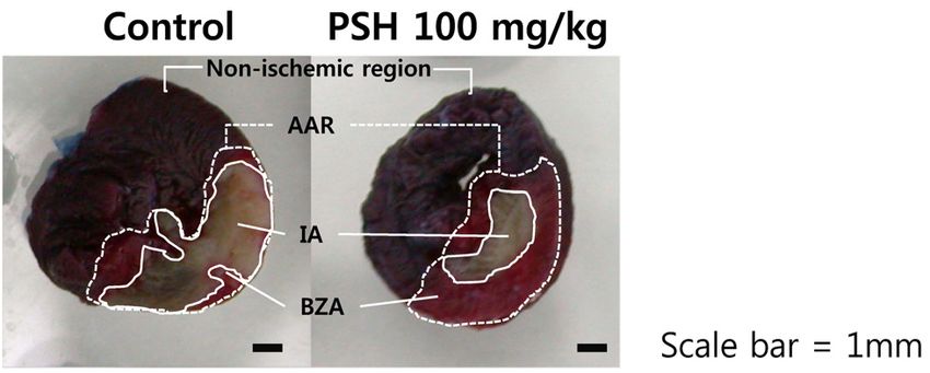
Fig. 2. Effect of PSH supplementation on infarct size. Rats underwent 30 min
2A). AAR, IA, and BZA were defined as the area without ischemia through ligation of LAD, followed by 3 h reperfusion through release of the
infiltration of Evans blue dye, the area without TTC stain, and ligation. (A) Evans blue dye was infused into the heart after LAD was re-ligated. The heart
was harvested and cut into four slices. The slices were stained with TTC. AAR, IA, and
the area equivalent to (AAR-IA), respectively (Fig. 2A). To BZA were determined as the area without infiltration of Evans blue dye, the area without
determine whether PSH supplementation reduces myocardial TTC stain, and the area equivalent to (AAR-IA), respectively. (B) IS, the ratio of IA to AAR,
injury, we assessed the infarct size (IS, the ratio of IA to AAR), and RS, the ratio of AAR to LVA, are presented. In the PSH-treated groups, PSH (1,
10, or 100 mg/kg/d) supplements were fed for 3 days prior to LAD ligation. In the control
which represents degree of cell death in the AAR. We observed group, no PSH was administered prior to LAD ligation. The number of rats used in the
that supplementation with 10 or 100 mg/kg/d significantly control and PSH-treated (1, 10, or 100 mg/kg/d) groups were 6 per group. Values are
expressed as means ± SEM. * P < 0.05, when compared to control group. PSH, psyllium
reduces the IS, compared with the control group (39.8 ± 3.3 and seed husk; LAD, left anterior descending coronary artery; TTC, 2,3,5- triphenyltetrazolium
38.3 ± 4.9%, respectively, versus 55.1 ± 3.5%; P < 0.05) (Fig. 2B). chloride; AAR, area at risk; IA, infarct area; BZA, border zone area; IS, infarct size; RS,
risk size; LVA; left ventricular area.
To confirm reproducibility of the experiments, we next assessed
the risk size (RS, the ratio of AAR to LVA). The RS of PSH-treated
groups was not significantly different from that of the control against I/R injury during the postocclusion steps.
groups (P > 0.05), indicating that the surgical procedure to
generate I/R injury was reliable (Fig. 2B). Taken together, these PSH supplementation reduces apoptosis
results indicate that PSH intake over certain dosages protects As AX supplementation reduces myocardial injury by inhibiting
(A) (B)
Fig. 3. Effect of PSH supplementation on the formation of caspase-3 (CASP3) and expression of BCL-2 and BAX. (A) Western blots of CASP3 (cleaved caspase-3 generated
from procaspase-3), BCL-2, and BAX in the AAR. Protein levels were measured by Western blotting for the sham, control, and PSH-treated (100 mg/kg/d) groups. ERK1 was used as
the loading control. (B) Quantitative analysis of CASP3 (cleaved caspase-3), BCL-2, and BAX. C-CASP3 (CASP3)/ERK1 and BCL-2/BAX ratios are presented. The ratios were calculated
by setting the control group value (0 mg/kg/d of PSH) at 1. The number of rats used in the sham, control and PSH-treated (100 mg/kg/d) groups were 6 per group. Values are expressed
as means ± SEM. CASP3, caspase-3; C-CASP3, cleaved caspase-3; PSH, psyllium seed husk; AAR, area at risk.Sun Ha Lim and Jongwon Lee 5
apoptosis [26], we investigated whether PSH supplementation expression of BCL-2 and BAX, which are located upstream of
also protects against myocardial injury by inhibiting apoptosis, CASP3 in the apoptotic cascade [31]. We observed that PSH
using CASP3 as a biomarker [30]. Supplementation with PSH supplementation tended to increase the ratio of BCL-2 with
(100 mg/kg/d) tends to reduce levels of CASP3 (cleaved CASP3 anti-apoptotic activities, and BAX with pro-apoptotic activities
generated from procaspase-3) as compared with the control (BCL-2/BAX) as compared to the control group, suggesting that
group, suggesting that PSH supplementation results in decreased PSH supplementation contributes towards inhibiting apoptosis
apoptosis (Fig. 3A and 3B). To support the conclusion, we (Fig. 3B) [31,32]. Taken together, our results indicate that PSH
investigated whether PSH supplementation modulated the intake protects against myocardial I/R injury by inhibiting the
(A)
(B)
Fig. 4. Effect of PSH supplementation on the expression of NRF2, GSTM2, and SOD2. Western blots of NRF2, GSTM2, and SOD2 in the AAR are presented. (A) Protein levels
were measured by Western blotting for the sham, control, and PSH-treated (100 mg/kg/d) groups. ERK1 was used as a loading control. (B) Quantitative analysis of NRF2, GSTM2, and
SOD2: NRF2/ERK1, GSTM2/ERK1, and SOD2/ERK1 ratios are presented. The ratios were calculated by setting the control group value (0 mg/kg/d of PSH) at 1. The number of rats
used in the sham, control and PSH-treated (100 mg/kg/d) groups were 6 per group. Values are expressed as means ± SEM. *** P < 0.001, and * P < 0.05 vs. control group. PSH, psyllium
seed husk; NRF2, nuclear factor erythroid 2-related factor 2; GSTM2, glutathione S-transferase mu 2; SOD2, superoxide dismutase 2; AAR, area at risk.
(A)
(B)
Fig. 5. Effect of PSH supplementation on the expression of SIRT (1-7). Western blots of SIRT (1-7) in the AAR are presented. (A) Protein levels were measured by Western blotting
for the sham, control, and PSH-treated (100 mg/kg/d) groups. ERK1 was used as the loading control. (B) Quantitative analysis of SIRT (1-7): SIRT (1-7)/ERK1 ratios are presented. The
ratios were calculated by setting the control group value (0 mg/kg/d of PSH) at 1. The number of rats used in the sham, control and PSH-treated (100 mg/kg/d) groups were 6 per
group. Values are expressed as means ± SEM. ** P < 0.01 and * P < 0.05 vs. control group. PSH, psyllium seed husk; SIRT, sirtuins; AAR, area at risk.6 Myocardial protection through psyllium intake
(A) (B)
Fig. 6. Effect of PSH supplementation on the expression of PKAβ, pCREB, and CREB. Western blots of PKAβ, pCREB, and CREB in the AAR are presented. (A) Protein levels
were measured by Western blotting for the sham, control, and PSH-treated (100 mg/kg/d) groups. ERK1 was used as the loading control. (B) Quantitative analysis of PKAβ/ERK1,
pCREB/ERK1, and CREB/ERK1 ratios are presented. The ratios were calculated by setting the control group value (0 mg/kg/d of PSH) at 1. The number of rats used in the sham,
control and PSH-treated (100 mg/kg/d) groups were 6 per group. Values are expressed as means ± SEM. ** P < 0.01 and * P < 0.05 vs. control group. PSH, psyllium seed husk; PKAβ,
protein kinase Aβ; pCREB, phosphorylated cAMP response element-binding protein; AAR, area at risk.
apoptotic cascades. PKA expression. PSH supplementation significantly increases
PKAβ, a catalytic subunit of PKA, compared to the control (Fig.
PSH supplementation upregulates expression of antioxidant 6B). Taken together, the results indicate that PSH suppleme-
enzymes through NRF2 ntation upregulates PKA expression, which contributes to
We next investigated whether PSH supplementation upregulates enhancing the phosphorylation of CREB.
the levels of GSTM2. PSH supplementation significantly upregu-
lated GSTM2 expression compared with the control group (Fig. DISCUSSION
4A and 4B). In addition, PSH supplementation tended to
upregulate the SOD2 expression, which is responsible for the In this study, we determined that PSH supplementation
-
removal of O2 in the mitochondria. These results indicate that reduces myocardial I/R injury by inhibiting the apoptotic
PSH supplementation eliminates ROS through upregulation of cascades through reduction of CASP3 generation and increase
antioxidant enzymes. As both GSTM2 and SOD2 are target of BCL-2/BAX ratio in a rat I/R model. Inhibition of apoptosis
genes of NRF2 transcription factor [20,21], we investigated might be mediated by from upregulation of PKA expression
whether PSH supplementation upregulates NRF2 expression, to reduction of CASP3 generation (Fig. 7). In the cascades, PSH
and observed significant increase in NRF2 expression compared supplementation first upregulates the PKA expression, which
with the control (Fig. 4B). Taken together, our results indicate contributes to the enhancement of CREB phosphorylation to
that PSH supplementation promotes the NRF2 expression, which generate pCREB. This theory is supported by previous studies
contributes to upregulating gene expression of antioxidant which have reported that higher levels of PKA [24] and pCREB
enzymes. [23], and activation of PKA-CREB axis [34] are associated with
myocardial protection against I/R injury by inhibiting apoptosis
PSH supplementation upregulates expression of sirtuins through reduction of CASP3 generation and increase of
The effect of PSH supplementation was investigated on the BCL-2/BAX ratio. Therefore, our studies indicate that PSH supple-
modulation of gene expression of sirtuins (Fig. 5A). Of the mentation contributes to myocardial protection by upregulation
known sirtuins (1-7), SIRT5 (a mitochondrial sirtuin), and SIRT6 of PKA expression and enhancement of CREB phosphorylation.
and SIRT7 (nuclear sirtuins) [22] were significantly upregulated Of the 7 mammalian sirtuins (1-7), PSH supplementation
by PSH supplementation (Fig. 5B). These results suggest that upregulates the expression of SIRT5, SIRT6, and SIRT7. These
+
PSH supplementation upregulates some sirtuin genes, which findings indicate that PSH enhances the NAD -dependent
might contribute to upregulating the NRF2 expression. deacetylation of proteins including histones, in both mito-
chondria and nuclei, since SIRT5 is located in the mitochondria,
PSH supplementation promotes phosphorylation of CREB and and SIRT6 and SIRT7 are located in the nuclei [22]. Furthermore,
upregulates expression of protein kinase A (PKA) it has been reported that higher levels of SIRT6 protect against
Finally, we investigated whether PSH supplementation increases myocardial I/R injury by inhibiting apoptosis through reduction
the pCREB level (Fig. 6A). Indeed, PSH supplementation resulted of ROS generation, due to the upregulated antioxidant enzymes
in significant increases in the pCREB level compared to the such as SOD2 [35]. In addition, higher levels of SIRT7 contribute
control, although CREB levels remained unaffected (Fig. 6B). to enhancing wound healing through increased scar formation
Moreover, as phosphorylation of CREB is mediated by PKA [33], after MI [36]. Finally, higher levels of SIRT5 protect the cardio-
we investigated whether PSH supplementation modulates the myocytes from oxidative stress by inhibiting apoptosis [37].Sun Ha Lim and Jongwon Lee 7
toxicity (Fig. 7). Based on these results, we propose that PSH
supplementation protects against myocardial I/R injury through
upregulation of PKA expression, CREB phosphorylation, sirtuin
expression, NRF2 expression, antioxidant enzymes expression,
and subsequent reduction of ROS toxicity followed by reduction
of apoptosis (Fig. 7).
The findings from this study indicating that PSH supple-
mentation protects against myocardial I/R injury support our
hypothesis that consumption of any foods containing high level
of AX and/or arabinose (such as PSH) can be effective in
protecting against myocardial injury. In this study, supple-
mentation with PSH containing approximately 65% AX and 20%
arabinose [28] showed efficacies at a dosage of 10 mg/kg/d.
Previously, we showed that supplementation with AX containing
approximately 40% arabinose exerts efficacies at a dosage of
5 mg/kg/d [26]. We have also reported that supplementation
with apple pectin containing approximately 1.5% arabinose
shows efficacy at a dosage of 100 mg/kg/d [10]. Therefore, this
suggests that consumption of lesser amount of foods containing
higher levels of AX or arabinose are required to exhibit their
efficacies. An underlying mechanism by which PSH consumption
protects against myocardial injury through arabinose can be
presented, as previously described [26]. Briefly, once PSH is
ingested, AX present in PSH is hydrolyzed to arabinose and
Fig. 7. A proposed, underlying mechanism for the myocardial protection xylose by microbiota inhabiting the large intestine. In turn, the
through PSH supplementation. PSH supplementation protects against myocardial I/R
injury through upregulation of PKA expression, CREB phosphorylation, sirtuin expression, arabinose generated is absorbed into the body and protects
NRF2 expression, expression of antioxidant enzymes such as GSTM2 and SOD2, and the heart against ischemic injury.
subsequent reduction of ROS toxicity, followed by reduction of apoptosis through increase
of BCL-2/BAX ratio and subsequent reduction of CASP3 generation. PKA, protein kinase
Regardless of underlying mechanisms, the findings from this
A; pCREB, phosphorylated cAMP response element-binding protein; SIRT5, 6, 7, sirtuins study reveal that short-term PSH intake protects against
5, 6, 7; NRF2, nuclear factor erythroid 2-related factor 2; GSTM2, glutathione S-transferase myocardial I/R injury even after coronary arteries are occluded,
mu 2; SOD2, superoxide dismutase 2; CASP3, caspase-3; MI, myocardial infarction; PSH,
psyllium seed husk; I/R, ischemia/reperfusion; ROS, reactive oxygen species. indicating that PSH intake can prevent MI by blocking the
postocclusion steps (Fig. 1). It has already been shown that
Taken together, PSH supplementation upregulates expression long-term PSH consumption prevents MI by blocking the
of some sirtuins such as SIRT5, SIRT6, and SIRT7, which preocclusion steps, because PSH consumption was effective in
contributes to protecting against myocardial I/R injury through reducing risk factors for coronary ischemic disease such as
elimination of ROS and subsequent enhancement of wound hyperlipidemia [11,12], diabetes [14], and hypertension [15],
healing. Upregulation of the sirtuins through PSH suppleme- which promote the formation of atherosclerotic plaques
ntation might be mediated by binding of pCREB to the binding responsible for narrowing the coronary arteries (Fig. 1). Overall,
site CRE located on promoters of the sirtuins (SIRT6 gene we conclude that PSH consumption prevents MI by inhibiting
promoter has putative pCREB binding site) [38] (Fig. 7). PSH both the preocclusion and postocclusion steps.
supplementation also upregulates the expression of NRF2 and Approximately 10 g/d of PSH was administered in various
its target genes such as GSTM2 and SOD2 [20,21]. In our clinical trials to assess the efficacies in reducing the risk factors
previous study, we showed that supplementation with wheat [11,14,15]. Based on these findings, the US Food and Drug
extract (WE) containing AX as an active component reduces the Administration (FDA) approved health claims of reducing
MDA generation and upregulates GSTM2 [26]. SOD2 is involved cardiovascular disease for PSH at dosages of over 8 g/d [43].
-
in removing O2 in the mitochondria through conversion of O2
- However, results from the current study indicate that PSH
into H2O2 [18]. On the other hand, GSTM2 is involved in supplementation to the rats protected myocardial I/R injury at
detoxification of HNE and MDA, the peroxidative byproducts dosages over 10 mg/kg/d. The dosage is equivalent to 100 mg/d
of PUFA attributed to ROS, through conjugation of the for a 60 kg person, based on calculations using a conversion
byproducts with glutathione [19]. In addition, NRF2 expression factor provided by the FDA [44]. Thus, we postulate that the
is regulated by SIRT6 [39,40] through deacetylation of histones dosage required to show efficacies for blocking postocclusion
[41]. The NRF2 gene promoter also has the pCREB binding site, steps is much smaller than that required for blocking
implicating that CREB is also involved in the upregulation of preocclusion steps, which will therefore not cause any safety
NRF2 gene expression [42]. Taken together, these findings concerns for consuming PSH to reduce myocardial injury.
suggest that PSH supplementation upregulates NRF2 expression In conclusion, the results from our study reveal that PSH
by pCREB and the sirtuins, which in turn results in upregulation consumption reduces myocardial I/R injury in rats by inhibiting
of antioxidant enzymes and subsequent elimination of ROS the apoptotic cascades through modulation of gene expression
of several genes located upstrem of apoptosis, at a safe dosage8 Myocardial protection through psyllium intake
when converted to human beings. Our findings indicate that agent in animal, human and poultry. Int J Pharmacol 2017;13:690-7.
PSH consumption prevents MI by blocking the postocclusion 14. Gibb RD, McRorie JW Jr, Russell DA, Hasselblad V, D'Alessio DA.
steps. We belive that PSH has the potential to be developed Psyllium fiber improves glycemic control proportional to loss of
as a functional food for preventing MI, through blockade of glycemic control: a meta-analysis of data in euglycemic subjects,
both the preocclusion and postocclusion steps. patients at risk of type 2 diabetes mellitus, and patients being
treated for type 2 diabetes mellitus. Am J Clin Nutr 2015;102:
CONFLICT OF INTEREST 1604-14.
15. Khan K, Jovanovski E, Ho HV, Marques AC, Zurbau A, Mejia SB,
The authors declare no potential conflicts of interests. Sievenpiper JL, Vuksan V. The effect of viscous soluble fiber on
blood pressure: a systematic review and meta-analysis of randomized
ORCID controlled trials. Nutr Metab Cardiovasc Dis 2018;28:3-13.
16. Obata K, Ikeda K, Yamasaki M, Yamori Y. Dietary fiber, psyllium,
Sun Ha Lim: https://orcid.org/0000-0002-8371-4740 attenuates salt-accelerated hypertension in stroke-prone sponta-
Jongwon Lee: https://orcid.org/0000-0001-9372-9850 neously hypertensive rats. J Hypertens 1998;16:1959-64.
17. Raedschelders K, Ansley DM, Chen DD. The cellular and molecular
REFERENCES origin of reactive oxygen species generation during myocardial
ischemia and reperfusion. Pharmacol Ther 2012;133:230-55.
1. Lim SH, Lee J. Xyloglucan intake attenuates myocardial injury by 18. Zhang Y, Martin SG. Redox proteins and radiotherapy. Clin Oncol
inhibiting apoptosis and improving energy metabolism in a rat (R Coll Radiol) 2014;26:289-300.
model of myocardial infarction. Nutr Res 2017;45:19-29. 19. Ayala A, Muñoz MF, Argüelles S. Lipid peroxidation: production,
2. Lim SH. Larch arabinogalactan attenuates myocardial injury by metabolism, and signaling mechanisms of malondialdehyde and
inhibiting apoptotic cascades in a rat model of ischemia- 4-hydroxy-2-nonenal. Oxid Med Cell Longev 2014;2014:360438.
reperfusion. J Med Food 2017;20:691-9. 20. Zhou S, Sun W, Zhang Z, Zheng Y. The role of Nrf2-mediated
3. Kim MY, Lim SH, Lee J. Intake of hot water-extracted apple protects pathway in cardiac remodeling and heart failure. Oxid Med Cell
against myocardial injury by inhibiting apoptosis in an Longev 2014;2014:260429.
ischemia/reperfusion rat model. Nutr Res 2014;34:951-60. 21. Murphy KE, Park JJ. Can co-activation of Nrf2 and neurotrophic
4. Lim SH, Kim MJ, Han MJ, Kim Y, Lee J. Prevention of ischemic signaling pathway slow Alzheimer's disease? Int J Mol Sci
diseases and cognitive disorders through wheat consumption. In: 2017;18:1168.
Duncan LT, editor. Advances in Health and Disease. Hauppauge 22. Matsushima S, Sadoshima J. The role of sirtuins in cardiac disease.
(NY): Nova Science Publishers, Inc.; 2018. p.1-66. Am J Physiol Heart Circ Physiol 2015;309:H1375-89.
5. Soler EP, Ruiz VC. Epidemiology and risk factors of cerebral ischemia 23. Yu W, Xu M, Zhang T, Zhang Q, Zou C. Mst1 promotes cardiac
and ischemic heart diseases: similarities and differences. Curr ischemia-reperfusion injury by inhibiting the ERK-CREB pathway
Cardiol Rev 2010;6:138-49. and repressing FUNDC1-mediated mitophagy. J Physiol Sci 2019;69:
6. Hajar R. Risk factors for coronary artery disease: historical perspectives. 113-27.
Heart Views 2017;18:109-14. 24. Zhang Y, Wang XL, Zhao J, Wang YJ, Lau WB, Yuan YX, Gao EH,
7. Hurtubise J, McLellan K, Durr K, Onasanya O, Nwabuko D, Ndisang Koch WJ, Ma XL. Adiponectin inhibits oxidative/nitrative stress
JF. The different facets of dyslipidemia and hypertension in during myocardial ischemia and reperfusion via PKA signaling. Am
atherosclerosis. Curr Atheroscler Rep 2016;18:82. J Physiol Endocrinol Metab 2013;305:E1436-43.
8. Park KH, Park WJ. Endothelial dysfunction: clinical implications in 25. Arai AE. Healing after myocardial infarction: a loosely defined
cardiovascular disease and therapeutic approaches. J Korean Med process. JACC Cardiovasc Imaging 2015;8:680-3.
Sci 2015;30:1213-25. 26. Lim SH, Kim Y, Yun KN, Kim JY, Jang JH, Han MJ, Lee J. Plant-based
9. Maiolino G, Rossitto G, Caielli P, Bisogni V, Rossi GP, Calò LA. The foods containing cell wall polysaccharides rich in specific active
role of oxidized low-density lipoproteins in atherosclerosis: the monosaccharides protect against myocardial injury in rat
myths and the facts. Mediators Inflamm 2013;2013:714653. myocardial infarction models. Sci Rep 2016;6:38728.
10. Lim SH, Kim MY, Lee J. Apple pectin, a dietary fiber, ameliorates 27. Van Craeyveld V, Delcour JA, Courtin CM. Ball milling improves
myocardial injury by inhibiting apoptosis in a rat model of extractability and affects molecular properties of psyllium (Plantago
ischemia/reperfusion. Nutr Res Pract 2014;8:391-7. ovata Forsk) seed husk arabinoxylan. J Agric Food Chem 2008;56:
11. Wei ZH, Wang H, Chen XY, Wang BS, Rong ZX, Wang BS, Su BH, 11306-11.
Chen HZ. Time- and dose-dependent effect of psyllium on serum 28. Lim SH, Kim MJ, Lee J. Intake of psyllium seed husk reduces white
lipids in mild-to-moderate hypercholesterolemia: a meta-analysis of matter damage in a rat model of chronic hypoperfusion. Nutr Res
controlled clinical trials. Eur J Clin Nutr 2009;63:821-7. 2019; https://doi.org/10.1016/j.nutres.2019.04.002
12. Ribas SA, Cunha DB, Sichieri R, Santana da Silva LC. Effects of 29. Lim SH, Lee J. Protection of the brain through supplementation
psyllium on LDL-cholesterol concentrations in Brazilian children and with larch arabinogalactan in a rat model of vascular dementia.
adolescents: a randomised, placebo-controlled, parallel clinical trial. Nutr Res Pract 2017;11:381-7.
Br J Nutr 2015;113:134-41. 30. Porter AG, Jänicke RU. Emerging roles of caspase-3 in apoptosis.
13. Xing LC, Santhi D, Shar AG, Saeed M, Arain MA, Shar AH, Bhutto Cell Death Differ 1999;6:99-104.
ZA, Katar MU, Manzoor R, El-Hack ME, Alagawany M, Dhama K, Ling 31. Youle RJ, Strasser A. The BCL-2 protein family: opposing activities
MC. Psyllium husk (Plantago ovata) as a potent hypocholesterolemic that mediate cell death. Nat Rev Mol Cell Biol 2008;9:47-59.Sun Ha Lim and Jongwon Lee 9
32. Edlich F. BCL-2 proteins and apoptosis: recent insights and genes in GeneCards. Database (Oxford) 2017;2017:1.
unknowns. Biochem Biophys Res Commun 2018;500:26-34. 39. Pan H, Guan D, Liu X, Li J, Wang L, Wu J, Zhou J, Zhang W, Ren
33. Sands WA, Palmer TM. Regulating gene transcription in response R, Zhang W, Li Y, Yang J, Hao Y, Yuan T, Yuan G, Wang H, Ju Z,
to cyclic AMP elevation. Cell Signal 2008;20:460-6. Mao Z, Li J, Qu J, Tang F, Liu GH. SIRT6 safeguards human
34. Zhao D, Feng P, Sun Y, Qin Z, Zhang Z, Tan Y, Gao E, Lau WB, mesenchymal stem cells from oxidative stress by coactivating NRF2.
Ma X, Yang J, Yu S, Xu X, Yi D, Yi W. Cardiac-derived CTRP9 protects Cell Res 2016;26:190-205.
against myocardial ischemia/reperfusion injury via calreticulin- 40. Zhang W, Wei R, Zhang L, Tan Y, Qian C. Sirtuin 6 protects the
dependent inhibition of apoptosis. Cell Death Dis 2018;9:723. brain from cerebral ischemia/reperfusion injury through NRF2
35. Wang XX, Wang XL, Tong MM, Gan L, Chen H, Wu SS, Chen JX, activation. Neuroscience 2017;366:95-104.
Li RL, Wu Y, Zhang HY, Zhu Y, Li YX, He JH, Wang M, Jiang W. 41. Liao CY, Kennedy BK. SIRT6, oxidative stress, and aging. Cell Res
SIRT6 protects cardiomyocytes against ischemia/reperfusion injury 2016;26:143-4.
by augmenting FoxO3α-dependent antioxidant defense mechanisms. 42. Impey S, McCorkle SR, Cha-Molstad H, Dwyer JM, Yochum GS, Boss
Basic Res Cardiol 2016;111:13. JM, McWeeney S, Dunn JJ, Mandel G, Goodman RH. Defining the
36. Araki S, Izumiya Y, Rokutanda T, Ianni A, Hanatani S, Kimura Y, CREB regulon: a genome-wide analysis of transcription factor
Onoue Y, Senokuchi T, Yoshizawa T, Yasuda O, Koitabashi N, regulatory regions. Cell 2004;119:1041-54.
Kurabayashi M, Braun T, Bober E, Yamagata K, Ogawa H. Sirt7 43. Jenkins DJ, Kendall CW, Vuksan V, Vidgen E, Parker T, Faulkner D,
contributes to myocardial tissue repair by maintaining transforming Mehling CC, Garsetti M, Testolin G, Cunnane SC, Ryan MA, Corey
growth factor-β signaling pathway. Circulation 2015;132:1081-93. PN. Soluble fiber intake at a dose approved by the US Food and
37. Liu B, Che W, Zheng C, Liu W, Wen J, Fu H, Tang K, Zhang J, Xu Drug Administration for a claim of health benefits: serum lipid risk
Y. SIRT5: a safeguard against oxidative stress-induced apoptosis in factors for cardiovascular disease assessed in a randomized
cardiomyocytes. Cell Physiol Biochem 2013;32:1050-9. controlled crossover trial. Am J Clin Nutr 2002;75:834-9.
38. Fishilevich S, Nudel R, Rappaport N, Hadar R, Plaschkes I, Iny Stein 44. Nair AB, Jacob S. A simple practice guide for dose conversion
T, Rosen N, Kohn A, Twik M, Safran M, Lancet D, Cohen D. between animals and human. J Basic Clin Pharm 2016;7:27-31.
GeneHancer: genome-wide integration of enhancers and targetYou can also read