Omega 7 oil increases telomerase activity and accelerates healing of grafted burn and donor site wounds - Nature
←
→
Page content transcription
If your browser does not render page correctly, please read the page content below
www.nature.com/scientificreports
OPEN Omega‑7 oil increases telomerase
activity and accelerates healing
of grafted burn and donor site
wounds
Yosuke Niimi1,2,5, Dannelys Pérez‑Bello1,5, Koji Ihara2, Satoshi Fukuda1, Sam Jacob3,
Clark R. Andersen4, Tuvshintugs Baljinnyam1, Jisoo Kim1, Suzan Alharbi1,
Donald S. Prough1 & Perenlei Enkhbaatar1*
This study investigated the efficacy of Omega-7 isolated from the sea buckthorn oil (Polyvit Co., Ltd,
Gangar Holding, Ulaanbaatar, Mongolia) in ovine burn wound healing models. In vitro, proliferation
(colony-forming rate) and migration (scratch) assays using cultured primary ovine keratinocytes
were performed with or without 0.025% and 0.08% Omega-7, respectively. The colony-forming rate
of keratinocytes in the Omega-7 group at 72 and 96 h were significantly higher than in the control
(P < 0.05). The percentage of closure in scratch assay in the Omega-7 group was significantly higher
than in the control at 17 h (P < 0.05). In vivo, efficacy of 4% Omega-7 isolated from buckthorn oil
was assessed at 7 and 14 days in grafted ovine burn and donor site wounds. Telomerase activity,
keratinocyte growth factor, and wound nitrotyrosine levels were measured at day 14. Grafted sites:
Un-epithelialized raw surface area was significantly lower and blood flow was significantly higher in
the Omega-7-treated sites than in control sites at 7 and 14 days (P < 0.05). Telomerase activity and
levels of keratinocyte growth factors were significantly higher in the Omega-7-treated sites after
14 days compared to those of control (P < 0.05). The wound 3-nitrotyrosine levels were significantly
reduced by Omega-7. Donor sites: the complete epithelialization time was significantly shorter and
blood flow at day 7 was significantly higher in the Omega-7-treated sites compared to control sites
(P < 0.05). In summary, topical application of Omega-7 accelerates healing of both grafted burn
and donor site wounds. Omega-7 should be considered as a cost-efficient and effective supplement
therapy for burn wound healing.
In the U.S., 486,000 burn injuries requiring medical treatment occur a nnually1. The average daily hospital cost
of surviving patients in the U.S., is $81792. Delayed wound healing of severely burned patients and limited avail-
ability of donor skin remain challenging problems3. Thus, an efficient and robust therapy for healing of both
grafted burn and donor sites is needed.
The wound healing process is dynamic and has three overlapping phases (i.e., inflammation, tissue formation,
and tissue remodeling) that occur in a specific time and duration4. Optimal bio-physiologic events of the heal-
ing process involve the following: appropriate inflammation; angiogenesis; re-epithelialization; mesenchymal
cell differentiation, proliferation, and migration; and collagen remodeling—cross-linking and strength5. These
wound healing events are affected by local factors, such as cytokines (i.e., keratinocyte growth factor, vascular
endothelial growth factor, fibroblast growth factor, transforming growth factor beta, and interleukin-1), blood
cells, extracellular matrix, and parenchymal cells.
Telomeres and telomerases play essential roles in the control of cell proliferation. Telomerase is a ribonucleo-
protein complex that extends telomeres. It is known that telomerase maintains telomere length in various c ells6.
When cells divide, telomere length is shortened with each round of DNA replication leading to senescence7,8.
1
Department of Anesthesiology, Medical Branch, University of Texas, 301 University Blvd, Galveston,
TX 77555‑1102, USA. 2Department of Plastic and Reconstructive Surgery, Tokyo Women’s Medical University,
8‑1, Kawada‑cho, Shinjuku, Tokyo, Japan. 3Department of Pathology, Shriners Hospitals for Children, 815
Market St, Galveston, TX 77550, USA. 4Department of Biostatistics, Medical Branch, University of Texas, 301
University Blvd, Galveston, TX 77555‑1102, USA. 5These authors contributed equally: Yosuke Niimi and Dannelys
Pérez-Bello. *email: peenkhba@utmb.edu
Scientific Reports | (2021) 11:975 | https://doi.org/10.1038/s41598-020-79597-0 1
Vol.:(0123456789)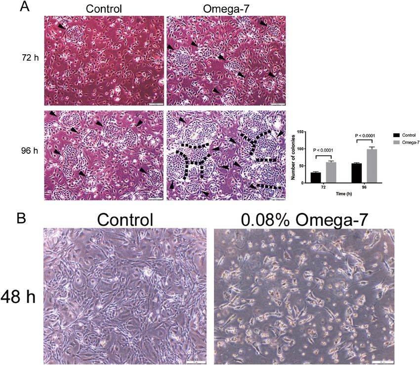
www.nature.com/scientificreports/
Skin cell telomeres may be particularly susceptible to accelerated shortening due to excess generation of reac-
tive oxygen species (ROS) in burn wounds which damage cellular DNA and negatively affect p roliferation9,10. The
11
healthy length of telomeres is maintained by a number of factors, including telomerase . Although telomerase
is known to be activated in the epidermis and it plays a significant role in the maintenance of skin cell functions
and proliferation, it is not known if telomerase activity is negatively affected by burns12.
Various treatment approaches have been proposed to improve healing of both grafted burn and donor site
wounds, including topical medications and tissue engineered materials (i.e., fibroblast growth f actor13, artificial
dermis templates14, cultured epithelial allograft15, and/or cellular therapy such as mesenchymal stem cells16,
induced pluripotent stem cells17, and embryonic stem cells18). However, high cost, long culture time, and ethical
issues often prevent their use in clinical practice.
There are several reports on the use of natural products, such as Allium sativum, Aloe vera, and Centella
asiatica for treatment of burn wounds. Previously, we have reported the effects of sea buckthorn (Hippophae
rhamnoides L.) oil on grafted burn wound healing19–21. Sea buckthorn is a wild berry of the Elaeagnaceae family
growing at an elevation of 2500–4300 m in Europe and Asia22. The plant has been used extensively in traditional
medicine in Asia for treating burn wounds for more than 1000 y ears23. Omega-7 unsaturated oils constitute
approximately 48% of sea buckthorn oil21. Although sea buckthorn oil has been shown to promote skin and
mucosal epithelialization24,25, effects of Omega-7 in wound healing are not known. In the present study, we have
tested the hypothesis that Omega-7 improves healing of both grafted burn and donor site wounds.
Results
Number of colonies in cultured keratinocytes and keratinocyte wound closure rate
in vitro. The proliferation and migration rates of cultured keratinocytes were assessed by the colony counting
and scratch assays, respectively. The number of keratinocyte colonies in the Omega-7 group treated with 0.025%
of Omega-7 at 72 and 96 h (60.8 ± 3.3, and 98.8 ± 6.8, respectively) was significantly higher than that of the con-
trol group (30.5 ± 2.8, and 57.4 ± 1.9, respectively) (P < 0.0001) (Fig. 1A). Omega-7 with dose of 0.08% negatively
affected the cells (Fig. 1B).
The percentage of cultured keratinocyte wound closure (scratch assay) in the Omega-7 group at 17 h after
scratch (81.8 ± 4.9) was significantly higher than that of the control group (58.9 ± 2) (P = 0.005) (Fig. 2A).
Omega-7 with doses of greater than 0.1% negatively affected the cell migration (Fig. 2B).
Un‑epithelialized raw surface area in grafted burn wounds and time (number of days) of com-
plete re‑epithelialization of donor sites in vivo. All grafted sites had no complications such as infec-
tion or hematoma. On the day of the surgery, the sizes of the un-epithelialized raw surface areas of the grafted
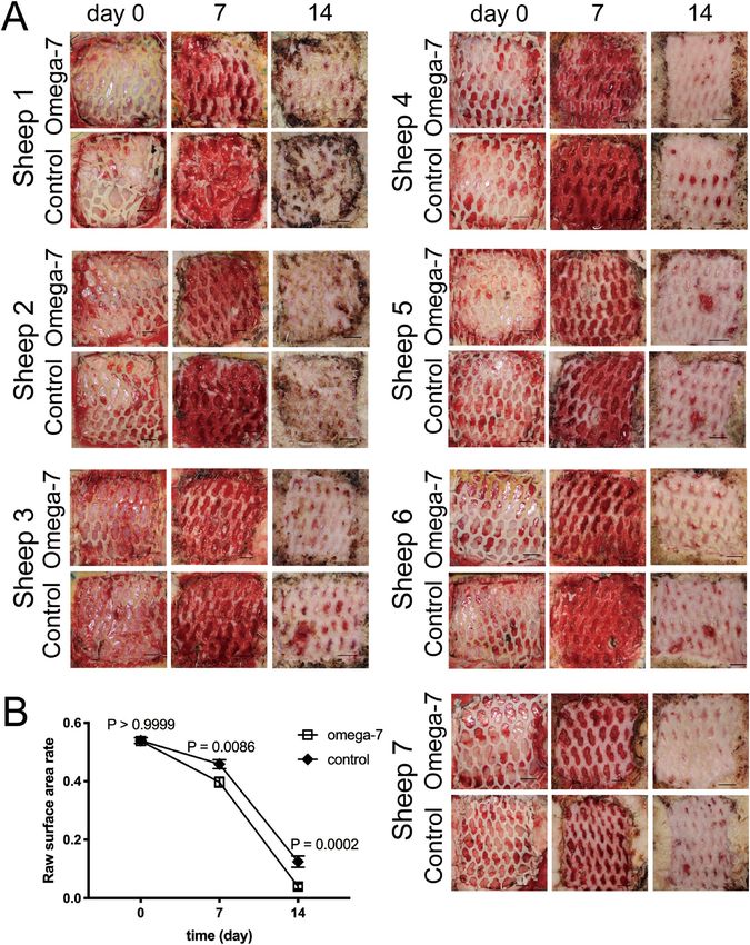
sites were comparable in the Omega-7 (0.54 ± 0.01) and control (0.54 ± 0.01) groups (P > 0.999). At post opera-
tion days (POD) 7 and 14, the size of the un-epithelialized raw surface area in the Omega-7 group (0.40 ± 0.02,
and 0.04 ± 0.01, respectively) was significantly less than those of the control group (0.46 ± 0.02 [P = 0.0086] and
0.13 ± 0.02 [P = 0.0002], respectively) (Fig. 3).
Donor sites had no complications such as infection, bleeding or ulceration. The complete epithelialization
time in donor sites treated with Omega-7 (9.1 ± 1.0 days) was significantly shorter than of the control group
(12.6 ± 0.5 days, P = 0.021) (Fig. 4A).
Re‑epithelialized epidermal thickness of donor sites in vivo. The re-epithelialized epidermal thick-
ness of donor sites in Omega-7 group (111.4 ± 7.4 μm) was slightly higher than in control (91.2 ± 6.0 μm) group
at POD14; however, no significant difference was found between the groups (P = 0.095, Fig. 4B).
Blood flow of grafted and donor sites in vivo. Table 1 illustrates the wound blood flow of each indi-
vidual sheep. In summary, the blood flow, in the grafted sites, was significantly higher in the Omega-7 group
at POD7 and POD14 (190.0 ± 16.9, and 137.2 ± 8.0 Perfusion Units [PU], respectively) than that of the con-
trol group (153.3 ± 21.2, and 100.2 ± 8.9 PU, respectively) (P = 0.047) (Fig. 5, left panel). In the donor sites, the
blood flow in the Omega-7 group at POD7 (398.0 ± 43.0 PU) was significantly greater than that of the control
group (303.3 ± 43.5 PU) (P = 0.034). However, the blood flow at POD14 was comparable between the Omega-7
(184.1 ± 14.5 PU) and control (143.6 ± 17.7 PU) groups (Fig. 5, right panel).
Number of blood vessels in grafted skin burn wounds at POD14 in vivo. Raw data on number of
blood vessels in each individual sheep are shown in Table 2. In summary, the number of wound blood vessels
semi-quantified using MMP-2 staining was significantly higher in the Omega-7 group (80.1 ± 6.4) than those in
the control group (55.3 ± 3.9, P = 0.007) (Fig. 6).
KGF, 3‑nitrotyrosine, and telomerase activity levels in grafted skin tissue at POD14. The level
of KGF in skin tissue was significantly higher in the treatment group compared to the control group (0.31 ± 0.07
vs. 0.21 ± 0.07 relative unit, P = 0.038) (Fig. 7A). The level of 3-nitrotyrosine in skin tissue was significantly
reduced in the treatment group compared to the control group (0.36 ± 0.05 vs. 0.61 ± 0.11 relative unit, P = 0.035)
(Fig. 7B). The telomerase activity was significantly higher in Omega-7-treated grafted burn sites than in control
sites (47.1 ± 9.2 vs. 9.0 ± 0.9, P = 0.007) (Fig. 7C).
Scientific Reports | (2021) 11:975 | https://doi.org/10.1038/s41598-020-79597-0 2
Vol:.(1234567890)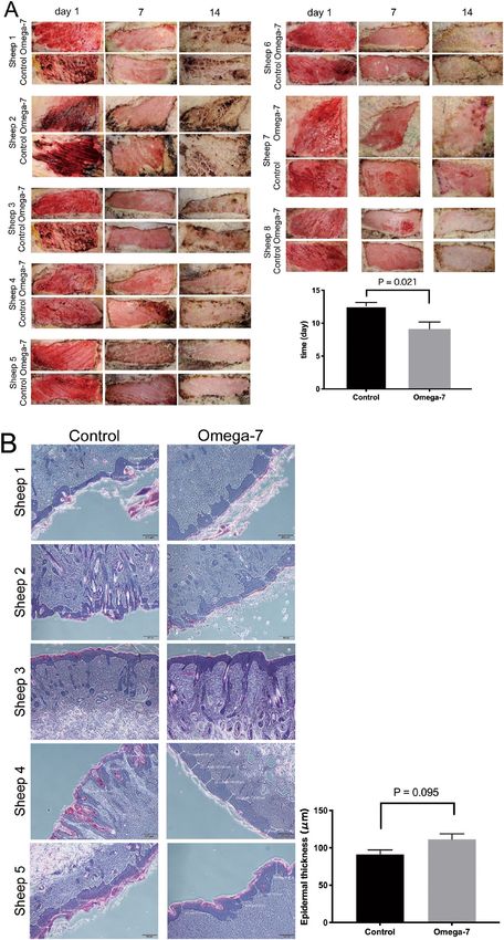
www.nature.com/scientificreports/
Figure 1. Effects of Omega-7 on the cultured keratinocyte colony-forming rate. (A) Effects of 0.025% omega-7
on cultured keratinocyte colony-forming rate (proliferation) at 72 and 96 h. (B) Effects of 0.08% of Omega-7
on cultured keratinocyte colony-forming rate at 48 h. Omega-7 with dose of 0.08% negatively affected the cells.
Keratinocyte colony counting assay was performed using 24,000 cells/cm2 sheep primary keratinocytes in
passage 2. Cells were treated with Omega-7 at time 0 h. The colonies were counted in 12 dishes (6 treated and
6 controls). Data are expressed as mean ± SEM. Scale bar in micrographs: 200 μm. Open circles indicate cells
treated with Omega-7 and closed circles represent control cells. Photographs taken and compiled, by authors,
in Adobe Photoshop CC 2020 (https://www.adobe.com/jp/products/photoshop.html) without changing the
content of images themselves.
Discussion
In the present study, we have tested the efficacy of Omega-7 unsaturated oils isolated from natural Sea buckthorn
pulp oil in a clinical relevant ovine model of grafted burn wound healing.
The key findings of the present study are as follows: (1) Topical application of Omega-7 increased blood flow
as well as the number of blood vessels and accelerated wound healing; (2) Omega-7 increased telomerase activity
in grafted burn wounds in vivo; and (3) Omega-7 promoted keratinocyte proliferation (colony-forming rate)
and migration (scratch assay) in vitro models of wound healing.
The importance of neovascularization for wound healing is well k nown26. Treatment with Omega-7 signifi-
cantly increased the number of blood vessels and blood flow in the wound beds. This is consistent with findings
by Upadhyay et al. who reported that sea buckthorn (SBT) oil containing abundant Omega-7 increased MMP-2
and MMP-9, which are known as important factors for n eovascularization4 in rat burn wound m odels24. We
have previously shown that SBT oil increases blood flow in ovine grafted burn w ounds21, which was supported
odels27.
by findings by Seven et al. in rat burn m
To understand mechanisms of increased blood flow as well as neovascularization, we have measured KGF,
also known as fibroblast growth factor (FGF)-7 and found it significantly increased by treatment with Omega-7.
Scientific Reports | (2021) 11:975 | https://doi.org/10.1038/s41598-020-79597-0 3
Vol.:(0123456789)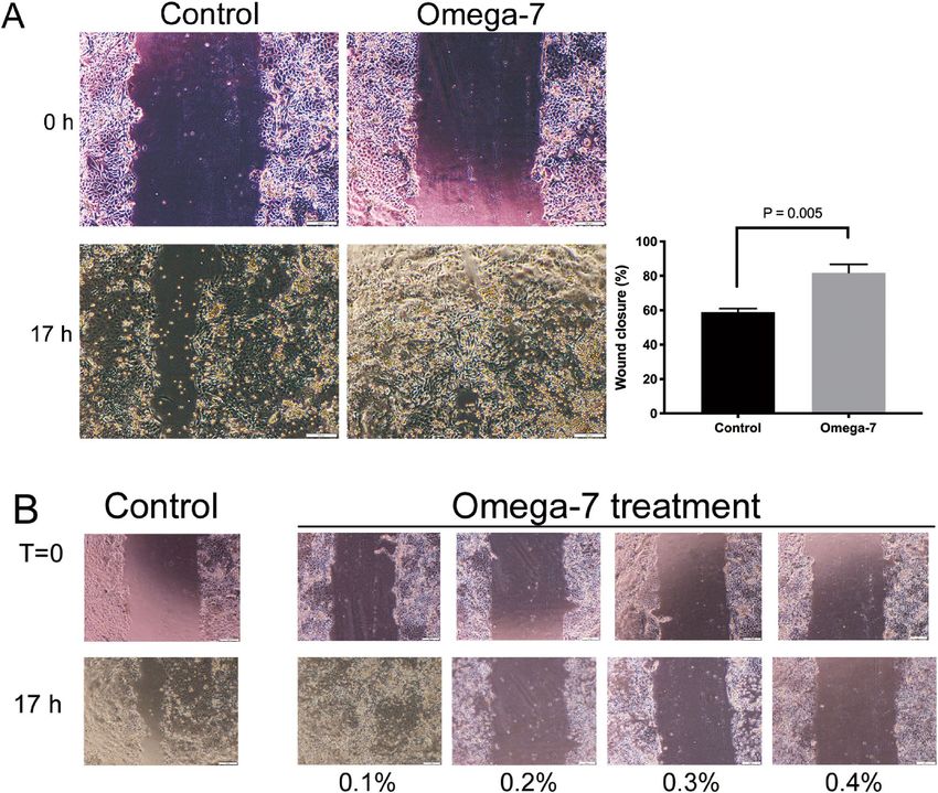
www.nature.com/scientificreports/
Figure 2. Cultured keratinocyte migration assay. (A) Effects of 0.08% Omega-7 on the cultured keratinocyte
migration rate 17 h after the scratch. (B) Effects of Omega-7 (0.1, 0.2, 0.3, and 0.4%) on cultured keratinocyte
migration rate. The scratch was performed when the cells were grown confluently. The Omega-7 was added to
the confluently grown cells immediately after the scratch. Omega-7 had a negative effect on the keratinocyte
migration with dose greater than 0.1%. The cells were assessed in 12 dishes (6 treated and 6 controls). Data are
expressed as mean ± SEM. Scale bar in micrographs: 200 μm. Photographs taken and compiled, by authors,
in Adobe Photoshop CC 2020 (https://www.adobe.com/jp/products/photoshop.html) without changing the
content of images themselves.
Niu et al. reported that KGF is important during the later stages of neovascularization when luminal spaces and
basement membranes are being developed. KGF is known as a potent mitogen for vascular endothelial cells,
and stimulates endothelial cells to produce a protease of urokinase-type plasminogen activator, which acts
on neovascularization28. In our present study, although the direct link remains unknown, Omega-7 may have
increased blood flow via increasing KGF.
On the other hand, cell proliferation and migration is important for wound re-epithelialization. Our in vitro
study results show that Omega-7 treatment accelerated keratinocyte colony-forming rate and improved their
migration. Various cytokines have been reported to promote keratinocyte m igration29. Among them, KGF plays
an important role for re-epithelialization by stimulating proliferation and migration of k eratinocytes29. As men-
tioned, the topical application of Omega-7 increased wound KGF, suggesting that Omega-7 may have also
increased keratinocyte proliferation and migration, and accelerated wound re-epithelialization via increasing
KGF.
In the present study, we also demonstrated that topical application of Omega-7 upregulated telomerase activ-
ity in grafted wound keratinocytes. Counter et al. reported that the epidermis telomere length, after cultured
keratinocytes sheet grafting in burn patients, is shortened compared to those of normal epidermis in the same
patient30. This may be one of the mechanisms leading to long term complications such as graft fragility or blis-
ter formation. Buckingham et al. indicated that telomerase is activated in the epidermis as it is needed for cell
Scientific Reports | (2021) 11:975 | https://doi.org/10.1038/s41598-020-79597-0 4
Vol:.(1234567890)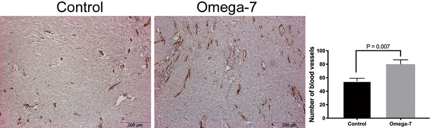
www.nature.com/scientificreports/
Figure 3. Effects of Omega-7 on ovine grafted burn wound healing. (A) Pictures of control and Omega-7-
treated wounds in individual sheep at 0, 7 and 14 days. (B) Wound un-epithelialized surface area was measured
by ultrasound and semi-quantified by Image J software. The raw surface area was measured in seven sheep.
Each sheep had two treated and two untreated sites. Open circles indicate raw surface area in Omega-7, whereas
closed circles indicate raw surface area in control sites. Data are expressed as mean ± SEM. Photographs taken
and compiled, by authors, in Adobe Photoshop CC 2020 (https://www.adobe.com/jp/products/photoshop.html)
without changing the content of images themselves.
Scientific Reports | (2021) 11:975 | https://doi.org/10.1038/s41598-020-79597-0 5
Vol.:(0123456789)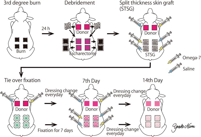
www.nature.com/scientificreports/
Figure 4. Day of complete epithelialization and epithelial thickness of donor sites. (A) The complete epithelialization of the donor
sites was assessed by ultrasound examination. The assay was performed in eight sheep (8 treated and 8 control sites). The graph shows
the complete epithelialization time (days) in donor sites. (B) In five sheep, the re-epithelialized epidermal thickness of donor sites was
measured in five-places in each wound site. Data are expressed as mean ± SEM. Scale bar in the macroscopic image (A) is 5 mm and in
the micrograph (B) is 200 μm. Photographs taken and compiled, by authors, in Adobe Photoshop CC 2020 (https://www.adobe.com/
jp/products/photoshop.html) without changing the content of images themselves.
Scientific Reports | (2021) 11:975 | https://doi.org/10.1038/s41598-020-79597-0 6
Vol:.(1234567890)www.nature.com/scientificreports/
Sheep # 1 2 3 4 5 6 7
Grafted site 7 days
Control 96.25 ± 15.75 219 ± 47 151.5 ± 31.5 89.5 ± 23.5 210 ± 37
Omega7 148.5 ± 17.5 128.5 ± 33.5 207.5 ± 65.5 239.5 ± 25.5 169.5 ± 1.5 246.5 ± 22.5
Grafted site; 14 days
Control 71 ± 2.0 107.5 ± 11.5 85.5 ± 4.5 142 ± 20 125 ± 16 70 ± 1.0
Omega7 115 ± 2.0 145.5 ± 24.5 160 ± 6.0 155.5 ± 21.5 149.5 ± 1.5 97.5 ± 0.5
Donor site; 7 days
Control 162 372 202 296 446 342
Omega7 344 563 383 292 320 486
Donor site; 14 days
Control 81 86 166 140 181 207 144
Omega7 121 176 228 198 212 207 147
Table 1. Blood flow of grafted burn and donor sites at 7 and 14 days. Data are shown as mean ± SEM.
Figure 5. Blood flow of grafted burn and donor sites at 7 and 14 days. Blood flow was measured in six sheep
(12 treated and 12 control sites) at grafted sites, and in seven sheep (7 treated and 7 control sites) at donor sites.
Left panel: Blood flow in grafted burn wound sites. Right panel: Blood flow in donor sites. Data are expressed as
mean ± SEM.
Sheep 1 Sheep 2 Sheep 3 Sheep 4 Sheep 5 Sheep 6 Sheep 7
Control 49 ± 1.6 74 ± 7.9 54 ± 5.6 57 ± 13.6 61.4 ± 1.0 43 ± 8.5 48 ± 7.1
Omega7 66 ± 8.5 98.8 ± 11.8 70.4 ± 5.3 105.2 ± 8.5 87.8 ± 8.3 63.6 ± 9.1 70.4 ± 8.0
Table 2. Number of blood vessels in grafted wounds at 14 days. Data are shown as mean ± SEM.
proliferation and damage repair31. Taken together, the results of the present and previous studies suggest that the
topical application of Omega-7 may have promoted keratinocyte proliferation via increasing telomerase activity
and accelerating wound re-epithelialization.
It should be noted that oxidative damage is a major cause determining the rate of loss of telomeric DNA
and telomere shortening12. Parihar et al. reported increased oxidative stress in burn patients32. It is known that
SBT reduces oxidative stress in blood cells and some organs33,34. In the present study, we report that Omega-7
significantly reduced the wound bed 3-nitrotyrisine. In addition, KGF is known to increase the transcription of
factors involved in the detoxification of oxidative stress35. In brief, previous studies strongly suggest a possible
Scientific Reports | (2021) 11:975 | https://doi.org/10.1038/s41598-020-79597-0 7
Vol.:(0123456789)www.nature.com/scientificreports/
Figure 6. Number of blood vessels in grafted wounds. The number of vessels was counted using MMP-2
staining in grafted wounds at 14 days in eight sheep (8 treated and 8 control sites). Data are expressed as
mean ± SEM. Scale bar in micrographs: 200 μm. Photographs taken and compiled, by authors, in Adobe
Photoshop CC 2020 (https://www.adobe.com/jp/products/photoshop.html) without changing the content of
images themselves.
involvement of reduced oxidative stress, KGF and telomorese activity in burn wound healing. Although, we
were not able report causative effects of those factors, we speculate that Omega-7 may have improved wound
healing via inhibiting oxidative stress and promoting KGF and telomerase activity. Further studies should focus
on interlink between these factors.
Even though the current study shed some valuable insights into the role of Omega-7 in wound healing,
there are a few limitations: (1) We did not directly measure the telomere size; (2) Although we have shown that
Omega-7 attenuates 3-nitrotyrosine levels, precise mechanisms by which Omega-7 increased KGF and telomer-
ase activity remain unknown. Additionally, we did not investigate exact mechanisms of how telomerase activity
promoted keratinocyte proliferation and migration. As mentioned, the present study did not show a direct link
between telomere size and telomerase activity. Sarin et al. showed that telomerase promoted cell proliferation
of quiescent, resting multipotent stem cells in the hair follicle bulge region through a non-canonical p athway36.
Thus, we do not exclude the possibility that Omega-7-induced telomerase activity may have promoted cell
proliferation and migration by multiple mechanisms; (3) No studies have been performed to explore a possible
direct link between KGF and telomerase activity; (4) Because of limited availability of the reagents/kits, KGF and
telomerase activity have not been measured in all 8 sheep; (5) In the present study, we were not able to measure
half-life time of Omega-7 (previously, it was shown to be 9 h 37). We have also not performed its inhibition assay.
Further studies are warranted to investigate these underlying mechanistic aspects, and perform cytotoxicity
assays to reveal possible side effects; and (6) Finally, present study did not consider possible impact of wound
dressing on the healing process. Previous studies reported potential impacts of various wound d ressing38–41.
Zhao et al. demonstrated that nanocomposite cryogels as injectable shape memory hemostatic dressings not only
promoted the wound healing process but also exerted hemostatic effects compared to the gauze dressing38. El
Fawal e al. reported antimicrobial activities of novel wound dressing with hydroxyethyl cellulose-based hydrogel
membranes39.
Nevertheless, taken together, the results of present and previous studies support our notion that Omega-7
significantly increases the wound bed cell telomerase activity and KGF, and accelerates grafted burn wound heal-
ing, possibly by inhibiting burn-induced oxidative stress. Sheep are frequently used to mimic clinical scenarios
of human disease and treatment because the anatomy of their organs (e.g. skin and subcutaneous tissue, lung,
and nerves) are similar to those in h umans42–47. Therefore, we believe that our findings in this study are highly
translational and capable of being imported into clinical practice, as the clinically relevant ovine model resembles
all aspects of wound healing in burn patients, i.e., escharectomy within 24 h after burn, pressure bandage coverage
of grafted wounds for a week, daily wound washing and changing of dressings, daily wound closure assessment,
and continuous hemodynamic monitoring and resuscitation in an ICU setting.
Conclusions
Topical application of Omega-7 accelerates healing of grated burn wounds in ovine model. Further studies are
warranted to explore mechanistic aspects underlying its salutary effects.
Materials and methods
Preparation of Omega‑7. Omega-7 was isolated from sea buckthorn pulp oil (Polyvit Co., Ltd., Gan-
gar Holding, Ulaanbaatar, Mongolia) using Florisil column chromatography method. Then, it was dissolved in
100% ethanol and 0.1% v/v DMSO. The pH was adjusted to 5.22 with NaOH. For in vitro studies, concentra-
tions of 0.025% and 0.08% were used for colony counting and migration assays, respectively. These doses were
chosen based on initial toxicity studies. For colony counting, we have evaluated effects of Omega-7 in concen-
tration ranges of 0.025% and 0.08% and 0.025% was chosen as an optimal dose because the colonies were not
formed with dose of 0.08% (Fig. 1B). For the migration assay, we assessed concentration ranges of 0.08% to 0.4%
Scientific Reports | (2021) 11:975 | https://doi.org/10.1038/s41598-020-79597-0 8
Vol:.(1234567890)www.nature.com/scientificreports/
Figure 7. Keratinocyte growth factor (KGF), nitrotyrosine, and telomerase activity in grafted burn wounds at
14 days. (A) KGF were measured in four sheep (4 treated and 4 control sites). The level of KGF in skin tissue
was significantly higher in the treatment group compared to the control group (0.21 ± 0.07 vs. 0.31 ± 0.07 relative
unit). Full-length blots/gels are presented in Supplementary Fig. 1. (B) Nitrotyrosine were measured in eight
sheep (8 treated and 8 control sites). The level of nitrotyrosine in skin tissue was significantly reduced in the
treatment group compared to the control group (0.36 ± 0.05 vs. 0.61 ± 0.11 relative unit). Full-length blots/gels
are presented in Supplementary Fig. 2. (C) Telomerase activity were measured in five sheep (6 Treated and 6
control sites). Telomerase activity measured by Telomeric Repeat Amplification Protocol (TRAP) and Gel-Based
Telomerase Detection at 14 days after skin graft. The telomerase activity was significantly higher in Omega-7
grafted burn sites than that of control sites (47.1 ± 9.2 vs. 9.0 ± 0.9). The pictures show the telomerase activity
of non-heat-treated samples in each group. Full-length blots/gels are presented in Supplementary Fig. 3. S-IC:
signal from the internal standard in non-heat-treated samples. Data is expressed as mean ± SEM. Photographs
taken and compiled, by authors, in Adobe Photoshop CC 2020 (https://www.adobe.com/jp/products/photoshop.
html) without changing the content of images themselves.
and found that 0.08% was the most effective dose. Concentrations exceeding 0.1% negatively affected the cells
(Fig. 2B). For in vivo studies, 4% Omega-7 diluted in 0.9% normal saline (0.1 mL/cm2) was used.
Isolation and culture of ovine keratinocytes. Ovine hair was removed by using 3-min hair removal
cream. Skin was thoroughly washed before the harvesting. Then, the epidermis with 0.007 inches thickness was
taken by dermatome and washed twice under sterile conditions with PBS 1X containing 200U/ml penicillin and
200 μg/ml streptomycin (Gibco by Life Technologies) and trimmed into 0.2 × 0.2 cm pieces. Then, it was digested
in 0.25% trypsin-2.21 mM EDTA (Corning) solution at 37 °C and 15×g for 30 min in a processing unit from
Ingeneron Incorporated. The trypsinized tissue was strongly shacked and left rest for few minutes. The super-
natant was transferred to another tube (KC1) through a cell strainer of 70 µm where the digestion was stopped
by addition of equal volume of DMEM high-glucose medium (Gibco by Life Technologies) that contained 10%
fetal bovine serum (FBS) (Corning), 100U/ml of penicillin and 100 μg/ml streptomycin, relative to the volume
of trypsin. Then, keratinocyte isolation tube was filled up with 0.25% trypsin-2.21 mM EDTA (Corning) solu-
tion and returned to the processing unit for another 30 min. The suspension (KC1) was centrifuged at 200×g
for 5 min, the supernatant was discarded, and the cell pellet was re-suspended in complete keratinocyte growth
medium. The cells were kept on the incubator (5% CO2) at 37˚C until the tissue processing is completed.
Scientific Reports | (2021) 11:975 | https://doi.org/10.1038/s41598-020-79597-0 9
Vol.:(0123456789)www.nature.com/scientificreports/
Keratinocyte colony counting (proliferation) assay. Keratinocytes in passage 2 were seeded (24,000
cells/cm2) in either complete growth medium containing 0.1% DMSO (control group); or complete growth
medium with 0.025% Omega-7 dissolved in 0.1% DMSO (treatment group) onto a fresh feeder layer of mitomy-
cin C-treated 3T3 fi broblasts48 that had been prepared 24 h earlier at the same cell density as the keratinocytes in
6 well plates. The cells were cultured for 96 h at the same conditions described above. The number of colonies in
5 fields per well were counted at 72 and 96 h after treatment.
Keratinocyte migration (scratch) assay. Confluent monolayers of ovine keratinocytes in passage 2 were
scratched using a 100 μl pipet tip on a 6 well plate. Keratinocyte medium containing 0.1% DMSO with (Omega-7
group; n = 4) or without (Control group; n = 4) 0.08% Omega-7 were added to the plate. Just after scratch (t = 0)
and 17 h after scratch (t = 17), 5 to 8 digital photographs per well were taken. The wound area at each time point
(i.e., At=0 and A
t=17) was calculated using ImageJ software version 1.50 (National Institutes of Health, Bethesda,
MD). The wound closure percentage was calculated by the following equation:
At = 0 − At = 17
Percent of wound closure (%) = × 100
At = 0
Animal care. The Institutional Animal Care and Use Committee (IACUC) at the University of Texas Medical
Branch approved this study, including the number of animals used. The guidelines of the National Institutes of
Health for the care and use of experimental animals were carefully followed. Animals were individually housed
in metabolic cages. Burn injury and skin grafting procedures were performed under deep isoflurane anesthesia
and buprenorphine analgesia. After these procedures, sheep were studied in a conscious state for 14 days. Post-
surgical analgesia was provided by long acting buprenorphine.
Surgical preparation and experimental protocol. Eight female Merino sheep (31.5 ± 4.5 kg, approxi-
mately 3 years old; Talley Ranch, Bastrop, TX) were used in this study. The animals were chronically instru-
mented for hemodynamic monitoring with a right femoral artery catheter and a 7-French Swan-Ganz thermodi-
lution catheter under aseptic conditions. Two full-thickness flame burn sites of 25 cm2 were made on both sides
of dorsum with 2 cm distance between sites and 3 cm apart from the spine under anesthesia and analgesia. For
the induction of burn sites, we have used a metal sheet with 25 cm2 open space in the middle. Underneath the
metal sheet, a burn resistant cloth was instilled to prevent the heat to the adjacent tissues. The burn was induced
by fire flame, which was applied approximately 3 s until the skin shrinkage stops. The method was previously
established in our laboratory that the full-thickness skin burn was confirmed by the histological a nalysis49.
Twenty-four hours after the burn, the eschar was excised down to the fascia. Split-thickness skin grafts 0.03
inches thick were harvested from sites remote from the burned sites to be meshed and grafted to the wounds.
0.1 ml/cm2 of 4% Omega-7 in 0.9% saline was topically applied to one of two randomly selected autografted sites
(Omega-7 group). The remaining site received the same amount of 0.9% saline as a control (control group). The
donor sites were also randomly allocated to 0.1 ml/cm2 of 4% Omega-7 or 0.9% saline. After the initial treatment,
the grafted sites were covered with tie-over dressings removed at postoperative day (POD) 7. At the 7 days after
the grafting, the wounds were gently washed daily with a sterile saline without anesthesia. Thereafter, the wounds
were treated with Omega-7 or 0.9% saline, and covered with polyurethane foam for 7 days (total duration of the
study is 14 days) (Fig. 8). No wound debridement was done.
The donor site wounds were covered with polyurethane film dressing (Tegaderm, 3 M, MN) and treated
daily with 4.0% Omega-7 or 0.9% saline starting at POD1. Fourteen days after surgery, animals were deeply
anesthetized and euthanized by intravenous administration of xylazine (3.0 mg/kg), ketamine (40 mg/kg), and
buprenorphine (0.01 mg/kg) following IACUC approved protocols and American Veterinary Medical Associa-
tion Guidelines for Euthanasia.
Planimetric wound healing assessments. After the initial autografting and during each dressing
change, standardized digital photographs of the wound fields were taken. A calibrated benchmark was posi-
tioned adjacent to each wound when photographs were taken. Photographs were processed using ImageJ. In
grafted sites, for each wound at POD 0, 7, and 14, the total wound area ( AT) and open mesh interstices of auto-
grafts (un-epithelialized raw surface area, AU) were measured. The proportion of the total wound surface area
that was un-epithelialized (AR) was calculated by the: AR = AU/AT.
The time required for complete epithelialization of donor sites were also determined. Un-epithelialized area
was identified by high resolution ultrasound and semiquantified using image J as described in our previous
study21.
Blood flow measurement in grafted and donor sites. The blood flow was measured in both treated
and untreated autograft and donor sites of six sheep using a LASER Doppler device (PeriFlux System 5000 Mod-
elPF5001, Perimed AB, Sweden) at POD 7 and 14. Two locations were randomly chosen in each site for blood
flow measurements and the means were analyzed.
Histology. Wound biopsies were taken at two different locations from each donor site. Four-micrometer-
thick cross-sections through the wound center were stained with hematoxylin and eosin (HE) according to
standard histologic procedures. Images were acquired with an Olympus CKX41 microscope. The thickness of
the epithelial layer of the donor site in each group was measured using software (CellSens Standard 1.11, Olym-
Scientific Reports | (2021) 11:975 | https://doi.org/10.1038/s41598-020-79597-0 10
Vol:.(1234567890)www.nature.com/scientificreports/
Figure 8. Experimental design of clinically relevant ovine grafted burn wound healing model. The third-degree
burn was induced by fire flame (above left). 24 h after the burn, the eschar was excised down to the fascia. Split-
thickness skin grafts 0.03 inches thick were harvested from sites remote from the burned sites to be meshed and
grafted to the wounds (above center). 0.1 ml/cm2 of 4% Omega-7 in 0.9% saline was topically applied to one of
two randomly selected autografted sites (Omega-7 group). The remaining site received the same amount of 0.9%
saline as a control (control group). The donor sites were also randomly allocated to 0.1 ml/cm2 of 4% Omega-7
or 0.9% saline (above right). After the initial treatment, the grafted sites were covered with tie-over dressings
removed at postoperative day (POD) 7 (below left). At the 7 days after the grafting, the wounds were gently
washed daily with a sterile saline without anesthesia (below center). Thereafter, the wounds were treated with
Omega-7 or 0.9% saline, and covered with polyurethane foam for 7 days (14 days total duration of experiments).
The donor site wounds were covered with polyurethane film dressing and treated daily with 4.0% Omega-7 or
0.9% saline starting at POD1 for 14 days (below right). The study design was drawn using Adobe illustrator by
the first author (https://www.adobe.com/jp/products/illustrator.html).
pus, Tokyo). In the images taken at a magnification of 400, five measurements per field (total four fields per
sheep) were acquired by an independent masked pathologist.
Quantification of blood vessels. Immunolocalization of matrix metallopeptidase 2 (MMP-2), as a well-
known marker for the angiogenesis50–52, was determined for semi-quantification of blood vessels using 1:200
diluted monoclonal antibody against human MMP-2 antigen (MS-806-P1, Thermo Fisher Scientific, MI) follow-
ing previously established protocol53. Five images per slide were taken at a magnification of 100, and the number
of MMP-2-stained vessels in each area was counted by a masked pathologist using Image J.
Western blot analysis. We measured 3-nitrotyrosine and keratinocyte growth factors (KGF) in grafted
skin tissue at POD14 in each group. The 3-nitrotyrosine was measured using an automated capillary West-
ern blot analyzer (Wes, ProteinSimple, CA) system (n = 8). KGF was measured by a conventional Western blot
method (n = 4). The skin tissue was homogenized, lysed, and the protein levels were measured using anti-3-ni-
trotyrosine antibody (06-284; MilliporeSigma, MA) and anti-KGF antibody (ab131162; Abcam, MA).
Telomerase activity. Telomerase activity was measured in grafted skin tissue at POD14 in each group.
Skin tissue (30 mg) was homogenized as described by Smith et al.54. All sample extracts were evaluated for heat
sensitivity by incubation at 85 °C for 30 min. The telomeric repeat amplification protocol (TRAP) assay was
performed according to the TRAPeze kit (S7700, EMD Millipore, MA, USA). 100 ng/μl of each sample and 2
Scientific Reports | (2021) 11:975 | https://doi.org/10.1038/s41598-020-79597-0 11
Vol.:(0123456789)www.nature.com/scientificreports/
units of DNA Polymerase (Nova Taq, Millipore-Novagen) were used per reaction. The following parameters
were measured: (1) primer-dimer/PCR contamination control (r0); (2) telomerase quantitation control template
(r); (3) the signal of the region of the gel corresponding to the TRAP product ladder bands in non-heat-treated
( x ); (4) heat-treated sample extracts ( x0); (5) the signal from the internal standard in non-heat-treated samples
(c); and (6) the telomerase quantitation control template (cR ). The total product generated (TPG) was calculated
by the following equation:
(x − x0)/c
TPG = × 100
(r − r0)/cR
Statistical analysis. Values are expressed as means ± SEM. The un-epithelialized area and blood flow were
modeled with relation to the treatment group and time by mixed effects ANOVA. Differences between groups at
each time point were estimated by Hommel-adjusted contrasts using R statistical s oftware55. Other analyses were
performed by paired t-test with GraphPad Prism version 7.0c for Mac OS X (GraphPad Software, CA). The sig-
nificance level was set at P < 0.05.
Received: 19 February 2020; Accepted: 10 December 2020
References
1. American Burn Association. Burn Incidence and Treatment in the United States: 2016. http://ameriburn.org/resources_factsheet.
php (2016).
2. American Burn Association. National Burn Repository 2016 Report Dataset version 12.0.
3. Wendt, J. R., Ulich, T. & Rao, P. N. Long-term survival of human skin allografts in patients with immunosuppression. Plast. Reconstr.
Surg. 113, 1347–1354 (2004).
4. Singer, A. J. & Clark, R. A. Cutaneous wound healing. N. Engl. J. Med. 341, 738–746. https://doi.org/10.1056/NEJM19990902341
1006 (1999).
5. Gosain, A. & DiPietro, L. A. Aging and wound healing. World J. Surg. 28, 321–326. https://doi.org/10.1007/s00268-003-7397-6
(2004).
6. Masutomi, K. et al. Telomerase maintains telomere structure in normal human cells. Cell 114, 241–253. https://doi.org/10.1016/
S0092-8674(03)00550-6 (2003).
7. Armanios, M. Syndromes of telomere shortening. Annu. Rev. Genom. Hum. Genet. 10, 45–61. https://doi.org/10.1146/annurev-
genom-082908-150046 (2009).
8. Harley, C. B., Futcher, A. B. & Greider, C. W. Telomeres shorten during ageing of human fibroblasts. Nature 345, 458–460. https
://doi.org/10.1038/345458a0 (1990).
9. Wieczor, M. & Czub, J. Telomere uncapping by common oxidative guanine lesions: Insights from atomistic models. Free Radic.
Biol. Med. 148, 162–169. https://doi.org/10.1016/j.freeradbiomed.2020.01.006 (2020).
10. Tamer, T. M. et al. Chitosan/hyaluronan/edaravone membranes for anti-inflammatory wound dressing: In vitro and in vivo evalu-
ation studies. Mater. Sci. Eng. C. 90, 227–235. https://doi.org/10.1016/j.msec.2018.04.053 (2018).
11. Greider, C. W. Telomere length regulation. Annu. Rev. Biochem. 65, 337–365. https://doi.org/10.1146/annurev.bi.65.070196.00200
5 (1996).
12. von Zglinicki, T. Oxidative stress shortens telomeres. Trends Biochem. Sci. 27, 339–344 (2002).
13. Branski, L. K., Herndon, D. N. & Barrow, R. E. Total Burn Care 5th edn, 1–7 (Elsevier, Amsterdam, 2018).
14. Heimbach, D. et al. Artificial dermis for major burns. A multi-center randomized clinical trial. Ann. Surg. 208, 313–320 (1988).
15. Barret, J. P., Wolf, S. E., Desai, M. H. & Herndon, D. N. Cost-efficacy of cultured epidermal autografts in massive pediatric burns.
Ann. Surg. 231, 869–876 (2000).
16. Lewis, C. J. Stem cell application in acute burn care and reconstruction. J. Wound Care 22, 7–8. https://doi.org/10.12968/
jowc.2013.22.1.7 (2013).
17. Takahashi, K. et al. Induction of pluripotent stem cells from adult human fibroblasts by defined factors. Cell 131, 861–872. https
://doi.org/10.1016/j.cell.2007.11.019 (2007).
18. Lee, K. B. et al. Topical embryonic stem cells enhance wound healing in diabetic rats. J. Orthop. Res. 29, 1554–1562. https://doi.
org/10.1002/jor.21385(2011).
19. Burusapat, C., Supawan, M., Pruksapong, C., Pitiseree, A. & Suwantemee, C. Topical aloe vera gel for accelerated wound healing
of split-thickness skin graft donor sites: A double-blind, randomized, controlled trial and systematic review. Plast. Reconst. Surg.
142, 217–226. https://doi.org/10.1097/PRS.0000000000004515 (2018).
20. Bahramsoltani, R., Farzaei, M. H. & Rahimi, R. Medicinal plants and their natural components as future drugs for the treatment
of burn wounds: an integrative review. Arch. Dermatol. Res. 306, 601–617. https://doi.org/10.1007/s00403-014-1474-6 (2014).
21. Ito, H. et al. Healing efficacy of sea buckthorn (Hippophae rhamnoides L.) seed oil in an ovine burn wound model. Burns 40,
511–519. https://doi.org/10.1016/j.burns.2013.08.011 (2014).
22. Rousi, A. The genus Hippophaë L. A taxonomic study. Ann. Bot. Fenn. 8, 177–227 (1971).
23. Suleyman, H. et al. Beneficial effects of Hippophae rhamnoides L. on nicotine induced oxidative stress in rat blood compared with
vitamin E. Biol. Pharm. Bull. 25, 1133–1136 (2002).
24. Upadhyay, N. K., Kumar, R., Siddiqui, M. S. & Gupta, A. Mechanism of wound-healing activity of Hippophae rhamnoides L. Leaf
extract in experimental burns. Evid. Based Complement. Alternat. Med. 2011, 659705. https: //doi.org/10.1093/ecam/nep189 (2011).
25. Upadhyay, N. K. et al. Safety and healing efficacy of Sea buckthorn (Hippophae rhamnoides L.) seed oil on burn wounds in rats.
Food. Chem. Toxicol. 47, 1146–1153. https://doi.org/10.1016/j.fct.2009.02.002 (2009).
26. Guo, S. & Dipietro, L. A. Factors affecting wound healing. J. Dent. Res. 89, 219–229. https://doi.org/10.1177/0022034509359125
(2010).
27. Seven, B. et al. Hippophae rhamnoides L. and dexpanthenol-bepanthene on blood flow after experimental skin burns in rats using
Xe-133 clearance technique. Hellenic J. Nucl. Med. 12, 55–58 (2009).
28. Niu, J. et al. Keratinocyte growth factor/fibroblast growth factor-7-regulated cell migration and invasion through activation of
NF-kappaB transcription factors. J. Biol. Chem. 282, 6001 (2007).
Scientific Reports | (2021) 11:975 | https://doi.org/10.1038/s41598-020-79597-0 12
Vol:.(1234567890)www.nature.com/scientificreports/
29. Barrientos, S., Stojadinovic, O., Golinko, M. S., Brem, H. & Tomic-Canic, M. Growth factors and cytokines in wound healing.
Wound Repair. Regen. 16, 585–601. https://doi.org/10.1111/j.1524-475X.2008.00410.x (2008).
30. Counter, C. M., Press, W. & Compton, C. C. Telomere shortening in cultured autografts of patients with burns. Lancet 361,
1345–1346. https://doi.org/10.1016/S0140-6736(03)13042-5 (2003).
31. Buckingham, E. M. & Klingelhutz, A. J. The role of telomeres in the ageing of human skin. Exp. Dermatol. 20, 297–302. https://
doi.org/10.1111/j.1600-0625.2010.01242.x (2011).
32. Parihar, A., Parihar, M. S., Milner, S. & Bhat, S. Oxidative stress and anti-oxidative mobilization in burn injury. Burns 34, 6–17.
https://doi.org/10.1016/j.burns.2007.04.009 (2007).
33. Olas, B., Kontek, B., Malinowska, P., Zuchowski, J. & Stochmal, A. Hippophae rhamnoides L. fruits reduce the oxidative stress in
human blood platelets and plasma. Oxid. Med. Cell. Long. 2016, 4692486. https://doi.org/10.1155/2016/4692486 (2016).
34. Cho, C. H. et al. Sea buckthorn (Hippophae rhamnoides L.) leaf extracts protect neuronal PC-12 cells from oxidative stress. J.
Microbiol. Biotechnol. 27, 1257–1265. https://doi.org/10.4014/jmb.1704.04033 (2017).
35. Raja, R., Sivamani, K., Garcia, M. S. & Isseroff, R. R. Wound re-epithelialization: modulating keratinocyte migration in wound
healing. Front. Biosci. 12, 2849–2868 (2007).
36. Sarin, K. Y. et al. Conditional telomerase induction causes proliferation of hair follicle stem cells. Nature 436, 1048–1052. https://
doi.org/10.1038/nature03836 (2005).
37. Pfrang, C., Shiraiwa, M. & Poschl, U. Chemical ageing and transformation of diffusivity in semi-solid multi-component organic
aerosol particles. Atmos. Chem. Phys. 11, 7343–7354 (2011).
38. Zhao, X., Guo, B., Wu, H., Liang, Y. & Ma, P. X. Injectable antibacterial conductive nanocomposite cryogels with rapid shape
recovery for noncompressible hemorrhage and wound healing. Nat. Commun. 9, 2784. https://doi.org/10.1038/s41467-018-04998
-9 (2018).
39. El Fawal, G. F., Abu-Serie, M. M., Hassan, M. A. & Elnouby, M. S. Hydroxyethyl cellulose hydrogel for wound dressing: Fabrica-
tion, characterization and in vitro evaluation. Int. J. Biol. Macromol. 111, 649–659. https://doi.org/10.1016/j.ijbiomac.2018.01.040
(2018).
40. He, J., Liang, Y., Shi, M. & Guo, B. Anti-oxidant electroactive and antibacterial nanofibrous wound dressings based on poly(ε-
caprolactone)/quaternized chitosan-graft-polyaniline for full-thickness skin wound healing. Chem. Eng. J. 385, 123464. https://
doi.org/10.1016/j.cej.2019.123464 (2020).
41. Tamer, T. M. et al. Enhancement of wound healing by chitosan/hyaluronan polyelectrolyte membrane loaded with glutathione:
in vitro and in vivo evaluations. J. Biotechnol. 310, 103–113. https://doi.org/10.1016/j.jbiotec.2020.02.002 (2020).
42. Enkhbaatar, P. et al. Pathophysiology, research challenges, and clinical management of smoke inhalation injury. Lancet 388,
1437–1446. https://doi.org/10.1016/S0140-6736(16)31458-1 (2016).
43. Niimi, Y. et al. Surgical anatomy of ovine facial and hypoglossal nerves for facial nerve reconstruction and regeneration research:
An experimental study in sheep. Microsurgery https://doi.org/10.1002/micr.30405 (2019).
44. Niimi, Y. et al. Surgical anatomy of the ovine sural nerve for facial nerve regeneration and reconstruction research. Sci. Rep. 9,
10564. https://doi.org/10.1038/s41598-019-46661-3 (2019).
45. Niimi, Y., Fukuda, S., Alharbi, S., Prough, D. S. & Enkhbaatar, P. Polyurethane foam for skin graft fixation in clinical-relevant
ovine burn wound model for wound repair and regeneration research. Regen. Ther. 14, 341–343. https://doi.org/10.1016/j.
reth.2020.04.007 (2020).
46. Fukuda, S. et al. Blood carboxyhemoglobin elimination curve, half-lifetime, and arterial-venous differences in acute phase of
carbon monoxide poisoning in ovine smoke inhalation injury model. Biochem. Biophys. Res. Commun. https://doi.org/10.1016/j.
bbrc.2020.03.048 (2020).
47. Baljinnyam, T. et al. Cutaneous burn diminishes beneficial effect of intravenously administered mesenchymal stem cells on acute
lung injury induced by smoke inhalation in sheep. Burns https://doi.org/10.1016/j.burns.2020.05.012 (2020).
48. Blacker, K. L., Williams, M. L. & Goldyne, M. Mitomycin C-treated 3T3 fibroblasts used as feeder layers for human keratinocyte
culture retain the capacity to generate eicosanoids. J. Invest. Dermatol. 89, 536–539 (1987).
49. Sakurai, H., Nozaki, M., Traber, L. D., Hawkins, H. K. & Traber, D. L. Microvascular changes in large flame burn wound in sheep.
Burns 28, 3–9 (2002).
50. Caley, M. P., Martins, V. L. & O’Toole, E. A. Metalloproteinases and wound healing. Adv. Wound Care 4, 225–234. https://doi.
org/10.1089/wound.2014.0581 (2015).
51. Bergers, G. et al. Matrix metalloproteinase-9 triggers the angiogenic switch during carcinogenesis. Nat. Cell. Biol. 2, 737–744. https
://doi.org/10.1038/35036374 (2000).
52. Ulrich, D., Lichtenegger, F., Unglaub, F., Smeets, R. & Pallua, N. Effect of chronic wound exudates and MMP-2/-9 inhibitor on
angiogenesis in vitro. Plast. Reconstr. Surg. 116, 539–545. https://doi.org/10.1097/01.prs.0000173447.81513.7a (2005).
53. Zhang, B., Yan, L., Tsang, P. C. & Moses, M. A. Matrix metalloproteinase-2 (MMP-2) expression and regulation by tumor necrosis
factor alpha (TNFalpha) in the bovine corpus luteum. Mol. Reprod. Dev. 70, 122–132. https://doi.org/10.1002/mrd.20196 (2005).
54. Smith, P. K. et al. Measurement of protein using bicinchoninic acid. Anal. Biochem. 150, 76–85 (1985).
55. R: A Language and Environment For Statistical Computing (R Foundation for Statistical Computing, Vienna, 2012).
Acknowledgements
The authors thank the staff of the Translational Intensive Care Unit at the University of Texas Medical Branch
for their valuable assistance in conducting these studies, and Ms. Clemmie White-Matthews for her editorial
assistance in preparing this article. This project was supported by Shriners of North America grant no. 84050
(Enkhbaatar, P), Itoe Okamoto Scholarship grant no. 98, and the Japan Society for the Promotion of Science
KAKENHI grant no. 20K18423.
Author contributions
P.E. developed the concept. Y.N., and D.P. designed the study. Y.N., D.P., K.I., S.F., C.R.A., T.B., J.K., and S.A.
performed experiments, and analyzing the data. S.J. performed histological sections. Y.N., D.P., and P.E. wrote
the manuscript. D.S.P. provided expert advice and reviewed the manuscript. All authors approved the final ver-
sion of the manuscript.
Competing interests
The authors declare no competing interests.
Additional information
Supplementary Information The online version contains supplementary material available at https://doi.
org/10.1038/s41598-020-79597-0.
Scientific Reports | (2021) 11:975 | https://doi.org/10.1038/s41598-020-79597-0 13
Vol.:(0123456789)www.nature.com/scientificreports/
Correspondence and requests for materials should be addressed to P.E.
Reprints and permissions information is available at www.nature.com/reprints.
Publisher’s note Springer Nature remains neutral with regard to jurisdictional claims in published maps and
institutional affiliations.
Open Access This article is licensed under a Creative Commons Attribution 4.0 International
License, which permits use, sharing, adaptation, distribution and reproduction in any medium or
format, as long as you give appropriate credit to the original author(s) and the source, provide a link to the
Creative Commons licence, and indicate if changes were made. The images or other third party material in this
article are included in the article’s Creative Commons licence, unless indicated otherwise in a credit line to the
material. If material is not included in the article’s Creative Commons licence and your intended use is not
permitted by statutory regulation or exceeds the permitted use, you will need to obtain permission directly from
the copyright holder. To view a copy of this licence, visit http://creativecommons.org/licenses/by/4.0/.
© The Author(s) 2021
Scientific Reports | (2021) 11:975 | https://doi.org/10.1038/s41598-020-79597-0 14
Vol:.(1234567890)You can also read