Inhibition of Rac1 GTPase Decreases Vascular Oxidative Stress, Improves Endothelial Function, and Attenuates Atherosclerosis Development in Mice
←
→
Page content transcription
If your browser does not render page correctly, please read the page content below
ORIGINAL RESEARCH
                                                                                                                                            published: 06 August 2021
                                                                                                                                      doi: 10.3389/fcvm.2021.680775

                                            Inhibition of Rac1 GTPase Decreases
                                            Vascular Oxidative Stress, Improves
                                            Endothelial Function, and Attenuates
                                            Atherosclerosis Development in Mice
                                            Sebastian Zimmer 1*, Philip Roger Goody 1 , Matthias Oelze 2 , Alexander Ghanem 3 ,
                                            Cornelius F. Mueller 1 , Ulrich Laufs 4 , Andreas Daiber 2 , Felix Jansen 1 , Georg Nickenig 1 and
                                            Sven Wassmann 5,6
                                            1
                                              Department of Internal Medicine II, Heart Center Bonn, University Hospital Bonn, Bonn, Germany, 2 Zentrum für
                                            Kardiologie - Kardiologie I, Universitätsmedizin der Johannes Gutenberg-Universität, Mainz, Germany, 3 Department of
                                            Internal Medicine II - Cardiology and Medical Intensive Care, Asklepius Hospital Nord - Heidberg, Hamburg, Germany,
                                            4
                                              Department of Cardiology, University Hospital Leipzig, Leipzig, Germany, 5 Cardiology Pasing, Munich, Germany,
                                            6
                                              Department of Inernal Medicine III, Saarlang University Medical Center, Homburg, Germany
                           Edited by:
                Masuko Ushio-Fukai,
                                            Aims: Oxidative stress and inflammation contribute to atherogenesis. Rac1 GTPase
     Augusta University, United States
                                            regulates pro-oxidant NADPH oxidase activity, reactive oxygen species (ROS) formation,
                       Reviewed by:
                       Chieko Mineo,        actin cytoskeleton organization and monocyte adhesion. We investigated the vascular
    University of Texas Southwestern        effects of pharmacological inhibition of Rac1 GTPase in mice.
       Medical Center, United States
                          Nhat Tu Le,       Methods and Results: We treated wild-type and apolipoprotein E-deficient (ApoE−/− )
Houston Methodist Research Institute,
                                            mice with Clostridium sordellii lethal toxin (LT), a Rac1 inhibitor, and assessed
                        United States
                                            vascular oxidative stress, expression and activity of involved proteins, endothelial
                 *Correspondence:
                    Sebastian Zimmer        function, macrophage infiltration, and atherosclerosis development. LT-treated wild-type
        sebastian.zimmer@ukbonn.de          mice displayed decreased vascular NADPH oxidase activity and ROS production.
                                            Therapeutic LT doses had no impact on behavior, food intake, body weight, heart
                    Specialty section:
          This article was submitted to     rate, blood pressure, vascular and myocardial function, differential blood count, and
          Atherosclerosis and Vascular      vascular permeability. ApoE−/− mice were fed a cholesterol-rich diet and were
                              Medicine,
                a section of the journal
                                            treated with LT or vehicle. LT treatment led to decreased aortic Rac1 GTPase
  Frontiers in Cardiovascular Medicine      activity, NADPH oxidase activity and ROS production, but had no impact on
           Received: 15 March 2021          expression and membrane translocation of NADPH oxidase subunits and RhoA GTPase
            Accepted: 13 July 2021          activity. LT-treated mice showed improved aortic endothelium-dependent vasodilation,
          Published: 06 August 2021
                                            attenuated atherosclerotic lesion formation and reduced macrophage infiltration of
                             Citation:
       Zimmer S, Goody PR, Oelze M,         atherosclerotic plaques. Concomitant treatment of cholesterol-fed ApoE−/− mice with
      Ghanem A, Mueller CF, Laufs U,        LT, the specific synthetic Rac1 inhibitor NSC 23766 or simvastatin comparably reduced
   Daiber A, Jansen F, Nickenig G and
Wassmann S (2021) Inhibition of Rac1
                                            aortic Rac1 activity, NADPH oxidase activity, oxidative stress, endothelial dysfunction,
GTPase Decreases Vascular Oxidative         atherosclerosis development, and macrophage infiltration.
Stress, Improves Endothelial Function,
       and Attenuates Atherosclerosis       Conclusions: These findings identify an important role of the small GTPase Rac1 in
                Development in Mice.        atherogenesis and provide a potential target for anti-atherosclerotic therapy.
   Front. Cardiovasc. Med. 8:680775.
     doi: 10.3389/fcvm.2021.680775          Keywords: atherosclerosis, endothelial function, oxidative stress, free radicals, Rac1, GTPases

Frontiers in Cardiovascular Medicine | www.frontiersin.org                        1                                           August 2021 | Volume 8 | Article 680775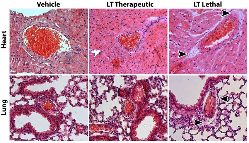
Zimmer et al.                                                                                                               Rac1 in Atherosclerosis

INTRODUCTION                                                               in vascular smooth muscle cells after stimulation of Rac1 by
                                                                           angiotensin II can be completely inhibited by LT (22).
Reactive oxygen species (ROS) are thought to be involved in the               We hypothesized that in vivo inhibition of Rac1 GTPase
pathogenesis of atherosclerosis because pathological conditions            results in reduced NADPH oxidase activity and thus diminished
such as hypertension, hypercholesterolemia and diabetes are                ROS production in the arterial wall, and that chronic Rac1
associated with an increase in vascular ROS production and                 inhibition in a mouse model of atherosclerosis will improve
predispose for cardiovascular events. In fact, increased levels            endothelial function and reduce atherosclerosis development
of ROS have been demonstrated in all layers of the diseased                and macrophage infiltration in atherosclerotic lesions. In the
arterial wall and within atherosclerotic plaques. Furthermore,             present work we have used specific inhibitors of Rac1 activity
there is ample evidence that oxidative stress is directly involved         to investigate the role of the GTPase in atherosclerosis using a
in the pathogenesis of endothelial dysfunction and ultimately              mouse model and conclude that Rac1 inhibition could indeed be
atherosclerosis development by numerous molecular and cellular             an important therapeutic principle.
mechanisms (1–6).
   Under physiological conditions, ROS formation and
elimination are delicately balanced in the vascular wall.                  METHODS
Enhanced activity of pro-oxidant enzymes and/or reduced
                                                                           The Methods section of this manuscript can be found in the
activity of anti-oxidant enzymes, however, lead to oxidative
                                                                           Supplementary Material.
stress. A major source of ROS in vascular cells is NADPH
oxidase and this is recognized to be involved in vascular
pathology (7). This enzyme consists of two membrane-bound                  RESULTS
subunits p22-phox and Nox1 or Nox2, two cytosolic subunits
p67-phox and p47-phox and the small GTPase Rac1. Nox4 is                   Proof of Concept and Dose Finding Study
another subunit that is present in vascular cells (8–11). These            The first experiment was designed as a proof of concept and
subunits assemble on activation to form the functional enzyme,             dose finding study. To determine the highest tolerated dose of LT
which then catalyses an electron transfer to molecular oxygen              with a relevant effect on vascular ROS production, we treated 12-
producing superoxide radicals.                                             week-old wild-type mice with 0.1 or 1.0 µg LT/week continuously
   NADPH oxidase is activated by many important modulators                 via osmotic mini-pumps for 7 days. We then measured vascular
of vascular cell activity (12) including endothelin-1 (11), vascular       ROS production and NADPH oxidase activity. Mice that received
endothelial growth factor (13), transforming growth factor-β               vehicle or 0.1 µg LT/week behaved normally and had a sustained
(14), angiotensin II (15), and mechanical stimuli (16, 17). The            food and water consumption. Treatment with this lower dose of
small GTPase Rac1 plays a central common role in activation                LT resulted in decreased vascular ROS production (100 ± 29% vs.
of NADPH oxidase. It interacts with other subunits and tethers             49 ± 25%, n = 3 per group) and significantly reduced NADPH
the complex of cytosolic subunits to the cell membrane (18,                oxidase activity (100 ± 19% vs. 47 ± 7%, p < 0.05 vs. vehicle, n =
19). Expression of a dominant negative Rac1 allele/mutant in               3 per group) compared to controls. Mice treated with the higher
human aortic endothelial cells and vascular smooth muscle cells,           dose showed no external signs of illness but were apathetic and
respectively, abolishes ROS production by the NADPH oxidase                had a reduced food and water intake. Therefore, 0.1 µg LT/week
(20, 21). Rac1 expression and activity is further stimulated by            was used in all of the following experiments.
angiotensin II, an effect that can be prevented by 3-hydroxy-
3-methylglutaryl coenzyme A reductase inhibitors (e.g., statins)           Short-Term Rac1 Inhibition and Endothelial
that inhibit geranylgeranylation of the GTPase (22). In addition,          Function
ROS production in response to statin withdrawal appears to be              To investigate whether Rac1 GTPase inhibition with LT and
caused by enhanced activation and anchoring of Rac1 to the                 the associated reduction in vascular oxidative stress affects
plasma membrane (23).                                                      endothelial dysfunction in mice, we first studied endothelial
   Rac1 GTPase, a member of the rho family of small GTPases,               function in atherosclerotic mice treated with LT. ApoE−/− mice
also plays an important role in organization of the actin                  were fed a cholesterol-rich, high-fat diet for 7 weeks and were
cytoskeleton. Actin modulates cell shape, cell-cell contacts and           treated with 0.1 µg LT/week or vehicle during the last 7 days of
interaction with adhesion molecules. Cellular motility depends             diet. Wild-type mice that received high-fat, cholesterol-rich diet
on a rapid response to dynamic signals, and ROS have emerged               showed normal vasodilation in intact aortic ring preparations.
as key mediators. All Rho family members, but especially Rac1              ApoE−/− mice developed severely impaired endothelium-
regulate actin cytoskeleton dynamics (24, 25). It has been shown           dependent vasodilation, indicating endothelial dysfunction. Rac1
that Rac1 is involved in monocyte adhesion to the endothelium              GTPase inhibition with LT significantly improved endothelium-
by modulation of the actin cytoskeleton but not ROS release (26).          dependent vasodilation in these ApoE−/− mice (maximal
   Rac1 GTPase can be specifically inhibited by Clostridium                relaxation: wild-type + vehicle 81 ± 7%, ApoE−/− + vehicle 16
sordellii lethal toxin (LT). LT is a glucosyltransferase that              ± 2%, ApoE−/− + LT 48 ± 11%, p < 0.05 vs. ApoE+vehicle, p <
selectively uses UDP-glucose as a co-substrate to glucosylate              0.05 vs. wild-type, n = 5 per group). Endothelium-independent
Rac1 (27). LT effectively inhibits Rac1 in vascular cells. We              vasorelaxation was not affected by LT treatment (maximal
have previously demonstrated that increased ROS production                 relaxation: wild-type + vehicle 122 ± 10%, ApoE−/− + vehicle

Frontiers in Cardiovascular Medicine | www.frontiersin.org             2                                    August 2021 | Volume 8 | Article 680775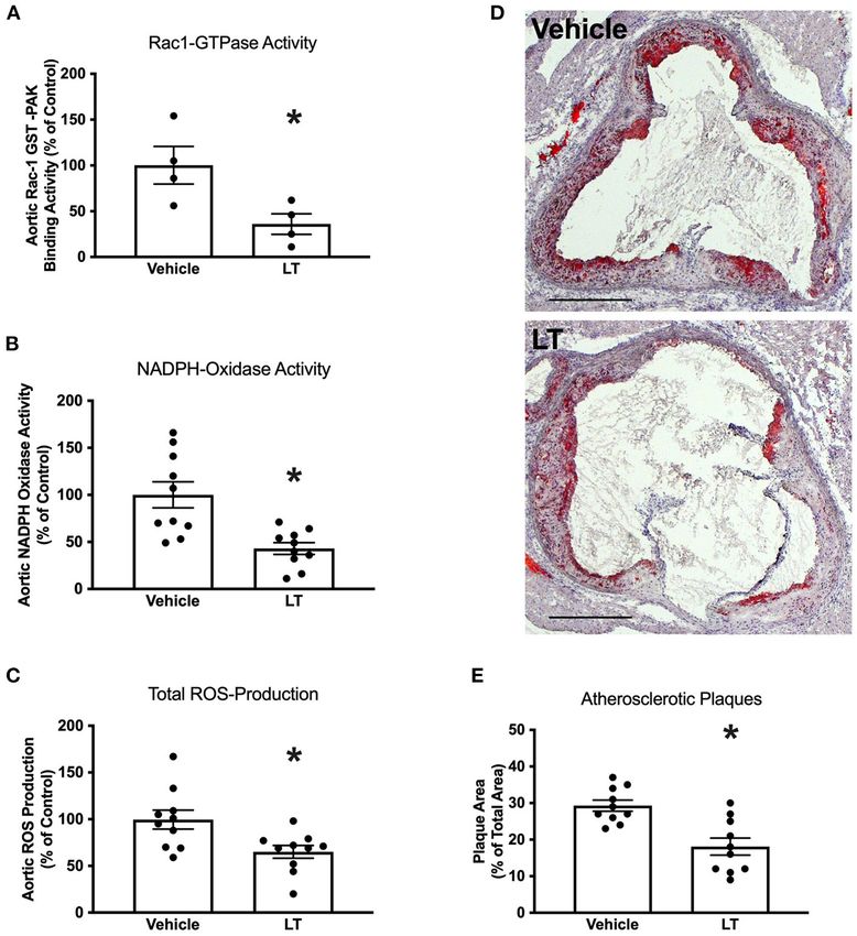
Zimmer et al.                                                                                                                                               Rac1 in Atherosclerosis

TABLE 1 | Assessment of Clostridium sordellii lethal toxin (LT) toxicity.

                                                                                                Vehicle                                 LT                                LT lethal

                                           Body weight (g)                                     25.4 ± 0.5                          26.1 ± 0.8                            25.3 ± 0.8
                                           Serum albumin (g/l)                                 18.9 ± 1.3                          16.9 ± 1.2                           11.3 ± 2.1*
Blood pressure                             Systolic (mmHg)                                    121.5 ± 6.8                         122.6 ± 4.7                               n.a.
                                           Diastolic (mmHg)                                    81.3 ± 7.0                          81.8 ± 3.9                               n.a.
                                           Heart rate (bpm)                                    733 ± 36                             740 ± 29                                n.a.
Blood count                                Lymphocyte (%)                                       85 ± 5.6                           88.2 ± 3.2                            53 ± 5.7*
                                           Neutrophil (%)                                      8.2 ± 3.8                            6.8 ± 0.8                           15.5 ± 0.7*
                                           Monocyte (%)                                        3.4 ± 1.7                            3.2 ± 1.6                           31.5 ± 6.4*
                                           Eosinophil (%)                                      1.6 ± 1.1                            0.6 ± 0.9                              0±0
                                           Basophil (%)                                        1.8 ± 1.3                            1.2 ± 1.3                            0.5 ± 0.7
Wet/Dry ratio                              Lung                                                4.2 ± 0.3                           4.2 ± 0.24                           4.48 ± 0.1*
                                           Heart                                              4.08 ± 0.13                         4.06 ± 0.36                          4.42 ± 0.18*
Echocardiography                           LVEDV (µl)                                          43.7 ± 6.9                          43.7 ± 3.1                               n.a.
                                           SV (µl)                                             35.4 ± 2.1                          38.9 ± 2.1                               n.a.
                                           LVM (mg)                                           133.2 ± 30.7                        142.3 ± 26.3                              n.a.
                                           EF (%)                                             82.3 ± 11.6                         82.5 ± 13.7                               n.a.
Endothelial function                       LogIC50 (M Carbachol)                              −5.2 ± 0.33                        −5.25 ± 0.14                               n.a.
                                           LogIC50 (M Nitro)                                  −5.36 ± 0.38                       −5.43 ± 0.51                               n.a.

Wild-type mice were treated for 4 weeks with vehicle or LT in the therapeutic dose of 0.1 µg LT/week via osmotic mini-pumps or received a lethal dose of LT (150 ng/kg i.p.), and
body weight, heart rate, blood pressure, endothelial function, echocardiographic parameters, differential blood count and markers of vascular permeability were evaluated. LVEDV, left
ventricular end-diastolic volume; SV, stroke volume; LVM, left ventricular mass; EF, ejection fraction. *p < 0.05.

116 ± 12%, ApoE−/− + LT 117 ± 15%, n = 5 per group), neither                                   of the heart and lung (Table 1). Another sign of LT toxicity
was KCl- or phenylephrine-induced vasoconstriction (n = 5 per                                  is perivascular edema (28). Histological analysis of heart and
group, data not shown).                                                                        lung tissues showed perivascular edema exclusively in mice that
                                                                                               received a lethal but not therapeutic dose of LT (Figure 1).
                                                                                               These results demonstrate that long-term, therapeutic-dose LT
Assessment of LT Toxicity                                                                      treatment does not exert toxicity in our mouse model.
To significantly influence atherosclerotic plaque development,
longer treatments with LT were required. Because high doses
of LT lead to increased vascular permeability, tissue edema and                                Long-Term Rac1 Inhibition, Oxidative
death in mice (28), we first investigated the potential toxic effects                          Stress, and Atherosclerosis
of long-term, therapeutic-dose LT treatment. Wild-type mice                                    Next, the effect of long-term Rac1 GTPase inhibition by LT
were treated with either vehicle or 0.1 µg LT/week for 4 weeks via                             on atherosclerosis development was tested. Twelve week-old
osmotic mini-pumps. Consistent with the previous experiments,                                  ApoE−/− mice were fed a high-fat, cholesterol-rich diet and
long-term LT treatment resulted in a significant reduction of                                  were concomitantly treated with 0.1 µg LT/week or vehicle for
vascular ROS production and NADPH oxidase activity (n = 5                                      7 weeks. Aortic Rac1 GTPase activity was significantly reduced
per group, data not show). The mice had no external signs of                                   after treatment with LT (100 ± 22% vs. 36 ± 13%, p < 0.05
acute or chronic illness. Behavior, body weight as well as food                                vs. vehicle, n = 4 per group; Figure 2A). Consistently, aortic
and water consumption remained unchanged in both groups.                                       NADPH oxidase activity (100 ± 20% vs. 43 ± 10%, p < 0.05 vs.
There were no signs of cardiovascular toxicity; specifically blood                             vehicle, n = 10 per group; Figure 2B) and aortic ROS production
pressure, heart rate, cardiac and endothelial function were not                                (100 ± 13% vs. 65 ± 5%, p < 0.05 vs. vehicle, n = 10 per group;
altered by LT treatment (Table 1). Histological analysis of the                                Figure 2C) were significantly decreased in the LT-treated animal
heart did not reveal apparent changes (Figure 1). Indicators                                   group. To investigate whether Rac1 inhibition in the aortic
of increased vascular permeability and tissue edema, including                                 wall affects atherosclerosis, we assessed atherosclerotic plaque
serum albumin concentration and wet/dry ratio of the heart and                                 formation in the aortic root of these mice. Figures 2D,E reveal
lung, did not differ between the groups (Table 1). Both vehicle                                that atherosclerotic lesion formation was significantly reduced
and LT-treated mice had similar differential leukocyte blood                                   in ApoE−/− mice treated with LT compared to vehicle-treated
counts without evidence of an inflammatory process (Table 1).                                  animals (29 ± 2% vs. 18 ± 4%, p < 0.05 vs. vehicle, n = 10 per
In contrast, when wild-type mice received a lethal dose of LT                                  group). To validate that the observed effects were not mediated
(150 ng/kg i.p.), they were soon apathetic, died within 12 h,                                  by a change in cholesterol levels, we measured total plasma
had significantly lower serum albumin concentrations, altered                                  cholesterol concentrations after the 7-week high-fat, cholesterol-
differential leukocyte blood counts and higher wet/dry ratios                                  rich diet. There was no significant difference between ApoE−/−

Frontiers in Cardiovascular Medicine | www.frontiersin.org                                3                                               August 2021 | Volume 8 | Article 680775
Zimmer et al. Rac1 in Atherosclerosis
FIGURE 1 | Assessment of Clostridium sordellii lethal toxin (LT) toxicity. The main characteristic of LT toxicity is increased vascular permeability with perivascular
edema. Representative HE stainings of heart and lung tissue from mice treated with vehicle or a therapeutic dose of LT for 4 weeks show no signs of tissue edema. In
contrast, mice that received a lethal dose of LT display edema in the connective tissue surrounding larger arteries (arrow heads).
mice that received vehicle and those treated with LT (1,318 ± 46 0.05 vs. vehicle, n = 4–7 per group; Figure 4A). There have
mg/dl vs. 1,414 ± 51 mg/dl, n = 10 per group). been reports that LT can glucosylate other members of the Rho
family of small GTPases to some degree, as for example RhoA.
NADPH Oxidase Subunit Expression However, only simvastatin but not LT or NSC 23766 significantly
To assess whether LT in addition to inhibition of Rac1 GTPase inhibited aortic RhoA GTPase activity in the ApoE−/− mice
activity also influences NADPH oxidase expression, NADPH (vehicle 100 ± 8%, simvastatin 53 ± 9%, p < 0.05 vs. vehicle,
oxidase subunits were analyzed in aortic tissue of the above- NSC 93 ± 13%, LT 81 ± 6%, n = 5–8 per group; Figure 4B). All
mentioned ApoE−/− mice. There were no significant differences three compounds significantly reduced aortic NADPH oxidase
in protein expression levels of the NADPH oxidase subunits activity (vehicle 100 ± 19%, simvastatin 47 ± 11%, NSC 47
Rac1, p47-phox, p67-phox, Nox1, and Nox2 between the ± 12%, LT 42 ± 8%, all p < 0.05 vs. vehicle, n = 3–5 per
groups (Figures 3A,B). Furthermore, there was no difference group; Figure 4C) and aortic ROS production (vehicle 100 ±
in Rac1, p47-phox, or p67-phox expression in cytosolic and 16%, simvastatin 54 ± 9%, NSC 57 ± 8%, LT 44 ± 8%, all p
membrane protein fractions, indicating that there were no < 0.05 vs. vehicle, n = 6–9 per group; Figure 4D). Treatment
changes in membrane translocation of these cytosolic subunits with these Rac1 GTPase inhibitors improved endothelium-
(Figures 3A,B). Moreover, neither Nox4 (1.0 ± 0.15 2−11Ct vs. dependent vasodilation (maximal relaxation: vehicle 46 ± 8%,
1.2 ± 0.24 2−11Ct , n = 2–3 per group; Figure 3C left) nor simvastatin 66 ± 5%, NSC 81 ± 7%, LT 68 ± 5%, all p
p22-phox (1.0 ± 0.32 2−11Ct vs. 0.9 ± 0.05 2−11Ct , n = 5 < 0.05 vs. vehicle, n = 5 per group; Figure 4E) but had no
per group; Figure 3C right) mRNA expression was altered by effect on endothelium-independent vasorelaxation (Figure 4F)
LT treatment. or phenylephrine-induced vasoconstriction (data not shown).
Importantly, treatment with simvastatin, NSC 23766 or LT
Comparison of Different Rac1 Inhibitors significantly attenuated atherosclerotic plaque development in
Statins have been shown to reduce Rac1 GTPase activity in vitro ApoE−/− mice to a similar degree (vehicle 34 ± 3%, simvastatin
and in vivo (22, 29), and a specific synthetic small-molecule 17 ± 3%, NSC 20 ± 3%, LT 17 ± 2%, all p < 0.05 vs. vehicle,
Rac1 GTPase inhibitor, NSC 23766, has recently been developed n = 3–5 per group; Figures 5A,B). Because Rac1 inhibition
(30, 31). To test whether these Rac1 inhibitors have comparable diminishes monocyte adhesion to endothelial cells in vitro, we
vascular in vivo effects as LT, ApoE−/− mice were fed a high-fat, studied macrophage infiltration of atherosclerotic plaques in
cholesterol-rich diet for 7 weeks and were concomitantly treated these ApoE−/− mice. Simvastatin, NSC 23766 and LT treatment
with vehicle, simvastatin, NSC 23766 or LT. Serum lipoprotein significantly reduced macrophage infiltration of atherosclerotic
profiles have been previously investigated and no significant plaques to a similar degree compared to vehicle-treated mice
effects were measured (32). All three treatments significantly (vehicle 41 ± 3%, simvastatin 29 ± 5%, NSC 15 ± 3%,
inhibited aortic Rac1 GTPase activity (vehicle 100 ± 21%, LT 22 ± 2%, all p < 0.05 vs. vehicle, n = 3–4 per group;
simvastatin 44 ± 21%, NSC 32 ± 14%, LT 24 ± 6%, all p < Figures 5C,D).
Frontiers in Cardiovascular Medicine | www.frontiersin.org 4 August 2021 | Volume 8 | Article 680775Zimmer et al. Rac1 in Atherosclerosis
FIGURE 2 | Long-term Rac1 inhibition, oxidative stress and atherosclerosis. (A) Aortic homogenates of ApoE−/− mice that were fed a cholesterol-rich diet for 7
weeks and were concomitantly treated with 0.1 µg LT/week via osmotic mini-pumps showed reduced Rac1 GTPase activity compared to vehicle-treated mice in a
Rac1 GST-PAK pull-down assay (*p < 0.05, n = 4 per group). (B,C) LT-treated ApoE−/− mice displayed reduced aortic NADPH oxidase activity (B, *p < 0.05, n = 10
per group) and aortic ROS production (C, *p < 0.05, n = 10 per group), as measured by lucigenin-enhanced chemiluminescence and L-012 chemiluminescence,
respectively. (D) Representative histological cross-sections of the aortic root stained with oil red O to display atherosclerotic plaques. LT-treated ApoE−/− mice
showed decreased atherosclerotic plaque formation. (E) Quantification of atherosclerotic plaque formation, expressed as plaque area in percent of total area (*p <
0.05, n = 10 per group). Scale bars indicate 0.5 mm.
DISCUSSION humans and animals after bacterial infection, which is mediated
by a severe increase in vascular permeability (28, 33, 34). We
The data in this study demonstrate that long-term tested two dosing regimens of LT and found that the lower
inhibition of Rac1 GTPase exerts atheroprotective effects in was well-tolerated by mice, yet it was sufficient to significantly
hypercholesterolemic ApoE−/− mice. Systemic application of inhibit Rac1 GTPase activity in the aortic wall. In contrast
Clostridium sordellii lethal toxin or the specific synthetic small- to treatment with a lethal dose of LT, which caused severe
molecule inhibitor NSC 23766 effectively inhibit Rac1 GTPase pulmonary and cardiac edema, this therapeutic dose of LT had no
activity in the aorta which in turn significantly reduces NADPH impact on behavior, food intake, body weight, heart rate, blood
oxidase activity and results in decreased oxidative stress in the pressure, vascular and myocardial function, differential blood
vessel wall. Long-term Rac1 inhibition is ultimately associated count and vascular permeability. It is therefore possible to treat
with improved endothelial function, reduced atherosclerotic mice systemically with LT over long durations and investigate the
plaque formation and diminished macrophage infiltration. effects of chronic Rac1 inhibition without inducing acute illness.
Intraperitoneal injection of high doses of LT in mice induces LT is an established model of specific inhibition of Rac1
symptoms similar to fatal toxic shock syndrome observed in GTPase activity, and the toxin is highly effective in vascular cells.
Frontiers in Cardiovascular Medicine | www.frontiersin.org 5 August 2021 | Volume 8 | Article 680775Zimmer et al. Rac1 in Atherosclerosis FIGURE 3 | NADPH oxidase subunit expression. (A) Western blot analysis of NADPH oxidase subunit protein expression in cytosolic and membrane fractions of aortic homogenates of cholesterol-fed ApoE−/− mice treated with vehicle or LT. There was no difference in Rac1, Nox1, Nox2, p47-phox, and p67-phox expression. (B) Densitometric quantification of the Western analyses. Expression of genes-of-interest was normalized to housekeeping gene expression. These values were normalized to membrane protein expression of vehicle-treated mice (n = 2 per group). For each subunit, membrane expression is displayed on the left, cytosolic expression on the right. (C) Real-time PCR analysis of Nox4 and p22-phox subunit mRNA expression in aortic homogenates of cholesterol-fed ApoE−/− mice treated with vehicle or LT. Neither aortic Nox4 nor p22-phox expression was altered by LT treatment in these mice (n = 2–3 independent measurements (pooled from n=2-3 mice per sample) per group). However, there have been reports that LT can glucosylate other vascular smooth muscle cells and that Rac1 inhibition via LT members of the rho family of small GTPases to some degree, as or overexpression of the dominant-negative RacN17 mutant for example RhoA. RhoA has been shown to play an important decreases ROS production (21, 22). We now demonstrate that regulatory role in vascular cells (24). Our data show that LT LT treatment exerts this effect in vivo when applied systemically profoundly inhibits activation of Rac1 GTPase but does not affect in mice. Rac1 inhibition causes diminished NADPH oxidase RhoA GTPase activation in aortic tissue of ApoE−/− mice. In activity in the vessel wall, which in turn leads to reduced addition, treatment of ApoE−/− mice with the specific synthetic ROS formation and thus decreased oxidative stress. LT inhibits small-molecule Rac1 GTPase inhibitor NSC 23766, which does Rac1 activity by direct glucosylation of the GTPase. However, not exert effects on other rho family proteins (30, 31), profoundly to evaluate whether LT has additional effects on the NADPH inhibited Rac1 GTPase but not RhoA GTPase activity and led oxidase complex, we investigated NADPH oxidase subunit to identical results as treatment with LT. Although effects on expression and membrane translocation in aortic homogenates other Rho family proteins cannot be fully excluded, these findings of vehicle or LT-treated ApoE−/− mice. There was no significant corroborate the notion that LT acts predominantly on Rac1 difference in Nox1, Nox2, Nox4, p22-phox, p47-phox, p67- GTPase in the vascular system of our animal model. phox, and Rac1 subunit expression. Moreover, comparison of We have previously shown that overexpression of the Rac1, p47-phox, and p67-phox expression in cytosolic and constitutively active mutant RacL61 increases ROS release in membrane protein fractions revealed that there were no changes Frontiers in Cardiovascular Medicine | www.frontiersin.org 6 August 2021 | Volume 8 | Article 680775
Zimmer et al.                                                                                                                                         Rac1 in Atherosclerosis

 FIGURE 4 | Vascular effects of different Rac1 inhibitors: GTPase activity, oxidative stress and endothelial function. To investigate the vascular effects of different Rac1
 GTPase inhibitors, ApoE−/− mice were fed a cholesterol-rich diet for 7 weeks and were concomitantly treated with vehicle, LT (0.1 µg LT/week), NSC 23766 (10
 mg/kg/d), or simvastatin (20 mg activated simvastatin/kg/d). All three compounds significantly inhibited aortic Rac1 GTPase activity (A, Rac1 GTPase activity G-LISA,
 *p < 0.05 vs. vehicle, n = 4–7 per group), but only simvastatin significantly decreased aortic rhoA GTPase activity (B, rhoA GTPase activity G-LISA, *p < 0.05 vs.
 vehicle, n = 5–8 per group). All three compounds significantly reduced aortic NADPH oxidase activity (C, lucigenin-enhanced chemiluminescence, *p < 0.05 vs.
 vehicle, n = 3–5 per group) and aortic ROS production (D, L-012 chemiluminescence, *p < 0.05 vs. vehicle, n = 6–9 per group) and significantly improved
 endothelium-dependent vasodilation (E, organ chamber experiments with isolated aortic segments, *p < 0.05 vs. vehicle, n = 5 per group) compared to
 vehicle-controls. Endothelium-independent vasodilation was not affected by the Rac1 inhibitors (F, n = 5 per group).

in membrane translocation of these subunits. Inhibition of                                LT-mediated reduction of vascular oxidative stress in our animal
NADPH oxidase activity through specific inhibition of Rac1                                model. Although effects on other pro- and antioxidant systems
GTPase is a principle mechanism of action underlying the                                  cannot be excluded, the above notion is further supported by our

Frontiers in Cardiovascular Medicine | www.frontiersin.org                            7                                             August 2021 | Volume 8 | Article 680775
Zimmer et al. Rac1 in Atherosclerosis
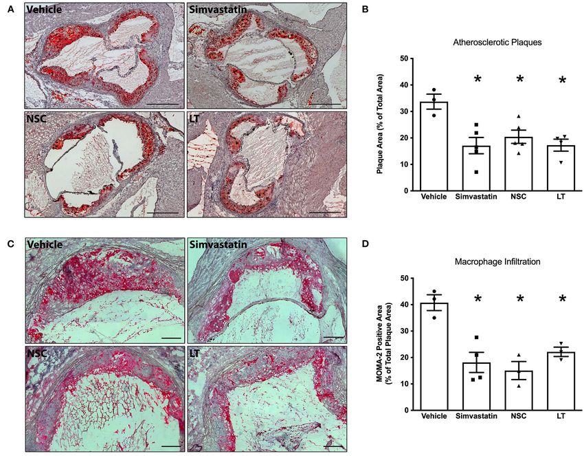
FIGURE 5 | Vascular effects of different Rac1 inhibitors: atherosclerosis and macrophage infiltration. ApoE−/− mice were fed a cholesterol-rich diet for 7 weeks and
were concomitantly treated with vehicle, LT (0.1 µg LT/week), NSC 23766 (10 mg/kg/d), or simvastatin (20 mg activated simvastatin/kg/d). All three compounds
significantly reduced atherosclerotic plaque formation in the aortic root compared to vehicle-control. (A) Representative histological cross-sections. Scale bars indicate
0.5 mm. (B) Quantification of atherosclerotic plaque formation, expressed as plaque area in percent of total area (*p < 0.05 vs. vehicle, n = 3–5 per group).
Furthermore, all three compounds significantly reduced macrophage infiltration of atherosclerotic plaques in these mice. (C) Representative MOMA-2
immunohistochemical staining using a monoclonal rat anti-mouse MOMA-2 antibody. Scale bars indicate 0.15 mm. (D) Quantification of macrophages infiltrating
aortic atherosclerotic plaques (*p < 0.05 vs. vehicle, n = 3–4 per group).
data derived from specific pharmacological Rac1 inhibition with of oxidative stress in atherosclerosis, but rather implies that
NSC 23766. the antioxidative agents used were not capable of significantly
Oxidative stress has been implicated in the pathogenesis modifying oxidative stress at the appropriate site and time.
of atherosclerosis (35). All common risk factors associated Instead of scavenging an excess of ROS, primarily reducing the
with coronary artery disease such as elevated LDL cholesterol, formation of ROS may be a more efficient approach to decrease
diabetes, hypertension, and cigarette smoking are associated with vascular oxidative stress and therefore impact on vascular
increased ROS levels within the arterial wall. In fact, it has been pathologies. Our data suggest that inhibiting a major source of
demonstrated that increased oxidative stress leads to endothelial ROS in the vessel wall, NADPH oxidase, is an effective method
dysfunction by countering the vasoprotective and vasodilating to decrease vascular oxidative stress and improve endothelial
effects of nitric oxide (36, 37). It was speculated that decreasing function in an established animal model of atherosclerosis.
the oxidative stress burden would reduce cardiovascular events Endothelial dysfunction is not only an early and crucial stage of
but clinical trials with antioxidant therapies failed to show atherosclerosis development, but is also associated with increased
reductions in event rates (38). The lack of benefit seen in risk for cardiovascular events (39). ApoE −/− mice develop
these clinical trials does not, however, disprove the central role atherosclerotic lesions in response to a cholesterol-rich diet and
Frontiers in Cardiovascular Medicine | www.frontiersin.org 8 August 2021 | Volume 8 | Article 680775Zimmer et al. Rac1 in Atherosclerosis
are widely used to study the pathogenesis of atherosclerosis (40). extent of the observed, and sometimes contradictory, effects (46).
Typically, atherosclerotic lesions are more pronounced in the Interestingly, prevention of Rac1 prenylation in macrophages
aortic root and the proximal aorta than in more distal parts by deleting GTPase 1, the prenylase responsible for Rac1
(41). Importantly, we provide evidence that long-term Rac1 modification, leads to significant inflammatory response in
inhibition significantly reduces atherosclerotic plaque formation macrophages and to rheumatoid arthritis (46). The authors
in this animal model. Our finding is in agreement with other suggest that this is due to increased activation of unprenylated
studies demonstrating diminished atherosclerosis on genetic Rac1 (i.e., generation of the GTP-bound form of the GTPase)
inhibition of NADPH oxidase and reduction of vascular oxidative and that effector interactions involved in innate immunity are
stress (42). enhanced. This would presumably also apply to statins and
Rac1 GTPase not only exerts effects on ROS generation recent findings confirm that disruption of Rac1 regulation via
but also on organization of the actin cytoskeleton, which is, statins leads to increased atherosclerotic plaque calcification (47).
among others, important for monocyte adhesion (24). Monocyte However, these effects would not be induced by the modes of
adhesion to the endothelium, migration, accumulation, and inhibition used in the present study, thus avoiding the potential
activation of monocytes in atherosclerotic lesions is a central negative effects of statins.
part of the inflammatory process involved in atherogenesis Although specific Rac1 inhibition with LT or NSC 23766 was
(39). We have previously demonstrated that Rac1 mediates not superior to statin treatment, our findings are decisive because
actin rearrangement, lamellipodia formation and adhesion of they identify Rac1 GTPase as a key molecule in atherogenesis and
human monocytes in vitro independently of ROS (26). We provide novel proof of concept that specifically inhibiting this
now provide evidence of reduced macrophage infiltration of molecule is sufficient to diminish the atherosclerotic process in
atherosclerotic plaques after long-term Rac1 inhibition, further our animal model and therefore may improve vascular health.
supporting the importance of Rac1 GTPase for monocyte Thus, Rac-1 GTPase may represent an interesting target for
function in vivo and development of atherosclerosis. It should future anti-atherosclerotic drug development. Further detailed
be noted that a further contribution of Rac1 to atherogenesis in vitro investigations are necessary to establish the exact
in the vascular endothelium is by coupling of Rac1 activation mechanisms of the observed effects upon Rac1 inhibition.
to LDL transport across endothelial monolayers via SR-B1
(scavenger receptor, class B type 1) (43). Physiological or chronic DATA AVAILABILITY STATEMENT
activation of Rac1 is achieved through an exchange of guanosine
diphosphate (GDP) to guanosine triphosphate, which is catalyzed The datasets generated and/or analyzed during the current
by the exchange factor Vav2. Vav2 is phosphorylated and thus study are available from the corresponding author on
activated by activated vascular endothelial growth factor (VEGF). reasonable request.
Phosphorylation of Vav2 seems to be regulated by Src and Src
kinase activity is required for activation of Rac1 (44). Several
other Rac1 GEFs, including Tiam or DOCK proteins, could also ETHICS STATEMENT
be important (43, 45).
The animal study was reviewed and approved by Landesamt für
The findings of our study help to identify Rac1 GTPase as
Natur, Umwelt und Verbraucherschutz Nordrhein-Westfalen,
an important protagonist in atherogenesis. Inhibition of Rac1
Germany Godesberger Allee 136, 53175 Bonn.
GTPase by either Clostridium sordellii lethal toxin or the specific
small-molecule inhibitor NSC 23766 is associated with reduced
vascular NADPH oxidase activity and oxidative stress, decreased AUTHOR CONTRIBUTIONS
macrophage infiltration, improved endothelial function and
ultimately diminished atherosclerosis development. In our SZ, GN, and SW conceived and designed the project. SZ, PG,
animal model, the vascular and ultimately atheroprotective MO, AG, CM, AD, and FJ performed experiments. SZ, PG, MO,
actions of LT and NSC 23766 were as efficacious as those AG, CM, UL, AD, FJ, GN, and SW analyzed and interpreted
of simvastatin. This is important because the specific Rac1 the data. All authors contributed to writing and editing of
inhibitors LT and NSC 23766 do not display lipid-lowering the manuscript.
properties or other pleiotropic effects attributed to statins.
Additionally, the mode of action of statins on Rac1 is different FUNDING
and involves prevention of geranylgeranylation of Rac1 at its
C-terminus. This is expected to prevent membrane association This study was supported by Deutsche Forschungsgemeinschaft,
of Rac1, a prerequisite for its activation of the NADPH European Vascular Genomics Network, Canada Foundation for
oxidase. Statins also activate Rac1 via induction of AMP- Innovation, Canada Research Chairs and Canadian Institutes of
activated protein kinase (AMPK) and liver kinase B1 (LKB1) Health Research. PG was supported by the Else Kröner-Fresenius
phosphorylation (45). However, statins were also shown to Stiftung (2014_Kolleg.05), SZ, PG, FJ, and GN were supported by
inhibit NADPH oxidase activity in a Rac1 independent manner the Deutsche Forschungsgemeinschaft (DFG, German Research
in endothelial cells, which demonstrates that multiple pathways Foundation)—Grant No. 397484323—TRR 259, FJ was further
are regulated after statin treatment and might help explain the supported by the Corona foundation.
Frontiers in Cardiovascular Medicine | www.frontiersin.org 9 August 2021 | Volume 8 | Article 680775Zimmer et al. Rac1 in Atherosclerosis
ACKNOWLEDGMENTS SUPPLEMENTARY MATERIAL
The excellent technical assistance of Isabel Paez-Maletz, The Supplementary Material for this article can be found
Catharina Peseke, Annika Bohner, and Omar Arfa is greatly online at: https://www.frontiersin.org/articles/10.3389/fcvm.
appreciated. LT was kindly provided by Klaus Aktories. 2021.680775/full#supplementary-material
REFERENCES 18. Diekmann D, Abo A, Johnston C, Segal A, Hall A. Interaction of Rac with
p67phox and regulation of phagocytic NADPH oxidase activity. Science.
1. Darley-Usmar V, Halliwell B. Blood radicals: reactive nitrogen species, reactive (1994) 265:531–3. doi: 10.1126/science.8036496
oxygen species, transition metal ions, and the vascular system. Pharmaceut 19. Sarfstein R, Gorzalczany Y, Mizrahi A, Berdichevsky Y, Molshanski-Mor S,
Res. (1996) 13:649–62. doi: 10.1023/A:1016079012214 Weinbaum C, et al. Dual role of Rac in the assembly of NADPH oxidase,
2. Laursen JB, Rajagopalan S, Galis Z, Tarpey M, Freeman BA, Harrison DG. tethering to the membrane and activation of p67phox a study based on
Role of superoxide in angiotensin II-induced but not catecholamine-induced mutagenesis of p67phox-Rac1 chimeras. J Biol Chem. (2004) 279:16007–
hypertension. Circulation. (1997) 95:588–93. doi: 10.1161/01.CIR.95.3.588 16. doi: 10.1074/jbc.M312394200
3. Li P-F, Dietz R, von Harsdorf R. Reactive oxygen species induce 20. Hu Q, Zheng G, Zweier JL, Deshpande S, Irani K, Ziegelstein RC. NADPH
apoptosis of vascular smooth muscle cell. Febs Lett. (1997) 404:249– oxidase activation increases the sensitivity of intracellular Ca2+ stores to
52. doi: 10.1016/S0014-5793(97)00093-8 inositol 1,4,5-trisphosphate in human endothelial cells. J Biol Chem. (2000)
4. Burtenshaw D, Kitching M, Redmond EM, Megson IL, Cahill PA. Reactive 275:15749–57. doi: 10.1074/jbc.M000381200
Oxygen Species (ROS), intimal thickening, and subclinical atherosclerotic 21. Laufs U, Adam O, Strehlow K, Wassmann S, Konkol C, Laufs K, et al.
disease. Front Cardiovasc Med. (2019) 6:89. doi: 10.3389/fcvm.2019.00089 Down-regulation of Rac-1 GTPase by estrogen. J Biol Chem. (2003) 278:5956–
5. Nowak WN, Deng J, Ruan XZ, Xu Q. Reactive oxygen species 62. doi: 10.1074/jbc.M209813200
generation and atherosclerosis. Arterioscler Thromb Vasc Biol. (2017) 22. Wassmann S, Laufs U, Bäumer AT, Müller K, Konkol C, Sauer H, et al.
37:e41–52. doi: 10.1161/ATVBAHA.117.309228 Inhibition of geranylgeranylation reduces angiotensin II-mediated free radical
6. Sahoo S, Meijles DN, Pagano PJ. NADPH oxidases: key modulators in aging production in vascular smooth muscle cells: involvement of angiotensin
and age-related cardiovascular diseases? Clin Sci Lond Engl. (2016) 130:317– AT1 receptor expression and Rac1 GTPase. Mol Pharmacol. (2001) 59:646–
35. doi: 10.1042/CS20150087 54. doi: 10.1124/mol.59.3.646
7. Konior A, Schramm A, Czesnikiewicz-Guzik M, Guzik TJ. NADPH 23. Vecchione C, Brandes RP. Withdrawal of 3-hydroxy-3-methylglutaryl
oxidases in vascular pathology. Antioxid Redox Signal. (2013) 20:2794– coenzyme a reductase inhibitors elicits oxidative stress and
814. doi: 10.1089/ars.2013.5607 induces endothelial dysfunction in mice. Circ Res. (2002)
8. Dusi S, Donini M, Rossi F. Mechanisms of NADPH oxidase activation in 91:173–9. doi: 10.1161/01.RES.0000028004.76218.B8
human neutrophils: p67phox is required for the translocation of rac 1 but 24. Burridge K, Wennerberg K. Rho and Rac take center stage. Cell. (2004)
not of rac 2 from cytosol to the membranes. Biochem J. (1995) 308:991– 116:167–79. doi: 10.1016/S0092-8674(04)00003-0
4. doi: 10.1042/bj3080991 25. Sit S-T, Manser E. Rho GTPases and their role in organizing the actin
9. Rinckel LA, Faris SL, Hitt ND, Kleinberg ME. Rac1 disrupts p67phox/p40phox cytoskeleton. J Cell Sci. (2011) 124:679–83. doi: 10.1242/jcs.064964
binding: a novel role for Rac in NADPH oxidase activation. Biochem Bioph Res 26. Friedrich EB, Clever YP, Wassmann S, Hess C, Nickenig G. 17Beta-estradiol
Co. (1999) 263:118–22. doi: 10.1006/bbrc.1999.1334 inhibits monocyte adhesion via down-regulation of Rac1 GTPase. J Mol Cell
10. Brandes RP, Schröder K. Composition and functions of vascular nicotinamide Cardiol. (2005) 40:87–95. doi: 10.1016/j.yjmcc.2005.10.007
adenine dinucleotide phosphate oxidases. Trends Cardiovas Med. (2008) 27. Just I, Selzer J, Hofmann F, Green GA, Aktories K. Inactivation of Ras by
18:15–19. doi: 10.1016/j.tcm.2007.11.001 Clostridium sordellii lethal toxin-catalyzed glucosylation. J Biol Chem. (1996)
11. Duerrschmidt N, Wippich N, Goettsch W, Broemme H-J, Morawietz H. 271:10149–53. doi: 10.1074/jbc.271.17.10149
Endothelin-1 induces NAD(P)H oxidase in human endothelial cells. Biochem 28. Geny B, Khun H, Fitting C, Zarantonelli L, Mazuet C, Cayet N,
Bioph Res Co. (2000) 269:713–7. doi: 10.1006/bbrc.2000.2354 et al. Clostridium sordellii lethal toxin kills mice by inducing a major
12. Schröder K, Weissmann N, Brandes RP. Organizers and activators: cytosolic increase in lung vascular permeability. Am J Pathol. (2007) 170:1003–
Nox proteins impacting on vascular function. Free Radical Bio Med. (2017) 17. doi: 10.2353/ajpath.2007.060583
109:22–32. doi: 10.1016/j.freeradbiomed.2017.03.017 29. Adam O, Frost G, Custodis F, Sussman MA, Schäfers H-J, Böhm M, et al.
13. Ushio-Fukai M, Alexander RW. Reactive oxygen species as mediators of Role of Rac1 GTPase activation in atrial fibrillation. J Am Coll Cardiol. (2007)
angiogenesis signaling. Role of NAD(P)H oxidase. Mol Cell Biochem. (2004) 50:359–67. doi: 10.1016/j.jacc.2007.03.041
264:85–97. doi: 10.1023/B:MCBI.0000044378.09409.b5 30. Gao Y, Dickerson JB, Guo F, Zheng J, Zheng Y. Rational design and
14. Hu T, RamachandraRao SP, Siva S, Valancius C, Zhu Y, Mahadev K, et al. characterization of a Rac GTPase-specific small molecule inhibitor. Proc Natl
Reactive oxygen species production via NADPH oxidase mediates TGF-β- Acad Sci USA. (2004) 101:7618–23. doi: 10.1073/pnas.0307512101
induced cytoskeletal alterations in endothelial cells. Am J Physiol Renal. (2005) 31. Akbar H, Cancelas J, Williams DA, Zheng J, Zheng Y. Rational design and
289:F816–25. doi: 10.1152/ajprenal.00024.2005 applications of a Rac GTPase–specific small molecule inhibitor. Methods
15. Desideri G, Bravi MC, Tucci M, Croce G, Marinucci MC, Santucci A, Enzymol. (2006) 406:554–65. doi: 10.1016/S0076-6879(06)06043-5
et al. Angiotensin II inhibits endothelial cell motility through an AT1- 32. Schierwagen R, Maybüchen L, Hittatiya K, Klein S, Uschner FE, Braga TT,
dependent oxidant-sensitive decrement of nitric oxide availability. Arterioscler et al. Statins improve NASH via inhibition of RhoA and Ras. Am J Physiol
Thromb Vasc Biol. (2003) 23:1218–23. doi: 10.1161/01.ATV.0000078521. Gastr L. (2016) 311:G724–33. doi: 10.1152/ajpgi.00063.2016
51319.65 33. Boehm C, Gibert M, Geny B, Popoff MR, Rodriguez P. Modification
16. Hwang J, Saha A, Boo YC, Sorescu GP, McNally JS, Holland SM, et al. of epithelial cell barrier permeability and intercellular junctions
Oscillatory shear stress stimulates endothelial production of from p47 phox by Clostridium sordellii lethal toxins. Cell Microbiol. (2006)
-dependent NAD(P)H oxidases, leading to monocyte adhesion. J Biol Chem. 8:1070–85. doi: 10.1111/j.1462-5822.2006.00687.x
(2003) 278:47291–8. doi: 10.1074/jbc.M305150200 34. Waschke J, Drenckhahn D, Adamson RH, Curry FE. Role of adhesion
17. Brandes RP, Weissmann N, Schröder K. Nox family NADPH oxidases in and contraction in Rac 1-regulated endothelial barrier function in
mechano-transduction: mechanisms and consequences. Antioxid Redox Sign. vivo and in vitro. Am J Physiol Hear Circ Physiol. (2004) 287:H704–
(2014) 20:887–98. doi: 10.1089/ars.2013.5414 11. doi: 10.1152/ajpheart.01076.2003
Frontiers in Cardiovascular Medicine | www.frontiersin.org 10 August 2021 | Volume 8 | Article 680775Zimmer et al. Rac1 in Atherosclerosis
35. Yang X, Li Y, Li Y, Ren X, Zhang X, Hu D, et al. Oxidative stress- 45. Servitja J-M, Marinissen MJ, Sodhi A, Bustelo XR, Gutkind JS. Rac1 function
mediated atherosclerosis: mechanisms and therapies. Front Physiol. (2017) is required for Src-induced transformation. J Biol Chem. (2003) 278:34339–
8:600. doi: 10.3389/fphys.2017.00600 46. doi: 10.1074/jbc.M302960200
36. Sawada N, Salomone S, Kim H-H, Kwiatkowski DJ, Liao JK. Regulation of 46. Akula MK, Ibrahim MX, Ivarsson EG, Khan OM, Kumar IT, Erlandsson M,
endothelial nitric oxide synthase and postnatal angiogenesis by Rac1. Circ Res. et al. Protein prenylation restrains innate immunity by inhibiting Rac1 effector
(2008) 103:360–8. doi: 10.1161/CIRCRESAHA.108.178897 interactions. Nat Commun. (2019) 10:3975. doi: 10.1038/s41467-019-11606-x
37. Cai H, Harrison DG. Endothelial dysfunction in cardiovascular diseases: the 47. Healy A, Berus JM, Christensen JL, Lee C, Mantsounga C, Dong W,
role of oxidant stress. Circ Res. (2000) 87:840–4. doi: 10.1161/01.RES.87.10.840 et al. Statins disrupt macrophage Rac1 regulation leading to increased
38. Steinhubl SR. Why have antioxidants failed in clinical trials? Am J Cardiol. atherosclerotic plaque calcification. Arterioscler Thromb Vasc Biol. (2020)
(2008) 101:S14–9. doi: 10.1016/j.amjcard.2008.02.003 40:714–32. doi: 10.1161/ATVBAHA.119.313832
39. Epstein FH, Ross R. Atherosclerosis — an inflammatory disease. New Engl J
Med. (1999) 340:115–26. doi: 10.1056/NEJM199901143400207 Conflict of Interest: The authors declare that the research was conducted in the
40. Osada J, Joven J, Maeda N. The value of apolipoprotein E knockout mice for absence of any commercial or financial relationships that could be construed as a
studying the effects of dietary fat and cholesterol on atherogenesis. Curr Opin potential conflict of interest.
Lipidol. (2000) 11:25–9. doi: 10.1097/00041433-200002000-00004
41. Wassmann S, Czech T, Eickels M van, Fleming I, Böhm M, Nickenig G. Publisher’s Note: All claims expressed in this article are solely those of the authors
Inhibition of diet-induced atherosclerosis and endothelial dysfunction in and do not necessarily represent those of their affiliated organizations, or those of
apolipoprotein E/angiotensin II type 1A receptor double-knockout mice.
the publisher, the editors and the reviewers. Any product that may be evaluated in
Circulation. (2004) 110:3062–7. doi: 10.1161/01.CIR.0000137970.47771.AF
this article, or claim that may be made by its manufacturer, is not guaranteed or
42. Barry-Lane PA, Patterson C, Merwe M van der, Hu Z, Holland SM, Yeh ETH,
endorsed by the publisher.
et al. p47phox is required for atherosclerotic lesion progression in ApoE–/–
mice. J Clin Invest. (2001) 108:1513–22. doi: 10.1172/JCI200111927
Copyright © 2021 Zimmer, Goody, Oelze, Ghanem, Mueller, Laufs, Daiber, Jansen,
43. Huang L, Chambliss KL, Gao X, Yuhanna IS, Behling-Kelly E, Bergaya S,
Nickenig and Wassmann. This is an open-access article distributed under the terms
et al. SR-B1 drives endothelial cell LDL transcytosis via DOCK4 to promote
of the Creative Commons Attribution License (CC BY). The use, distribution or
atherosclerosis. Nature. (2019) 569:565–9. doi: 10.1038/s41586-019-1140-4
reproduction in other forums is permitted, provided the original author(s) and the
44. Garrett TA, Buul JDV, Burridge K. VEGF-induced Rac1 activation in copyright owner(s) are credited and that the original publication in this journal
endothelial cells is regulated by the guanine nucleotide exchange factor Vav2. is cited, in accordance with accepted academic practice. No use, distribution or
Exp Cell Res. (2007) 313:3285–97. doi: 10.1016/j.yexcr.2007.05.027 reproduction is permitted which does not comply with these terms.
Frontiers in Cardiovascular Medicine | www.frontiersin.org 11 August 2021 | Volume 8 | Article 680775You can also read