Toll-Like Receptor 4 Protects Against Clostridium perfringens Infection in Mice - Frontiers
←
→
Page content transcription
If your browser does not render page correctly, please read the page content below
ORIGINAL RESEARCH
                                                                                                                                              published: 08 March 2021
                                                                                                                                       doi: 10.3389/fcimb.2021.633440

                                               Toll-Like Receptor 4 Protects
                                               Against Clostridium perfringens
                                               Infection in Mice
                                               Masaya Takehara *, Keiko Kobayashi and Masahiro Nagahama *

                                               Department of Microbiology, Faculty of Pharmaceutical Sciences, Tokushima Bunri University, Yamashiro-cho, Japan

                                               Toll-like receptor 4 (TLR4) has been reported to protect against Gram-negative bacteria
                                               by acting as a pathogen recognition receptor that senses mainly lipopolysaccharide (LPS)
                                               from Gram-negative bacteria. However, the role of TLR4 in Gram-positive bacterial
                                               infection is less well understood. Clostridium perfringens type A is a Gram-positive
                                               bacterium that causes gas gangrene characterized by severe myonecrosis. It was
                                               previously demonstrated that C. perfringens q-toxin is a TLR4 agonist, but the role of
                                               TLR4 in C. perfringens infection is unclear. Here, TLR4-defective C3H/HeJ mice infected
                                               with C. perfringens showed a remarkable decrease in survival rate, an increase in viable
                           Edited by:          bacterial counts, and accelerated destruction of myofibrils at the infection site compared
                       Grace Yi Chen,
 University of Michigan, United States         with wild-type C3H/HeN mice. These results demonstrate that TLR4 plays an important
                         Reviewed by:          role in the elimination of C. perfringens. Remarkable increases in levels of inflammatory
                 Rksubbarao Malireddi,         cytokines, such as interleukin-1b (IL-1b), interleukin-6 (IL-6), and granulocyte colony-
St. Jude Children’s Research Hospital,
                           United States
                                               stimulating factor (G-CSF), were observed in C. perfringens-infected C3H/HeN mice,
                          Ashu Sharma,         whereas the increases were limited in C3H/HeJ mice. Generally, increased G-CSF
    University at Buffalo, United States       accelerates granulopoiesis in the bone marrow and the spleen to exacerbate neutrophil
                  *Correspondence:             production, resulting in elimination of bacteria. The number of neutrophils in the spleen
                    Masaya Takehara
           mtakehara@ph.bunri-u.ac.jp          was increased in C. perfringens-infected C3H/HeN mice compared with non-infected
                 Masahiro Nagahama             mice, while the increase was lower in C. perfringens-infected C3H/HeJ mice.
           nagahama@ph.bunri-u.ac.jp
                                               Furthermore, DNA microarray analysis revealed that the mutation in TLR4 partially
                    Specialty section:
                                               affects host gene expression during C. perfringens infection. Together, our results
          This article was submitted to        illustrate that TLR4 is crucial for the innate ability to eliminate C. perfringens.
        Microbes and Innate Immunity,
                a section of the journal       Keywords: TLR4, Clostridium (C.) perfringens, innate immunity, Granulopoiesis, G-CSF
               Frontiers in Cellular and
                 Infection Microbiology

       Received: 25 November 2020
        Accepted: 03 February 2021
                                               INTRODUCTION
         Published: 08 March 2021
                                               Clostridium perfringens type A is a spore-forming Gram-positive anaerobic bacterium that causes
                                  Citation:    gas gangrene in both humans and animals (Songer, 1996; Petit et al., 1999). The disease is a life-
        Takehara M, Kobayashi K and
                                               threatening necrotizing soft tissue infection that leads to myonecrosis, shock, multiple organ failure,
Nagahama M (2021) Toll-Like Receptor
          4 Protects Against Clostridium
                                               and death of patients (Bryant, 2003; Titball, 2005). C. perfringens sepsis often develops extremely
              perfringens Infection in Mice.   rapidly, accompanied by intravascular hemolysis and metabolic acidosis and has a high mortality
Front. Cell. Infect. Microbiol. 11:633440.     rate (Hifumi, 2020). The only current therapeutic measures are hyperbaric oxygen therapy and use
       doi: 10.3389/fcimb.2021.633440          of antibiotics, but these treatments are not sufficient to prevent disease progression in some patients

Frontiers in Cellular and Infection Microbiology | www.frontiersin.org               1                                       March 2021 | Volume 11 | Article 633440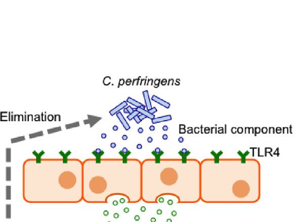
Takehara et al.                                                                                                     TLR4 and C. perfringens Infection

(Stephens, 1996; Bryant and Stevens, 2010). More effective                   recognition of structural components of microorganisms;
therapeutic strategies are urgently required.                                pattern recognition receptors, such as toll-like receptors
    C. perfringens type A has been reported to produce multiple              (TLRs), are responsible for this recognition (Boettcher et al.,
virulence factors. Many studies have focused on the biological               2014; Manz and Boettcher, 2014; Wirths et al., 2014). Among the
and biochemical activities of a-toxin (or phospholipase C),                  TLR family members, TLR4 has been shown to recognize the
which has both phospholipase C (PLC) and sphingomyelinase                    Gram-negative bacterial endotoxin lipopolysaccharide (LPS) and
(SMase) activities (Bryant, 2003; Sakurai et al., 2004). Using a C.          to protect against Gram-negative bacteria (Takeuchi et al., 1999;
perfringens infection mouse model, an a-toxin-deficient strain                Beutler, 2000). TLR2 has been identified as pivotal for the
was demonstrated to exhibit delayed spread of muscle necrosis                recognition of Gram-positive bacteria, and a cell wall
(Ellemor et al., 1999). It is supposed that a-toxin is involved in           component, peptidoglycan (PGN), is known to be its ligand
the spread of infection by multiple mechanisms. a-Toxin                      (Takeuchi et al., 1999; Dziarski, 2003; Texereau et al., 2005). LPS
enhances intravascular cell aggregation and impairs the host                 or PGN stimulates TLR signaling and increases the production of
immune response by impeding inflammatory cell infiltration to                  granulocyte colony-stimulating factor (G-CSF), a glycoprotein
the site of infection (Bryant et al., 2000; Bahl and Dürre, 2001;            that influences the proliferation, survival, and differentiation of
Ochi et al., 2002; Bryant et al., 2006). We previously reported that         neutrophils, leading to the acceleration of granulopoiesis
a-toxin inhibits neutrophil production by interfering with                   (Boettcher et al., 2012; Takehara et al., 2017). Thus, TLR2 and
differentiation in the bone marrow, leading to the impairment                TLR4 have been demonstrated to protect against Gram-positive
of innate host immunity (Takehara et al., 2016a; Takehara et al.,            or Gram-negative bacterial infection by sensing the respective
2016b). These results indicate that a-toxin is involved in                   structural components peculiar to each bacterium. However, the
avoiding host immune clearance. Additionally, a-toxin induces                role of TLR4 in Gram-positive bacterial infection is less well
death of endothelial cells, suggesting that it disturbs peripheral           understood because the bacteria do not contain LPS.
circulation (Takehara et al., 2020a). The a-toxin-mediated                      C. perfringens q-toxin was demonstrated to be a TLR4 agonist
intravascular cell aggregation also promotes vascular occlusion              (Park et al., 2004). This finding suggests that TLR4 contributes to
(Bryant et al., 2000; Hickey et al., 2008). Moreover, a-toxin                the recognition of C. perfringens. Additionally, we reported that
inhibits the differentiation of erythroid progenitors, which might           a-toxin amplifies LPS-induced inflammatory responses
lead to hypogloburia (Takagishi et al., 2017). These results                 (Takehara et al., 2019a). However, the role of TLR4 in C.
suggest that a-toxin affects the tissue microcirculation, leading            perfringens infection is unclear. In this study, we investigated
to ischemic conditions in the muscle tissue. Recently, we                    whether TLR4 mutation influences susceptibility to C.
reported that a-toxin inhibits the differentiation of myoblasts,             perfringens and found that it plays an important role in
which might impair muscle tissue repair (Takehara et al., 2020b).            elimination. Our findings provide a novel perspective for
Thus, a-toxin is involved in disease progression via various                 understanding the host defense mechanism against
mechanisms. Besides a-toxin, q-toxin (or perfringlysin O),                   C. perfringens.
which is a pore-forming and cholesterol-dependent cytolysin,
contributes to the progression of myonecrosis (Rood, 1998;
Verherstraeten et al., 2015). It was reported that q-toxin-                  METHODS
deficient strains show delayed spread of muscle necrosis
(Ellemor et al., 1999). q-toxin also enhances intravascular cell             Mice
aggregation, leading to vascular occlusion (Bryant et al., 1993;             Mice aged more than 8 weeks were used for all experiments. All
Hickey et al., 2008). Additionally, C. perfringens produces other            mice were from SLC (Shizuoka, Japan). The Animal Care and
toxins and enzymes including a collagenase, hyaluronidase,                   Use Committee of Tokushima Bunri University approved the
sialidases, and the cysteine protease a-clostripain (Rood, 1998;             animal experiments, and the procedures were performed in
Shimizu et al., 2002). Thus, the infection process and virulence             accordance with institutional guidelines. The institutional
factors have become increasingly clear, but our understanding                guidelines conform to the Fundamental Guidelines for Proper
remains limited regarding the host immune exclusion                          Conduct of Animal Experiment and Related Activities in
mechanism after C. perfringens infection.                                    Academic Research Institutions under the jurisdiction of the
    We previously showed that neutrophil elimination by anti-                Ministry of Education, Culture, Sports, Science and Technology,
Ly6G treatment increases viable cell counts after injection of an            2006. The mice were kept in a specific pathogen-free animal
a-toxin-deficient mutant of C. perfringens into skeletal muscle,              facility at Tokushima Bunri University.
suggesting that neutrophils play a key role in the elimination of
C. perfringens (Takehara et al., 2016a). Neutrophils phagocytose             Reagents and Strain
and kill pathogenic bacteria and are important innate immune                 Fluorescein isothiocyanate (FITC)-, phycoerythrin (PE)- and
cells comprising the first line of host defense against invading              allophycocyanin (APC)-conjugated specific antibodies against
microorganisms (Dale et al., 2008; Amulic et al., 2012;                      mouse CD11b (clone M1/70), Ly-6G (clone 1A8), NK1.1 (clone
Kolaczkowska and Kubes, 2013). Generally, granulopoiesis                     PK136) or CD3 epsilon (clone 145-2C11), an antibody against
accelerates to replenish neutrophils during bacterial infection.             CD31 (clone MEC 13.3), and purified rat anti-mouse CD16/CD32
This so-called emergency granulopoiesis is initiated by the                  (Fc Block) were purchased from BD Biosciences (CA, USA).

Frontiers in Cellular and Infection Microbiology | www.frontiersin.org   2                                  March 2021 | Volume 11 | Article 633440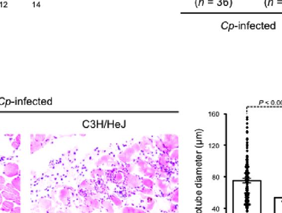
Takehara et al. TLR4 and C. perfringens Infection
Antibody against interleukin-1b (IL-1b) (clone 11E5) was from were isolated 24 h after the infection. Each isolated muscle was
Santa Cruz Biotechnology (CA, USA). Alexa Fluor 488 goat cut into small pieces of 2–4 mm in PBS and dissociated in a
anti-rat IgG and Chromeo 546 goat anti-mouse IgG were gentleMACS C tube (Miltenyi Biotec, Bergisch Gladbach,
obtained from Abcam (MA, USA). All other chemicals were of Germany) using a gentleMACS dissociator (Miltenyi Biotec,
the highest grade available from commercial sources. Wild-type Bergisch Gladbach, Germany). Measurements of G-CSF,
C. perfringens Strain 13 was used in this study. interleukin-6 (IL-6) and IL-1b levels in the supernatant were
performed using mouse Quantikine ELISA kits in accordance
Bacterial Culture and Infection with the manufacturer’s instructions (R&D Systems, MN, USA).
Bacterial culture and infection were performed as previously
described (Takehara et al., 2016a). C. perfringens strain 13 was Quantification of C. perfringens CFUs in
grown in TGY (tryptone, glucose, and yeast extract) medium in Femoral Muscle
anaerobic conditions at 37°C. Exponentially growing bacteria Mice were intramuscularly injected with 1 × 10⁷ CFU of
were harvested, washed, re-suspended in TGY medium, and C. perfringens, and C. perfringens-infected femoral muscles
injected into the femoral muscles of mice. Residual bacteria were were isolated 72 h after the infection. To quantify C.
serially diluted, plated on brain heart infusion agar plates, and perfringens CFUs, the isolated muscles were cut into small
cultured anaerobically at 37°C to quantify colony-forming units pieces of 2–4 mm in TGY medium and dissociated in a
(CFUs). Survival of mice intramuscularly injected with 5 × 10⁷ gentleMACS C tube using a gentleMACS dissociator as
CFU of C. perfringens was monitored. described previously (Takehara et al., 2016a). The supernatants
were serially diluted, plated on BHI agar plates, and cultured
Immunohistochemistry anaerobically at 37°C.
Immunohistochemistry was performed as previously described
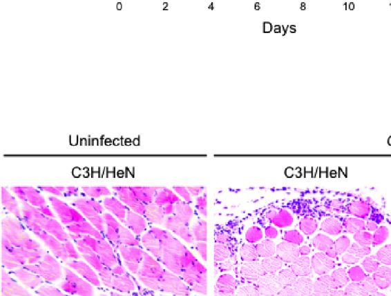
with some modifications (Takehara et al., 2019a). Mice were Myotube Morphology Analysis
intramuscularly injected with 1 × 10⁷ CFU of C. perfringens, and Myotube morphology was analyzed as previously described
C. perfringens-infected femoral muscles were isolated 24 h after (Takehara et al., 2019b). Mice were intramuscularly injected
the infection. Samples were embedded in OCT compound with 1 × 10⁷ CFU of C. perfringens, and C. perfringens-infected
(Sakura Finetek Japan, Tokyo, Japan), and cryosectioning of muscles were isolated 24 h after infection. The isolated muscles
the frozen tissue was performed using a cryostat microtome were fixed in 4% paraformaldehyde and embedded in paraffin.
(Leica, IL, USA). Sections were blocked using Blocking One To visualize muscle fibers, paraffin sections were cut from the
Histo (Nacalai Tesque, Inc., Kyoto, Japan), and incubated with tissue and stained with hematoxylin and eosin. Pictures of
primary antibodies. Finally, the sections were incubated for 1 h the muscle fibers were taken using a digital camera, and the
with secondary antibodies. The antibodies were diluted in diameters of muscle fibers were measured using DS-L4 (Nikon,
DAKO Antibody Diluent (DAKO, Glostrup, Denmark). Nuclei Tokyo, Japan). The diameters of 100 muscle fibers were
were stained with 4′,6-diamino-2-phenylindole (DAPI). Images measured for each condition.
were captured on a confocal laser-scanning fluorescence
microscope (Nikon A1, Nikon instruments, Tokyo, Japan). Microarray Analysis
Mice were intramuscularly injected with 1 × 10⁷ CFU of
Flow Cytometry Analysis C. perfringens, and C. perfringens-infected femoral muscles
Mice were intramuscularly injected with 1 × 10⁷ CFU of were isolated 1.5 h after the infection. The isolated muscles
C. perfringens, and spleens were isolated 72 h after the were cut into small pieces of 2–4 mm in lysis buffer RLT of an
infection. To isolate spleen cells, each isolated spleen was RNeasy mini kit (QIAGEN, Hilden, Germany) and dissociated in
crushed in phosphate-buffered saline (PBS) supplemented with a gentleMACS M tube (Miltenyi Biotec, Bergisch Gladbach,
2% heat-inactivated fetal bovine serum (FBS; AusGeneX, Germany) using a gentleMACS dissociator. Total RNA was
QLD, Australia), and filtered through a 40 mm mesh. Red extracted using the RNeasy mini kit, and the quality of purified
blood cells were hemolyzed with lysis buffer (ACK lysing RNA was assessed by Filgen Incorporated (Aichi, Japan).
buffer; GIBCO, NY, USA). The number of living cells was Microarray analysis was also performed by Filgen Incorporated
counted after trypan blue staining. using the Clariom S Assay for mice (Thermo Fisher Scientific
Flow cytometry analysis was performed as described Incorporated, MA, USA) and GeneChip Scanner 3000 7G
previously (Takehara et al., 2016a). Briefly, cells were labeled (Thermo Fisher Scientific Incorporated, MA, USA). Scan data
with antibodies diluted in PBS containing 2% FBS after blocking were analyzed using a software package (Expression Console
Fc-receptors with purified rat anti-mouse CD16/CD32. The Software; Thermo Fisher Scientific Incorporated, MA, USA).
labeled cells were analyzed using a Guava easyCyte (Millipore,
MA, USA), and data were analyzed using FlowJo software (Tree Statistical Analysis
Star, OR, USA). All statistical analyses were performed with Easy R (Saitama
Medical Center, Jichi Medical University) (Kanda, 2013).
ELISA Differences between two groups were evaluated using two-
Mice were intramuscularly injected with 1 × 10⁷ CFU of tailed Student’s t-test. One-way analysis of variance (ANOVA)
C. perfringens, and C. perfringens-infected femoral muscles followed by the Tukey test was used to evaluate differences
Frontiers in Cellular and Infection Microbiology | www.frontiersin.org 3 March 2021 | Volume 11 | Article 633440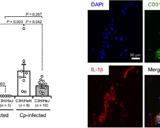
Takehara et al.                                                                                                                     TLR4 and C. perfringens Infection

among three or more groups. Differences were considered to be                        injected with C. perfringens, but the severity was greater in C3H/
significant for values of P
Takehara et al. TLR4 and C. perfringens Infection
B
A
FIGURE 2 | TLR4 signaling accelerates expression of inflammatory cytokines during C. perfringens infection. C3H/HeN and C3H/HeJ mice were intramuscularly
injected with 1 × 10⁷ CFU of C. perfringens Strain 13 (Cp-infected), or TGY medium as a control (Uninfected). At 24 h after the infection, IL-1b and IL-6 levels in the
muscle were determined (A), or the muscle was subjected to immunohistochemical analysis with antibodies against CD31 and IL-1b (B). One-way ANOVA was
employed to assess significance. Values are mean ± standard error.
perfringens-infected muscle from C3H/HeN mice, while the upregulated in C. perfringens-infected mice, so our results
increase was limited in that from C3H/HeJ mice, suggesting corroborate the previous report (Low et al., 2018). Next, a
that TLR4 plays a role in the regulation of granulopoiesis during comparison of gene expression levels between C3H/HeN and
C. perfringens infection (Figure 3A). During systemic C3H/HeJ mice was performed. In C. perfringens-infected C3H/
Escherichia coli infection, stimulation of TLR4 mobilizes HeJ mice, 351 genes were upregulated more than 1.5-fold, and
CD150+CD48−Lineage−/lowSca1+cKit+ hematopoietic stem cells 258 genes were downregulated less than 1.5-fold compared with
(HSCs) to the spleen to give rise to neutrophils, which C. perfringens-infected C3H/HeN mice (Figure 4B). The genes
contributes to the host defense (Burberry et al., 2014). The are listed in Supplementary Table S2. Among the 179 genes
number of CD11b+Ly-6G+ neutrophils was greatly increased in upregulated by C. perfringens infection, 21 genes were
spleens from C. perfringens-infected C3H/HeN mice compared downregulated in C3H/HeJ mice compared with C3H/HeN
with uninfected control mice, whereas the increase was limited in mice (Figure 4C). Also, 18 genes were upregulated in C3H/
C. perfringens-infected C3H/HeJ mice (Figure 3B). Meanwhile, HeJ mice compared with C3H/HeN mice among the 82 genes
the numbers of NK1.1−CD3+ T cells and NK1.1+CD3− natural downregulated by C. perfringens infection (Figure 4C). The
killer cells were similar in both mouse lines (Figure 3B). genes are listed in Supplementary Table S3. These results
Together, our results indicate that TLR4 signaling plays an demonstrate that TLR4 signaling is partially involved in
important role in the regulation of G-CSF-mediating changes in host gene expression associated with C.
granulopoiesis during C. perfringens infection, and this perfringens infection.
mechanism probably contributes to the replenishment of
neutrophils and the elimination of bacteria (Figure 3C).
TLR4 Signaling Is Partially Involved in DISCUSSION
Changes in Host Gene Expression C. perfringens type A is a Gram-positive bacterium that causes
Associated With C. perfringens Infection gas gangrene characterized by severe myonecrosis (Songer, 1996;
It was previously demonstrated that 1,055 host genes are Petit et al., 1999). It has been demonstrated using TLR2-deficient
upregulated, and 386 host genes are downregulated in response mice that TLR2 contributes to the host defense against Gram-
to C. perfringens infection by RNA sequencing (Low et al., 2018). positive bacterial infection. TLR2-deficient mice are greatly
In that study, RNA was extracted from mice at 1.5 h post- susceptible to Staphylococcus aureus, showing impaired
infection because disease progression in murine models is rapid. clearance of the infected bacteria (Takeuchi et al., 2000; Hoebe
In the present study, RNA was also extracted from C. et al., 2005). Similarly, TLR2-deficiency reduces the elimination
perfringens-infected muscle at 1.5 h post-infection, and host of Streptococcus pneumoniae, resulting in increased susceptibility
gene expression was measured by DNA microarray analysis. In to the bacteria (Echchannaoui et al., 2002). In the clinical setting,
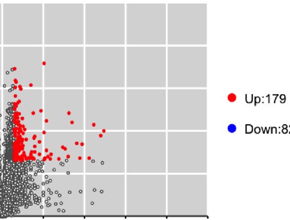
C. perfringens-infected C3H/HeN mice, 179 genes were some polymorphisms in the Tlr2 gene, which attenuates TLR2
upregulated more than 1.5-fold, and 82 genes were signaling, increase the risk of severe tuberculosis caused by
downregulated less than 1.5-fold compared with uninfected Mycobacterium tuberculosis (Ben-Ali et al., 2004; Ogus et al.,
control mice (Figure 4A). Host immunity genes, such as 2004). Moreover, TLR2 polymorphisms affect immune responses
CXCL2, Trem1, CXCL1, Il1b, and CXCL3, were significantly to Mycobacterium leprae (Kang et al., 2002; Bochud et al., 2003).
upregulated in C. perfringens-infected mice (Supplementary Thus, TLR2 has been demonstrated to play an important role in
Table S1). These genes were already reported to be the recognition and elimination of Gram-positive bacteria.
Frontiers in Cellular and Infection Microbiology | www.frontiersin.org 5 March 2021 | Volume 11 | Article 633440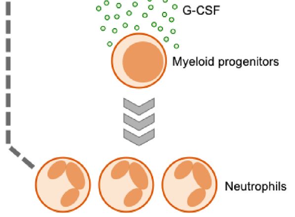
Takehara et al.                                                                                                                      TLR4 and C. perfringens Infection

                                      A                                                  B

                                      C

 FIGURE 3 | TLR4 signaling accelerates production of neutrophils. C3H/HeN and C3H/HeJ mice were intramuscularly injected with 1 × 10⁷ CFU of C. perfringens
 Strain 13 (Cp-infected), or TGY medium as a control (Uninfected). (A) At 24 h after infection, G-CSF levels in the muscle were determined. (B) At 72 h after the
 infection, spleen cells were isolated from the mice, and flow cytometry analysis was performed using a Guava easyCyte. The proportions of CD11b+Ly-6G+
 neutrophils, NK1.1−CD3+ T cells and NK1.1+CD3− natural killer cells are shown. (C) Model of accelerated production of neutrophils in C. perfringens-infected host
 through TLR4 signaling activation. One-way ANOVA was employed to assess significance. Values are mean ± standard error.

However, the role of TLR4 in Gram-positive bacterial infection is                     Norazmi, 2013). It was also reported that M. tuberculosis itself
less well understood. The present study found that TLR4-                              binds to TLR4 (Sepehri et al., 2019). Anthrolysin O (ALO) from
defective C3H/HeJ mice infected with C. perfringens have a                            Bacillus anthracis can activate TLR4, inducing an apoptotic
remarkably lower survival rate, an increase in viable bacterial                       response in bone marrow-derived macrophages (BMDMs)
counts, and accelerated destruction of myofibrils at the infection                     (Park et al., 2004). ALO is a cholesterol-dependent cytolysin
site compared with wild-type C3H/HeN mice. These results                              (CDC), and the other CDCs, such as C. perfringens q-toxin,
indicate that TLR4 plays an important role in the elimination                         Listeria monocytogenes listeriolysin O, and Streptococcus
of C. perfringens and the host defense against the bacteria.                          pyogenes streptolysin O, can activate BMDMs through TLR4
Similar to our results, TLR4 was shown to contribute to host                          signaling (Park et al., 2004). Thus, some Gram-positive bacteria
protection against M. tuberculosis infection (Park et al., 2020).                     contain TLR4 ligands, and it might be possible that activation of
Thus, it is important to pay attention to the function of TLR4                        TLR4 at the time of Gram-positive bacterial infection is a
during Gram-positive bacterial infection.                                             common phenomenon.
    Gram-positive bacteria do not contain LPS, but some ligands                          Pattern recognition receptors, such as TLRs, are responsible
of TLR4 have been identified from these bacteria. Within the                           for the recognition of structural components of microorganisms,
components of M. tuberculosis, lipomannans and heat shock                             and their activation affects the expression of inflammatory
proteins bind to TLR4 and activate immune cells (Hossain and                          cytokines (O’Neill, 2002). Among the TLR family members,

Frontiers in Cellular and Infection Microbiology | www.frontiersin.org            6                                         March 2021 | Volume 11 | Article 633440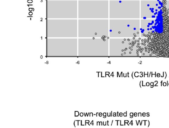
Takehara et al.                                                                                                                       TLR4 and C. perfringens Infection

                                   A

                                   B

                               C

 FIGURE 4 | Differentially expressed genes between C3H/HeN and C3H/HeJ mice during C. perfringens infection. C3H/HeN and C3H/HeJ mice were intramuscularly
 injected with 1 × 10⁷ CFU of C. perfringens Strain 13 (Cp-infected), or TGY medium as a control (Uninfected). At 1.5 h after infection, muscles were isolated from the
 mice, and total RNAs from the tissues were subjected to DNA microarray analysis as described in Methods. (A) Volcano plot depicting information about differentially
 expressed genes between C. perfringens-infected and uninfected muscles from C3H/HeN mice. (B) Volcano plot depicting information about differentially expressed
 genes in C. perfringens-infected muscles between C3H/HeN and C3H/HeJ mice. (C) Venn diagrams for microarray data.

TLR4 has been shown to recognize the Gram-negative bacterial                           2017). G-CSF promotes the proliferation and differentiation
endotoxin LPS, and TLR2 has been identified as pivotal for the                          of neutrophils and their progenitor cells (Demetri and Griffin,
recognition of cell wall components such as PGN (Takeuchi                              1991). G-CSF-deficient mice exhibit chronic neutropenia and
et al., 1999; Beutler, 2000; Dziarski, 2003; Texereau et al., 2005).                   reduced infection-driven granulopoiesis. Granulopoiesis is
During Gram-negative bacterial infection, LPS stimulates the                           also impaired in G-CSF receptor-deficient mice (Lieschke
production of G-CSF, which is a glycoprotein, leading to the                           et al., 1994). Thus, G-CSF plays a key role in regulating
acceleration of granulopoiesis, and this phenomenon depends on                         granulopoiesis during bacterial infection, and TLR2 and TLR4
TLR4-expressing endothelial cells (Boettcher et al., 2014).                            are essential to the host defense mechanism. The present study
Similarly, PGN is suggested to stimulate the production of                             observed remarkable increases in the levels of inflammatory
G-CSF in TLR2-expressing endothelial cells (Takehara et al.,                           cytokines IL-1b, IL-6, and G-CSF, in C. perfringens-infected

Frontiers in Cellular and Infection Microbiology | www.frontiersin.org             7                                         March 2021 | Volume 11 | Article 633440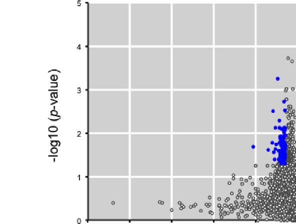
Takehara et al.                                                                                                                                    TLR4 and C. perfringens Infection

C3H/HeN mice. The cytokines would be secreted from                                             DATA AVAILABILITY STATEMENT
endothelial cells but not infiltrating immune cells because it
was demonstrated that the infiltration of immune cells is                                       The raw data supporting the conclusions of this article will be
impaired by a-toxin in C. perfringens-infected muscle                                          made available by the authors, without undue reservation.
(Takehara et al., 2016a). The result shown in Figure 2B
reinforces this idea. Moreover, the cytokines were only
partially increased in C3H/HeJ mice. The results suggest that                                  ETHICS STATEMENT
TLR2 plays a complementary role to TLR4 during C. perfringens
infection. It might be that TLR2 and TLR4, signaling                                           The animal study was reviewed and approved by Animal Care
cooperatively, regulate the production of G-CSF to control                                     and Use Committee of Tokushima Bunri University.
granulopoiesis effectively and strictly. Otherwise, further
studies will be needed to unveil the functional difference
between TLR2 and TLR4 signaling on the regulation of                                           AUTHOR CONTRIBUTIONS
neutrophil production.
   It has been reported that activation of TLRs should be                                      MT and MN designed the study and supervised experiments. MT
properly regulated to avoid tissue damage by excessive                                         performed experiments and analyses, and wrote the manuscript.
inflammation (Kawai and Akira, 2010). Ubiquitin ligases,                                        KK contributed to the design of in vivo studies. All authors
which are splice variants for adaptors and transcriptional                                     contributed to the article and approved the submitted version.
regulators, have been identified as negative regulators of TLR
signaling (Kawai and Akira, 2010). We recently reported that a-
toxin amplifies LPS-induced inflammatory responses and                                           FUNDING
increases the lethal toxicity of LPS, which is dependent on
TLR4 (Takehara et al., 2019a). In the previous report, it was                                  This work was supported by a Grant-in-Aid for Scientific
unclear whether it is meaningful to speculate on the role of a-                                Research from the Ministry of Education, Culture, Sports,
toxin in the TLR4-mediated inflammatory response in C.                                          Science, and Technology of Japan (grant number 18K07129).
perfringens infection, because C. perfringens do not contain
LPS. In the present study, we showed that TLR4 plays an
important role in the elimination of C. perfringens, indicating                                ACKNOWLEDGMENTS
that TLR4 signaling is activated during infection. As described
above, q-toxin was reported to be a TLR4 agonist (Park et al.,                                 We thank Hiroto Bandou and Daiki Yamane for providing
2004). Together, it might be possible that q-toxin activates TLR4,                             technical assistance.
and that the excessive activation of TLR4 by a-toxin contributes
to the characteristics of C. perfringens infection, such as the
destruction of muscle, shock, multiple organ failure, systemic                                 SUPPLEMENTARY MATERIAL
inflammation, and death of patients. In conclusion, our results
indicate that TLR4 plays an important role in the elimination                                  The Supplementary Material for this article can be found online
of C. perfringens, which provides a novel perspective for                                      at: https://www.frontiersin.org/articles/10.3389/fcimb.2021.
understanding the pathogenesis of C. perfringens.                                              633440/full#supplementary-material

REFERENCES                                                                                     Boettcher, S., Gerosa, R. C., Radpour, R., Bauer, J., Ampenberger, F.,
                                                                                                  Heikenwalder, M., et al. (2014). Endothelial cells translate pathogen signals
Amulic, B., Cazalet, C., Hayes, G. L., Metzler, K. D., and Zychlinsky, A. (2012).                 into G-CSF-driven emergency granulopoiesis. Blood 124, 1393–1403. doi:
   Neutrophil function: from mechanisms to disease. Annu. Rev. Immunol. 30,                       10.1182/blood-2014-04-570762
   459–489. doi: 10.1146/annurev-immunol-020711-074942                                         Bryant, A. E., and Stevens, D. L. (2010). Clostridial myonecrosis: new insights in
Bahl, H., and Dürre, P. (2001). Clostridia : biotechnology and medical applications.,             pathogenesis and management. Curr. Infect. Dis. Rep. 12, 383–391. doi:
   (Weinheim Chichester: Wiley-VCH).                                                              10.1007/s11908-010-0127-y
Ben-Ali, M., Barbouche, M. R., Bousnina, S., Chabbou, A., and Dellagi, K. (2004).              Bryant, A. E., Bergstrom, R., Zimmerman, G. A., Salyer, J. L., Hill, H. R., Tweten, R. K.,
   Toll-like receptor 2 Arg677Trp polymorphism is associated with susceptibility                  et al. (1993). Clostridium perfringens invasiveness is enhanced by effects of theta
   to tuberculosis in Tunisian patients. Clin. Diagn. Lab. Immunol. 11, 625–626.                  toxin upon PMNL structure and function: the roles of leukocytotoxicity and
   doi: 10.1128/CDLI.11.3.625-626.2004                                                            expression of CD11/CD18 adherence glycoprotein. FEMS Immunol. Med.
Beutler, B. (2000). Tlr4: central component of the sole mammalian LPS sensor.                     Microbiol. 7, 321–336. doi: 10.1111/j.1574-695X.1993.tb00414.x
   Curr. Opin. Immunol. 12, 20–26. doi: 10.1016/S0952-7915(99)00046-1                          Bryant, A. E., Chen, R. Y., Nagata, Y., Wang, Y., Lee, C. H., Finegold, S., et al.
Bochud, P. Y., Hawn, T. R., and Aderem, A. (2003). Cutting edge: a Toll-like receptor 2           (2000). Clostridial gas gangrene. II. Phospholipase C-induced activation of
   polymorphism that is associated with lepromatous leprosy is unable to mediate                  platelet gpIIbIIIa mediates vascular occlusion and myonecrosis in Clostridium
   mycobacterial signaling. J. Immunol. 170, 3451–3454. doi: 10.4049/jimmunol.170.7.3451          perfringens gas gangrene. J. Infect. Dis. 182, 808–815. doi: 10.1086/315757
Boettcher, S., Ziegler, P., Schmid, M. A., Takizawa, H., Van Rooijen, N., Kopf, M.,            Bryant, A. E., Bayer, C. R., Aldape, M. J., Wallace, R. J., Titball, R. W., and Stevens,
   et al. (2012). Cutting edge: LPS-induced emergency myelopoiesis depends on                     D. L. (2006). Clostridium perfringens phospholipase C-induced platelet/
   TLR4-expressing nonhematopoietic cells. J. Immunol. 188, 5824–5828. doi:                       leukocyte interactions impede neutrophil diapedesis. J. Med. Microbiol. 55,
   10.4049/jimmunol.1103253                                                                       495–504. doi: 10.1099/jmm.0.46390-0

Frontiers in Cellular and Infection Microbiology | www.frontiersin.org                     8                                             March 2021 | Volume 11 | Article 633440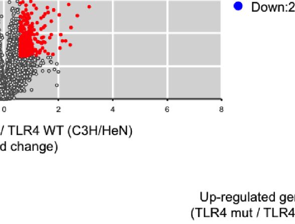
Takehara et al.                                                                                                                                        TLR4 and C. perfringens Infection

Bryant, A. E. (2003). Biology and pathogenesis of thrombosis and procoagulant activity                 optimal host protection against highly virulent Mycobacterium tuberculosis K
    in invasive infections caused by group A streptococci and Clostridium perfringens.                 infection. Virulence 11, 430–445. doi: 10.1080/21505594.2020.1766401
    Clin. Microbiol. Rev. 16, 451–462. doi: 10.1128/CMR.16.3.451-462.2003                           Petit, L., Gibert, M., and Popoff, M. R. (1999). Clostridium perfringens: toxinotype and
Burberry, A., Zeng, M. Y., Ding, L., Wicks, I., Inohara, N., Morrison, S. J., et al.                   genotype. Trends Microbiol. 7, 104–110. doi: 10.1016/S0966-842X(98)01430-9
    (2014). Infection mobilizes hematopoietic stem cells through cooperative                        Poltorak, A., He, X., Smirnova, I., Liu, M. Y., Van Huffel, C., Du, X., et al. (1998).
    NOD-like receptor and Toll-like receptor signaling. Cell Host Microbe 15,                          Defective LPS signaling in C3H/HeJ and C57BL/10ScCr mice: mutations in
    779–791. doi: 10.1016/j.chom.2014.05.004                                                           Tlr4 gene. Science 282, 2085–2088. doi: 10.1126/science.282.5396.2085
Dale, D. C., Boxer, L., and Liles, W. C. (2008). The phagocytes: neutrophils and                    Rood, J. I. (1998). Virulence genes of Clostridium perfringens. Annu. Rev.
    monocytes. Blood 112, 935–945. doi: 10.1182/blood-2007-12-077917                                   Microbiol. 52, 333–360. doi: 10.1146/annurev.micro.52.1.333
Demetri, G. D., and Griffin, J. D. (1991). Granulocyte colony-stimulating factor                     Sakurai, J., Nagahama, M., and Oda, M. (2004). Clostridium perfringens alpha-
    and its receptor. Blood 78, 2791–2808. doi: 10.1182/blood.V78.11.2791.                             toxin: characterization and mode of action. J. Biochem. 136, 569–574. doi:
    bloodjournal78112791                                                                               10.1093/jb/mvh161
Dziarski, R. (2003). Recognition of bacterial peptidoglycan by the innate immune                    Sepehri, Z., Kiani, Z., Kohan, F., and Ghavami, S. (2019). Toll-Like Receptor 4 as
    system. Cell Mol. Life Sci. 60, 1793–1804. doi: 10.1007/s00018-003-3019-6                          an Immune Receptor Against Mycobacterium tuberculosis: A Systematic
Echchannaoui, H., Frei, K., Schnell, C., Leib, S. L., Zimmerli, W., and Landmann, R.                   Review. Lab. Med. 50, 117–129. doi: 10.1093/labmed/lmy047
    (2002). Toll-like receptor 2-deficient mice are highly susceptible to Streptococcus              Shimizu, T., Ohtani, K., Hirakawa, H., Ohshima, K., Yamashita, A., Shiba, T., et al.
    pneumoniae meningitis because of reduced bacterial clearing and enhanced                           (2002). Complete genome sequence of Clostridium perfringens, an anaerobic
    inflammation. J. Infect. Dis. 186, 798–806. doi: 10.1086/342845                                     flesh-eater. Proc. Natl. Acad. Sci. U.S.A. 99, 996–1001. doi: 10.1073/
Ellemor, D. M., Baird, R. N., Awad, M. M., Boyd, R. L., Rood, J. I., and Emmins, J. J.                 pnas.022493799
    (1999). Use of genetically manipulated strains of Clostridium perfringens reveals               Songer, J. G. (1996). Clostridial enteric diseases of domestic animals. Clin.
    that both alpha-toxin and theta-toxin are required for vascular leukostasis to                     Microbiol. Rev. 9, 216–234. doi: 10.1128/CMR.9.2.216
    occur in experimental gas gangrene. Infect. Immun. 67, 4902–4907. doi:                          Stephens, M. B. (1996). Gas gangrene: potential for hyperbaric oxygen therapy.
    10.1128/IAI.67.9.4902-4907.1999                                                                    Postgrad. Med. 99, 217–220, 224. doi: 10.1080/00325481.1996.11946109
Hickey, M. J., Kwan, R. Y., Awad, M. M., Kennedy, C. L., Young, L. F., Hall, P.,                    Takagishi, T., Takehara, M., Seike, S., Miyamoto, K., Kobayashi, K., and
    et al. (2008). Molecular and cellular basis of microvascular perfusion deficits                     Nagahama, M. (2017). Clostridium perfringens alpha-toxin impairs
    induced by Clostridium perfringens and Clostridium septicum. PloS Pathog. 4,                       erythropoiesis by inhibition of erythroid differentiation. Sci. Rep. 7, 5217.
    e1000045. doi: 10.1371/journal.ppat.1000045                                                        doi: 10.1038/s41598-017-05567-8
Hifumi, T. (2020). Spontaneous non-traumatic Clostridium perfringens sepsis. Jpn.                   Takehara, M., Takagishi, T., Seike, S., Ohtani, K., Kobayashi, K., Miyamoto, K.,
    J. Infect. Dis. 73, 177–180. doi: 10.7883/yoken.JJID.2019.382                                      et al. (2016a). Clostridium perfringens alpha-Toxin Impairs Innate Immunity
Hoebe, K., Georgel, P., Rutschmann, S., Du, X., Mudd, S., Crozat, K., et al. (2005).                   via Inhibition of Neutrophil Differentiation. Sci. Rep. 6, 28192. doi: 10.1038/
    CD36 is a sensor of diacylglycerides. Nature 433, 523–527. doi: 10.1038/                           srep28192
    nature03253                                                                                     Takehara, M., Takagishi, T., Seike, S., Oishi, K., Fujihara, Y., Miyamoto, K., et al.
Hossain, M. M., and Norazmi, M. N. (2013). Pattern recognition receptors and                           (2016b). Clostridium perfringens alpha-Toxin Impairs Lipid Raft Integrity in
    cytokines in Mycobacterium tuberculosis infection–the double-edged sword?                          Neutrophils. Biol. Pharm. Bull. 39, 1694–1700. doi: 10.1248/bpb.b16-00444
    BioMed. Res. Int. 2013, 179174. doi: 10.1155/2013/179174                                        Takehara, M., Seike, S., Takagishi, T., Kobayashi, K., and Nagahama, M. (2017).
Kanda, Y. (2013). Investigation of the freely available easy-to-use software ‘EZR’ for                 Peptidoglycan accelerates granulopoiesis through a TLR2- and MyD88-
    medical statistics. Bone Marrow Transplant. 48, 452–458. doi: 10.1038/bmt.2012.244                 dependent pathway. Biochem. Biophys. Res. Commun. 487, 419–425. doi:
Kang, T. J., Lee, S. B., and Chae, G. T. (2002). A polymorphism in the toll-like                       10.1016/j.bbrc.2017.04.077
    receptor 2 is associated with IL-12 production from monocyte in lepromatous                     Takehara, M., Seike, S., Sonobe, Y., Bandou, H., Yokoyama, S., Takagishi, T., et al.
    leprosy. Cytokine 20, 56–62. doi: 10.1006/cyto.2002.1982                                           (2019a). Clostridium perfringens alpha-toxin impairs granulocyte colony-
Kawai, T., and Akira, S. (2010). The role of pattern-recognition receptors in innate                   stimulating factor receptor-mediated granulocyte production while triggering
    immunity: update on Toll-like receptors. Nat. Immunol. 11, 373–384. doi:                           septic shock. Commun. Biol. 2, 45. doi: 10.1038/s42003-019-0280-2
    10.1038/ni.1863                                                                                 Takehara, M., Sonobe, Y., Bandou, H., Kobayashi, K., and Nagahama, M. (2019b).
Kolaczkowska, E., and Kubes, P. (2013). Neutrophil recruitment and function in                         Granulocyte Colony-Stimulating Factor Does Not Influence Clostridium
    health and inflammation. Nat. Rev. Immunol. 13, 159–175. doi: 10.1038/nri3399                       Perfringens alpha-Toxin-Induced Myonecrosis in Mice. Toxins (Basel) 11.
Lieschke, G. J., Grail, D., Hodgson, G., Metcalf, D., Stanley, E., Cheers, C., et al. (1994).          doi: 10.3390/toxins11090509
    Mice lacking granulocyte colony-stimulating factor have chronic neutropenia,                    Takehara, M., Bandou, H., Kobayashi, K., and Nagahama, M. (2020a). Clostridium
    granulocyte and macrophage progenitor cell deficiency, and impaired neutrophil                      perfringens a-toxin specifically induces endothelial cell death by promoting
    mobilization. Blood 84, 1737–1746. doi: 10.1182/blood.V84.6.1737.1737                              ceramide-mediated apoptosis. Anaerobe 65, 102262. doi: 10.1016/
Low, L. Y., Harrison, P. F., Gould, J., Powell, D. R., Choo, J. M., Forster, S. C., et al.             j.anaerobe.2020.102262
    (2018). Concurrent Host-Pathogen Transcriptional Responses in a Clostridium                     Takehara, M., Kobayashi, K., and Nagahama, M. (2020b). Clostridium perfringens
    perfringens Murine Myonecrosis Infection. MBio 9:e00473-18. doi: 10.1128/                          a-toxin inhibits myogenic differentiation of C2C12 myoblasts. Anaerobe 65,
    mBio.00473-18                                                                                      102265. doi: 10.1016/j.anaerobe.2020.102265
Manz, M. G., and Boettcher, S. (2014). Emergency granulopoiesis. Nat. Rev.                          Takeuchi, O., Hoshino, K., Kawai, T., Sanjo, H., Takada, H., Ogawa, T., et al.
    Immunol. 14, 302–314. doi: 10.1038/nri3660                                                         (1999). Differential roles of TLR2 and TLR4 in recognition of gram-negative
Ochi, S., Miyawaki, T., Matsuda, H., Oda, M., Nagahama, M., and Sakurai, J.                            and gram-positive bacterial cell wall components. Immunity 11, 443–451. doi:
    (2002). Clostridium perfringens alpha-toxin induces rabbit neutrophil                              10.1016/S1074-7613(00)80119-3
    adhesion. Microbiology 148, 237–245. doi: 10.1099/00221287-148-1-237                            Takeuchi, O., Hoshino, K., and Akira, S. (2000). Cutting edge: TLR2-deficient and
Ogus, A. C., Yoldas, B., Ozdemir, T., Uguz, A., Olcen, S., Keser, I., et al. (2004). The               MyD88-deficient mice are highly susceptible to Staphylococcus aureus
    Arg753GLn polymorphism of the human toll-like receptor 2 gene in tuberculosis                      infection. J. Immunol. 165, 5392–5396. doi: 10.4049/jimmunol.165.10.5392
    disease. Eur. Respir. J. 23, 219–223. doi: 10.1183/09031936.03.00061703                         Texereau, J., Chiche, J. D., Taylor, W., Choukroun, G., Comba, B., and Mira, J. P.
O’Neill, L. A. (2002). Toll-like receptor signal transduction and the tailoring of                     (2005). The importance of Toll-like receptor 2 polymorphisms in severe
    innate immunity: a role for Mal? Trends Immunol. 23, 296–300.                                      infections. Clin. Infect. Dis. 41 Suppl 7, S408–S415. doi: 10.1086/431990
Park, J. M., Ng, V. H., Maeda, S., Rest, R. F., and Karin, M. (2004). Anthrolysin O                 Titball, R. W. (2005). Gas gangrene: an open and closed case. Microbiology 151,
    and other gram-positive cytolysins are toll-like receptor 4 agonists. J. Exp. Med.                 2821–2828. doi: 10.1099/mic.0.28248-0
    200, 1647–1655. doi: 10.1084/jem.20041215                                                       Verherstraeten, S., Goossens, E., Valgaeren, B., Pardon, B., Timbermont, L.,
Park, J., Kim, H., Kwon, K. W., Choi, H. H., Kang, S. M., Hong, J. J., et al. (2020).                  Haesebrouck, F., et al. (2015). Perfringolysin O: The Underrated Clostridium
    Toll-like receptor 4 signaling-mediated responses are critically engaged in                        perfringens Toxin? Toxins (Basel) 7, 1702–1721. doi: 10.3390/toxins7051702

Frontiers in Cellular and Infection Microbiology | www.frontiersin.org                          9                                            March 2021 | Volume 11 | Article 633440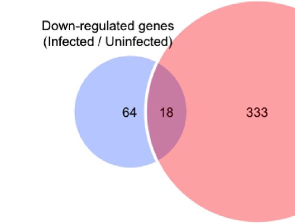
Takehara et al.                                                                                                                                   TLR4 and C. perfringens Infection

Wirths, S., Bugl, S., and Kopp, H. G. (2014). Neutrophil homeostasis and its regulation        Copyright © 2021 Takehara, Kobayashi and Nagahama. This is an open-access article
  by danger signaling. Blood 123, 3563–3566. doi: 10.1182/blood-2013-11-516260                 distributed under the terms of the Creative Commons Attribution License (CC BY).
                                                                                               The use, distribution or reproduction in other forums is permitted, provided the
Conflict of Interest: The authors declare that the research was conducted in the                original author(s) and the copyright owner(s) are credited and that the original
absence of any commercial or financial relationships that could be construed as a               publication in this journal is cited, in accordance with accepted academic practice. No
potential conflict of interest.                                                                 use, distribution or reproduction is permitted which does not comply with these terms.

Frontiers in Cellular and Infection Microbiology | www.frontiersin.org                    10                                           March 2021 | Volume 11 | Article 633440
You can also read