Histological and molecular classification of breast cancer: what do we know? - Mastology
←
→
Page content transcription
If your browser does not render page correctly, please read the page content below
REVIEW ARTICLE
                                               DOI: 10.29289/25945394202020200024

                 Histological and molecular classification
                   of breast cancer: what do we know?
                                 Renan Gomes do Nascimento1*              , Kaléu Mormino Otoni2

    ABSTRACT
Breast cancer is the neoplasm most diagnosed malignancy and the leading cause of mortality among women on a global scale.
A profound increase in the understanding and clinical management of breast cancer has occurred over the past two decades,
which has led to significant progress in prevention, early detection, and personalized breast cancer therapy. However, the biggest
obstacle still faced in clinical practice is the complete understanding of intertumoral and intratumoral heterogeneity, in addition
to the mechanisms of multiple drug resistance in the systemic treatment of the disease. In view of this, many studies focus on
analyzing morphological and, mainly, molecular patterns of breast cancer, with the purpose of grouping these tumors into classes
or entities to assist in clinical management, in the elaboration of epidemiological and functional studies, and in the performance of
clinical trials. The most common special histological types of breast cancer include: medullary carcinoma, metaplastic carcinoma,
apocrine carcinoma, mucinous carcinoma, cribriform carcinoma, tubular carcinoma, neuroendocrine carcinoma, classic lobular
carcinoma, and pleomorphic lobular carcinoma, in addition to the non-specific type of invasive ductal carcinoma, which constitutes
the majority of newly diagnosed cases. As to their molecular aspect, intrinsic subtypes were identified based on global studies
of gene expression profiles. Today, four molecular subgroups are widely reproduced and well established in the clinical routine,
namely: Luminal A, Luminal B, HER2 +, and Triple Negative. Thus, the present article aims to briefly address the histological and
molecular classification of breast cancer.
KEYWORDS: breast cancer; classification; neoplasms.

INTRODUCTION                                                            approximately 66,280 new cases of breast cancer annually,
Cancer has become one of the main causes of morbidity and               with an estimated risk of 61.61 cases per 100 thousand women.
mortality on a global scale in recent decades, as a result largely      Without considering non-melanoma skin cancer, this type of
due to demographic, economic and epidemiological transitions1,2.        malignancy is the second most incident in the general population
Among the female population, breast cancer is the most com-             and the most incident among the female population in Brazil, rep-
mon malignancy in the world (154 out of 185 countries), except          resenting 29.7% of all cancer cases in this population, surpassing
in West Africa, where cervical cancer prevailed. In 2018, a total of    the world average, estimated at 24.2%5. It is known by the scien-
2.1 million women were diagnosed with breast cancer, approxi-           tific community that the morphological and molecular aspects
mately one new case diagnosed every 18 seconds. In addition,            of breast cancer have been thoroughly explored and that these
breast cancer also represents the highest cancer mortality rates        studies sought further clarification of the tumor heterogeneity
in women across the globe (103 out of 185 countries), with roughly      of breast cancer. Therefore, this article aims to briefly address
626,600 deaths due to the disease, with the main exceptions             the current status of the histological and molecular classifica-
being the countries of Northern Europe, South America North             tion of breast cancer. For that to be accomplished, articles were
and Sub-Saharan Africa, where the main causes of death were             searched in the PubMed database without language restrictions.
due to cervical and/or lung cancer2-4.                                  The search terms “breast cancer” were used in combination with
    In Brazil, according to the latest publication for the 2020–2021    specific terms that cover the different histological and molecular
biennium, produced by the National Cancer Institute (INCA),             subtypes, as appropriate. We selected publications widely over

1
 Faculty of Medicine, Universidade de São Paulo – São Paulo (SP), Brazil.
2
 ProNutrir Nutritional Support and Chemotherapy – Fortaleza (CE), Brazil.
*Corresponding author: renanfarmaceutico@outlook.com
Conflict of interests: nothing to declare.
Received on: 04/29/2019. Accepted on: 06/26/2020.

                                                        Mastology 2020;30:e20200024                                                  1
Nascimento RG, Otoni KM
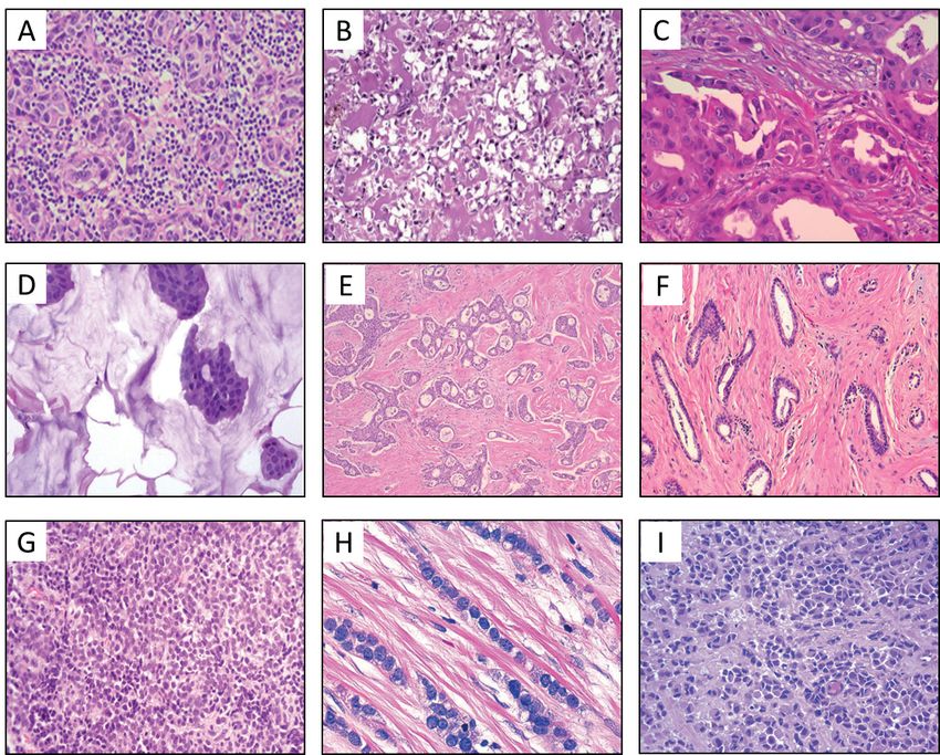
the last five years, and did not exclude older, commonly refer- Metaplastic carcinoma
enced and highly regarded publications. We also searched the This histological subtype is characterized by the dominant com-
reference lists of articles identified by this search strategy and ponent of metaplastic differentiation, representing approximately
selected those that we deemed relevant. 1% of all cases and affecting women, mainly in post-menopause14.
This group of tumors shows aggressive biological behavior and an
often lymph node involvement15. Morphologically, it is a poorly
HISTOLOGICAL CLASSIFICATION differentiated heterogeneous tumor that contains ductal car-
For the morphological study of breast cancer, we must under- cinoma cells mixed with other histological elements, such as
stand whether the tumor is limited to the epithelial compo- squamous cells, spindle cells or other mesenchymal differen-
nent of the breast or has invaded the surrounding stroma, and tiation, such as chondroid cells, bone cells, and myoepithelial
whether this tumor appeared in the mammary ducts or lobes6. cells (Figure 1B)12,15.
However, in histopathological practice, cell type characteristics,
number of cells, type and location of secretion, immunohisto- Apocrine carcinoma
chemical profile and architectural characteristics determine if It constitutes about 1% to 4% of all cases, with prominent apocrine
the tumor is ductal or lobular, in addition to its sub-classifica- differentiation comprising at least 90% of tumor cells7. This sub-
tions, rather than its precise location in the mammary tissue7,8. type is generally of high histological grade, with poor prognosis
About 50% to 80% of newly diagnosed breast cancer cases are and affects a wide age group, but it is more commonly seen in
called invasive ductal carcinoma (IDC); the rest of the cases are postmenopausal women16. Microscopically, tumor cells are large,
classified as invasive lobular carcinoma (ILC)9. IDCs can be clas- with an abundant granular eosinophilic cytoplasm, positive for
sified as “no specific type” because these tumors do not present PAS (Periodic acid-reactive Schiff) staining and prominent nucle-
sufficient morphological characteristics to be determined as a oli; in addition, bizarre tumor cells with multilobulated nuclei
characteristic histological type; they can also be recognized as can also be observed (Figure 1C)12,17.
a “special type” if they present sufficient distinctive characteris-
tics, and particular cellular and molecular behavior9,10. The most Mucinous carcinoma
common special types of breast cancer include: medullary car- It is a special subtype of breast cancer, also known as colloid,
cinoma, metaplastic carcinoma, apocrine carcinoma, mucinous gelatinous, mucous and mucoid carcinoma, responsible for 2%
carcinoma, cribriform carcinoma, tubular carcinoma, neuroen- of all newly diagnosed cases11. This subtype has been associ-
docrine carcinoma, classic lobular carcinoma, and pleomorphic ated with a favorable prognosis and often affects women over
lobular carcinoma10. 60 years of age18. Morphologically, these tumors have abundant
amounts of extracellular mucin, surrounding small clusters of
Invasive ductal carcinoma tumor cells with different growth patterns and with mild nuclear
no specific type (IDC-NST) atypia (Figure 1D)12,19.
The histological subtype IDC-NST is the most common, constitut-
ing about 40% to 75% of all invasive breast carcinomas. Usually, it Cribriform carcinoma
has a wide scope of morphological variation and clinical behav- Special subtype associated to a good prognosis, generally
ior10. Tumor cells are pleomorphic, with protruding nucleoli and affecting patients who are approximately 50 years old and
numerous mitoses. Areas of necrosis and calcifications can be constituting about 1% to 3.5% of all breast cancer cases 6 .
detected in more than half of the cases7,10. Cribriform carcinoma has almost no evidence of regional or
distant metastasis7. Microscopically, this subtype presents
Medullary carcinoma islands of uniform tumor cells, with low-grade atypia, crib-
Special subtype of invasive breast carcinoma, responsible for riform appearance in 90% of the tumor and often associated
approximately 5% of all cases, and associated with better clin- with DCIS (Ductal carcinoma in situ) without well-defined
ical results and lower rates of involvement in axillary lymph stromal invasion (Figure 1E)20.
nodes11. It usually affects patients between 30 and 40 years old
and is often associated with mutations in the BRCA1 germline Tubular carcinoma
(Breast cancer gene 1)10. Microscopically, it is a well-circum- Well-differentiated subtype, occurring in women between 50
scribed carcinoma, composed of large and pleomorphic tumor and 60 years of age and constituting about 2% of all newly diag-
cells, with a syncytial growth pattern, frequent mitotic fig- nosed cases11. Most tubular carcinomas are associated to a
ures and prominent lymphoplasmacytic infiltrate (Figure 1A). wide range of potentially premalignant proliferative lesions21.
Other commonly seen features include spindle cell metaplasia This subtype is characterized by the proliferation of prominent
and giant tumor cells12,13. tubules (> 90%), which can be angled, oval or elongated, with a
2 Mastology 2020;30:e20200024Histological and molecular classification of breast cancer: what do we know
disorganized disposition and open lumen covered by a single Invasive lobular carcinoma
layer of epithelium, usually without presentation of necrosis and It is the second largest biologically distinct carcinoma, representing
mitosis (Figure 1F)12,22. about 5% to 15% of all newly diagnosed cases and generally affect-
ing women of advanced age11. The classic form of the ILC is charac-
Neuroendocrine carcinoma terized by the presence of small tumor cells with little atypia, uni-
It constitutes about 0.5% to 5% of all cases of breast cancer formly distributed throughout the stroma in a concentric pattern
and commonly occurs in older ages10. This type of tumor has (Figure 1H)10. Among pleomorphic ILC, tumor cells have a hyper-
characteristics similar to neuroendocrine tumors of the gas- chromatic and eccentric nucleus, prominent mitoses and apocrine.
trointestinal tract and lung, consistently expressing the mark- Histiocytic or signet ring cells can be observed (Figure 1I) and
ers chromogranin A and synaptophysin in more than 50% they are more likely to have TP53 mutations (Tumor protein 53)25.
of neoplastic cells23 . Morphologically, there is an infiltrative
growth pattern with solid aggregates of tumor cells arranged
in alveolar, trabecular or rosette patterns, and peripheral pali- MOLECULAR CLASSIFICATION
sades can also be observed12 . Neoplastic cells can be of differ- We now know that breast cancer represents a biologically and
ent sizes and generally have fine eosinophilic granular cyto- phenotypically heterogeneous collection of diseases with dif-
plasm (Figure 1G)24. ferent clinical and treatment response behaviors26. In this era of
Figure 1. Morphological variants representative of the main subtypes of invasive breast carcinomas. (A) medullary carcinoma; (B)
metaplastic carcinoma; (C) apocrine carcinoma; (D) mucinous carcinoma; (E) cribriform carcinoma; (F) tubular carcinoma; (G) neuroen-
docrine carcinoma; (H) classic lobular carcinoma; and (I) pleomorphic lobular carcinoma.
Mastology 2020;30:e20200024 3Nascimento RG, Otoni KM
modern medicine, only the morphological classification (nuclear who would benefit from adjuvant chemotherapy40,41. The PAM50
grade, tubular grade, mitotic index, histological grade, and archi- trial (Prosigna) is a classifier for breast cancer subtypes. It also
tectural characteristics) and the clinical pathological parameters assesses a patient’s risk for distant recurrence of the disease
(tumor size, lymph node involvement, metastasis), are insufficient and the likelihood of efficacy of neoadjuvant chemotherapy40,41.
to predict the real behavior of breast tumor pathophysiology10,27. Molecular subtyping changed our view of breast cancer, with
Thus, many studies focus on analyzing the molecular patterns the possibility of stratifying this neoplasm in different entities
of breast cancer in order to group these tumors into classes or that require specific treatments and different monitoring strat-
entities to assist in clinical management, in the preparation of egies, in addition to a better understanding of the pathophysi-
epidemiological and functional studies and in the performance ological pattern and clinical prognosis. Next, we briefly present
of clinical trials28-34. the different molecular subtypes of breast cancer.
The pioneering work by Perou, Sorlie and colleagues at the
beginning of this millennium classified breast cancer molecularly Luminal A
into distinct subgroups, based on similarities in gene expression This molecular subtype is the most common and comprises
profiles, using the cDNA microarray technique31,33,34. Thus, these approximately half of newly diagnosed breast cancer cases7.
studies demonstrated that there are breast cancer subtypes with According to the last update of St. Gallen in 2013, the immuno-
differences in gene expression patterns, reflecting the individual histochemical profile of this subtype was defined as: ER+ (≥ 1%),
phenotype, disease prognosis and systemic treatment planning35. high expression of PR (≥ 20%), HER2- (≤ 10%), and low levels of
Based on comprehensive gene expression profile studies, four Ki-67 (< 14%)42. In addition, these tumors have characteristics of
clinically relevant molecular subtypes were revealed: Luminal luminal epithelial cells of the breast, such as the high expression
A, Luminal B, enriched HER2 (HER2+), and Triple Negative (TN) of cytokeratin’s 7/8/18/1943. They include a wide range of low his-
(36). The groups of genes responsible mainly for the segregation tological grade variants, such as IDC-NST, tubular, cribriform,
of the molecular subtypes of breast cancer are genes related to mucinous, and classic ILC6,43. This subtype has been associated
the expression of estrogen receptors (ER), progesterone receptors with a highly favorable prognosis, with a more indolent clini-
(PR), HER2 (Human epidermal growth factor receptor 2), and cell cal course, and generally shows less lymph node involvement44.
proliferation regulator (Ki-67)1. The Immunohistochemical (IHC) Nonetheless, due to the positive status of hormone receptors,
panel with these four biomarkers (ER/PR/HER2/Ki-67) has been patients benefit from endocrine therapies, either with selective
considered efficient and significant in the stratification of these estrogen receptor modulators (tamoxifen) or with aromatase
molecular entities6,35. However, the growing need to improve risk inhibitors (anastrozole) (Table 1)45.
stratification and accurate prognosis determination, in addi-
tion to an accurate understanding of tumor biology, led to the Luminal B
development of many multigenic assays, such as Oncotype DX, Responsible for approximately 20% to 30% of invasive breast
Prosigna PAM50 and Mammaprint36-39. The signature of 70 genes cancer cases26. This subtype can be categorized immunophe-
(Mammaprint) and of 21 genes (Oncotype DX) are being used in notypically into Luminal B (HER-): ER+ (≥ 1%), PR- or < 20%,
patients with ER+ disease at an early clinical stage to distin- HER2- (≤ 10%) and high levels of Ki-67 (≥ 20%); or Luminal B
guish women who may have the greatest risk of recurrence and (HER2+): ER+ (≥ 1%), HER2+ (> 10%) and any level of PR and
Table 1. Classification of molecular subtypes of breast cancer and therapies.
Luminal B
Molecular Subtypes Luminal A HER2+ TN
(HER2-) (HER2+)
ER+ ER+ ER+ ER- ER-
PR+ PR- PR-/+ PR- PR-
Biomarkers
HER2- HER2- HER2+ HER2+ HER2-
Ki67low Ki67high Ki67low/high Ki67high Ki67high
Frequency of Cases (%) 40–50 20–30 15–20 10–20
Well Differentiated Little Differentiated Little Differentiated
Histological Grade Moderately Differentiated (Grade II)
(Grade I) (Grade III) (Grade III)
Prognosis Good Intermediate Poor Poor
Endocrine
Endocrine Target Therapy Chemotherapy
Response to Therapies Endocrine Chemotherapy
Chemotherapy Chemotherapy PARP Inhibitors
Target Therapy
ER: estrogen receptor; PR: progesterone receptor; HER2: human epidermal growth factor receptor 2.
4 Mastology 2020;30:e20200024Histological and molecular classification of breast cancer: what do we know
Ki-6742,46. The expression of low molecular weight cytokeratin’s tumors, according to the Ki-67 index (> 30%)42. Most TN tumors
from luminal epithelial cells is a rule26. This molecular entity gen- manifest as the IDC-NST histological type. However, they also
erally presents a moderate histological grade, including most of include variants of medullary, metaplastic and apocrine carci-
the IDC-NST and associated with an intermediate prognosis, nomas26. These tumors are generally more prevalent in patients
with greater likelihood of locoregional recurrence when com- with BRCA1 mutations and young women, with a higher histo-
pared to Luminal A44,47. Luminal B subtype is understood as the logical grade, risk of loco-regional recurrence, contralateral dis-
most aggressive form of ER+ breast cancer cases and often does ease and systemic relapse52. Many gene expression profile stud-
not show benefits for hormone therapy (Table 1)27 (EXCLUDED). ies have been carried out to better understand the heterogeneity
Luminal B subtype is understood as the most aggressive form of this particularly aggressive form of breast cancer. Thus, TN
of hormone-dependent breast cancer cases, requiring additional tumors can be further divided into seven other distinct entities,
treatments to hormonal therapy, such as chemotherapy (when including two basal-like types (BL1 and BL2), with a basal pat-
HER2 +/-) or targeted target therapy (when HER2 +) (Table 1)27. tern of gene expression, but showing differences in the immune
The main difference in the molecular aspect between the two response; one of the luminal androgen receptor type (LAR), which
luminous subgroups is the increased expression of genes related presents differential expression of genes involved in androgen
to cell proliferation, such as NSEP1 (Nuclease sensitive element metabolism; one of the immunomodulatory type (IM), which
binding protein 1) and cyclin E1 (CCNE1), in addition to the acti- presents important changes in the expression of genes involved
vation of certain alternative pathways of growth factors, such in immunological signaling pathways; one of the claudin-low
as PI3K (Phosphatidyllinositol 3-Kinase) and Src (Proto-oncogene types (CL), characterized by the low expression of cellular junc-
sarcoma) in Luminal B breast tumors36. tion proteins (claudins 3, 4 and 7, in addition to E-cadherin); and
two of the mesenchymal type, namely, mesenchymal itself (M)
HER2+ and mesenchymal stem-like (MSL), both with positive regulation
It represents 15% to 20% of newly diagnosed breast cancer cases48. of the signaling pathways involved in EMT (epithelial mesenchy-
This subtype is characterized by a high expression of HER2 (> 10%), mal transition), but differing in the signaling of genes associated
negativity for ER (< 1%) and PR (< 20%), and high expression of to stem cells and angiogenic factors29,30,32,53. Despite its simple
Ki-67 (> 20%)42. In addition to the immunophenotypic charac- definition, this subtype has been a challenge for the clinic, due
terization routinely used to assess the status of HER2 in breast to its morphological, molecular and clinical heterogeneity and
cancer, the FISH (Fluorescence in situ hybridization) technique has the lack of targeted therapies54. Non-surgical treatment of the
also been employed to assess gene amplification49. According to TN subtype has been limited to platinum-based chemotherapy
the latest clinical practice guidelines provided by the American and PARP (Poly ADP-ribose polymerase) inhibitors for patients
Society of Clinical Oncology (ASCO), if the IHC result shows with BRCA1 and 2 mutations27,55.
complete staining of the cell membrane with strong marking, Although great advances have occurred in high-performance
the diagnosis is positive for HER2; if staining of low to moderate molecular techniques and bioinformatics during the last decades,
intensity is observed, it will be necessary to use the FISH assay which allowed refinement in the stratification of breast cancer,
with an additional observer to confirm positivity, and, finally, molecular tests are still evolving, arising important questions:
in cases with negative marking the complete weak staining of • How many subtypes of this malignant neoplasm are there?
the membrane, the diagnosis can be confirmed as negative for • Which molecular classification system is more robust?
HER250. HER2 overexpression occurs almost exclusively in the • Are the classifications able to illustrate intratumoral
ILC pleomorphic variant27. The amplification of the gene and heterogeneity and clonal evolution?
the elevated expression of the HER2 protein has been related to • How should we interpret breast cancer subtypes?;
tumors of greater histological grade, high proliferative index and • Is it possible for different classification schemes in clinical
propensities to metastasis, leading to short disease-free survival practice to exist56,57?
and worse prognosis26. However, these tumors may respond well
to drugs that block HER2 activity, especially humanized mono- These questions will be answered over the next years.
clonal antibodies (Trastuzumab) and molecular receptor tyro- The accumulation of knowledge around cellular and molec-
sine kinase inhibitors (Lapatinib)35,51. ular biology, clinical behavior and therapeutic response, added
to the emergence of new drugs and new treatment modalities,
Triple negative undoubtedly brought a greater understanding and quality
This class of tumors constitutes from 10% to 20% of all breast in the management of breast cancer36 . All the improvements
cancer cases35. This subtype is characterized by the lack of expres- obtained so far are a great achievement for humanity and
sion of the hormone receptors ER (< 1%) and PR (< 20%) and the occurred thanks to the contributions of many researchers
oncoprotein HER2 (≤ 10%); moreover, they are highly proliferative around the world1,58 .
Mastology 2020;30:e20200024 5Nascimento RG, Otoni KM
CONCLUSION resistance are still poorly understood. However, anti-apoptotic
Despite great advances in the stratification of breast cancer sub- resistance, ATP-dependent drug efflux pumps, changes in drug
types, the greatest obstacle currently found in clinical oncology targets, epigenetic changes, EMT and miRNAs make up impor-
is the complete understanding of intertumoral heterogeneity tant factors for failures in anti-cancer therapies. In this context,
(illustrated by tumor size, regional lymph node status, distant hundreds of other candidates for biomarkers have been investi-
metastases and differences in survival), especially the intratu- gated and studied for potential implications for diagnosis, prog-
moral heterogeneity (illustrated by histological and biomolecu- nosis, drug targets and predictor of therapeutic response, “jus-
lar variability, chromosomal, genomic, metabolic and epigen- tifying regular reviews”.
etic changes, in addition to cellular plasticity and the tumor
microenvironment), which impacts the adversity of diagnosis
and accurate prognosis, and weakening strategies in personal- AUTHORS’ CONTRIBUTION
ized medicine. In addition, resistance to multiple drugs (RMD) R.G.N.: Conceptualization, investigation, methodology, project
is considered the biggest obstacle in the systemic treatment of administration, supervision, validation, visualization, writ-
breast cancer, making the disease often uncontrollable and lead- ing – review & editing.
ing to high mortality rates. The mechanisms underlying drug K.M.O.: Formal analysis, investigation, writing – review & editing.
REFERENCES
1. Lukong KE. Understanding breast cancer – The long and 11. Akram M, Iqbal M, Daniyal M, Khan AU. Awareness and
winding road. BBA Clin. 2017;7(1):64-77. http://dx.doi. current knowledge of breast cancer. Biol Res. 2017;50(1):33.
org/10.1016/j.bbacli.2017.01.001 https://doi.org/10.1186/s40659-017-0140-9
2. Bray F, Ferlay J, Soerjomataram I, Siegel RL, Torre LA, Jernal 12. Provenzano E, Ulaner GA, Chin SF. Molecular Classification
A. Global Cancer Statistics 2018: GLOBOCAN Estimates of of Breast Cancer. PET Clin. 2018;13(3):325-38. https://doi.
Incidence and Mortality Worldwide for 36 Cancers in 185 org/10.1016/j.cpet.2018.02.004
Countries. CA Cancer J Clin. 2018;68(6):394-424. https://doi. 13. Zangouri V, Akrami M, Tahmasebi S, Talei A, Hesarooeih AG.
org/10.3322/caac.21492 Medullary breast carcinoma and invasive ductal carcinoma:
3. Ferlay J, Colombet M, Soerjomataram I, Mathers C, Parkin A review study. Iran J Med Sci. 2018;43(4):365-71. https://doi.
DM, Piñeros M, et al. Estimating the global cancer incidence org/10.21859/mci-supp-100
and mortality in 2018: GLOBOCAN sources and methods. Int J 14. Sinn HP, Kreipe H. A brief overview of the WHO classification
Cancer. 2019;144(8):1941-53. https://doi.org/10.1002/ijc.31937 of breast tumors, 4th edition, focusing on issues and updates
4. Torre LA, Islami F, Siegel RL, Ward EM, Jemal A. Global cancer in from the 3rd edition. Breast Care. 2013;8(2):149-54. https://
women: Burden and trends. Cancer Epidemiol Biomarkers Prev. dx.doi.org/10.1159%2F000350774
2017;26(4):444-57. https://doi.org/10.1158/1055-9965.epi-16-0858 15. Schwartz TL, Mogal H, Papageorgiou C, Veerapong
5. Brasil. Ministério da Saúde. Instituto Nacional de Câncer J, Hsueh EC. Metaplastic breast cancer: Histologic
José Alencar Gomes da Silva. Estimativa 2020: Incidência de characteristics, prognostic factors and systemic treatment
Câncer no Brasil. Brasil: INCA; 2019. 120 p. strategies. Exp Hematol Oncol. 2013;2(1):31. https://dx.doi.
org/10.1186%2F2162-3619-2-31
6. Vuong D, Simpson PT, Green B, Cummings MC, Lakhani SR.
Molecular classification of breast cancer. Virchows Arch. 16. Vranic S, Schmitt F, Sapino A, Costa JL, Reddy S, Castro M,
2014;465(1):1-14. https://doi.org/10.1007/s00428-014-1593-7 et al. Apocrine carcinoma of the breast: A comprehensive
review. Histol Histopathol. 2013;28(11):1393-409. https://doi.
7. Makki J. Diversity of breast carcinoma: Histological subtypes org/10.14670/hh-28.1393
and clinical relevance. Clin Med Insights Pathol. 2015;8(1):23-
17. Vranic S, Feldman R, Gatalica Z. Apocrine carcinoma of the
31. https://dx.doi.org/10.4137%2FCPath.S31563
breast: A brief update on the molecular features and targetable
8. Nounou MI, ElAmrawy F, Ahmed N, Abdelraouf K, Goda S, biomarkers. Bosn J Basic Med Sci. 2017;17(1):9-11. https://doi.
Syed-Sha-Qhattal H. Breast cancer: Conventional diagnosis org/10.17305/bjbms.2016.1811
and treatment modalities and recent patents and technologies.
18. Marrazzo E, Frusone F, Milana F, Sagona A, Gatzemeier W,
Breast Cancer Basic Clin Res. 2015;9(Suppl. 2):17-34. https://
Barbieri E, et al. Mucinous breast cancer: A narrative review
dx.doi.org/10.4137%2FBCBCR.S29420
of the literature and a retrospective tertiary single-centre
9. Henry NL, Cannon-Albright L. Breast Cancer Histologic analysis. Breast. 2020;49(1):87-92. https://doi.org/10.1016/j.
Subtypes Show Excess Familial Clustering. Wiley Cancer. breast.2019.11.002
2019;125(18):3131-8. https://doi.org/10.1002/cncr.32198 19. Dumitru A, Procop A, Iliesiu A, Tampa M, Mitrache L,
10. Masood S. Breast Cancer Subtypes: Morphologic and Biologic Costache M, et al. Mucinous Breast Cancer: a Review Study
Characterization. Womens Health. 2016;12(1):103-19. https:// of 5 Year Experience from a Hospital-Based Series of Cases.
doi.org/10.2217%2Fwhe.15.99 Maedica (Buchar). 2015;10(1):14-8.
6 Mastology 2020;30:e20200024Histological and molecular classification of breast cancer: what do we know
20. Cong Y, Qiao G, Zou H, Lin J, Wang X, Li X, et al. Invasive Proc Natl Acad Sci U S A. 2001;98(19):10869-74. https://doi.
cribriform carcinoma of the breast: A report of nine cases and org/10.1073/pnas.191367098
a review of the literature. Oncol Lett. 2015;9(4):1753-8. https:// 34. Sorlie T, Tibshirani R, Parker J, Hastie T, Marron JS, Nobel
dx.doi.org/10.3892%2Fol.2015.2972 A, et al. Repeated observation of breast tumor subtypes in
21. Zhang WW, Wu SG, Ling YH, Sun JY, Long ZQ, Hua X, et al. independent gene expression data sets. Proc Natl Acad Sci.
Clinicopathologic characteristics and clinical outcomes of pure 2003;100(14):8418-23. https://doi.org/10.1073/pnas.0932692100
type and mixed type of tubular carcinoma of the breast: A single- 35. Tsang JYS, Tse GM. Molecular Classification of Breast Cancer.
institution cohort study. Cancer Manag Res. 2018;10(1):4509- Adv Anat Pathol. 2020;27(1):27-35. https://doi.org/10.1097/
15. https://dx.doi.org/10.2147%2FCMAR.S177046 pap.0000000000000232
22. Fritz P, Bendrat K, Sonnenberg M, Trautmann C, Ott G, 36. Harbeck N, Penault-Llorca F, Cortes J, Gnant M, Houssami
Heidemann E, et al. Tubular breast cancer. A retrospective N, Poortmans P, et al. Breast cancer. Nat Rev Dis Prim.
study. Anticancer Res. 2014;34(7):3647-56. 2019;5(1):66. https://doi.org/10.1038/s41572-019-0111-2
23. Jurčić P, Kruslin B, Gatalica Z, Sanati S, Vranic S. Breast 37. Parker JS, Mullins M, Cheung MCU, Leung S, Voduc D, Vickery
carcinoma with neuroendocrine features: a brief review. T, et al. Supervised risk predictor of breast cancer based on
Endocr Oncol Metab. 2016;2(2):138-45. https://dx.doi. intrinsic subtypes. J Clin Oncol. 2009;27(8):1160-7. https://
org/10.21040/eom/2016.2.2.6 dx.doi.org/10.1200%2FJCO.2008.18.1370
24. Li Y, Du F, Zhu W, Xu B. Neuroendocrine carcinoma of 38. Paik S, Shak S, Tang G, Kim C, Baker J, Cronin M, et al. A
the breast: A review of 126 cases in China. Chin J Cancer. multigene assay to predict recurrence of tamoxifen-treated,
2017;36(1):45. https://dx.doi.org/10.1186%2Fs40880-017-0211-x node-negative breast cancer. N Engl J Med. 2004;351(27):2817-
25. Thomas M, Kelly ED, Abraham J, Kruse M. Invasive 26. https://doi.org/10.1056/nejmoa041588
lobular breast cancer: A review of pathogenesis, diagnosis, 39. Vijver MJV, He Y, Veer L, Dai H, Hart AAM, Voskuil DW, et al. A
management, and future directions of early stage disease. gene-expression signature as a predictor of survival in breast
Semin Oncol. 2019;46(2):121-32. https://doi.org/10.1053/j. cancer. N Engl J Med. 2002;347(25):1999-2009. https://doi.
seminoncol.2019.03.002 org/10.1056/nejmoa021967
26. Feng Y, Spezia M, Huang S, Yuan C, Zeng Z, Zhang L, et al. 40. Huang S, Murphy L, Xu W. Genes and functions from breast
Breast cancer development and progression: Risk factors, cancer signatures. BMC Cancer. 2018;18(1):473. https://doi.
cancer stem cells, signaling pathways, genomics, and org/10.1186/s12885-018-4388-4
molecular pathogenesis. Genes Dis. 2018;5(2):77-106. https://
41. Vieira AF, Schmitt F. An update on breast cancer multigene
doi.org/10.1016/j.gendis.2018.05.001
prognostic tests-emergent clinical biomarkers. Front Med.
27. Fragomeni SM, Sciallis A, Jeruss JS. Molecular Subtypes and 2018;5(1):248. https://dx.doi.org/10.3389%2Ffmed.2018.00248
Local-Regional Control of Breast Cancer. Surg Oncol Clin N Am.
42. Goldhirsch A, Winer EP, Coates AS, Gelber RD, Piccart-
2018;27(1):95-120. https://dx.doi.org/10.1016%2Fj.soc.2017.08.005
Gebhart M, Thürlimann B, et al. Personalizing the treatment
28. Burstein MD, Tsimelzon A, Poage GM, Covington KR, Fuqua of women with early breast cancer: Highlights of the st gallen
SAW, Savage MI, et al. Comprehensive Genomic Analysis international expert consensus on the primary therapy of early
Identifies Novel Subtypes and Targets of Triple-negative breast Cancer 2013. Ann Oncol. 2013;24(9):2206-23. https://
Breast Cancer. Clin Cancer Res. 2015;21(7):1688-98. https://doi. dx.doi.org/10.1093%2Fannonc%2Fmdt303
org/10.1158/1078-0432.ccr-14-0432
43. Gao JJ, Swain SM. Luminal A Breast Cancer and Molecular
29. Lehmann BD, Bauer JA, Chen X, Sanders ME, Chakravarthy Assays: A Review. Oncologist. 2018;23(5):556-65. https://
AB, Shyr Y, et al. Identification of human triple-negative dx.doi.org/10.1634%2Ftheoncologist.2017-0535
breast cancer subtypes and preclinical models for selection of
44. Hashmi AA, Aijaz S, Khan SM, Mahboob R, Irfan M, Zafar NI,
targeted therapies. J Clin Invest. 2011;121(7):2750-67. https://
et al. Prognostic parameters of luminal A and luminal B intrinsic
dx.doi.org/10.1172%2FJCI45014
breast cancer subtypes of Pakistani patients. World J Surg Oncol.
30. Lehmann BD, Jovanovic B, Chen X, Estrada M V, Johnson 2018;16(1):1-6. https://doi.org/10.1186/s12957-017-1299-9
N, Shyr Y, et al. Refinement of Triple-Negative Breast
45. Rocca A, Farolfi A, Maltoni R, Carretta E, Melegari E,
Cancer Molecular Subtypes : Implications for Neoadjuvant
Ferrario C, et al. Efficacy of endocrine therapy in relation to
Chemotherapy Selection. PLoS One. 2016;11(6):e0157368.
progesterone receptor and Ki67 expression in advanced breast
https://doi.org/10.1371/journal.pone.0157368
cancer. Breast Cancer Res Treat. 2015;152(1):57-65. https://doi.
31. Perou CM, Sorlie T, Eisen MB, van de Rijn M, Jeffrey SS, Rees CA, org/10.1007/s10549-015-3423-2
et al. Molecular portraits of human breast tumours. Nature.
46. Cheang MCU, Chia SK, Voduc D, Gao D, Leung S, Snider J,
2000;406(6797):747-52. https://doi.org/10.1038/35021093
et al. Ki67 index, HER2 status, and prognosis of patients with
32. Prat A, Perou CM. Deconstructing the molecular portraits luminal B breast cancer. J Natl Cancer Inst. 2009;101(10):736-
of breast cancer. Mol Oncol. 2011;5(1):5-23. https://dx.doi. 50. https://dx.doi.org/10.1093%2Fjnci%2Fdjp082
org/10.1016%2Fj.molonc.2010.11.003
47. Tsoutsou PG, Vozenin MC, Durham AD, Bourhis J. How could
33. Sorlie T, Perou CM, Tibshirani R, Aas T, Geisler S, Johnsen breast cancer molecular features contribute to locoregional
H, et al. Gene expression patterns of breast carcinomas treatment decision making? Crit Rev Oncol Hematol.
distinguish tumor subclasses with clinical implications. 2017;110(1):43-8. http://dx.doi.org/10.1016/j.critrevonc.2016.12.006
Mastology 2020;30:e20200024 7Nascimento RG, Otoni KM
48. Cho N. Molecular subtype and imaging phenotype of breast 53. Sabatier R, Finetti P, Guille A, Adelaide J, Chaffanet M, Viens
cancer. Ultrasonography. 2016;35(4):281-8. http://dx.doi. P, et al. Claudin-low breast cancers: Clinical, pathological,
org/10.14366/usg.16030 molecular and prognostic characterization. Mol Cancer.
49. Desai NV, Torous V, Parker J, Auman JT, Rosson GB, Cruz C, et al. 2014;13(1):228. https://doi.org/10.1186/1476-4598-13-228
Intrinsic molecular subtypes of breast cancers categorized as HER2- 54. Russnes HG, Lingjærde OC, Børresen-Dale AL, Caldas C. Breast
positive using an alternative chromosome 17 probe assay. Breast Cancer Molecular Stratification: From Intrinsic Subtypes to
Cancer Res. 2018;20(1):75. https://doi.org/10.1186/s13058-018-1005-z Integrative Clusters. Am J Pathol. 2017;187(10):2152-62. https://
50. Wolff AC, Hammond MEH, Allison KH, Harvey BE, Mangu PB, doi.org/10.1016/j.ajpath.2017.04.022
Bartlett JMS, et al. Human epidermal growth factor receptor 2 55. Yam C, Mani S, Moulder S. Targeting the Molecular Subtypes
testing in breast cancer: American society of clinical oncology/ of Triple Negative Breast Cancer: Understanding the Diversity
college of American pathologists clinical practice guideline to Progress the Field. Oncologist. 2017;22(9):1086-93. https://
focused update. J Clin Oncol. 2018;36(20):2105-22. https://doi. dx.doi.org/10.1634/theoncologist.2017-0095
org/10.1200/jco.2018.77.8738
56. Rakha EA, Green AR. Molecular classification of breast cancer:
51. Llombart-Cussac A, Cortés J, Paré L, Galván P, Bermejo B, what the pathologist needs to know. Pathology. 2017;49(2):111-
Martínez N, et al. HER2-enriched subtype as a predictor 9. https://doi.org/10.1016/j.pathol.2016.10.012
of pathological complete response following trastuzumab
and lapatinib without chemotherapy in early-stage HER2- 57. Song Q, Merajver SD, Li JZ. Cancer classification in the genomic
positive breast cancer (PAMELA): an open-label, single-group, era: five contemporary problems. Hum Genomics. 2015;9(1):27.
multicentre, phase 2 trial. Lancet Oncol. 2017;18(4):545-54. https://doi.org/10.1186/s40246-015-0049-8
http://dx.doi.org/10.1016/S1470-2045(17)30021-9 58. Ades F, Tryfonidis K, Zardavas D. The past and future of
52. Kumar P, Aggarwal R. An overview of triple-negative breast breast cancer treatment - From the papyrus to individualised
cancer. Arch Gynecol Obstet. 2016;293(2):247-69. https://doi. treatment approaches. Ecancermedicalscience. 2017;11(1):746.
org/10.1007/s00404-015-3859-y https://dx.doi.org/10.3332/ecancer.2017.746
© 2020 Brazilian Society of Mastology
This is an open access article distributed under the terms of the Creative Commons license.
8 Mastology 2020;30:e20200024You can also read