Characteristics and outcome of breast cancer-related microangiopathic haemolytic anaemia: a multicentre study
←
→
Page content transcription
If your browser does not render page correctly, please read the page content below
Alhenc-Gelas et al. Breast Cancer Research              (2021) 23:9
https://doi.org/10.1186/s13058-021-01386-y

 RESEARCH ARTICLE                                                                                                                                     Open Access

Characteristics and outcome of breast
cancer-related microangiopathic haemolytic
anaemia: a multicentre study
Marion Alhenc-Gelas1, Luc Cabel1,2, Frederique Berger3, Suzette Delaloge4, Jean-Sebastien Frenel5, Christelle Levy6,
Nelly Firmin7, Sylvain Ladoire8, Isabelle Desmoulins9, Pierre-Etienne Heudel9, Florence Dalenc10, Delphine Loirat1,
Coraline Dubot1, Perrine Vuagnat1, Elise Deluche4, Meriem Mokdad-Adi4, Anne Patsouris5, Josselin Annic5,
Lounes Djerroudi11, Marion Lavigne11, Jean-Yves Pierga1,12, Paul Coppo13,14 and Francois-Clement Bidard1,2*

  Abstract
  Background: Cancer-related microangiopathic haemolytic anaemia (MAHA) is a rare but life-threatening
  paraneoplastic syndrome. Only single cases or small series have been reported to date. We set up a retrospective
  multicentre study focusing on breast cancer-related MAHA.
  Methods: Main inclusion criteria were known diagnosis of breast cancer, presence of schistocytes and either low
  haptoglobin or cytopenia and absence of any causes of MAHA other than breast cancer, including gemcitabine- or
  bevacizumab-based treatment. Patient characteristics, treatments and outcome were retrieved from digital medical records.
  Results: Individual data from 54 patients with breast cancer-related MAHA were obtained from 7 centres. Twenty-three
  (44%) patients had a breast tumour with lobular features, and most primary tumours were low grade (grade I/II, N = 39,
  75%). ER+/HER2−, HER2+ and triple-negative phenotypes accounted for N = 33 (69%), N = 7 (15%) and N = 8 (17%) cases,
  respectively. All patients had stage IV cancer at the time of MAHA diagnosis. Median overall survival (OS) was 28 days
  (range 0–1035; Q1:10, Q3:186). Independent prognostic factors for early death (≤ 28 days) were PS > 2 (OR = 7.0 [1.6; 31.8]),
  elevated bilirubin (OR = 6.9 [1.1; 42.6]), haemoglobin < 8.0 g/dL (OR = 3.7 [0.9; 16.7]) and prothrombin time < 50% (OR = 9.1
  [1.2; 50.0]). A score to predict early death displayed a sensitivity of 86% (95% CI [0.67; 0.96]), a specificity of 73% (95% CI
  [0.52; 0.88]) and an area under the curve of 0.90 (95% CI [0.83; 0.97]).
  Conclusions: Breast cancer-related MAHA appears to be a new feature of invasive lobular breast carcinoma. Prognostic
  factors and scores may guide clinical decision-making in this serious but not always fatal condition.
  Keywords: Microangiopathic haemolytic anaemia, Breast cancer, Survival, Prognostic factors

* Correspondence: fcbidard@curie.fr
1
 Department of Medical Oncology, Institut Curie, Paris and Saint Cloud,
France
2
 UVSQ, Université Paris-Saclay, 35 rue Dailly, Saint Cloud, 92210, France
Full list of author information is available at the end of the article

                                          © The Author(s). 2021 Open Access This article is licensed under a Creative Commons Attribution 4.0 International License,
                                          which permits use, sharing, adaptation, distribution and reproduction in any medium or format, as long as you give
                                          appropriate credit to the original author(s) and the source, provide a link to the Creative Commons licence, and indicate if
                                          changes were made. The images or other third party material in this article are included in the article's Creative Commons
                                          licence, unless indicated otherwise in a credit line to the material. If material is not included in the article's Creative Commons
                                          licence and your intended use is not permitted by statutory regulation or exceeds the permitted use, you will need to obtain
                                          permission directly from the copyright holder. To view a copy of this licence, visit http://creativecommons.org/licenses/by/4.0/.
                                          The Creative Commons Public Domain Dedication waiver (http://creativecommons.org/publicdomain/zero/1.0/) applies to the
                                          data made available in this article, unless otherwise stated in a credit line to the data.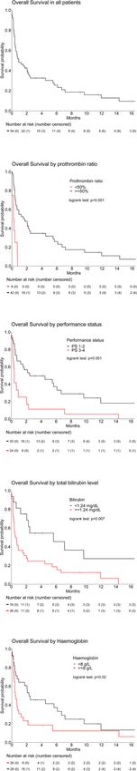
Alhenc-Gelas et al. Breast Cancer Research   (2021) 23:9                                                            Page 2 of 10

Background                                                          criteria were patients with histologically proven breast
Thrombotic microangiopathy (TMA) is a rare syndrome                 cancer and MAHA diagnosed between 1995 and 2019 in
combining diffuse microvessel thrombosis and mechanical             participating centres. Ineligibility criteria were MAHA at-
haemolytic anaemia, often associated with thrombocytopenia          tributed to a cause other than breast cancer determined
[1]. It can result in severe, life-threatening organ dysfunction,   by treating physician and patients treated with either gem-
especially affecting the kidneys and central nervous system.        citabine or bevacizumab at the time of or during the 6
TMA is a causally heterogeneous syndrome related to several         months prior to MAHA diagnosis.
conditions including thrombotic thrombocytopenic purpura
(TTP) and haemolytic-uraemic syndrome (HUS), which are              Case search
primarily caused by a functional deficiency of ADAMTS 13            In July 2018, a call for participants was sent to 13 French
(an enzyme involved in the degradation of Von Willebrand            cancer centres, outlining the study’s objectives, and list-
Factor) activity and Shiga toxin or complement dysregula-           ing the data that needed to be collected for the study.
tion, respectively. TMA may also occur following exposure           Participating centres were encouraged, whenever possible,
to certain drugs [1], including bevacizumab and gemcitabine,        to automatically screen their patient files by means of
two antineoplastic agents that have been approved for meta-         computerised searches using the following key words (in
static breast cancer. TMA has also been observed in patients        French): “microangiopathie(s) thrombotique(s); micro-
with solid tumours: cancer-related microangiopathic haemo-          angiopathie(s) thrombotique(s); micro angiopathie(s)
lytic anaemia (MAHA) is a rare paraneoplastic syndrome,             thrombotique(s); schistocyte(s)”. Computerised screening
first described as a clinicopathological entity in 1979 by          of laboratory registries was also performed, searching for
Antman et al. [2]. Pathogenesis of cancer-related MAHA              blood counts with elevated schistocytes. Senior medical
remains unknown; three mechanisms might be involved: (i)            oncologists manually reviewed all cases retrieved by com-
mechanical lysis of red blood cells, related to tumour micro-       puterised search to confirm the diagnosis of breast
emboli in micro-vessels; (ii) inflammatory syndrome follow-         cancer-related MAHA. In centres in which computerised
ing activation of endothelial cells by circulating tumour cells;    screening was deemed unfeasible, physician-based case
(iii) activation of the coagulation cascade (high tissue factor     declaration was also accepted. Case collection was closed
expression by endothelial and tumour cells; mucins secretion        in February 2019.
by tumour cells; von Willebrand Factor release caused by
long-lasting bone marrow metastasis) [3–9]. Over the last           Statistics
40 years, single cases or small retrospective series of cancer-     Data requested from participating centres are listed in
related MAHA have been reported, with very poor survival            Additional file 1, Supp Mat 1A. The PRONOPALL
[4–7]. Apart from lymphomas, most cases were reported in            score, a validated prognostic score in oncology patients
patients with adenocarcinoma, while very few cases have             [17], was obtained from collected data. Because of the
been reported with squamous cell carcinomas. In 2012,               rarity of breast cancer-related MAHA, no data apart
Lechner et al. performed a literature search and compiled           from those required for the diagnosis of MAHA were
168 published cases of cancer-related MAHA; gastric and             considered to be mandatory. The call for participants
breast adenocarcinomas were the two most common pri-                indicated that the study would investigate patient
mary tumour types in patients with cancer-related MAHA,             characteristics, response to treatment, outcomes and
accounting for 26% and 21% of compiled cases, respectively          prognostic factors. In the absence of robust breast
[10]. To date, despite a handful of case reports [5, 11–14],        cancer-related MAHA data in the literature, no hypoth-
breast cancer-related MAHA remains a very poorly known              esis could be formulated concerning the number of cases
condition. We therefore undertook a multicentre retrospect-         needed to achieve any of the study’s objectives; we did
ive study to specifically identify breast cancer-related MAHA       not hierarchise objectives into primary or secondary and
characteristics, outcomes and prognostic factors.                   this exploratory study did not have a predefined power.
                                                                      The patients’ clinical characteristics are expressed as
Methods                                                             numbers and proportions; the Chi-squared test or Fisher’s
This study was approved by the Institut Curie review                exact test was used to compare categorical variables.
board; a waiver of informed consent was granted be-                 Median follow-up was estimated using the Kaplan-Meier
cause of the retrospective nature of the work.                      method.
                                                                      Overall survival (OS) was determined from the date of
Eligibility criteria                                                MAHA diagnosis until the date of death or last follow-
The presence of schistocytes (> 0.5%) and either low                up. Survival curves were established by the Kaplan-
haptoglobin or cytopenia (anaemia, thrombocytopenia or              Meier method. Biological and clinical factors were tested
both) were mandatory for a diagnosis of MAHA, in agree-             using a log-rank test in univariate analysis. The survival
ment with current guidelines [15, 16]. Other eligibility            time variable was binarised into 2 categories: “death
Alhenc-Gelas et al. Breast Cancer Research (2021) 23:9 Page 3 of 10
before or at 28 days” versus “death after 28 days”. The computerised search, and ten cases were submitted by
Hmisc package was used for imputation of missing data three centres based on the physician’s memory (Fig. 1).
using the “aregImpute” function and to perform univari- All patients were female. Median follow-up was 30.2
ate and multivariate logistic regressions using “fit.mul- months (range 1.8–34).
t.impute” with “lrm” as modelling function [18, 19]. Primary tumour characteristics, and clinical and laboratory
Factors considered useful according to clinical consider- features at MAHA diagnosis are shown in Tables 1 and 2,
ations or with p value less than 0.2 in univariate analysis respectively. Invasive lobular adenocarcinoma or mixed
were included in a stepwise top-down procedure using adenocarcinoma with overt lobular component tumours was
the Akaike information criterion (AIC) and the likeli- observed in N = 23 patients (44.2%). Few breast cancers were
hood ratio test as a criterion for variable selection. The high-grade tumours (grade III, N = 13, 25.0%). Oestrogen
accuracy of the final model was verified by controlling receptor-positive/HER2-negative, HER2-positive, and triple-
calibration and discrimation with the RMS package. The negative phenotypes accounted for N = 33 (68.7%), N = 7
model with the lower value of Brier Score and the higher (14.6%) and N = 8 (16.7%) breast cancers, respectively.
value of R2 was selected as the final model to ensure the At breast cancer-related MAHA diagnosis, median age
best discrimination. Calibration was assessed by visual was 57 years (range 33–91) and all patients displayed
examination of the calibration plot generated after boot- breast cancer metastases. The median interval between
strap resampling. A prognostic score was constructed diagnosis of breast cancer metastases and onset of breast
and weighted with β-coefficients estimation in the final cancer-related MAHA was 16.7 months (range 0–143.6);
model. The discriminatory capacity of the score, which Kaplan-Meier Survival curves corresponding to time
represents the probability of dying within 28 days after from the first cancer diagnosis and from the first metas-
the diagnosis of MAHA, was estimated by calculating tasis until the development of MAHA are presented in
the sensitivity and specificity of the score with their 95% Additional file 2, Supp Mat 2. None of the patients re-
confidence intervals (95% CI). A ROC curve was dis- ceived mitomycin-C, gemcitabine or bevacizumab at the
played and the area under the curve (AUC) was calcu- time or during the 6 months prior to MAHA for meta-
lated using the ROCit package [20]. The 95% CI of the static breast cancer. In N = 15 patients (28.0%), breast
AUC was estimated by bootstrap. cancer-related MAHA was diagnosed either simultan-
All statistical analyses were performed with R software eously or within 2 months of the diagnosis of metastatic
(version 3.6.2). This report was written in accordance breast cancer. Twenty-nine patients (53.7%) had three or
with the REMARK guidelines. more metastatic sites. Metastatic sites were mainly the
bone (N = 44, 81.5%) and liver (N = 35, 64.8%), followed
Results by the bone marrow (N = 11, 78.5%) and lung (N = 8,
Patients and tumour characteristics 14.8%). At MAHA diagnosis, 30 patients (55.6%) had a
Fifty-four cases of breast cancer-related MAHA from performance status (PS) ≤ 2; 9 patients (16.7%) presented
seven centres were included in this study. Forty-four clinical features of bleeding (ecchymosis, purpura, epistaxis,
cases were retrieved from four centres by means of haematoma, haematuria and/or brain haemorrhage); 11
Fig. 1 Study flow chartAlhenc-Gelas et al. Breast Cancer Research (2021) 23:9 Page 4 of 10
Table 1 Primary tumour characteristics (N = 54) elevated total bilirubin (≥ 1.24 mg/dL); Elevated bilirubin
Characteristics N % was not associated with presence of liver metastasis (Khi
Primary tumour size 2 test, p = 0.18). Other laboratory parameters at breast
T1–2 32 61.5
cancer-related MAHA diagnosis are shown in Table 2.
Clinicopathological characteristics were not significantly
T3-T4 20 38.5
different between cases retrieved by in silico screening
NA 2 and those reported by physicians (not shown).
Primary tumour histological type
IC-NST 29 55.8 Survival and prognostic factors
a
Mixed with lobular component or ILC 23 44.2 Median overall survival (OS) was 4.0 weeks (95% CI [2.3;
NA 2
10.7]); Fig. 2a).Three-, 6- and 12-month survival rates
were 32.6% (95% CI [22.1; 48.1]), 25.6% (95% CI [15.9;
Nodal status
41.2]) and 12.8% (95% CI [5.7; 28.4]), respectively. The
N0 14 26.9 23 patients who received no anti-tumour therapy after
N+ 38 73.1 MAHA diagnosis had a median OS of 10 days (range 0–
NA 2 203; Q1: 5.5; Q3:18). Twenty-one patients received one
Histological grade line of anti-tumour therapy and achieved a median OS
Grade I–II 39 75.0
of 47 days (range 0–353; Q1: 15; Q3: 98). Ten patients
received two or more successive lines of anti-tumour
Grade III 13 25.0
therapy and achieved a median OS of 290 days (range
NA 2 52–1035; Q1: 149; Q3: 524). Only yen patients were still
IHC profile alive after 6 months, and only 5 were still alive at or after
HR+/HER2− 33 68.7 1 year. Noteworthy, outcomes are dramatically poor
HER2+ 7 14.6 compared to general metastatic breast cancer population
HR−/HER2− 8 16.7
without TMA [22]. To identify patients that could be eli-
gible for palliative care, we then assessed factors associ-
NAb 6
ated with overall survival less than 4 weeks. In univariate
NA not available, IHC immunohistochemistry, HR hormone receptors, IC-NST
invasive carcinoma of no special type, ILC invasive lobular carcinoma
logistic regression analysis, PS 3/4 (odds ratio (OR) = 6.0,
a
In mixed ductal–lobular carcinoma, the lobular component constitutes ≥ 50% 95% CI [1.8; 19.8]), one or more prior lines of treatment
of the tumour (OR = 2.9 [1.0; 8.7]), elevated bilirubin (OR = 7.3 [7.8;
b
These 6 cases were diagnosed before 2000 and have missing HER2+ status
30.6]), haemoglobin < 8 g/dL (OR = 4.0 [1.3; 12.5]), pro-
(20.8%) experienced dyspnoea, and 15 patients (28.3%) thrombin time < 50% (OR = 4.5 [0.9; 25.0]) and a short
presented neurological symptoms (confusion, headache, survival according to the PRONOPALL score (after im-
dizziness, gait disorders, aphasia and/or somnolence), unre- plementation of missing data) (OR = 3.7 [0.8; 16.8]) were
lated to presence of brain metastasis (not shown). associated with a higher risk of death within 4 weeks of
All patients had thrombocytopenia and anaemia. For MAHA diagnosis (Table 2). Fourteen clinical character-
14 patients, elevated schistocytes (mandatory inclusion istics among the 24 available were included in the
criterium) was reported in medical files, but exact counts multivariate analysis based on their p value less than
were not available. Among 40 patients with available 0.20 or for clinical rationale (Additional file 1, Supp
schistocyte counts, six (15.0%), 24 (25.0%) and 10 Mat 1B). In multivariate analysis, PS 3/4 (OR = 7.0 [1.6;
(25.0%) patients had schistocyte counts of 0.5 to 0.9%, 31.8]), elevated bilirubin (OR = 6.9 [1.1;42.6]), haemo-
1.0 to 4.9% and 5.0% or higher, respectively. The clinical globin < 8 g/dL (OR = 3.7 [0.9; 16.7]) and prothrombin
characteristics and outcome of the six patients with the time < 50% (OR = 9.1 [1.2; 50.0]) remained significantly
lowest schistocyte counts were not significantly different associated with a higher risk of death within 4 weeks of
from those of the other patients (not shown). Erythro- MAHA diagnosis (Table 2). The corresponding survival
blastemia and myelemia were commonly observed in 40 curves are shown in Fig. 2b–e. The PRONOPALL score
(85.1%) and 38 (90.5%) patients, respectively. Coagula- being not an independent prognostic factor for early
tion disorders (prothrombin time < 50%, platelets < 50G/ death, we combined the independent prognostic factors
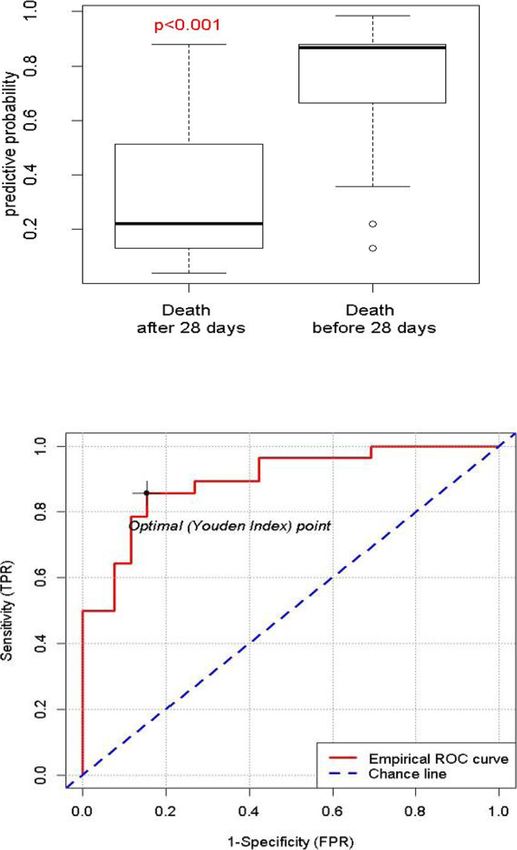
L, fibrinogen < 1 g/L) were observed in six patients into a breast cancer MAHA survival score (Fig. 3a).
(11%), suggesting possible disseminated intravascular Applied to our retrospective cohort, this score dis-
coagulation (DIC), according to the ISTH-DIC score (at played a sensitivity for early death (< 4 weeks OS) of
least a score of 5) [21]. Impaired renal function, defined 0.86 (95% CI [0.67; 0.96]), a specificity of 0.73 (95% CI
as glomerular filtration rate < 60 ml/min, was observed [0.52; 0.88]) and an area under the curve (AUC) of 0.90
in 13 patients (34.2%). Thirty-six (69.2%) patients had (95% CI [0.83; 0.97], Fig. 3).Alhenc-Gelas et al. Breast Cancer Research (2021) 23:9 Page 5 of 10
Table 2 Clinical and laboratory features at MAHA diagnosis and association with 4-week survival
Univariate analysis Multivariate analysis
Factors N (%) OR 95% IC p value OR 95% IC p value
PS at MAHA diagnosis
PS 1–2 30 (55.6%) 1.0 0.003 1.0 0.01
PS 3–4 24 (44.4%) 6.0 [1.8; 19.8] 7.0 [1.6; 31.8]
Clinical bleeding
Yes 9 (16.7%) NS NS
No 45 (83.3%)
Neurological symptoms
Yes 15 (28.3%) NS NS
No 38 (71.7%)
NA 1
Dyspnoea
Yes 11 (20.8%) NS NS
No 42 (79.2%)
NA 1
Metastatic sites
1 28 (51.9%) 2.9 [1.0; 8.7]
Platelets
< 50 G/L 29 (53.7%) NS NS
≥ 50 G/L 25 (46.3%)
Haemoglobin
< 8 g/L 26 (48.1%) 4.0 [1.3; 12.5] 0.02 3.7 [0.9; 16.7] 0.08
≥ 8 g/L 28 (51.9%) 1.0 1.0
Schistocytes
> 5.0% 10 (25.0%) NS NS
0.5–5.0% 30 (75.0%)
NA 14
Erythroblastemia
Yes 40 (85.1%) NS NS
No 7 (14.9%)
NA 7
Myelemia
Yes 38 (90.5%) NS NS
No 4 (9.5%)Alhenc-Gelas et al. Breast Cancer Research (2021) 23:9 Page 6 of 10
Table 2 Clinical and laboratory features at MAHA diagnosis and association with 4-week survival (Continued)
Univariate analysis Multivariate analysis
Factors N (%) OR 95% IC p value OR 95% IC p value
NA 12
Prothrombin time
< 50% 8 (16.0%) 4.5 [0.9; 25.0] 0.07 9.1 [1.2; 50.0] 0.03
≥ 50% 42 (84.0%) 1.0 1.0
NA 4
Fibrinogen
≤ 2 g/L 15 (34.9) NS NS
> 2 g/L 28 (65.1)
NA 11
Glomerular filtration eate
> 60 mL/min 25 (65.8%) NS NS
30–60 mL/min 9 (23.7%) NS
< 30 mL/min 4 (10.5%) NS
NA 16
Total bilirubin level
< 1.24 mg/dL 16 (30.8%) 1.0 0.007 1.0
≥ 1.24 mg/dL 36 (69.2%) 7.3 [1.8; 30.6] 6.9 [1.1; 42.6] 0.04
NA 2
Pronopall scorec
Short survival 12 (31.6%) 3.7 [0.8; 16.8] 0.09
Intermediate/long survival 26 (68.4%) 1.0 NS
NA 16
NA not available, OR odds ratio, NS non-significant
a
Only 14 patients had a bone marrow examination (myelogram or bone marrow biopsy) at the time of BC-MAHA diagnosis
b
Others metastatic sites: cerebral, carcinomatous meningitis, node involvement
c
The pronopall score for early death among oncology patients was calculated according to Barbot et al., J Clin Oncol 2008, missing data being imputed
Discussion outcome was significantly different from those of the
To our knowledge, this is the first large cohort study of other patients.
breast cancer-related MAHA addressing the clinical and Our study did not estimate the breast cancer-related
laboratory characteristics at MAHA diagnosis and iden- MAHA incidence rate, as the number of metastatic
tifying survival prognostic factors. Fewer than 60 individ- breast cancer patients treated over the same time period
ual cases of breast cancer-related MAHA have been is unknown. However, for benchmarking purposes, in a
reported in the literature, mostly corresponding to single single participating institution (Institut Curie, Paris), 2
case reports [23]. The limited clinical and laboratory patients were diagnosed with breast cancer-related
data available in these reports and a very likely publica- MAHA and 91 patients were diagnosed with breast can-
tion bias (biased toward patients with exceptional sur- cer meningeal carcinomatosis between 2000 and 2007
vival) limit the value of case report compilations. [26]. Meningeal carcinomatosis has an estimated cumu-
Firstly, our study confirms that breast cancer-related lative incidence of < 5% in metastatic breast cancer pa-
MAHA is very rare: we identified 54 cases over the last tients [26, 27]. A cumulative incidence of ~ 0.1% is
20 years (1995–2018) in seven of the largest breast can- therefore likely for breast cancer-related MAHA among
cer centres in France. Patients with a schistocyte count metastatic breast cancer patients. However, the short
higher than 0.5% were included in our study, while some survival observed in our study suggests that many pa-
guidelines recommend a 1.0% cut-off for the diagnosis tients may die before MAHA is even diagnosed.
of mechanical haemolysis [24, 25]. However, only six of Secondly, regarding primary tumour characteristics, a
our patients had a schistocyte count between 0.5 and new finding of our study is the high prevalence of breast
1.0%, and neither their clinical characteristics nor their adenocarcinoma with either lobular histology or overtAlhenc-Gelas et al. Breast Cancer Research (2021) 23:9 Page 7 of 10
Fig. 2 Overall survival according to independent prognostic factors.
a Overall survival in all patients. b Overall survival by prothrombin
time. c Overall survival by performance status. d Overall survival by
total bilirubin level. d Overall survival by haemoglobin
lobular component (44.2%) compared to previous re-
ports describing the metastatic breast cancer population
(10–14%) [22, 28]. To our knowledge, this association
has not been previously demonstrated: while four of the
eight cases of breast cancer-related MAHA reported by
Regierer et al. were lobular adenocarcinoma, the histo-
logical subtype was missing in the 36 cases compiled in
the compilation of published cases by Lechner et al.
[10, 14]. Interestingly, lobular breast adenocarcinoma
and gastric adenocarcinoma, described as the leading
cause of cancer-associated MAHA, share many pheno-
typic and genotypic traits in common, such as low E-
Cadherin [29]. High mucin expression may also play a
direct role in the pathogenesis of cancer-related
MAHA, as it triggers platelet aggregation independently
of tissue factor secretion [9, 30]. Regarding immunohis-
tochemical profile, oestrogen receptor-positive/HER2-
negative, HER2-positive and triple-negative subtypes
frequency were similar to that observed in the general
metastatic breast cancer population [22].
In accordance with previous MAHA reports, all patients
had stage IV disease and many presented multiple
metastatic sites [31–34] with laboratory signs of bone
marrow involvement (myelemia, erythroblastemia) and/or
cytologically-proven bone marrow metastasis [10, 33–37,
38]. Degradation fibrin markers (such as D-Dimers) were
not available for most patients. However, coagulation dis-
orders observed for 6 patients suggest possible DIC, ac-
cording to ISTH-DIC criteria [21]. One of them presented
with a low fibrinogen (< 1 g/L) and high D-Dimers, sug-
gesting hyperfibrinolysis. To diagnose DIC in cancer, best
strategy should be a longitudinal biological parameters
monitoring including platelets, PT, fibrinogen and D-
Dimers [39]. Unfortunately, due to the retrospective
nature of our study, we were not able to perform it. Those
are serious limitations for defining DIC in our cohort.
Noteworthy, DIC can be responsible for biological disor-
ders such as hemolysis, thrombocytopenia and schisto-
cytes formation [40]. Then, it is almost impossible to
know whether TMA is the origin or the consequence of
coagulopathy. Establishing DIC frequency in a CR-MAHA
population is challenging and, in practice, hard to deter-
mine [41].
Moreover, in keeping with prior reports focused on
CR-MAHA [34], kidney and neurological disorders were
rare, compare to other MAHA’s causes.
OS was very poor with a median OS of 4.0 weeks, shorter
than that reported in some previous studies on cancer-
associated MAHA [10, 34, 42]. Although a difference inAlhenc-Gelas et al. Breast Cancer Research (2021) 23:9 Page 8 of 10 Fig. 3 Breast cancer MAHA survival predictive score. a predictive score of death within 28 days after MAHA diagnosis, according to observed outcome. b Predictive score according to the observed outcome. c ROC curve. Area under the curve (AUC = 0.90 (95% CI [0.83; 0.97]). Sensitivity for early death of 0.86 (95% CI [0.67; 0.96]). Specificity for early death of 0.73 (95% CI [0.62; 0.88]) survival specifically related to breast cancer-related MAHA To the best of our knowledge, no survival prognostic cannot be ruled out, this difference compared to previous factors have yet been identified for breast cancer-related studies could be primarily attributed to our study method: MAHA. In our study, altered performance status, abnor- most cases were retrieved by a systematic in silico search, mal prothrombin time, and elevated total bilirubin were while previous reports may be subject to declaration (to a the three strongest independent prognostic factors, while MAHA registry [34]) or positive publication [10] biases. low haemoglobin had a more marginal impact. These
Alhenc-Gelas et al. Breast Cancer Research             (2021) 23:9                                                                                Page 9 of 10

factors could be used to distinguish patients likely to                          Consent for publication
benefit from urgent antineoplastic therapy (the only ef-                         Not applicable.

fective treatment for CR-MAHA [34, 35, 38, 43–45])
                                                                                 Competing interests
from those who should preferably be referred for pallia-                         The authors declare that they have no competing interests
tive care. Of note, the proposed algorithm was not vali-
dated on an external series, due to the rarity of breast                         Author details
                                                                                 1
                                                                                  Department of Medical Oncology, Institut Curie, Paris and Saint Cloud,
cancer-related MAHA. Other limitations of our study                              France. 2UVSQ, Université Paris-Saclay, 35 rue Dailly, Saint Cloud, 92210,
include its retrospective nature, limited sample size and                        France. 3Institut Curie, Biometry Unit, Paris and Saint-Cloud, France.
                                                                                 4
a lack of a systematic TMA diagnosis strategy including                           Department of Cancer Medicine, Institut Gustave Roussy, Villejuif, France.
                                                                                 5
                                                                                  Department of Medical Oncology, Institut de Cancérologie de l’Ouest,
ADAMTS13 activity dosage to formally exclude idio-                               Saint-Herblain, France. 6Department of Medical Oncology, Centre François
pathic TTP. To prevent those bias, prospective studies                           Baclesse, Caen, France. 7Department of Medical Oncology, Institut du Cancer
should thus be performed to explore the incidence of                             de Montpellier, Institut de cancérologie de Montpellier INSERM U1194,
                                                                                 Montpellier, France. 8Department of Medical Oncology, Centre
CR-MAHA in metastatic breast cancer patients.                                    Georges-François Leclerc, Dijon, France. 9Department of Medical Oncology,
                                                                                 Centre Léon Bérard, Lyon, France. 10Department of Medical Oncology,
                                                                                 Institut Claudius Regaud, Institut Universitaire du Cancer de
Conclusions                                                                      Toulouse-Oncopole (IUCT-Oncopole), Toulouse, France. 11Department of
Our study substantiates the pathological, clinical and la-                       Pathology, Institut Curie, Paris, France. 12Université de Paris, Paris, France.
                                                                                 13
boratory profile of patients with breast cancer MAHA:                              Reference Center for Thrombotic Microangiopathies (CNR-MAT), AP-HP.SU,
                                                                                 INSERM UMRS, 1138 Paris, France. 14Sorbonne University, Paris, France.
patients with cancer of lobular or mixed-type histology
associated with direct or indirect signs of bone marrow                          Received: 24 August 2020 Accepted: 1 January 2021
involvement and possibly DIC. We confirm the dramat-
ically poor survival prognosis of breast cancer-related
                                                                                 References
MAHA but identify prognostic factors that may be use-
                                                                                 1. George JN, Nester CM. Syndromes of thrombotic microangiopathy. N Engl J
ful for treatment decision-making and any future clinical                            Med. 2014;371(7):654–66.
trial on breast cancer MAHA treatment.                                           2. Antman KH, Skarin AT, Mayer RJ, Hargreaves HK, Canellos GP.
                                                                                     Microangiopathic hemolytic anemia and cancer: a review. Medicine
                                                                                     (Baltimore). 1979;58(5):377–84.
Supplementary Information                                                        3. Brain MC, Dacie JV, Hourihane DO. Microangiopathic haemolytic anaemia:
The online version contains supplementary material available at https://doi.         the possible role of vascular lesions in pathogenesis. Br J Haematol. 1962;
org/10.1186/s13058-021-01386-y.                                                      8(4):358–74.
                                                                                 4. Versteeg HH, Spek CA, Peppelenbosch MP, Richel DJ. Tissue factor and
 Additional file 1: Supp Mat 1. Clinical and biological parameters                   cancer metastasis: the role of intracellular and extracellular signaling
 included in statistical analysis. 1A: Data requested from participating             pathways. Mol Med Camb Mass. 2004;10(1–6):6–11.
 centres. 1B: Predictive score construction according to statistical analysis.   5. Werner TL, Agarwal N, Carney HM, Rodgers GM. Management of cancer-
                                                                                     associated thrombotic microangiopathy: what is the right approach? Am J
 Additional file 2: Supp Mat 2. Kaplan-Meier survival curves corre-                  Hematol. 2007;82(4):295–8.
 sponding to 2A: Time from first cancer diagnosis until development of           6. Zwicker JI, Liebman HA, Neuberg D, Lacroix R, Bauer KA, Furie BC, et al.
 MAHA. 2B: Time from first metastasis until development of MAHA.                     Tumor-derived tissue factor-bearing microparticles are associated with
                                                                                     venous thromboembolic events in malignancy. Clin Cancer Res Off J Am
Abbreviations                                                                        Assoc Cancer Res. 2009;15(22):6830–40.
MAHA: Microangiopathic haemolytic anaemia; TMA: Thrombotic                       7. Marumo S, Sakaguchi M, Teranishi T, Higami Y, Koshimo Y, Kato M.
microangiopathy; PS: Performance status; AUC: Area under the curve; ROC              Pulmonary tumor thrombotic microangiopathy induced by ureteral
: Receiver operating characteristic; OS: Overall survival; OR: Odds ratio            carcinoma: a necropsy case report. Case Rep Oncol. 2014;7(2):605–10.
                                                                                 8. Oleksowicz L, Bhagwati N, DeLeon-Fernandez M. Deficient activity of von
                                                                                     Willebrand’s factor-cleaving protease in patients with disseminated
Acknowledgements
                                                                                     malignancies. Cancer Res. 1999;59(9):2244–50.
We thank Sandrine Malot for the technical assistance.
                                                                                 9. Wahrenbrock M, Borsig L, Le D, Varki N, Varki A. Selectin-mucin
                                                                                     interactions as a probable molecular explanation for the association of
Authors’ contributions                                                               Trousseau syndrome with mucinous adenocarcinomas. J Clin Invest.
FCB designed and supervised the study. All authors collected the data. FB            2003;112(6):853–62.
performed statistical analyses. MAG drafted the manuscript. All authors          10. Lechner K, Obermeier HL. Cancer-related microangiopathic hemolytic
critically reviewed and edited the manuscript and approved the final version.        anemia: clinical and laboratory features in 168 reported cases. Medicine
                                                                                     (Baltimore). 2012;91(4):195–205.
Funding                                                                          11. Himmelmann A, Schefer H. Microangiopathic haemolytic anaemia in a
This study was supported by a research grant from Novartis France to FC              patient with metastatic breast cancer. Br J Haematol. 2009;146(3):231.
Bidard’s institution and by Institut Curie SiRIC 2 (grant INCa-DGOS-4654).       12. Ataga KI, Graham ML. Microangiopathic hemolytic anemia associated with
                                                                                     metastatic breast carcinoma. Am J Hematol. 1999;61(4):254–5.
Availability of data and materials                                               13. Brain MC, Azzopardi JG, Baker LR, Pineo GF, Roberts PD, Dacie JV.
The datasets used and/or analysed during the current study are available             Microangiopathic haemolytic anaemia and mucin-forming adenocarcinoma.
from the corresponding author on reasonable request.                                 Br J Haematol. 1970;18(2):183–93.
                                                                                 14. Regierer AC, Kuehnhardt D, Schulz C-O, Flath B, Jehn CF, Scholz CW, et al.
Ethics approval and consent to participate                                           Breast cancer-associated thrombotic microangiopathy. Breast Care. 2011;
This study was approved by the Institut Curie review board; a waiver of              6(6):441–5.
informed consent was granted because of the retrospective nature of the          15. Coppo P, Veyradier A, Chantal Loirat C. Microangiopathies thrombotiques.
work.                                                                                Référentiels Hémostase/Société Française d’Hématologie [Internet].
Alhenc-Gelas et al. Breast Cancer Research (2021) 23:9 Page 10 of 10
16. Moake JL. Thrombotic microangiopathies. N Engl J Med. 2002;347(8):589–600. 40. Colman RW, Rubin RN. Disseminated intravascular coagulation due to
17. Barbot A-C, Mussault P, Ingrand P, Tourani J-M. Assessing 2-month clinical malignancy. Semin Oncol. 1990;17(2):172–86.
prognosis in hospitalized patients with advanced solid tumors. J Clin Oncol 41. Coppo P. Microangiopathies thrombotiques secondaires. Rev Méd Interne.
Off J Am Soc Clin Oncol. 2008;26(15):2538–43. 2017;38(11):731–6.
18. Harrell FE Jr Hmisc: Harrell Miscellaneous. R package version 4.2–0 [Internet]. 42. Elliott MA, Letendre L, Gastineau DA, Winters JL, Pruthi RK, Heit JA. Cancer-
2019. Disponible sur: https://CRAN.R-project.org/package=Hmisc associated microangiopathic hemolytic anemia with thrombocytopenia: an
19. Harrell FE Jr. Regression Modeling Strategies. R package version 5.1–3.1. important diagnostic consideration. Eur J Haematol. 2010;85(1):43–50.
[Internet]. 2019. Disponible sur: https://CRAN.R-project.org/package=rms 43. von Bubnoff N, Sandherr M, Schneller F, Peschel C. Thrombotic
20. Md Riaz Ahmed Khan and Thomas Brandenburger. ROCit: Performance thrombocytopenic purpura in metastatic carcinoma of the breast. Am J Clin
Assessment of Binary Classifier with Visualization. R package version 1.1.1. Oncol. 2000;23(1):74–7.
[Internet]. 2019. Disponible sur: https://CRAN.R-project.org/package=ROCit 44. Abdel Samie A, Sandritter B, Theilmann L. Severe microangiopathic
21. Levi M, Toh CH, Thachil J, Watson HG. Guidelines for the diagnosis and hemolytic anemia as first manifestation of a CUP syndrome. Rapid
management of disseminated intravascular coagulation. British Committee hematologic remission under polychemotherapy. Med Klin Munich Ger
for Standards in Haematology. Br J Haematol. 2009;145(1):24–33. 1983. 2004;99(3):148–53.
22. Gobbini E, Ezzalfani M, Dieras V, Bachelot T, Brain E, Debled M, et al. Time 45. Narita M, Nakao K, Ogino N, Emoto T, Nakahara M, Yumiba T, et al. A case of
trends of overall survival among metastatic breast cancer patients in the microangiopathic hemolytic anemia associated with breast cancer:
real-life ESME cohort. Eur J Cancer Oxf Engl 1990. 2018;96:17–24. improvement with chemoendocrine therapy. Breast Cancer Tokyo Jpn.
23. Takabatake D, Oishi K. Microangiopathic hemolytic anemia associated with 1997;4(1):39–42.
metastatic breast cancer: case report and literature review. SpringerPlus.
2016;5(1):684.
24. Lesesve J-F, Lecompte T, Alla F, Fenneteau O, Cynober T, Siest J-P, et al.
Publisher’s Note
Springer Nature remains neutral with regard to jurisdictional claims in
Reproductibility of the morphological identification of schisocytes and
published maps and institutional affiliations.
evaluation of non observer-dependent methods. Ann Biol Clin (Paris). 2005;
63(3):279–89.
25. Lesesve J-F, Crepin O, Siest J-P, Régnier F, Zeltner S. Evaluation of schistocytes
measurement guidelines. Ann Biol Clin (Paris). 2012;70(5):605–13.
26. Gauthier H, Guilhaume MN, Bidard FC, Pierga JY, Girre V, Cottu PH, et al.
Survival of breast cancer patients with meningeal carcinomatosis. Ann
Oncol. 2010;21(11):2183–7.
27. Niwińska A, Rudnicka H, Murawska M. Breast cancer leptomeningeal
metastasis: propensity of breast cancer subtypes for leptomeninges and the
analysis of factors influencing survival. Med Oncol Northwood Lond Engl.
2013;30(1):408.
28. Guiu S, Wolfer A, Jacot W, Fumoleau P, Romieu G, Bonnetain F, et al.
Invasive lobular breast cancer and its variants: how special are they for
systemic therapy decisions? Crit Rev Oncol Hematol. 2014;92(3):235–57.
29. Schrader KA, Masciari S, Boyd N, Wiyrick S, Kaurah P, Senz J, et al. Hereditary
diffuse gastric cancer: association with lobular breast cancer. Familial
Cancer. 2008;7(1):73–82.
30. Sack GH, Levin J, Bell WR. Trousseau’s syndrome and other manifestations of
chronic disseminated coagulopathy in patients with neoplasms: clinical,
pathophysiologic, and therapeutic features. Medicine (Baltimore). 1977;56(1):1–37.
31. Francis KK, Kalyanam N, Terrell DR, Vesely SK, George JN. Disseminated
malignancy misdiagnosed as thrombotic thrombocytopenic purpura: a
report of 10 patients and a systematic review of published cases. The
Oncologist. 2007;12(1):11–9.
32. Lohrmann H-P. Microangiopathic hemolytic anemia in metastatic
carcinoma: report of eight cases. Ann Intern Med. 1973;79(3):368.
33. Fontana S, Gerritsen HE, Kremer Hovinga J, Furlan M, Lämmle B.
Microangiopathic haemolytic anaemia in metastasizing malignant tumours
is not associated with a severe deficiency of the von Willebrand factor-
cleaving protease. Br J Haematol. 2001;113(1):100–2.
34. Oberic L, Buffet M, Schwarzinger M, Veyradier A, Clabault K, Malot S, et al.
Cancer awareness in atypical thrombotic microangiopathies. The
Oncologist. 2009;14(8):769–79.
35. Lin YC, Chang HK, Sun CF, Shih LY. Microangiopathic hemolytic anemia as
an initial presentation of metastatic cancer of unknown primary origin.
South Med J. 1995;88(6):683–7.
36. Spoormans I, Altintas S, Van den Brande J, Luijks A, Vermorken JB. Purpura
in a patient with disseminated breast cancer: a rapidly progressive cancer-
related thrombotic thrombocytopenic purpura. Ann Oncol Off J Eur Soc
Med Oncol. 2008;19(6):1204–7.
37. Forman RB, Benkel SA, Novik Y, Tsai H-M. Presence of ADAMTS13 activity in
a patient with metastatic cancer and thrombotic microangiopathy. Acta
Haematol. 2003;109(3):150–2.
38. Chang JC, Naqvi T. Thrombotic thrombocytopenic purpura associated with
bone marrow metastasis and secondary myelofibrosis in cancer. Oncologist.
2003;8(4):375–80.
39. Thachil J, Falanga A, Levi M, Liebman H, Di Nisio M. Management of cancer-
associated disseminated intravascular coagulation: guidance from the SSC
of the ISTH. J Thromb Haemost. 2015;13(4):671–5.You can also read