Prognostic Importance of Left Ventricular Global Longitudinal Strain in Patients with Severe Aortic Stenosis and Preserved Ejection Fraction
←
→
Page content transcription
If your browser does not render page correctly, please read the page content below
VALUVULAR HEART DISEASE

                          Prognostic Importance of Left Ventricular
                          Global Longitudinal Strain in Patients with
                           Severe Aortic Stenosis and Preserved
                                      Ejection Fraction
               Nicolas Thellier, MD, Alexandre Altes, MD, Ludovic Appert, MD, Camille Binda, MD, Blandine Leman, MD,
                       Wassima Marsou, MD, Nicolas Debry, MD, Camille Joly, Pierre-Vladimir Ennezat, MD, PhD,
               Christophe Tribouilloy, MD, PhD, and Sylvestre Marechaux, MD, PhD, Lille, Valenciennes, Grenoble, and Amiens,
                                                                  France

               Background: Impaired left ventricular (LV) speckle-tracking-derived global longitudinal strain (GLS) magnitude
               (GLS worse than 14.7%) has been associated with poor outcome in patients with severe aortic stenosis (AS)
               and preserved LV ejection fraction (EF).

               Objectives: To test the hypothesis that GLS magnitude # 15% obtained with vendor-independent speckle-
               tracking strain software may be able to identify patients with severe AS who are at higher risk of death, despite
               preserved LVEF and no or mild symptoms.

               Methods: GLS was retrospectively obtained in 332 patients with severe AS (aortic valve area indexed
               [AVAi] < 0.6 cm2/m2), no or mild symptoms, and LVEF $ 50%. Absolute values of GLS were collected. Survival
               analyses were carried out to study the impact of GLS magnitude on all-cause mortality.

               Results: During a median follow-up period of 42 (37-46) months, 105 patients died. On multivariate analysis,
               and after adjustment of known clinical and/or echocardiographic predictors of outcome and aortic valve
               replacement as a time-dependent covariate, GLS magnitude # 15% was independently associated with mor-
               tality during follow-up (all P < .01). Adding GLS magnitude # 15% (adjusted hazard ratio = 1.99 [1.17-3.38],
               P = .011) to a multivariate model including clinical and echocardiographic variables of prognostic importance
               (aortic valve replacement, aortic valve area, LV stroke volume index < 30 mL/m2, and LVEF 15% (hazard ratio = 2.10; 95% confidence interval,
               1.20-3.68; P = .009).

               Conclusions: In this series of patients with severe AS, no or mild symptoms, and LVEF $ 50%, GLS obtained
               with vendor-independent speckle-tracking strain software was an effective tool to identify patients with a poor
               outcome. Detection of myocardial dysfunction by identifying GLS magnitude < 15% in patients with severe AS,
               no or mild symptoms, and LVEF $ 50%, can aid in risk assessment. (J Am Soc Echocardiogr 2020;33:1454-
               64.)

               Keywords: Aortic stenosis, Surgery, Speckle-tracking, Echocardiography, Outcome

From the Universite   Lille Nord de France, GCS-Groupement des Ho       ^ pitaux de
l’Institut Catholique de Lille, Laboratoire d’e   chocardiographie, services de          Thellier and Altes contributed equally to this work.
cardiologies, Centre des Valvulopathies, Faculte   de Medecine et de Ma€ıeutique,       Conflicts of Interest: None.
Universite  Catholique de Lille, Lille (N.T., A.A., L.A., C.B., B.L., W.M., N.D.,        Reprint requests: Sylvestre Marechaux, MD, PhD, Cardiology Department, GCS-
S.M.), Centre Hospitalier de Valenciennes, Service de cardiologie, Valenciennes           Groupement des Ho    ^ pitaux de l’Institut Catholique de Lille, Faculte
                                                                                                                                                                  libre de
                        Lille Nord de France, GCS-Groupement des Ho
(N.T., B.L.), Universite                                                  ^ pitaux de     medecine/Universite Catholique de Lille, Rue du Grand But, 59160 Lomme,
l’Institut Catholique de Lille, Dele
                                    gation a
                                             la recherche clinique et l’innovation,      France (E-mail: Sylvestre.marechaux@yahoo.fr).
Lille (C.J.), Centre Hospitalier Universitaire de Grenoble, Grenoble (P.-V.E.),           0894-7317/$36.00
Centre Universitaire de Recherche en Sante       , Laboratoire MP3CV-EA 7517,
                                                                                          Copyright 2020 by the American Society of Echocardiography.
Universite  de Picardie, Amiens (N.T., CT., S.M.), and Centre Hospitalier
Universitaire d’Amiens, Amiens (C.T., S.M.), France.                                      https://doi.org/10.1016/j.echo.2020.07.002

1454

                         Downloaded for Anonymous User (n/a) at Brazilian Society of Cardiology from ClinicalKey.com by Elsevier on January 10, 2021.
                                  For personal use only. No other uses without permission. Copyright ©2021. Elsevier Inc. All rights reserved.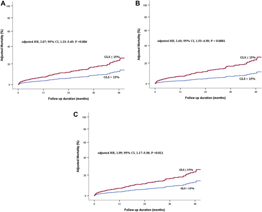
Journal of the American Society of Echocardiography                                                                                                  Thellier et al 1455
Volume 33 Number 12

          Abbreviations
                                   Calcific aortic stenosis (AS) is the                METHODS
                                   most prevalent valvular heart dis-
  AS = Aortic stenosis             ease in developed countries.1                       Study Population
  ASE = American Society of        The sole effective treatment for                       Consecutive patients ages $ 18 years with a diagnosis of severe AS
  Echocardiography                 patients with severe symptom-                       (defined as aortic valve area [AVA] # 1 cm2 and/or AVA normalized
                                   atic AS remains surgical or                         to body surface area [BSA] # 0.6 cm/m2), preserved LVEF $ 50%,
  AVA = Aortic valve area          transcatheter      aortic      valve                and no or minimal AS-related symptoms who attended the echocar-
  AVAi = Aortic valve area         replacement (AVR). American                         diography laboratory of the Groupement des H^        opitaux de l’Institut
  indexed                          and European guidelines alike                       Catholique de Lille, Lille Catholic University, from 2011 to 2018
                                   indicate that left ventricle (LV)                   were eligible for inclusion in the present study. Exclusion criteria
  AVR = Aortic valve
  replacement
                                   dysfunction, defined by left ven-                   were (1) moderate or greater aortic and/or mitral and/or tricuspid
                                   tricular (LV) ejection fraction                     regurgitation; (2) past or current symptoms of New York Heart
  BSA = Body surface area          (LVEF) < 50%, is a class I indica-                  Association class III-IV heart failure; (3) angina or syncope; (4) pros-
  CI = Confidence interval         tion for AVR, even in asymptom-                     thetic valve or supra- or subvalvular AS, congenital heart disease, or
                                   atic patients.2,3 As early as the                   dynamic LV outflow tract obstruction; (5) mitral stenosis; and (6) pa-
  CAD = Coronary artery            1970s, Dumesnil et al.4 reported                    tient refusal to participate in the study. The study population
  disease                          work on echocardiography                            comprised 332 patients who were followed for the duration of the
  EACVI = European                 showing that M-mode tracings                        study (2011-18). The present study is a retrospective analysis of a pro-
  Association of Cardiovascular    can reveal depressed LV longitu-                    spective registry.
  Imaging                          dinal systolic shortening despite                      Clinical and demographic data were collected at baseline. The
  EF = Ejection fraction           normal LVEF in AS patients                          Charlson comorbidity index, a summation of the patient’s individual
                                   compared with controls.4 One                        comorbidities, was calculated.13 The Charlson comorbidity index in-
  GLS = Global longitudinal        way to assess LV longitudinal                       cludes history of myocardial infarction, congestive heart failure, periph-
  strain                           shortening is to calculate LV                       eral artery disease, cerebrovascular disease, connective tissue disease,
  HR = Hazard ratio                global longitudinal strain (GLS)                    peptic ulcer disease, liver disease, diabetes mellitus, hemiplegia, mod-
                                   using speckle-tracking echocardi-                   erate or severe renal disease, solid tumor, leukemia, and lymphoma.
  ICC = Intraclass correlation     ography.5 In a recent participant
  coefficient                                                                          Coronary artery disease (CAD) was defined by documented history
                                   data meta-analysis, Magne et al.6                   of acute coronary syndromes, confirmation by coronary angiography
  IDI = Integrated                 elegantly demonstrated by pool-                     (reduction of normal diameter $ 50% in the left main coronary artery
  discrimination improvement       ing 10 studies that impaired                        and $70% in the right coronary artery, left anterior descending coro-
  LAV = Left atrial volume         GLS (defined as GLS < 14.7%)                        nary artery, left anterior descending coronary artery, or circumflex cor-
                                   was associated with a 2.5-fold                      onary artery), or history of coronary revascularization. Symptoms were
  LAVi = Left atrial volume        increased risk of mortality.                        ascertained by each patient’s personal cardiologist. Follow-up informa-
  indexed                          Recently, a 15% cutoff value                        tion was obtained retrospectively.
  LV = Left ventricular, ventricle has been suggested, which is                           The study was approved by an independent ethics committee and
                                   easier to remember and thus                         was conducted in accordance with institutional policies, national legal re-
  LVEF = Left ventricular          might allow a wider accep-
  ejection fraction                                                                    quirements, and the revised Declaration of Helsinki. Given the retro-
                                   tance.7,8 However, significant in-                  spective nature of the analysis, informed consent was waived. All
  LVH = Left ventricular           tervendor       variability     with                patients agreed to participate in the study when contacted for follow-up.
  hypertrophy                      significant differences in GLS
                                   values obtained with equipment                      Echocardiography
  NRI = Net reclassification
  improvement                      from different vendors, and also                       All patients underwent a comprehensive Doppler echocardiogra-
                                   between earlier or later (up-                       phy study, using commercially available ultrasound systems
  RAP = Right atrial pressure      graded) versions of the same                        (General Electric Vivid E9, Vivid E95, Vivid 7, General Electric
  RV = Right ventricular           software, may limit the general-                    HealthCare, Horten, Norway; Philips IE 33 and Epiq 7, Philips,
                                   ization of GLS use in clinical                      Andover, MA) by experienced echocardiographers. Aortic flow was
  SMD = Standardized mean          practice.9-12                                       recorded using continuous-wave Doppler, by imaging and nonimag-
  difference
                                       Hence, the present study was                    ing transducers, systematically in several acoustic windows (apical
  sPAP = Systolic pulmonary        designed to test the hypothesis                     five-chamber, right parasternal, suprasternal, epigastric).14 The highest
  artery pressure                  that an absolute value of                           aortic velocity was used to calculate aortic time-velocity integral and
  SV = Stroke volume               GLS # 15% obtained with                             mean pressure gradient. As recommended by current guidelines,
                                   vendor-independent          speckle-                wall (high-pass) filters were set at a high level and gain was decreased
  SVi = Stroke volume indexed      tracking strain software may be                     to optimize identification of the velocity curve from the spectrogram
                                   able to identify patients with se-                  envelope. The LV stroke volume (LV SV) was calculated by multi-
vere AS who are at higher risk of death, despite preserved LVEF                        plying the LV outflow tract area by the LV outflow tract time-
and no or only mild symptoms.                                                          velocity integral obtained by pulsed Doppler in the apical

                      Downloaded for Anonymous User (n/a) at Brazilian Society of Cardiology from ClinicalKey.com by Elsevier on January 10, 2021.
                               For personal use only. No other uses without permission. Copyright ©2021. Elsevier Inc. All rights reserved.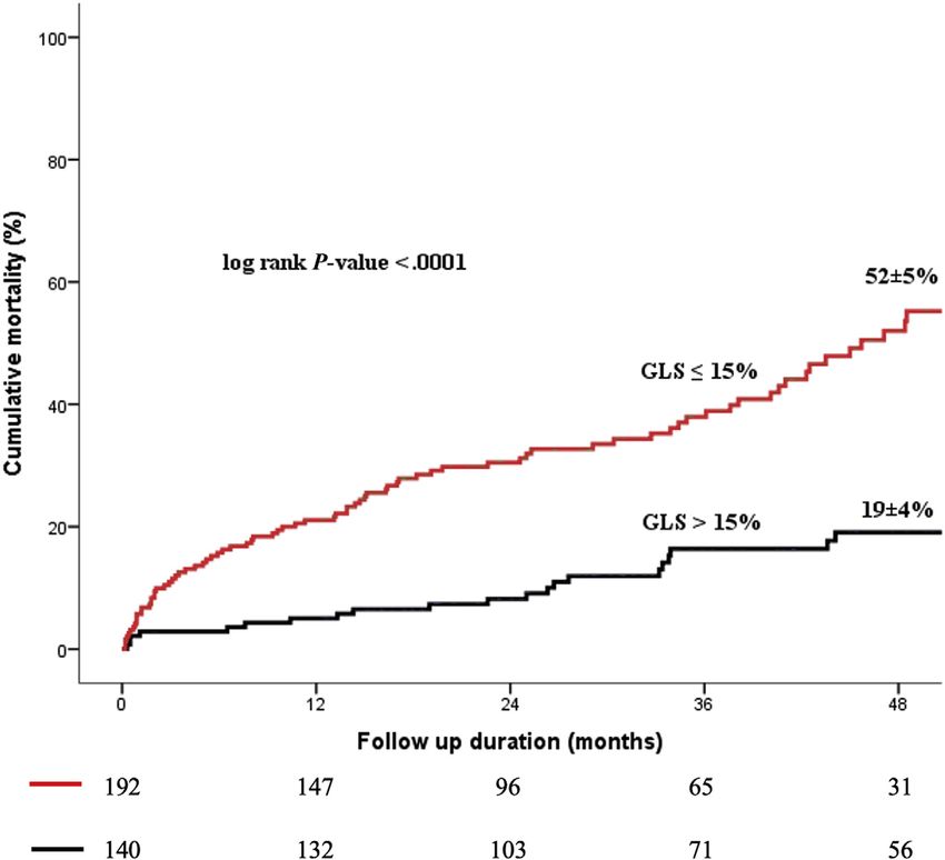
1456 Thellier et al Journal of the American Society of Echocardiography
December 2020
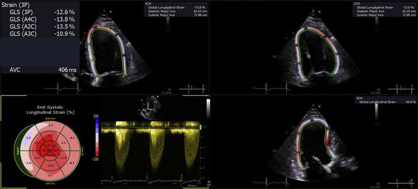
excursion 95 g/m2 in women and 115 g/m2 in men according to the automatic tracing of the endocardial border in the three apical views
American Society of Echocardiography (ASE) formula.15 Elevated (apical four-, three-, and two-chamber views). The videoloops were
filling pressure was defined by E/e’ > 14.7 Left atrial volume acquired with a minimal frame rate of 50 frames per second. After
(LAV) was calculated using the Simpson biplane method and frame-by-frame analysis during one cardiac cycle of the LV endocar-
indexed to BSA (LAVi). The transtricuspid pressure gradient was dial speckle-tracking, the software provides 16-segment regional seg-
recorded from any view with continuous-wave Doppler imaging mentation curves (six basal segments, six middle segments, four apical
and was used to determine the peak systolic pulmonary artery segments). Global longitudinal strain was defined as the average value
pressure (sPAP) using the modified Bernoulli equation of maximum deformation of each of the 16 segments during the sys-
(sPAP = 4Vmax2 + RAP), where V is the peak tricuspid regurgitation tole before aortic valve closure (Figure 1). Tracking adequacy was
velocity and RAP is the right atrial pressure. The RAP was assumed to checked visually, followed by manual adjustment of the endocardial
be 3, 8, or 15 mm Hg based on the diameter of the inferior vena cava border if considered suboptimal. Intra- and interobserver variability of
and importance of inspiratory collapse during a brief sniff, as recom- each GLS measurement was tested on a randomly selected set of 20
mended by current European Association of Cardiovascular echocardiograms from the study population.
Imaging (EACVI)/ASE guidelines.15 Moderate or greater right ven-
tricular (RV) dysfunction was determined by a multiparameter
Clinical Decision and Follow-Up
approach including semiquantitative assessment by visual examina-
tion and quantitative assessment using tricuspid annular systolic ve- After the initial medical management, treatment was conservative
locity (S’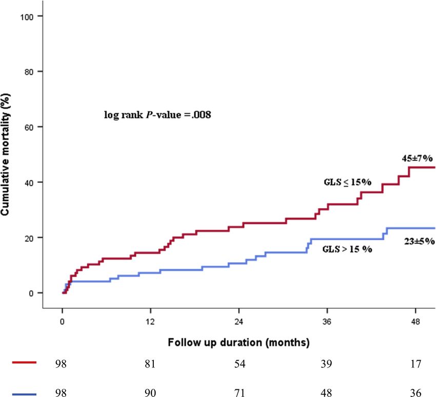
Journal of the American Society of Echocardiography                                                                                                  Thellier et al 1457
Volume 33 Number 12

The majority of patients were followed clinically and echocardio-                      the 95% bootstrap CIs of the 999 estimates, for which the lower and
graphically in our institution’s outpatient clinic. Others were followed               upper bounds were the 2.5th and 97.5th percentiles of the resampling
in public hospitals or private practices by referring cardiologists work-              distribution, respectively. Integrated discrimination improvement
ing in coordination with our tertiary center. Follow-up information                    (IDI) and net reclassification improvement (NRI) were determined
was obtained retrospectively. Events were ascertained by direct pa-                    to further describe the added utility of GLS when added to the multi-
tient interview and physical examination and/or via repeated                           variate model. The IDI measures the new model’s ability to improve
follow-up letters, questionnaires, and telephone calls to physicians,                  integrated sensitivity without compromising integrated specificity.
patients, and (if necessary) next of kin. The outcome variable of the                  The NRI measures the appropriateness of patient reclassification on
study was all-cause mortality. Clinical decisions regarding medical                    the basis of the probability of death at selected time points. Both
management and referral for surgery were made by the heart team                        NRI and IDI were computed at 48 months using the R package
with the approval of the patient’s cardiologist in accordance with cur-                survIDINRI. We conducted subgroup analyses (for GLS > and
rent practice guidelines.                                                              #15%) to determine the homogeneity of the GLS-mortality associa-
                                                                                       tion. First, we estimated the effect of GLS on mortality in each sub-
Statistical Analysis                                                                   group using a Cox univariate model and then formally tested for
   Continuous variables are expressed as median (25th-75th percen-                     first-order interactions, entering interaction terms separately for
tile) or mean 6 SD, and categorical variables are expressed as abso-                   each subgroup. Estimates of sensitivity, specificity, and positive and
lute numbers and percentages. To simplify interpretation of the                        negative predictive values for the GLS 15% and 14.7% cutoff values
results as well as the discussion, GLS values, although negative, are re-              were computed from time-dependent receiver operating curves using
ported as absolute values. The study population was divided accord-                    the timeROC package in R. Sensitivity analysis was also conducted to
ing to GLS > or #15%. The Pearson c2 statistic or Fisher’s exact test                  compare the occurrence of mortality during follow-up between pa-
was used to examine associations between the two groups and base-                      tients with GLS # 15% and >15% matched by age, sex, atrial fibrilla-
line categorical variables. Individual differences were compared using                 tion, Charlson comorbidity index, AVA, LV SVi, LVEF, and occurrence
Mann-Whitney U tests (with Bonferroni correction for multiple com-                     of mild-to-moderate RV dysfunction using a 5-to-1 digit-matching
parisons). The intraclass correlation coefficient (ICC) was used to ex-                propensity score greedy algorithm (MatchIT package in R). The
press variability. The ICC estimates and their 95% confidence interval                 nearest-neighbor matching method was used. Standardized mean dif-
(CI) were calculated based on a single rater/measurement, absolute-                    ferences (SMDs) before and after matching were estimated to assess
agreement, two-way fixed-effects model.                                                the quality of the propensity score matching procedure. Absolute
    Event rates 6 standard errors of the overall population and of two                 SMDs < 0.2 were considered an indicator of adequate balance and
groups were estimated according to the Kaplan-Meier method and                         thus sufficient bias reduction. The quality of the matching was visually
compared using two-sided log-rank tests. Univariate and multivariate                   assessed by the distribution of propensity scores (jitter plot of the dis-
analyses of time to events were performed using Cox proportional                       tance measure, QQplots, and histograms of propensity score density
hazards models. We did not use model-building techniques. For                          for observations before and after matching). A significance level of
each model, we retained covariates that we considered would                            0.05 was assumed for all tests. All P values are the results of two-
potentially have a prognostic impact on an epidemiologic basis to in-                  tailed tests. Data were analyzed with SPSS version 25.0 (IBM,
crease the external validity of the analyses. The effect of AVR on mor-                Armonk, NY), R version 3.5.3 (R Foundation for Statistical
tality was considered in all models and analyzed as a time-dependent                   Computing, Vienna, Austria), and MedCalc version 12.5.0
covariate.16 We thus tested the following models: model 1 including                    (MedCalc Software, Mariakerke, Belgium).
clinical factors (age, sex, body mass index, Charlson comorbidity index
[not including age], CAD, hypertension, atrial fibrillation); model 2
including classical echocardiographic factors of prognostic importance
                                                                                       RESULTS
previously included in an extra-aortic cardiac disorder staging7 (AVA,
LVH, LAVi > 34 mL/m2, sPAP $ 60 mm Hg, grade II diastolic dysfunc-                     Baseline Characteristics
tion, RV dysfunction $ moderate, LVEF < 60%, LV SV indexed                             The study population consisted of 332 patients with a median age of
[SVi] < 30 mL/m2); and model 3 including clinical and selected echo-                   79 (interquartile range, 71, 85 years), with 196 being women (59%,
cardiographic factors previously known to be strongly associated with                  Table 1). The clinical and echocardiographic characteristics of the study
outcome in AS (that is AVA, LV SVi < 30 mL/m2, and LVEF
1458 Thellier et al Journal of the American Society of Echocardiography
December 2020
Table 1 Demographic, clinical, and echocardiographic parameters according to GLS > and # 15%
Variable All (N = 332) GLS > 15 (n = 140) GLS # 15 (n = 192) Overall P value
Demographic and clinical characteristics
Age, years 79 [71;85] 76 [66;82] 82 [74;88] 60 mm Hg, E/
had more comorbidities as indicated by a higher Charlson’s comor- e’ > 14, RV dysfunction, LVEF < 60%, and LV SVi < 30 mL/m2)
bidity index. Aortic stenosis was more severe in these patients as indi- and AVR as a time-dependent covariate, GLS # 15% remained signif-
cated by a lower AVA but similar transaortic gradients and velocities. icantly associated with an increased risk of mortality: adjusted
Left ventricular EF was slightly lower in these patients as was LV SVi. HR = 2.63 (1.53–4.50; P < .001; Figure 3B). After adjustment for
Downloaded for Anonymous User (n/a) at Brazilian Society of Cardiology from ClinicalKey.com by Elsevier on January 10, 2021.
For personal use only. No other uses without permission. Copyright ©2021. Elsevier Inc. All rights reserved.Journal of the American Society of Echocardiography Thellier et al 1459
Volume 33 Number 12
Figure 2 Kaplan-Meier 48-month estimates of overall mortality according to GLS # 15% or GLS > 15%.
clinical parameters, AVA, LV SVi < 30 mL/m2, LVEF < 60%, and AVR DISCUSSION
as a time-dependent covariate, GLS # 15% remained significantly
associated with an increased risk of mortality: adjusted HR = 1.99 The present study clearly indicates that (1) GLS is frequently impaired
(1.17–3.38; P = .011; Figure 3C). Adding GLS # 15% to this multivar- despite preservation of LVEF in patients with severe AS and no or
iate model resulted in a significant improvement in the model fit, as mild symptoms, (2) vendor-independent software-measured GLS is
indicated by a lower Bayesian information criterion (Table 2), and bet- associated with a considerable increased risk of mortality during
ter model discrimination, as indicated by higher C statistics. At follow-up in this population, (3) GLS provides additional prognostic
48 months, adding GLS # 15% to the multivariate model resulted information over clinical and classical echocardiographic predictors
in improvements in reclassification indices (Table 2). Using GLS and of poor outcome in AS.
LVEF as continuous variables in this multivariate model did not alter
the relationship between GLS and mortality (adjusted HR per per- LV Dysfunction in AS
centage of absolute decrease in GLS 1.06 [1.0, 1.11]; P = .046). The
Left ventricular systolic dysfunction with LVEF < 50% occurs late in
association of GLS # 15% and mortality risk was consistent in all sub-
the course of AS, as LVEF may be maintained despite reduced
groups of patients with severe AS and no or mild symptoms
myocardial contractility and potentially irreversible alterations in
(Figure 4).
myocardial function owing to myocardial fibrosis by the use of pre-
load reserve leading to diastolic dysfunction17 or changes in LV geom-
Impact of GLS on Mortality in the Propensity-Matched etry including LV concentric hypertrophy and remodeling.18,19
Cohort Cramariuc et al.20 previously demonstrated that a higher degree of
The baseline characteristics of covariates used for propensity match- LVH and concentric remodeling is associated with decreased LV lon-
ing before and after matching are shown in Table 3. Between-group gitudinal deformation assessed by two-dimensional speckle-tracking
balance was obtained for all matched covariates. Ninety-eight pa- in patients with AS.20,21 In addition, impairment of LV longitudinal
tients with GLS # 15% were matched to 98 patients with GLS > shortening or strain correlates with the presence of symptoms in pa-
15%. The 48-month estimate of overall mortality remained signifi- tients with AS, is associated with LV myocardial fibrosis by cardiac
cantly higher in patients with GLS # 15% compared with those magnetic resonance imaging, and predicts elicited symptoms during
with GLS > 15% (45% 6 7% vs 23% 6 5%, log-rank P = .008; exercise testing in the subset of asymptomatic patients who are able
Figure 5), with a two-fold increased risk of mortality compared to exercise.22-26 Hence, the assessment of LV GLS may be helpful
with those with GLS > 15%: HR = 2.10 (1.20-3.68, P = .009). to identify patients with severe AS who present significant
Downloaded for Anonymous User (n/a) at Brazilian Society of Cardiology from ClinicalKey.com by Elsevier on January 10, 2021.
For personal use only. No other uses without permission. Copyright ©2021. Elsevier Inc. All rights reserved.1460 Thellier et al Journal of the American Society of Echocardiography
December 2020
Figure 3 (A) Cumulative hazard of overall mortality according to GLS # 15% or GLS > 15%, adjusted for age, sex, Charlson comor-
bidity index, CAD, hypertension, atrial fibrillation, body mass index, and AVR as a time-dependent covariate. (B) Cumulative hazard of
overall mortality according to GLS # 15% or GLS > 15%, adjusted for echocardiographic parameters including AVA, LVH, LAV
index $ 34 mL/m2, sPAP > 60 mm Hg, E/e’ > 14, RV dysfunction, LVEF < 60%, and LV SVi < 30 mL/m2 with AVR as a time-
dependent covariate. (C) Cumulative hazard of overall mortality according to GLS # 15% or GLS > 15%, adjusted for age, sex, Charl-
son comorbidity index, CAD, hypertension, atrial fibrillation, body mass index, AVA, LVEF < 60%, and LV SVi < 30 mL/m2 with AVR as
a time-dependent covariate.
Table 2 Cox multivariate models: predictive value, discrimination, and reclassification of the multivariate models with and without
GLS on overall mortality
Multivariate model Multivariate model with P value (model with vs
Overall mortality without GLS GLS model without GLS)
Adjusted HR (95% CI) 1.99 (1.17-3.38) .011
BIC 1045.436 1044.682
Harrell C statistic 0.824 0.832
C statistic difference (0.0002-0.0252)
(95% CI)
IDI Reference 0.021 (0.002-0.061) .020
NRI Reference 0.333 (0.032-0.476) .033
BIC, Bayesian information criterion.
Downloaded for Anonymous User (n/a) at Brazilian Society of Cardiology from ClinicalKey.com by Elsevier on January 10, 2021.
For personal use only. No other uses without permission. Copyright ©2021. Elsevier Inc. All rights reserved.Journal of the American Society of Echocardiography Thellier et al 1461
Volume 33 Number 12
Figure 4 HR and 95% CI for risk of overall mortality associated with GLS # 15% or GLS > 15% in subgroups of patients with severe
AS, preserved LVEF, and no or minimal symptoms. MPG, Mean pressure gradient; NYHA, New York Heart Association.
myocardial damage and who are at higher risk, despite preserved incomplete control of confounding factors. Nevertheless, the
LVEF and no or only mild symptoms. present study confirms the findings of this meta-analysis by showing
that a decrease in GLS below 15% (a cutoff value close to 14.7%) is
associated with a twofold increased risk of mortality in patients
Prognostic Impact of LV GLS in Asymptomatic AS without severe symptoms related to AS. Global longitudinal strain
In a recent participant data meta-analysis, Magne et al.6 observed a provides additional prognostic information over clinical but also
2.5-fold increased risk of mortality in patients with impaired GLS echocardiographic parameters. We demonstrated here that GLS #
(defined as GLS < 14.7%) compared with those with preserved 15% provides independent prognostic information over these clas-
GLS. It is, however, noteworthy that mortality was the primary sical echocardiographic indices. The prognostic impact of GLS #
endpoint in only three of these studies. In addition, the vast majority 15% was consistent in all subgroups of patients including those
of the studies included in this meta-analysis involved a single-vendor with LVEF $ 60% and those in a low-flow state despite having pre-
speckle-tracking strain software (General Electric). Hence, the use of served LVEF. Therefore, the present study demonstrates the impor-
GLS from other vendors would potentially result in different cutoff tance of assessing LV longitudinal function as part of the
values, which may lead to confusion. Moreover, in most of these multiparametric prognostic workup of patients with severe asymp-
studies, the majority of patients had symptomatic AS.27-29 In tomatic or mildly symptomatic AS. Importantly, the negative predic-
addition, the context of retrospective data pooling with limited tive value of GLS > 15% was high in the present report, thereby
statistical adjustment (age, sex, AVAi, LVEF) necessarily involved suggesting that these patients may be conservatively followed until
Downloaded for Anonymous User (n/a) at Brazilian Society of Cardiology from ClinicalKey.com by Elsevier on January 10, 2021.
For personal use only. No other uses without permission. Copyright ©2021. Elsevier Inc. All rights reserved.1462 Thellier et al Journal of the American Society of Echocardiography
December 2020
Table 3 Baseline characteristics according to GLS > and # 15% before and after propensity score matching
Entire cohort Matched cohort
Covariates GLS > 15 (n = 140) GLS # 15 (n = 192) SMD GLS > 15 (n = 98) GLS # 15 (n = 98) SMD
Age, years 73 6 13 80 6 13 0.650 75 6 10 77 6 11 0.137
Women 79 (56) 117 (61) 0.092 55 (56) 53 (54) 0.041
Atrial fibrillation 21 (15) 76 (40) 0.574 17 (17) 19 (19) 0.053
Charlson comorbidity 1.5 6 1.7 2.2 6 2 0.390 1.8 6 1.8 1.8 6 1.6 0.006
index
AVA, cm2 0.88 6 0.19 0.81 6 0.21 0.356 0.88 6 0.20 0.87 6 0.19 0.052
LV SVi, mL/m2 42 6 9 36 6 8 0.661 40 6 8 39 6 8 0.082
LVEF, % 63 6 6 61 6 7 0.322 62 6 6 61 6 7 0.128
RV dysfunction $ 6 (4) 40 (21) 0.516 5 (5) 5 (5)Journal of the American Society of Echocardiography Thellier et al 1463
Volume 33 Number 12
vendor (General Electric). Nagata et al.35 used the same vendor- ‘‘Delegation a la Recherche Clinique et l’Innovation’’ of the
independent software as in the present study in a similar population. Groupement des H^ opitaux de l’Institut Catholique de Lille; and
The present study confirms Nagata et al.’s finding by showing that this Gerald Pope, Medical and Scientific Translations.
vendor-independent speckle-tracking strain software enabled us to
calculate GLS values with a unique cutoff level regardless of the
manufacturer of the echocardiography equipment. This type of meth- REFERENCES
odology could be expected to favor more widespread acceptance of
GLS measurements in routine daily practice. However, it should be 1. Iung B, Delgado V, Rosenhek R, Price S, Prendergast B, Wendler O, et al.
acknowledged that identifying specific cutoff points remains an issue Contemporary presentation and management of valvular heart disease:
if upgrades to this software influence GLS values as previously re- the EURObservational Research Programme Valvular Heart Disease II
ported with vendor-specific software products.12 In addition, Survey. Circulation 2019; https://doi.org/10.1161/CIRCULATIO-
Nagata et al.12 previously reported in normal subjects that intervendor NAHA.119.041080.
agreement of GLS values using vendor-independent speckle-tracking 2. Baumgartner H, Falk V, Bax JJ, De Bonis M, Hamm C, Holm PJ, et al. 2017
software was only modest, with nonnegligible limits of agreement. ESC/EACTS guidelines for the management of valvular heart disease. Eur
Heart J 2017;38:2739-91.
Nevertheless, the intervendor agreement of GLS using vendor-
3. Nishimura RA, Otto CM, Bonow RO, Carabello BA, Erwin JP 3rd,
independent speckle-tracking software cannot be assessed in the pre- Fleisher LA, et al. 2017 AHA/ACC focused update of the 2014 AHA/
sent study, as patients did not undergo scanning with both GE and ACC guideline for the management of patients with valvular heart disease:
Philips systems. a report of the American College of Cardiology/American Heart Association
Task Force on clinical practice guidelines. Circulation 2017;135:e1159-95.
Limitations 4. Dumesnil JG, Shoucri RM, Laurenceau JL, Turcot J. A mathematical model
of the dynamic geometry of the intact left ventricle and its application to
This study was conducted in a single high-volume center with a dedi- clinical data. Circulation 1979;59:1024-34.
cated heart valve unit. Further multicenter studies are needed to confirm 5. Marechaux S. Speckle-tracking strain echocardiography: any place in
these findings. The use of all-cause mortality as an endpoint may repre- routine daily practice in 2014? Arch Cardiovasc Dis 2013;106:629-34.
sent a limitation compared with the use of cause-specific mortality. 6. Magne J, Cosyns B, Popescu BA, Carstensen HG, Dahl J, Desai MY, et al.
However, all-cause mortality remains an unbiased endpoint. Whereas Distribution and prognostic significance of left ventricular global longitudi-
echocardiograms were prospectively collected, speckle-tracking strain nal strain in asymptomatic significant aortic stenosis: an individual partic-
analysis and follow-up data were obtained retrospectively; hence, our ipant data meta-analysis. JACC Cardiovasc Imaging 2019;12:84-92.
study presents inherent limitations of this type of analysis. Cardiac mag- 7. Tastet L, Tribouilloy C, Marechaux S, Vollema EM, Delgado V, Salaun E,
et al. Staging cardiac damage in patients with asymptomatic aortic valve
netic resonance imaging was not available in the vast majority of the
stenosis. J Am Coll Cardiol 2019;74:550-63.
study population. Hence, we cannot provide data on LV myocardial 8. Lancellotti P, Vannan MA. Timing of intervention in aortic stenosis. N Engl
fibrosis. This study used an observational design, which implies that J Med 2020;382:191-3.
the baseline variables in the two groups determined on the basis of 9. Castel AL, Szymanski C, Delelis F, Levy F, Menet A, Mailliet A, et al. Pro-
the GLS value (#15% vs >15%) may have been imbalanced. spective comparison of speckle tracking longitudinal bidimensional strain
However, the final results remained unchanged after Cox multivariate between two vendors. Arch Cardiovasc Dis 2014;107:96-104.
analyses and propensity score matching performed to control the impact 10. Castel AL, Menet A, Ennezat PV, Delelis F, Le Goffic C, Binda C, et al.
of these differences on mortality. Very recently, Levy-Neuman et al.36 Global longitudinal strain software upgrade: implications for intervendor
observed that reduced exercise basal LS was associated with future car- consistency and longitudinal imaging studies. Arch Cardiovasc Dis 2016;
diovascular events in patients with moderate to severe asymptomatic 109:22-30.
11. Risum N, Ali S, Olsen NT, Jons C, Khouri MG, Lauridsen TK, et al. Vari-
AS. However, LS was not obtained during exercise in our study popula-
ability of global left ventricular deformation analysis using vendor depen-
tion. Lastly, a limitation of the study is that one cannot assess whether the dent and independent two-dimensional speckle-tracking software in
intervention (AVR) at various levels of reduced GLS can reverse the rates adults. J Am Soc Echocardiogr 2012;25:1195-203.
of adverse outcomes. 12. Nagata Y, Takeuchi M, Mizukoshi K, Wu VC, Lin FC, Negishi K, et al. In-
tervendor variability of two-dimensional strain using vendor-specific and
vendor-independent software. J Am Soc Echocardiogr 2015;28:630-41.
13. Charlson ME, Pompei P, Ales KL, MacKenzie CR. A new method of clas-
CONCLUSION sifying prognostic comorbidity in longitudinal studies: development and
validation. J Chronic Dis 1987;40:373-83.
In this series of patients with severe AS, no or mild symptoms, and 14. Baumgartner H, Hung J, Bermejo J, Chambers JB, Edvardsen T,
LVEF $ 50%, GLS obtained with vendor-independent speckle- Goldstein S, et al. Recommendations on the echocardiographic assess-
tracking strain software was an effective tool to identify patients ment of aortic valve stenosis: a focused update from the European Asso-
with a poor outcome. Detection of myocardial dysfunction by identi- ciation of Cardiovascular Imaging and the American Society of
fying GLS #15% in patients with severe AS, no or mild symptoms, Echocardiography. J Am Soc Echocardiogr 2017;30:372-92.
and LVEF $ 50% can aid in risk assessment. Further larger prospec- 15. Lang RM, Badano LP, Mor-Avi V, Afilalo J, Armstrong A, Ernande L, et al.
tive multicentric studies are needed to confirm the present findings. Recommendations for cardiac chamber quantification by echocardiogra-
phy in adults: an update from the American Society of Echocardiography
and the European Association of Cardiovascular Imaging. J Am Soc Echo-
cardiogr 2015;28:1-39.e14.
ACKNOWLEDGMENTS 16. Venables WN, Ripley BD. Modern Applied Statistics with S-PLUS. 3rd ed.
New York: Springer-Verlag; 1999.
We thank the Philips Company and especially Stephane Husson and 17. Krayenbuehl HP, Hess OM, Ritter M, Monrad ES, Hoppeler H. Left ventric-
Nicolas Victorin for providing the Tomtec Image Arena software; the ular systolic function in aortic stenosis. Eur Heart J 1988;9(Suppl E):19-23.
Downloaded for Anonymous User (n/a) at Brazilian Society of Cardiology from ClinicalKey.com by Elsevier on January 10, 2021.
For personal use only. No other uses without permission. Copyright ©2021. Elsevier Inc. All rights reserved.1464 Thellier et al Journal of the American Society of Echocardiography
December 2020
18. Aurigemma GP, Silver KH, Priest MA, Gaasch WH. Geometric changes 28. Kearney LG, Lu K, Ord M, Patel SK, Profitis K, Matalanis G, et al. Global
allow normal ejection fraction despite depressed myocardial shortening longitudinal strain is a strong independent predictor of all-cause mortality
in hypertensive left ventricular hypertrophy. J Am Coll Cardiol 1995;26: in patients with aortic stenosis. Eur Heart J Cardiovasc Imaging 2012;13:
195-202. 827-33.
19. Debry N, Marechaux S, Rusinaru D, Peltier M, Messika-Zeitoun D, 29. Kusunose K, Goodman A, Parikh R, Barr T, Agarwal S, Popovic ZB, et al.
Menet A, et al. Prognostic significance of left ventricular concentric remod- Incremental prognostic value of left ventricular global longitudinal strain in
elling in patients with aortic stenosis. Arch Cardiovasc Dis 2017;110: patients with aortic stenosis and preserved ejection fraction. Circ Cardio-
26-34. vasc Imaging 2014;7:938-45.
20. Cramariuc D, Gerdts E, Davidsen ES, Segadal L, Matre K. Myocardial 30. Sun JP, Lee AP, Wu C, Lam YY, Hung MJ, Chen L, et al. Quantification of
deformation in aortic valve stenosis: relation to left ventricular geometry. left ventricular regional myocardial function using two-dimensional
Heart 2010;96:106-12. speckle tracking echocardiography in healthy volunteers—a multi-center
21. Pibarot P, Dumesnil JG. Longitudinal myocardial shortening in aortic ste- study. Int J Cardiol 2013;167:495-501.
nosis: ready for prime time after 30 years of research? Heart 2010;96:95-6. 31. Negishi K, Lucas S, Negishi T, Hamilton J, Marwick TH. What is the pri-
22. Takeda S, Rimington H, Smeeton N, Chambers J. Long axis excursion in mary source of discordance in strain measurement between vendors: im-
aortic stenosis. Heart 2001;86:52-6. aging or analysis? Ultrasound Med Biol 2013;39:714-20.
23. Tongue AG, Dumesnil JG, Laforest I, Theriault C, Durand LG, Pibarot P. 32. Costa SP, Beaver TA, Rollor JL, Vanichakarn P, Magnus PC, Palac RT.
Left ventricular longitudinal shortening in patients with aortic stenosis: Quantification of the variability associated with repeat measurements of
Relationship with symptomatic status. J Heart Valve Dis 2003;12: left ventricular two-dimensional global longitudinal strain in a real-world
142-9. setting. J Am Soc Echocardiogr 2014;27:50-4.
24. Lafitte S, Perlant M, Reant P, Serri K, Douard H, DeMaria A, et al. Impact 33. Thomas JD, Badano LP. EACVI-ASE-industry initiative to standardize
of impaired myocardial deformations on exercise tolerance and prognosis deformation imaging: a brief update from the co-chairs. Eur Heart J Car-
in patients with asymptomatic aortic stenosis. Eur J Echocardiogr 2009;10: diovasc Imaging 2013;14:1039-40.
414-9. 34. Voigt JU, Pedrizzetti G, Lysyansky P, Marwick TH, Houle H, Baumann R, et al.
25. Azevedo CF, Nigri M, Higuchi ML, Pomerantzeff PM, Spina GS, Definitions for a common standard for 2D speckle tracking echocardiogra-
Sampaio RO, et al. Prognostic significance of myocardial fibrosis quantifi- phy: consensus document of the EACVI/ASE/Industry Task Force to stan-
cation by histopathology and magnetic resonance imaging in patients with dardize deformation imaging. J Am Soc Echocardiogr 2015;28:183-93.
severe aortic valve disease. J Am Coll Cardiol 2010;56:278-87. 35. Nagata Y, Takeuchi M, Wu VC, Izumo M, Suzuki K, Sato K, et al. Prog-
26. Weidemann F, Herrmann S, Stork S, Niemann M, Frantz S, Lange V, et al. nostic value of LV deformation parameters using 2D and 3D speckle-
Impact of myocardial fibrosis in patients with symptomatic severe aortic tracking echocardiography in asymptomatic patients with severe aortic
stenosis. Circulation 2009;120:577-84. stenosis and preserved LV ejection fraction. JACC Cardiovasc Imaging
27. Salaun E, Casalta AC, Donal E, Bohbot Y, Galli E, Tribouilloy C, et al. Api- 2015;8:235-45.
cal four-chamber longitudinal left ventricular strain in patients with aortic 36. Levy-Neuman S, Meledin V, Gandelman G, Goland S, Zilberman L,
stenosis and preserved left ventricular ejection fraction: analysis related Edri O, et al. The association between longitudinal strain at rest and stress
with flow/gradient pattern and association with outcome. Eur Heart J Car- and outcome in asymptomatic patients with moderate and severe aortic
diovasc Imaging 2018;19:868-78. stenosis. J Am Soc Echocardiogr 2019;32:722-9.
Downloaded for Anonymous User (n/a) at Brazilian Society of Cardiology from ClinicalKey.com by Elsevier on January 10, 2021.
For personal use only. No other uses without permission. Copyright ©2021. Elsevier Inc. All rights reserved.You can also read