Brain magnetic resonance imaging and cognitive alterations after ablation in patients with atrial fibrillation
←
→
Page content transcription
If your browser does not render page correctly, please read the page content below
www.nature.com/scientificreports
OPEN Brain magnetic resonance
imaging and cognitive alterations
after ablation in patients with atrial
fibrillation
Natsuko Kato1,2,7*, Kanako Muraga1,6,7, Yoshinori Hirata1, Akihiro Shindo1, Keita Matsuura1,
Yuichiro Ii1, Mariko Shiga1,2, Ken‑ichi Tabei1,2, Masayuki Satoh1,2, Satoshi Fujita3,
Tomoyuki Fukuma3, Yoshihiko Kagawa3, Eitaro Fujii3, Maki Umino4, Masayuki Maeda5,
Hajime Sakuma4, Masaaki Ito3 & Hidekazu Tomimoto1
Catheter ablation is an important non-pharmacological intervention for atrial fibrillation (AF), but
its effect on the incidence of asymptomatic cerebral emboli and long-term effects on cognitive
function remain unknown. We prospectively enrolled 101 patients who underwent AF ablation. Brain
magnetic resonance imaging (MRI) (72 patients) and neuropsychological assessments (66 patients)
were performed 1–3 days (baseline) and 6 months after ablation. Immediately after ablation,
diffusion-weighted MRI and 3-dimensional double inversion recovery (3D-DIR) detected embolic
microinfarctions in 63 patients (87.5%) and 62 patients (86.1%), respectively. After 6 months, DIR
lesions disappeared in 41 patients. Microbleeds (MBs) increased by 17%, and 65% of the de novo MBs
were exactly at the same location as the microinfarctions. Average Mini-Mental State Examination
scores improved from 27.9 ± 2.4 to 28.5 ± 1.7 (p = 0.037), and detailed neuropsychological assessment
scores showed improvement in memory, constructional, and frontal lobe functions. Ejection fraction,
left atrial volume index and brain natriuretic peptide level improved from baseline to 3–6 months after
ablation. Despite incidental microemboli, cognitive function was preserved 6 months after ablation.
Dementia has remarkable clinical and social impacts worldwide1, while symptoms of dementia, such as memory
deficits and executive dysfunction, interfere with activities of daily life. The two main causes of dementia are
Alzheimer’s disease (AD) and vascular dementia (VaD). Both AD and VaD have been reported as the extremes
of the same s pectrum2, and vascular risk factors are closely associated not only with VaD but also AD3.
Atrial fibrillation (AF) contributes to the incidence and outcome of ischemic stroke4 as well as the occurrence
of cognitive impairment/dementia5–9. These AF risks are increased in elderly individuals because the prevalence
of AF increases linearly with a ge10. Several studies have reported that the risk of dementia is higher with AF but
is downregulated by catheter a blation11 or administration of oral anticoagulant treatments for AF12.
AF is associated with an increased risk of dementia even in patients without a history of stroke, suggesting
that dementia may be caused by other factors than stroke, such as subclinical cerebrovascular disease13. Several
mechanisms have been proposed to explain the association between cognitive impairment and AF, including
chronic cerebral hypoperfusion, microembolism, cerebral microbleeds (MBs), vascular inflammation, and brain
atrophy8,14.
Cerebral MBs are mostly caused by small vessel disease, which refers to a group of conditions affecting the
cerebral vessels, comprising the small arteries, arterioles, capillaries, and postcapillary venules, and is closely
associated with dementia2. In patients with AF and small vessel disease, magnetic resonance imaging (MRI) of the
brain shows various vascular lesions, such as cerebral MBs, white matter hyperintensities, and lacunar i nfarcts15.
1
Department of Neurology, Mie University Graduate School of Medicine, 2‑174, Edobashi, Tsu, Mie,
Japan. 2Department of Dementia Prevention and Therapeutics, Mie University Graduate School of Medicine,
Tsu, Japan. 3Department of Cardiology and Nephrology, Mie University Graduate School of Medicine, Tsu,
Japan. 4Department of Radiology, Mie University Graduate School of Medicine, Tsu, Japan. 5Department of
Neuroradiology, Mie University Graduate School of Medicine, Tsu, Japan. 6Department of Neurologic Science,
Graduate School of Medicine, Nippon Medical School, Tokyo, Japan. 7These authors contributed equally: Natsuko
Kato and Kanako Muraga. *email: nkato27@med.mie-u.ac.jp
Scientific Reports | (2021) 11:18995 | https://doi.org/10.1038/s41598-021-98484-w 1
Vol.:(0123456789)www.nature.com/scientificreports/
Moreover, AF is related to a decrease in brain v olume16. Although these complex mechanisms may underlie the
association between AF and dementia, no prospective studies to date have been conducted to determine the
mechanism that leads to cognitive decline and dementia.
The mainstay of treatment in patients with AF includes prevention of ischemic stroke, administration of
anti-arrhythmic medications, and non-pharmacological intervention. Pharmacological treatment with rate- and
rhythm-control showed no significant differences in cognitive function over a 3-year follow-up period17. Catheter
ablation is an important non-pharmacological intervention for AF. Although clinical data suggests that catheter
ablation is superior to current pharmacological treatment, it is associated with procedural complications such
as periprocedural transient ischemic attack (TIA) and cerebrovascular accidents18. Moreover, the incidence rates
of asymptomatic cerebral emboli associated with catheter ablation for AF have been reported to range from 1.7
to 38% according to previous s tudies19.
The long-term effect of catheter ablation on cognitive function remains to be elucidated. Bunch et al.11 ret-
rospectively reviewed the medical records of patients with AF who underwent catheter ablation. They found
a significantly lower incidence and risk of dementia, including both VaD and AD, in AF patients treated with
catheter ablation than in those without ablation11. On the contrary, a prospective study by Kochhauser et al.20
found no significant differences in the neuropsychological findings before and immediately after or 6 months
after ablation. However, the effect of catheter ablation on microembolic infarctions was not evaluated in this
study, thereby making it difficult to determine its net effect on cognitive function. In this prospective study, we
evaluated whether catheter ablation has a protective effect on cognitive function by evaluating the incidence of
cerebral microembolism as well as the neuropsychological outcome after catheter ablation in patients with AF.
Methods
Study protocol. This prospective study was approved by the ethical review board of the Mie University
Hospital (permit number 3038), and all subjects provided written informed consent. All methods were per-
formed in accordance with the Declaration of Helsinki. A total of 101 patients who were admitted to the hospital
for AF ablation were recruited from the Department of Cardiology of Mie University Hospital between August
2018 and September 2019.
We obtained clinical, laboratory, and imaging data corresponding to baseline and follow-up timepoints after
catheter ablation. Patient characteristics including age, sex, hypertension, status of diabetes mellitus and dys-
lipidemia, history of stroke and/or TIA, and AF type were recorded. Cardiac function was assessed by measur-
ing ejection fraction (EF) and left atrial volume index (LAVI) using transthoracic echocardiography, and brain
natriuretic peptide (BNP) levels before ablation (baseline) and during the 3 to 6-month period after ablation
(follow-up). For the measurement of LAVI, we used a biplane (apical 2 chamber view and 4 chamber view)
modified Simpson method.
Ablation procedure. Catheter ablation was performed as described previously21. An electrophysiological
study was performed in the post-absorptive state under light sedation. Trans-esophageal echocardiography was
performed to exclude the possibility of LA appendage thrombus just before ablation in all patients. After internal
jugular and femoral vein punctures, a heparin bolus (100 U/kg) was administered, and continuous heparin infu-
sion provided thereafter to maintain an activated clotting time of 250–350 s. A diagnostic duodecapolar catheter
was placed in the coronary sinus via the jugular vein. Three long sheaths were inserted through the femoral
vein and introduced in the LA through a single transseptal puncture guided by intracardiac echocardiography.
Eicosapolar circumferential catheter (Lasso 2515, Biosense Webster, Diamond Bar, CA, USA) and multi-spline
mapping catheter (PentaRay, Biosense Webster, Diamond Bar, CA, USA) were introduced in the LA through
the transseptal long sheaths. All imaging was performed using a biplane flat-panel detector angiographic suite
(Allura Xper FD10/10 angio system; Philips Healthcare, Best, Netherlands). Electroanatomical mapping was
performed using the CARTO3 mapping system (Biosense Webster, Diamond Bar, CA, USA). Radiofrequency
ablation was performed with an irrigated catheter (EZ Steer Thermocool, Biosense Webster, Diamond Bar, CA,
USA) using 0.9% normal saline and a point-by-point technique. Extensive encircling pulmonary vein isolation
(EEPVI) was performed in patients with paroxysmal AF, and entrance and exit blocks documented in all cases
using Lasso2515 and PentaRay multipolar catheters. In addition to EEPVI, patients with persistent AF received
LA posterior wall isolation; additional linear ablation along the LA roof to connect the left superior pulmonary
vein to the right superior pulmonary vein and linear ablation along the LA floor to connect the inferior margin
of the left inferior pulmonary vein to the right inferior pulmonary vein were performed to gain a block into the
posterior wall. Bidirectional block was confirmed across all linear ablations using differential pacing techniques.
If common atrial flutter was induced by atrial burst or extra stimulus pacing, cavotricuspid isthmus line ablation
was performed in patients with both paroxysmal AF and persistent AF.
MRI protocol. The MRI studies were performed at 1–3 days (baseline) and 6 months after ablation (follow-
up) with a 3 T MR unit (Ingenia, Philips Medical System, The Netherlands) using a 32-channel phased-array
head coil. We used diffusion-weighted imaging (DWI), 3-dimentional (3D) fluid-attenuated inversion recov-
ery (3D-FLAIR), 3D double inversion recovery (3D-DIR), and 3D T1-weighted imaging (3D-T1WI) to detect
microemboli22,23. Acute microinfarctions were diagnosed with DWI sequences, while chronic microinfarctions
were evaluated with 3D-DIR, 3D-FLAIR, and 3D-T1WI sequences. To detect MBs, susceptibility-weighted
imaging (SWI) was used.
3D-DIR parameters were as follows: FOV, 250 mm; matrix, 208 × 163; thickness, 1.3 mm; TR (ms)/TE (ms),
5500/247; TI (ms), 2550/450; NEX, 2; and acquisition time, 5 min 13 s. 3D-FLAIR parameters were as follows:
FOV, 250 mm; matrix, 256 × 184; thickness, 1.14 mm; TR (ms)/TE (ms), 6000/390; TI, 2000 ms; NEX, 2; and
Scientific Reports | (2021) 11:18995 | https://doi.org/10.1038/s41598-021-98484-w 2
Vol:.(1234567890)www.nature.com/scientificreports/
acquisition time, 4 min 42 s. 3D-T1W imaging used turbo-field echo sequence and the following parameters:
FOV, 260 mm; matrix, 288 × 288; thickness, 0.9 mm; TR (ms)/TE (ms), 8.4/4.7; NEX, 1; FA, 10°; and acquisi-
tion time, 4 min 56 s. SWI parameters were as follows: FOV, 230 mm; matrix, 384 × 300; thickness, 2 mm; TR
(ms)/TE (ms), 31/7.2; NEX, 1; FA, 17°; and acquisition time, 4 min 52 s. And DWI parameters: FOV, 220 mm;
matrix, 112 × 168; thickness, 3 mm without gap; TR (ms)/TE (ms), 5800/87; b value = 1,000 s/mm2; NEX, 1; three
orthogonal diffusion directions; and acquisition time, 1 min 10 s.
All MR images were independently analyzed by two radiologists blinded to the patients’ clinical status.
Management of oral anticoagulants. All patients were taking direct oral anticoagulants (DOACs) or
warfarin before ablation. Warfarin was continued during the ablation procedure, and DOACs were omitted
only on the day of ablation and continued until 3 months after ablation in all patients. If there was no AF recur-
rence > 3 months after ablation, the CHADS2 score was calculated. If the CHADS2 score was 0 or 1, oral antico-
agulants were stopped. If the CHADS2 score was 2 to 6, oral anticoagulants were continued because of the higher
risk of cerebral infarction in these patients.
Neuropsychological assessment. To quantify intellectual function, the Mini-Mental State Examination
(MMSE) and the Japanese Raven’s colored progressive matrices (RCPM) were administered. RCPM measures
not only intelligence but also patients’ performance time, which reflects psychomotor speed. Memory was evalu-
ated using the Rivermead Behavioral Memory Test (RBMT), which consisted of immediate and delayed recall of
a short story. The assessment of constructional ability was based on the method described by Strub and B lack24.
A cube was shown to the examinees, and they were asked to draw it. Their drawing was scored by assigning one
of 4 possible grades (0: poor, 1: fair, 2: good, and 3: excellent). Mie Constructional Apraxia Scale (MCAS) was
also used for the assessment of constructional visuospatial a bility25. The MCAS was invented to assess construc-
tional disabilities by checking not only the shape of a drawn Necker cube but also the drawing process, with
larger scores corresponding to more severe symptoms. Frontal function was assessed on the basis of two types of
tasks: word fluency (WF) and trail-making test A,B (TMT-A,B). The WF test consisted of two domains: category
and letters. For the categorical WF test, subjects were asked to name as many animals/vegetables as possible in
one minute. For the letter WF test, subjects were asked to name objects that begin with each of 4 phonemes ka,
sa, ta, and te. We used the average scores of these 4 phonemes for statistical analysis. It is generally accepted that
the cognitive processing of categorical and letter WF is somewhat different; categorical WF is more reflective
of memory function than letter WF26. TMT is a test of visual scanning speed with two parts. Part A consists of
25 circles numbered from 1 to 25 distributed on a piece of paper. The task is to “connect the dots” as quickly as
possible. Part B consists of 25 circles with the numbers 1 to 13 and letters in sequence. The score corresponds to
the number of seconds required to finish each part.
Statistical analysis. For the analysis of differences in demographic characteristics, the Mann–Whitney U
test and the χ-squared test were used. Differences in neuropsychological scores and cardiac function between
the baseline and follow-up timepoints were analyzed using paired t-test or Mann–Whitney U test, and the
improvement in MMSE score according to the type of AF and past history was valuated using Mann–Whitney
U test. To test the correlations of neuropsychological test score changes with age, EF, LAVI, and BNP, Pearson’s
and Spearman’s correlation coefficient was used. Differences showing a p value < 0.05 were considered statisti-
cally significant. Statistical analyses were performed using IBM SPSS Statistics software version 24 (IBM Corp.,
Armonk, NY, United States) (“https://www.ibm.com/products/spss-statistics”).
Results
Patient characteristics. None of the patients showed neurological deficits immediately after ablation. At
baseline, 100 patients underwent MRI and 92 underwent neuropsychological assessments. One patient could
not undergo MRI because he previously underwent coil embolization for a cerebral arteriovenous malformation.
Nine patients were unable to undergo neuropsychological assessment because of lack of time for assessment
before discharge. At 6 months after ablation, 72 patients underwent MRI and 66 underwent neuropsychological
assessment. Twenty-seven patients dropped out, 2 declined to undergo MRI, and 8 declined the neuropsycho-
logical assessment (Fig. 1). We analyzed the data of a total of 74 patients who underwent MRI and/or neuropsy-
chological assessment both at baseline and 6 months after ablation. Sixty-four patients underwent both MRI and
neuropsychological assessment. Eight patients underwent only MRI because they did not have sufficient time to
finish the neuropsychological assessment after ablation. Two patients experienced claustrophobia while under-
going MRI at baseline and underwent only neuropsychological assessment after 6 months.
Cardiac function was evaluated in all patients before ablation, and repeated in 40 patients at 3–6 months after
ablation. Thirty-two out of 72 patients were lost to follow-up because they were discharged to the care of their
primary physicians at 1–2 months after ablation.
Patients’ baseline characteristics are shown in Table 1. The mean age at baseline was 68.3 ± 10.0 (range 32–86),
and 53 of the patients were men (71.6%). Forty-one patients (55.4%) had non-paroxysmal AF, and 33 paroxysmal
AF. All patients were using oral anticoagulants (DOAC:warfarin = 68:6) before ablation, and 52 patients (70.3%)
continued oral anticoagulant use even at 6 months after ablation. Nine patients (12.2%) had AF recurrence within
6 months after ablation, and all had been taking oral anticoagulants at the time of AF recurrence. Thirty-eight
patients had hypertension (51.3%), 10 had diabetes mellitus (13.5%), 22 had dyslipidemia (29.7%), and 5 had a
history of stroke or TIA (6.7%). None had any neurological abnormalities after ablation.
Scientific Reports | (2021) 11:18995 | https://doi.org/10.1038/s41598-021-98484-w 3
Vol.:(0123456789)www.nature.com/scientificreports/
Figure 1. Study protocol. We recruited 101 patients; 9 were excluded because they were unable to conduct
neuropsychological assessment and/or brain MRI. Therefore, one hundred patients underwent MRI and 92
underwent neuropsychological assessment at baseline. At 6 months after ablation, 27 patients dropped out, 2
declined to undergo MRI, and 8 declined to perform neuropsychological assessment. Finally, 72 and 66 patients
underwent MRI and neuropsychological assessment, respectively. MRI Magnetic resonance imaging.
Persistent AF Paroxysmal AF Total p
Sample size, n 41 (55.4%) 33 (44.6%) 74
Mean age, y 68.4 ± 9.5 [41–86] 68.1 ± 9.5 [32–84] 68.3 ± 10.0 [32–86] 0.935
Male, n (%) 33 (80.5) 20 (60.6) 53 (71.6) 0.052
History of ablation, n (%) 10 (24.4) 9 (23.3) 19 (25.6) 0.492
Recurrence of AF within 6 months after ablation (%) 5 (12.2) 4 (12.1) 9 (12.2) 0.639
Use of anticoagulant at 6 months after ablation (%) 34 (82.9) 18 (54.5) 52 (70.3) 0.011*
Hypertension, n (%) 22 (53.7) 16 (48.5) 38 (51.3) 0.417
Diabetes mellitus, n (%) 8 (19.5) 2 (6.1) 10 (13.5) 0.088
Dyslipidemia, n (%) 11 (26.8) 11 (33.3) 22 (29.7) 0.361
History of stroke/TIA, n (%) 1 (2.4) 4 (12.1) 5 (6.7) 0.119
Table 1. Demographic features of the patients. AF Atrial fibrillation, TIA Transient ischemic attack.
Brain MRI findings after ablation. Figure 2 shows two representative patterns of MRI findings. Case 1
is a female patient who had an embolic microinfarct detected in the right frontal cortex by DWI and 3D-DIR
after ablation. After 6 months, the lesion persisted on 3D-DIR. No signal changes suggesting hemorrhage were
observed on SWI. Case 2 is a male patient who had embolic microinfarctions detected by DWI and 3D-DIR after
ablation. The microinfarctions disappeared after 6 months, but SWI detected de novo MBs exactly at the same
location where the infarct was detected at baseline.
At baseline, DWI detected embolic microinfarctions in 63 out of 72 patients (87.5%), and 3D-DIR detected
embolic lesions in 62 patients (86.1%). After 6 months, the embolic microinfarctions detected by 3D-DIR disap-
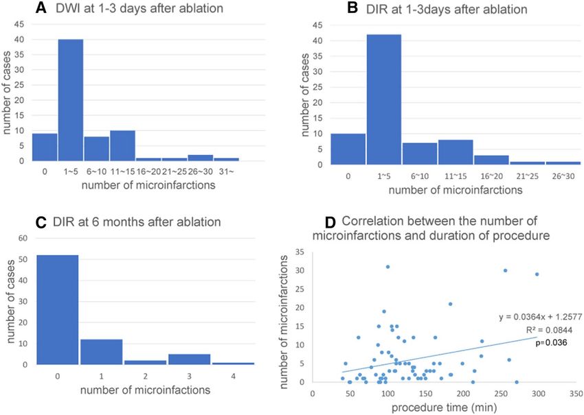
peared in 41 out of 62 patients (66.1%). Figure 3A–C shows the number of DWI/DIR positive lesions per case
at baseline and DIR positive lesions per case at 6 months after ablation. At baseline, one to five lesions were
most frequent. The median number of lesions was 3.0 (interquartile range, 6.25). Most lesions disappeared on
follow-up DIR images.
At baseline, DWI detected a total of 421 embolic microinfarctions (cortical: subcortical and deep = 361: 60),
and 3D-DIR detected 362 (cortical: subcortical and deep = 307: 55). Most lesions in cortical regions frequently
showed a diameter < 5 mm, which infrequently reached 10 mm. Follow-up DIR images after 6 months detected
35 emboli (cortical: subcortical and deep = 18: 17). The residual rate was 5.8% (18 of 307) in the cortical region,
and 30.9% (17 of 55) in subcortical regions (Table 2).
At baseline, SWI detected 577 MBs. Follow-up SWI after 6 months detected 98 new MBs. Sixty-four out of
98 MBs (65.3%) were exactly at the same location where microinfarctions were found at baseline.
Correlation between the number of microinfarctions and ablation parameters. The correlation
between the number of microinfarctions and duration of the ablation procedure was evaluated, where procedure
duration was defined as the time required from the start to end of the ablation. A mild positive correlation was
observed (p = 0.036, γ = 0.248) (Fig. 3D).
Scientific Reports | (2021) 11:18995 | https://doi.org/10.1038/s41598-021-98484-w 4
Vol:.(1234567890)www.nature.com/scientificreports/
Figure 2. Brain MR images. Case 1: Brain MR images of a female patient. DWI (A, B), 3D-DIR findings at
baseline (C) and after 6 months (D). An embolic infarct was detected by DWI and 3D-DIR after ablation. In
follow-up MRI, the lesion was still detected by 3D-DIR. Case 2: Brain MR images of a male patient. DWI (E, F),
3D-DIR (G, H), SWI (I, J) findings at baseline (left) and after 6 months (right). The embolic microinfarctions
detected by DWI and 3D-DIR after ablation disappeared at follow-up, but SWI detected de novo microbleeds
exactly at the same location where the infarcts were detected at baseline. DWI Diffusion-weighted imaging, 3D-
DIR 3-Dimensional double inversion recovery, SWI Susceptibility-weighted imaging.
Figure 3. Number of DWI (A) and DIR (B) positive lesions per case at baseline, and DIR positive lesions
per case at 6 months after ablation (C). At baseline, one to five lesions were most frequent, however, most
lesions disappear on follow-up DIR images. A mild positive correlation was observed between the number of
microinfarctions and duration of ablation procedure (p = 0.036) (D).
Correlation between MB number and CHADS2 score. We analyzed the correlation between the
number of new MBs detected by follow-up MRI and CHADS2 score but observed no significant correlation
(p = 0.75, γ = 0.038).
Neuropsychological findings after ablation. The MMSE score was 27.9 ± 2.4 at baseline. At 6 months
after ablation, the average MMSE score improved significantly to 28.5 ± 1.7 (p = 0.037). RBMT (both immediate
and delayed recall: p < 0.001 and p < 0.01, respectively), MCAS (p < 0.01), and TMT-A (p = 0.001) scores improved
Scientific Reports | (2021) 11:18995 | https://doi.org/10.1038/s41598-021-98484-w 5
Vol.:(0123456789)www.nature.com/scientificreports/
Baseline 6M p
MRI findings
DWI positive patients, n (%) 63 (87.5) 0 (0) Not applicable
DWI positive lesions, n 421 0
Cortical 361 0
Subcortical, deep 60 0
DIR positive patients, n (%) 62 (86.1) 21 (29.2)
DIR positive lesions, n 362 35
Cortical 307 18
Subcortical, deep 55 17
Neuropsychological test Normal range Baseline 6M p
MMSE ≧ 24 27.9 ± 2.39 28.5 ± 1.98 0.037*
RCPM Age dependent 31.4 ± 4.0 31.8 ± 3.1 0.503
RCPM time (s) Age dependent 391 ± 92.2 392 ± 116 0.69
RBMT immediate recall Age dependent 10.1 ± 3.4 14.3 ± 4.2 < 0.001*
RBMT delayed recall Age dependent 7.8 ± 2.98 13.1 ± 4.0 < 0.001*
Necker’s cube 2.3 ± 0.5 2.4 ± 0.6 0.157
MCAS ≤2 2.4 ± 1.4 1.8 ± 1.3 < 0.001*
TMT-A Age dependent 105 ± 31.2 97.8 ± 37.1 0.09
TMT-B Age dependent 160.2 ± 146 141.1 ± 61.2 0.098
Word fluency (vegetable) 15.3 ± 12.7 13.5 ± 4 0.436
Word fluency (animal) 16.2 ± 4.7 16.9 ± 4.6 0.228
Word fluency (letter) 6.5 ± 1.7 6.7 ± 1.9 0.17
Cardiac function Normal range Baseline 3–6 M
EF (%) 50–80 64.5 ± 7.2 67.2 ± 7.7 0.025*
LAVI (ml/m2) < 34 46.8 ± 16.9 40.9 ± 13.5 0.002*
BNP (pg/ml) 0–8.4 89.1 ± 70.9 62.5 ± 70.1 0.001*
Table 2. Comparison between findings at baseline and 6 months after ablation. DWI Diffusion-weighted
imaging, DIR Double inversion recovery, MCAS Mie constructional apraxia scale, EF Ejection fraction, LAVI
Left atrial volume index, BNP Brain natriuretic peptide.
significantly at 6 months after ablation (Table 2). To evaluate cognitive enhancement variability, we performed
correlation analysis between baseline scores and the change value of MMSE, RBMT immediate recall, RBMT
delayed recall, MCAS, TMT-A, which showed significant improvements at 6 months after ablation. Patients
with lower baseline cognitive function showed better improvement in almost all these scores (p < 0.01, p = 0.016,
p = 0.01, p < 0.01, p < 0.01, respectively.)
We evaluated the improvement in neuropsychological scores according to history of hypertension, diabetes
mellitus, dyslipidemia, and stroke/TIA. There was no significant difference in the changes of any scores between
patients with and without hypertension, diabetes mellitus, dyslipidemia, and history of stroke/TIA. Furthermore,
we analyzed the difference in cognitive changes between persistent and paroxysmal AF, with and without AF
recurrence, with and without oral anticoagulant use at 6 months after ablation, but no difference was observed.
Further, in 20 patients, MMSE was evaluated 1 day before ablation, and the score was 25–30 (average score:
27.9 ± 1.8). We found no significant differences between MMSE scores evaluated before and immediately after
catheter ablation (p = 0.14).
Cardiac function after ablation and correlation with neuropsychological findings. EF values
significantly increased (p = 0.025), whereas LAVI and BNP significantly decreased (p = 0.002, p = 0.001, respec-
tively) between baseline and at 3–6 months after ablation (Table 2). We compared changes in cardiac function
in patients with persistent and paroxysmal AF, with and without AF recurrence within 6 months after ablation,
and presence and absence of oral anticoagulant use at 6 months after ablation. No significant difference in car-
diac function changes was observed. No correlation was observed between EF percent increase and the changes
in MMSE scores or other findings. However, there were positive correlations between LAVI percent reduction
and WF changes (animal) (p = 0.04, γ = 0.331), between BNP reduction and WF changes (animal) (p ≦ 0.001,
γ = 0.546), and between BNP reduction and RBMT delayed recall (p = 0.026, γ = 0.351) (Fig. 4). In addition, there
was no correlation with any neuropsychological score change between the patients who showed LAVI/BNP
improvement or not.
Scientific Reports | (2021) 11:18995 | https://doi.org/10.1038/s41598-021-98484-w 6
Vol:.(1234567890)www.nature.com/scientificreports/
Figure 4. Correlation between cardiac function and neuropsychological findings (n = 40). There were positive
correlations between LAVI (%) reduction and word fluency changes (animal) (p = 0.04), between BNP (%)
reduction and word fluency changes (animal) (p ≤ 0.001), and between changes in BNP (%) and those in RBMT
delayed recall (p = 0.026). We calculated Spearman’s correlation coefficient. LAVI Left atrial volume index,
RBMT Rivermead behavioral memory test, BNP Brain natriuretic peptide, WF Word fluency.
Discussion
Our prospective study indicated that ablation has beneficial effects on overall neuropsychological scores despite
the incidental embolic microinfarctions caused by the procedure. After catheter ablation, there was an increase in
EF and a decrease in LAVI and BNP, which may be attributable to improved cardiac function, and which might
have led to a net beneficial effect on neuropsychological scores.
Both prospective and retrospective studies have examined the relationship between AF and cognitive
impairment27. AF can become a risk factor of dementia even in the absence of embolic stroke. One of the plau-
sible mechanisms by which AF induces dementia may be chronic cerebral hypoperfusion. It has been suggested
that chronic cerebral hypoperfusion is causally related to both AD and V aD28,29. AF causes cerebral hypoperfu-
sion through beat-to-beat variability and an overall reduced cardiac output owing to the lack of atrioventricular
synchrony30. Indeed, cerebral blood flow (CBF) is significantly lower in patients with persistent AF than in those
without31. Furthermore, the CBF level of patients with paroxysmal AF lies between persistent AF and sinus
rhythm11, and electrical cardioversion could restore CBF in patients with A F32. Cerebral hypoperfusion has been
correlated with brain atrophy and d ementia33, and AF per se is also associated with brain atrophy with a stronger
association in persistent/permanent AF than in paroxysmal A F16.
The other possible mechanism by which AF induces dementia is by remitting microembolism, which may
cause accumulation of cortical microinfarcts (CMIs) and subsequently, dementia34–36. Catheter ablation may
potentially suppress the chronic incidence of microembolism in AF37, while subsequent prevention of micro-
embolism may have a beneficial effect on cognitive dysfunction. However, this possibility could not explain the
improvement of neuropsychological scores encompassing overall cognitive domains in the present study, because
small vessel diseases, including CMI, usually contribute to frontal lobe d ysfunction38. Notably, in the present
study, improvements were not limited to scores related to frontal lobe function, but universally observed in every
cognitive domain including the temporal lobe. This result may indicate that improvement of cardiac output and
subsequent CBF might have led to the recovery of neuropsychological scores in patients with AF.
CMIs are caused by various pathological factors such as microembolism, arteriosclerosis, and cerebral amyloid
angiopathy35. In our study, catheter ablation caused microinfarctions in 85% of patients. This result contradicts
earlier published results where microinfarcts were detected in < 30% of patients. The MRI protocol used in this
study is very sensitive for detection of microlesions. Indeed, in our MR protocol, DWI slice thickness is 3 mm
with no gap. On the other hand, previous studies used a DWI slice thickness of 5 mm with 2 mm gap, so that
microinfarcts with 2–3 mm in size could go undetected. Nakamura et al. described that, compared with conven-
tional DWI, thin section (3 mm with no gap) DWI at 1.5 T permitted better lesion conspicuity and more precise
stroke diagnosis39. Moreover, we used a 3 T MR machine, with better signal-to-noise ratio than 1.5 T. Therefore,
Scientific Reports | (2021) 11:18995 | https://doi.org/10.1038/s41598-021-98484-w 7
Vol.:(0123456789)www.nature.com/scientificreports/
we consider that the higher percentage of patients with acute microinfarcts in this study was probably due to
the high resolution DWI employed.
Bergui et al. showed that silent embolic microinfarctions after AF ablation were more commonly found in
the cerebral cortex40. In our study, 80% of microinfarctions were found in the cerebral cortex. After 6 months, a
significantly higher proportion of microinfarctions disappeared from the cerebral cortex than from subcortical
regions (96.2% vs. 69.1%, respectively) on 3D-DIR images. This result is consistent with the findings of Terge
et al., who evaluated the cumulative incidence of acute CMI and found that all acute CMIs disappeared on follow-
up MRI (DWI, T1, FLAIR)41. Similarly, Havsteen et al. showed that cortical lesions in TIA disappeared more
frequently than those in subcortical ones and hypothesized that a strong leptomeningeal collateral circulation
in the cortical gray matter may prevent signs of persistent infarction in small gray matter lesions42.
Another notable finding is the numerical increase in MBs on follow-up MRIs. Previous studies reported a
higher incidence of MBs in patients with AF than in those without43, but the pathogenesis of MBs in AF and
the relationship between MBs and cognitive function remain unclear. In our study, there was a 17% increase in
the total number of MBs during the 6 months after ablation and most de novo MBs corresponded with embolic
microinfarctions detected at baseline. Previously, Ito et al. reported 3 cases of de novo lobar MBs transformed
from a small cortical i nfarction44. Our results imply that AF-related microemboli may cause CMI, subsequently
leading to MBs.
Our present study has several limitations. First, it did not include a control group of AF patients who did
not undergo ablation treatment. A memory clinic study showed a significantly higher prevalence of CMIs in
the brains of AF patients45, and therefore, patients with AF may have had preexisting CMIs. Our study may
indicate that most CMIs disappear and that only a small number remains. Second, our study did not perform
pre-ablation MRI, so we cannot confirm that all new microinfarctions and MBs detected after ablation were
caused by the ablation procedure. However, we conducted pre-ablation MRI in 13 out of 74 patients, none of
whom showed new microinfarctions. Moreover, we could not perform pre-ablation neuropsychological assess-
ment in all patients because of time constraints. We conducted a pre-ablation MMSE estimation in 21 patients,
among whom we found no significant changes between pre-ablation and post-ablation MMSE scores. We also
compared pre-ablation MMSE scores with 6-month follow-up scores in 16 patients (5 out of 21 patients who
underwent pre-ablation MMSE dropped out after 6 months), and found that follow-up scores significantly
improved (p = 0.023). Third, we did not monitor electrocardiography during neuropsychological assessments.
Many patients have AF episodes early after ablation, so the influence of arrythmia on neuropsychological scores
at 1–3 days after ablation is undeniable. Similarly, the sedation and stress caused by the ablation procedure
might also affect cognitive assessment scores, but none of the patients showed any signs of neuropsychological
alterations including delirium. As the average hospitalization period after an ablation procedure was 4–5 days,
we performed the examinations 1–3 days after ablation and confirmed absence of neuropsychological worsening
between pre-ablation period and immediately after ablation in 21 patients. If patients were too ill to perform
neuropsychological assessment, we postponed it to the next day or excluded the patients from the study. Fourth,
we could not measure cerebral perfusion by CBF single photon emission computed tomography or arterial spin
labeling MRI techniques. Further studies are needed to investigate the incidence of embolic microinfarctions in
patients with AF and CBF improvements after ablation.
In conclusion, our study showed preserved cognitive function at 6 months after ablation despite the incidence
of embolic microinfarctions. The improvement of neuropsychological scores across all cognitive domains might
indicate that ablation improves cognitive function by restoring cardiac function and mitigating chronic cerebral
hypoperfusion.
Data availability
Data are available upon reasonable request from the corresponding author.
Received: 16 March 2021; Accepted: 8 September 2021
References
1. Ferri, C. P. et al. Global prevalence of dementia: A Delphi consensus study. Lancet 366, 2112–2117 (2005).
2. Viswanathan, A., Rocca, W. A. & Tzourio, C. Vascular risk factors and dementia: How to move forward?. Neurology 72, 368–374
(2009).
3. Vijayan, M. & Reddy, P. H. Stroke, vascular dementia, and Alzheimer’s disease: Molecular links. J. Alzheimers Dis. 54, 427–443
(2016).
4. Marini, C. et al. Contribution of atrial fibrillation to incidence and outcome of ischemic stroke: results from a population-based
study. Stroke 36, 1115–1119 (2005).
5. Kalantarian, S., Stern, T. A., Mansour, M. & Ruskin, J. N. Cognitive impairment associated with atrial fibrillation: a meta-analysis.
Ann. Intern. Med. 158, 338–346 (2013).
6. Santangeli, P. et al. Atrial fibrillation and the risk of incident dementia: A meta-analysis. Heart Rhythm 9, 1761–1768 (2012).
7. Cacciatore, F. et al. Role of ventricular rate response on dementia in cognitively impaired elderly subjects with atrial fibrillation:
A 10-year study. Dement. Geriatr. Cogn. Disord. 34, 143–148 (2012).
8. Chen, L. Y. et al. Association of atrial fibrillation with cognitive decline and dementia over 20 years: The ARIC-NCS (Atherosclerosis
Risk in Communities Neurocognitive Study). J. Am. Heart Assoc. 7, e007301 (2018).
9. Ding, M. et al. Atrial fibrillation, antithrombotic treatment, and cognitive aging: A population-based study. Neurology 91, e1732–
e1740 (2018).
10. Feinberg, W. M., Blackshear, J. L., Laupacis, A., Kronmal, R. & Hart, R. G. Prevalence, age distribution, and gender of patients with
atrial fibrillation. Analysis and implications. Arch. Intern. Med. 155, 469–473 (1995).
11. Bunch, T. J. et al. Patients treated with catheter ablation for atrial fibrillation have long-term rates of death, stroke, and dementia
similar to patients without atrial fibrillation. J. Cardiovasc. Electrophysiol. 22, 839–845 (2011).
Scientific Reports | (2021) 11:18995 | https://doi.org/10.1038/s41598-021-98484-w 8
Vol:.(1234567890)www.nature.com/scientificreports/
12. Friberg, L. & Rosenqvist, M. Less dementia with oral anticoagulation in atrial fibrillation. Eur. Heart J. 39, 453–460 (2018).
13. Kwok, C. S., Loke, Y. K., Hale, R., Potter, J. F. & Myint, P. K. Atrial fibrillation and incidence of dementia: A systematic review and
meta-analysis. Neurology 76, 914–922 (2011).
14. Rivard, L. & Khairy, P. Mechanisms, clinical significance, and prevention of cognitive impairment in patients with atrial fibrillation.
Can. J. Cardiol. 33, 1556–1564 (2017).
15. Wardlaw, J. M., Smith, C. & Dichgans, M. Small vessel disease: Mechanisms and clinical implications. Lancet Neurol. 18, 684–696
(2019).
16. Stefansdottir, H. et al. Atrial fibrillation is associated with reduced brain volume and cognitive function independent of cerebral
infarcts. Stroke 44, 1020–1025 (2013).
17. Chung, M. K. et al. Functional status in rate-versus rhythm-control strategies for atrial fibrillation: Results of the Atrial Fibrilla-
tion Follow-Up Investigation of Rhythm Management (AFFIRM) Functional Status Substudy. J. Am. Coll. Cardiol. 46, 1891–1899
(2005).
18. Chinta, V. et al. Atrial fibrillation and deterioration in cognitive function. Curr. Probl. Cardiol. 44, 100386 (2019).
19. Merchant, F. M. & Delurgio, D. B. Catheter ablation of atrial fibrillation and risk of asymptomatic cerebral embolism. Pacing Clin.
Electrophysiol. 37, 389–397 (2014).
20. Kochhauser, S. et al. Neuropsychological impact of cerebral microemboli in ablation of atrial fibrillation. Clin. Res. Cardiol. 104,
234–240 (2015).
21. Kagawa, Y., Fujii, E., Fujita, S. & Ito, M. Association between left atrial reverse remodeling and maintenance of sinus rhythm after
catheter ablation of persistent atrial fibrillation. Heart Vessels 35, 239–245 (2020).
22. Ii, Y. et al. In vivo detection of cortical microinfarcts on ultrahigh-field MRI. J. Neuroimaging 23, 28–32 (2013).
23. Ii, Y. et al. Cortical microinfarcts in patients with multiple lobar microbleeds on 3 T MRI. J. Neurol. 266, 1887–1896 (2019).
24. Strub, R. L. & Black, F. W. The Mental Status Examination in Neurology 4th edn, 93–115 (FA Davis Company, 2000).
25. Satoh, M. et al. Improved Necker cube drawing-based assessment battery for constructional apraxia: The Mie Constructional
Apraxia Scale (MCAS). Dement. Geriatr. Cogn. Dis. Extra. 6, 424–436 (2016).
26. Satoh, M. et al. Physical exercise with music maintains activities of daily living in patients with dementia: Mihama-Kiho Project
Part 21. J. Alzheimers Dis. 57, 85–96 (2017).
27. Diener, H. C., Hart, R. G., Koudstaal, P. J., Lane, D. A. & Lip, G. Y. H. Atrial fibrillation and cognitive function: JACC review topic
of the week. J. Am. Coll. Cardiol. 73, 612–619 (2019).
28. de la Torre, J. C. Critically attained threshold of cerebral hypoperfusion: The CATCH hypothesis of Alzheimer’s pathogenesis.
Neurobiol. Aging. 21, 331–342 (2000).
29. Kawamura, J., Meyer, J. S., Terayama, Y. & Weathers, S. Leukoaraiosis correlates with cerebral hypoperfusion in vascular dementia.
Stroke 22, 609–614 (1991).
30. Aldrugh, S., Sardana, M., Henninger, N., Saczynski, J. S. & McManus, D. D. Atrial fibrillation, cognition and dementia: A review.
J. Cardiovasc. Electrophysiol. 28, 958–965 (2017).
31. Gardarsdottir, M. et al. Atrial fibrillation is associated with decreased total cerebral blood flow and brain perfusion. Europace 20,
1252–1258 (2018).
32. Petersen, P., Kastrup, J., Videbaek, R. & Boysen, G. Cerebral blood flow before and after cardioversion of atrial fibrillation. J. Cereb.
Blood Flow Metab. 9, 422–425 (1989).
33. Wirth, M. et al. Divergent regional patterns of cerebral hypoperfusion and gray matter atrophy in mild cognitive impairment
patients. J. Cereb. Blood Flow Metab. 37, 814–824 (2017).
34. White, L. et al. Cerebrovascular pathology and dementia in autopsied Honolulu-Asia Aging Study participants. Ann. N. Y. Acad.
Sci. 977, 9–23 (2002).
35. Kovari, E. et al. Cortical microinfarcts and demyelination significantly affect cognition in brain aging. Stroke 35, 410–414 (2004).
36. Kovari, E. et al. Cortical microinfarcts and demyelination affect cognition in cases at high risk for dementia. Neurology 68, 927–931
(2007).
37. Silva, R., Miranda, C. M., Liu, T., Tse, G. & Roever, L. Atrial fibrillation and risk of dementia: Epidemiology, mechanisms, and
effect of anticoagulation. Front. Neurosci. 13, 18 (2019).
38. Ueda, Y. et al. Neuropsychological features of microbleeds and cortical microinfarct detected by high resolution magnetic resonance
imaging. J. Alzheimers Dis. 53, 315–325 (2016).
39. Nakamura, H. et al. Effect of thin-section diffusion-weighted MR imaging on stroke diagnosis. AJNR Am. J. Neuroradiol. 26,
560–565 (2005).
40. Bergui, M. et al. Selective vulnerability of cortical border zone to microembolic infarct. Stroke 46, 1864–1869 (2015).
41. Ter Telgte, A. et al. Temporal dynamics of cortical microinfarcts in cerebral small vessel disease. JAMA Neurol. 77, 643–647 (2020).
42. Havsteen, I. et al. Small cortical grey matter lesions show no persistent infarction in transient ischaemic attack? A prospective
cohort study. BMJ Open 8, e018160 (2018).
43. Horstmann, S. et al. Prevalence of atrial fibrillation and association of previous antithrombotic treatment in patients with cerebral
microbleeds. Eur. J. Neurol. 22, 1355–1362 (2015).
44. Ito, A. et al. Small cortical infarcts transformed to lobar cerebral microbleeds: A case series. J. Stroke Cerebrovasc. Dis. 28, e30–e32
(2019).
45. Hilal, S. et al. Cortical cerebral microinfarcts predict cognitive decline in memory clinic patients. J. Cereb. Blood Flow Metab. 40,
44–53 (2020).
Acknowledgements
This work was supported by JSPS KAKENHI (Grant Number JP19H03572) and by a grant-in-aid for
Research on Intractable Disease from the Japanese Ministry of Health, Labour, and Welfare, Japan
(H30-Nanchitou[Nan]-Ippan-006).
Author contributions
N.K., and K.M.: draft of manuscript, acquisition of data, and analysis. A.S., E.F., and M.M. revision of manuscript,
interpretation of data, and study supervision. K.I.T. and S.M.: revision of manuscript and interpretation of data.
Y.H., K.M., Y.I., M.S., S.F., Y.K., T.F., H.S., and M.I.: acquisition of data and interpretation of data. H.T.: revision
of the manuscript, study concept and design, and study supervision.
Competing interests
The authors declare no competing interests.
Additional information
Correspondence and requests for materials should be addressed to N.K.
Scientific Reports | (2021) 11:18995 | https://doi.org/10.1038/s41598-021-98484-w 9
Vol.:(0123456789)www.nature.com/scientificreports/
Reprints and permissions information is available at www.nature.com/reprints.
Publisher’s note Springer Nature remains neutral with regard to jurisdictional claims in published maps and
institutional affiliations.
Open Access This article is licensed under a Creative Commons Attribution 4.0 International
License, which permits use, sharing, adaptation, distribution and reproduction in any medium or
format, as long as you give appropriate credit to the original author(s) and the source, provide a link to the
Creative Commons licence, and indicate if changes were made. The images or other third party material in this
article are included in the article’s Creative Commons licence, unless indicated otherwise in a credit line to the
material. If material is not included in the article’s Creative Commons licence and your intended use is not
permitted by statutory regulation or exceeds the permitted use, you will need to obtain permission directly from
the copyright holder. To view a copy of this licence, visit http://creativecommons.org/licenses/by/4.0/.
© The Author(s) 2021
Scientific Reports | (2021) 11:18995 | https://doi.org/10.1038/s41598-021-98484-w 10
Vol:.(1234567890)You can also read