Exacerbation of cardiovascular ageing by diabetes mellitus and its associations with acyl-carnitines
←
→
Page content transcription
If your browser does not render page correctly, please read the page content below
www.aging-us.com                                                              AGING 2021, Vol. 13, No. 11
                                                                                                     Research Paper
Exacerbation of cardiovascular ageing by diabetes mellitus and its
associations with acyl-carnitines
Fei Gao1,2, Jean-Paul Kovalik2,3, Xiaodan Zhao1, Vivian JM. Chow1, Hannah Chew1, Louis LY. Teo1,2,
Ru San Tan1,2, Shuang Leng1, See Hooi Ewe1,2, Hong Chang Tan2,3, Tsze Yin Tan2, Lye Siang Lee2,
Jianhong Ching2,5, Bryan MH. Keng1, Liang Zhong1,2, Woon-Puay Koh4, Angela S. Koh1,2
1
 National Heart Centre Singapore, Singapore
2
 Duke-NUS Medical School, Singapore
3
 Singapore General Hospital, Singapore
4
 Healthy Longevity Translational Research Programme, Yong Loo Lin School of Medicine, National University of
Singapore, Singapore
5
 KK Research Centre, KK Women's and Children's Hospital, Singapore

Correspondence to: Angela S. Koh, Jean-Paul Kovalik; email: angela.koh.s.m@singhealth.com.sg, jean-paul.kovalik@duke-
nus.edu.sg
Keywords: aging, diabetes, cardiovascular
Received: January 26, 2021         Accepted: May 17, 2021            Published: June 4, 2021

Copyright: © 2021 Gao et al. This is an open access article distributed under the terms of the Creative Commons Attribution
License (CC BY 3.0), which permits unrestricted use, distribution, and reproduction in any medium, provided the original
author and source are credited.

ABSTRACT

Objective: To demonstrate differences in cardiovascular structure and function between diabetic and non-
diabetic older adults. To investigate associations between acyl-carnitines and cardiovascular function as
indexed by imaging measurements.
Methods: A community-based cohort of older adults without cardiovascular disease underwent current
cardiovascular imaging and metabolomics acyl-carnitines profiling based on current and archived sera obtained
fifteen years prior to examination.
Results: A total of 933 participants (women 56%, n=521) with a mean age 63±13 years were studied. Old diabetics
compared to old non-diabetics had lower myocardial relaxation (0.8±0.2 vs 0.9±0.3, p=0.0039); lower left atrial
conduit strain (12±4.3 vs 14±4.1, p=0.045), lower left atrial conduit strain rate (-1.2±0.4 vs -1.3±0.5, p=0.042) and
lower ratio of left atrial conduit strain to left atrial booster strain (0.5±0.2 vs 0.7±0.3, p=0.0029). Higher levels of
archived short chain acyl-carnitine were associated with present-day impairments in myocardial relaxation (C5:1;
OR 1.03, p=0.011), worse left atrial conduit strain function (C5:1; OR 1.03, p=0.037). Increases in hydroxylated acyl-
carnitines were associated with worse left atrial conduit strain [(C4-OH; OR 1.05, p=0.0017), (C16:2-OH; OR 1.18,
p=0.037)]. Current, archived and changes in long chain acyl-carnitines were associated with cardiovascular
functions [(C16; OR 1.02, p=0.002), (C20:3; OR 1.01, p=0.014), (C14:3; OR 1.12, p=0.033), (C18:1; OR 1.01, p=0.018),
(C18:2; OR 1.01, p=0.028), (C20:4; OR 1.10, p=0.038)] (all p
cardiovascular system by these risk factors are known, Study (SCHS) [14] and directly from the local
the extent, of each risk factor, superimposed onto community. The current study sample consisted of men
cardiovascular ageing, is not readily appreciated. The and women who participated in the baseline CAS 2014-
extent to which diabetes mellitus modifies the effect of 2017 examination who had no self-reported history of
ageing on the heart, deserves deeper investigation. The physician-diagnosed cardiovascular disease (such as
clinical implication of ageing with or without diabetes coronary heart disease, atrial fibrillation), stroke or
may herald differences in cardiovascular phenotype, cancer. We studied the subjects in three groups,
outcomes and treatments. For example, age-associated comprising of young adults (age < 65 years old), and
decreases in left ventricular volumes, increases in left old adults (age ≥65 years), the latter old group was
ventricular mass index, and deteriorations in diastolic further categorized into diabetic and non-diabetic
function are commonly observed in heart failure among adults.
elderly patients [2]. The fact that heart failure can occur
without diabetes (or hypertension) as a main driver, Written informed consent was obtained from participants
implies an urgent need to differentiate the ageing upon enrolment. The SingHealth Centralised Institutional
phenotype, from phenotypes related to traditional risk Review Board (CIRC/2014/628/C) had approved the
factors [3]. study protocol.
The growing elderly population worldwide highlights Data acquisition
the need for unique strategies to confront unresolved
risks of heart failure burdens among the elderly [4]. The All participants were examined and interviewed on one
metabolome represents net profile of diverse chemicals study visit by trained study coordinators. Participants
that is influenced by genomics, transcriptomics and completed a standardized questionnaire that included
proteomic variability [5]. Metabolomic profiles are also medical history and coronary risk factors. Sinus rhythm
influenced by environmental exposure, diet and lifestyle status was ascertained by resting electrocardiogram.
[6–8]. Since metabolomics provides an integrated Clinical data were obtained on the same day as
profile, it may serve as a conglomerating tool for life assessment of echocardiography and serum collection.
course phenomena as heterogeneous as ageing.
Emerging data have demonstrated the utility of Echocardiography was performed using ALOKA α10
metabolomics in advancing understanding of with a 3.5 MHz probe. In each subject, standard
cardiovascular ageing [9–11], insulin resistance, and echocardiography, which included 2-D, M-mode, pulse
diabetes [12, 13]. Therefore, metabolomics may Doppler and tissue Doppler imaging, was performed in
represent a novel approach that dissects new the standard parasternal and apical (apical 4-chamber,
associations between cardiovascular ageing, diabetes apical 2-chamber and apical long) views, and three
and metabolic pathways. cardiac cycles were recorded. E/A ratio was computed
as a ratio of peak velocity flow in early diastole E (m/s)
Among a community cohort of participants with risk to peak velocity flow in late diastole by atrial
factors but free of cardiovascular disease, we illustrate contraction A (m/s).
structural and functional changes associated with
cardiovascular ageing and qualify the impact of diabetes Blood samples were collected on the day of
mellitus on cardiovascular ageing changes. In addition, echocardiography acquisition. Plasma levels of
by utilizing cross-sectional and archived metabolomic Galectin-3 (Gal-3) (ARCHITECT Galectin-3; produced
profiles across the participants’ lifespan, we by Fujirebio Diagnostics Inc for Abbott Laboratories)
hypothesize that metabolites may be associated with CV and B-type natriuretic peptide (BNP) (ARCHITECT
changes that differentiate between older adults with BNP; produced by Fujirebio Diagnostics Inc for Abbott
diabetes versus older adults without diabetes. Laboratories) were measured on the Abbott
ARCHITECT i2000SR analyzer.
MATERIALS AND METHODS
Cine cardiac magnetic resonance (CMR) scans were
Study population performed using balanced fast field echo sequence
(BFFE). All subjects were imaged on a 3T magnetic
The subjects were recruited from the Cardiac Ageing resonance imaging system (Ingenia, Philips Healthcare,
Study (CAS)9, a prospective study initiated in 2014 The Netherlands) with a dStream Torso coil (maximal
that examines characteristics and determinants of number of channels 32). Dedicated Qstrain software
cardiovascular function in elderly adults. CAS (version 2.0, Medis) was used in deriving LV and RV
participants were recruited from the prospective, longitudinal strain [15]. We developed an in-house
population-based cohort, the Singapore Chinese Health semi-automatic algorithm to track the distance (L)
www.aging-us.com 14786 AGINGbetween the left atrioventricular junction and a user- measured metabolites is presented in Supplementary
defined point at the mid posterior LA wall on standard Table 1. To identify serum metabolites correlations and
CMR 2- and 4-chamber views [9]. reduce the dimensionality of correlated metabolites, we
performed sparse principal component analysis
Metabolomics profiling (SPCA)9 using a penalized matrix decomposition on
data from current serum samples (Supplementary Table
Antecubital venous blood samples (20-30 ml) were 2). Metabolites with >25% of values below the lower
taken from consenting participants in the morning; limit of quantification were excluded from analysis
fasting was not required before blood collection. After (C24, C26 and C28 was excluded, hence a total of 66
collection, the blood samples were immediately placed metabolites were analyzed in the final sample). Other
on ice for transportation and were processed within 6 h missing metabolites were input with 0.01. In SPCA, we
to obtain serum samples, which were subsequently normalized the distributions of all metabolites by a
stored at −80° C. Additionally, archived blood samples logarithmic transformation. We assessed the component
obtained approximately 15 years prior to this metabolites within the significant PCA factors, between
assessment from subjects who had serum samples diabetic and non-diabetic using student t-test. For those
collected and stored at the time of enrolment were that show an association with pTable 1. Baseline clinical characteristics of the overall cohort.
p-value (old
Young Old non-diabetic Old diabetic P-value
Total (n=933) diabetic vs old
(n=418) (n=399) (n=116) (young vs old)
non-diabetic)
Clinical covariates
Age (years) 52 (10.6) 73 (4.4) 73 (4.3) 63 (12.9)Table 2. Cardiovascular characteristics of old non-diabetic vs old diabetic.
Old non-
Diabetic Univariate ~Adjusted
Echocardiography measurements diabetic
(n=116) p-value P-value
(n=399)
Interventricular septum thickness at end diastole (IVSD) (cm) 0.80 (0.1) 0.81 (0.2) 0.52 -
Interventricular septum thickness at end systole (IVSS) (cm) 1.3 (0.2) 1.2 (0.2) 0.76 -
Left ventricular internal diameter end diastole (LVIDD) (cm) 4.4 (0.6) 4.3 (0.6) 0.12 -
Left ventricular internal diameter end systole (LVIDS) (cm) 2.5 (0.5) 2.4 (0.5) 0.41 -
Left ventricular posterior wall end diastole (LVPWD) (cm) 0.76 (0.1) 0.77 (0.1) 0.16 -
Left ventricular posterior wall end systole (LVPWS) (cm) 1.4 (0.2) 1.5 (0.2) 0.28 -
Left ventricular outflow tract (LVOT) (cm) 2.1 (0.2) 2.0 (0.2) 0.26 -
Aortic diameter (AO) (cm) 3.0 (0.4) 3.1 (0.4) 0.084 -
Left atrium (LA) (cm) 3.6 (0.6) 3.7 (0.6) 0.55 -
Left ventricular ejection fraction (LVEF) (%) 74 (7.7) 73 (9.2) 0.11 -
Left ventricular fractional shortening (LVFS) (%) 44 (7.4) 42 (7.8) 0.12 -
Left ventricular mass (grams) 120 (49) 116 (40) 0.41 -
Left ventricular mass index (grams/m2) 74 (27) 70 (22) 0.14 -
Left atrial volume (ml) 35 (13) 36 (14) 0.45 -
Left atrial volume index (ml/m2) 21 (7.7) 22 (8.2) 0.90 -
Isovolumic relaxation time (IVRT) (ms) 103 (18) 103 (20) 0.98 -
Peak velocity flow in early diastole E (MV E peak) (m/s) 0.71 (0.2) 0.70 (0.2) 0.51 -
Peak velocity flow in late diastole by atrial contraction A (MV A
0.81 (0.2) 0.87 (0.2) 0.005 0.15
peak) (m/s)
Ratio of MV E peak velocity: MV A peak velocity 0.91 (0.3) 0.82 (0.2) 0.003 0.039
Mitral valve flow deceleration time (MV DT) (ms) 213 (40) 222 (42) 0.034 0.23
Right atrial pressure (mmHg) 5.0 (1.3) 4.7 (1.7) 0.36 -
Pulmonary artery systolic pressure (PASP) (mmHg) 28 (7.0) 25 (6.9) 0.005 0.001
Peak systolic septal mitral annular velocity (Septal S′) (m/s) 0.078 (0.02) 0.077 (0.01) 0.38 -
Peak early diastolic septal mitral annular velocity (Septal E’) (m/s) 0.074 (0.02) 0.067 (0.02) 0.0003 0.021
Septal mitral annular velocity during atrial contraction (Septal A’)
0.14 (0.6) 0.11 (0.02) 0.60 -
(m/s)
Peak systolic lateral mitral annular velocity (m/s) 0.10 (0.03) 0.10 (0.03) 0.10 -
Peak early diastolic lateral mitral annular velocity (m/s) 0.094 (0.02) 0.088 (0.02) 0.019 0.094
Lateral mitral annular velocity during atrial contraction (m/s) 0.12 (0.03) 0.13 (0.02) 0.51 -
Ratio of Peak velocity flow in early diastole E (MV E peak) velocity
10 (3.3) 11 (3.1) 0.022 0.34
to Peak early diastolic septal mitral annular velocity (Septal E’)
CMR measurements (n=187) (n=51)
LV global longitudinal strain (LVGLS) (%) -21 (2.9) -21 (2.9) 0.28 -
LV global circumferential strain (LVGCS) (%) -22 (3.8) -23 (3.1) 0.21 -
LV global radial strain (LVGRS) (%) 104 (25.1) 104 (19.5) 0.98 -
Right ventricular global longitudinal strain (RVGLS) (%) -31 (5.4) -31 (5.5) 0.84 -
LA reservoir strain (ɛs) (%) 31 (6.9) 31 (6.2) 0.98 -
LA conduit strain (ɛe) (%) 14 (4.1) 12 (4.3) 0.045 0.28
LA booster strain (ɛa) (%) 17 (4.7) 18 (3.9) 0.065 -
Reservoir strain rate (SRs) (1/s) 1.5 (0.5) 1.5 (0.4) 0.92 -
Conduit strain rate (SRe) (1/s) -1.3 (0.5) -1.2 (0.4) 0.042 0.30
Booster strain rate (SRa) (1/s) -2.2 (0.7) -2.3 (0.6) 0.19 -
Ratio of SRe/SRa 0.66 (0.3) 0.55 (0.2) 0.006 0.029
LAvolumemin (ml) 31 (12.6) 27 (10.1) 0.044 0.016
LAvolumemax (ml) 64 (18) 57 (17) 0.017 0.006
LA ejection fraction (%) 52 (8.9) 52 (7.2) 0.92 -
~adjusted for female, BMI, CV rf>2.
www.aging-us.com 14789 AGINGTable 3. Acyl-carnitine factors and significant components: comparisons between old non-diabetic vs old
diabetic.
Non-diabetic Diabetic p-value Adjusted Coef.
Acyl-carnitines Adjusted P-value*
(n=154) (n=53) (95% CI)*
PCA factors
X1 0.05 (2.7) -0.1 (2.7) 0.68 - -
X2 0.06 (2.0) -0.2 (2.6) 0.48 - -
X3 -0.04 (2.2) 0.1 (1.8) 0.61 - -
X4 -0.2 (2.1) 0.7 (1.9) 0.0080 1.0 (0.3, 1.7) 0.008
X5 -0.4 (2.0) 1.1 (2.6)(8.5 vs 5.9, p
Table 4. Association between current metabolites and cardiovascular function.
   i) Outcome: E/A2.

www.aging-us.com                                            14792                                           AGING
ii) Outcome: ɛe2.

metabolites thus reflects alterations in fatty acid                 alpha- and omega- fatty acid oxidation pathways [27].
oxidation, which is known to be associated with                     Notably, these metabolites have previously been shown
diabetes and cardiovascular disorders [26]. Older                   to be associated with increased risk of recurrent
subjects with diabetes also had higher levels of                    cardiovascular events as well as ischemic stroke [28].
dicarboxyl- and hydroxyl-carnitine metabolites. These               Since patients with diabetes are known to be at higher
metabolites are generally thought to be generated by                CV risk it is perhaps understandable that older subjects

www.aging-us.com                                          14793                                                    AGING
with diabetes would have increased levels of these (HFrEF), to heart failure with preserved ejection
metabolites. fraction (HFpEF), to non-heart failure (HF) controls,
long chain acyl-carnitine levels were greater in HFrEF
Interestingly, some of these acyl-carnitine metabolites than HFpEF, both of which were greater than non-HF
were associated with cardiovascular function. Higher controls [29]. Our observations now directly link long
levels of long chain acyl-carnitines were associated chain acyl-carnitines to imaging markers of diastolic
with impairments in myocardial relaxation as well as function, a pathophysiological disturbance that
with worse left atrial function. As earlier mentioned, predominates across the clinical heart failure
long-chain acyl-carnitines are linked to mitochondrial spectrum. In addition, levels of long chain acyl-
fatty acid oxidation pathways. This is one of the first carnitines obtained 15 years ago were associated with
reports that links fuel oxidation pathways to changes present-day abnormalities in these cardiovascular
in directly measured cardiac function, early functions. In tandem with baseline levels of long
disturbances in diastolic function. Previous reports chain acyl-carnitines from 15 years ago, interval
have noted links between long chain acyl-carnitines increase in long chain acyl-carnitines predicted
and heart failure [27]. Across the clinical spectrum abnormalities in myocardial relaxation and left atrial
from heart failure with reduced ejection fraction conduit strain.
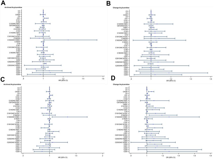
Figure 1. Acyl-carnitines and cardiovascular function. (A) Archived Acyl-carnitine and impaired myocardial relaxation. Blue circles and
lines represent unadjusted hazard ratios (HR) for one-unit increase in archived acyl-carnitine and its 95% confidence interval (95%CI) on
impaired myocardial relaxation. (B) Change in Acyl-carnitine and impaired myocardial relaxation. Blue circles and lines represent unadjusted
odds ratios (OR) for one-unit increase in archived acyl-carnitine and its 95% confidence interval (95%CI) on impaired myocardial relaxation.
(C) Archived Acyl-carnitine and impaired left atrial conduit strain. Blue circles and lines represent unadjusted hazard ratios (HR) for one-unit
increase in archived acylcarnitine and its 95% confidence interval (95%CI) on impaired myocardial relaxation. (D) Change in Acyl-carnitine and
impaired left atrial conduit strain. Blue circles and lines represent unadjusted odds ratios (OR) for one-unit increase in archived acyl-carnitine
and its 95% confidence interval (95%CI) on impaired myocardial relaxation.
www.aging-us.com 14794 AGINGWe also noted an association between increased levels day of blood collection were not important sources of
of short chain, hydroxylated- and dicarboxyl- acyl- variability in measurements of most metabolites [31].
carnitines and impaired LV and atrial function, Finally, we are unable to account for residual
confirming these observations in baseline levels confounding factors that may be present in such a
obtained 15 years ago and interval increases in these study design.
metabolites over time. These classes of fuel
intermediates are likely generated by the process of Despite these limitations, our observations lend
alpha- and omega oxidation [18, 19]. Short chain, biological basis to previous reports that have linked
hydroxylated- and dicarboxyl- acyl-carnitines were fuel oxidation pathways to cardiovascular outcomes
specifically higher among older adults with diabetes, [13]. Our results provide basis for future work that
highlighting the importance of fuel oxidation pathways explores the role of metabolite analysis in early
in the pathogenesis of diabetes, a connection which has detection, as a possible preventative strategy upstream
been well described [26]. These pathways may also in ageing.
represent important treatment targets to ameliorate
impact of diabetes on cardiovascular outcomes in older CONCLUSIONS
adults. Previous reports have noted associations
between the presence of atrial fibrillation and Distinct alterations in fuel oxidation pathways in short
generalized changes in metabolic pathways [30]. This is chain and long chain acyl-carnitines, di-carboxyl and
the first study to highlight alpha and omega oxidation, hydroxylated acyl-carnitines, were associated with
in association with altered left atrial function. present-day changes in cardiovascular function. These
alterations in cardiovascular function distinguished
We acknowledge study limitations. Our observational diabetic versus non-diabetic older adults. Targeting
study design does not imply causality between the distinct fuel oxidation pathways in older adults
metabolites and their present-day cardiovascular depending on diabetes status may provide greater
function. The metabolic perturbations among diabetic precision on therapeutic strategies. Investigations into
samples are complex, and while we could identify key acyl-carnitines early in the ageing trajectory may
pathways involved from their sera, our observational represent a window of opportunity to apply preventative
study design is unable to differentiate between and/or screening methods against deteriorations in
adaptive versus pathogenic responses. The lower cardiovascular health with ageing.
levels of certain long chain acyl-carnitines in the
diabetic samples, might hypothetically represent AUTHOR CONTRIBUTIONS
either an adaptive response or treatment effect. In
addition, metabolomics responses may differ between FG, JPK, RST, WPK, ASK contributed to the
study groups as a consequence of dietary or lifestyle conception and design of the study, advised on all
factors, which we did not account for in this analysis. statistical aspects, and interpreted the data. JPK, XDZ,
A prospective longitudinal clinical trial might clarify FG, SL, VC, HC, LYT, SHE, HCT, TYT, LSL, JC,
this, alongside adjustments for medication data and BMK, LZ performed data analyses. All authors
duration of co-morbidities, which we did not correct critically reviewed the manuscript. All authors approved
for as well. However, the generally low levels of the final draft for submission.
glycated haemoglobin among the groups suggest a
lower risk cohort. As a community-based study ACKNOWLEDGMENTS
focused on studying asymptomatic individuals prior to
disease manifestation, our results reflect We thank the staff of the imaging and biomarker
asymptomatic or preclinical phase of disease, laboratories for participating in the conduct of the study,
designed to look for upstream differences that are and Abbott for providing reagents and kits for certain
likely subtle. Even so, true relationships between the biomarker measurements.
groups, and their associations with metabolomics,
may only be underestimated, and unlikely CONFLICTS OF INTEREST
overestimated. While there may be analytic
differences between non-fasting serum samples The authors declare that they have no conflicts of
(which we used in our study) and fasting serum interest.
samples, large cohort studies face challenges in
getting community elderly participants to fast for FUNDING
prolonged periods of time. Based on the Health
Professionals Follow-up Study and Nurses’ Health The Cardiac Aging Study has received funding support
Study, fasting, season of blood collection, and time of from the National Medical Research Council of Singapore
www.aging-us.com 14795 AGING(NMRC/TA/0031/2015, MOH-000153), Hong Leong 7. Cruickshank-Quinn CI, Mahaffey S, Justice MJ, Hughes
Foundation, Duke-NUS Medical School, Estate of Tan Sri G, Armstrong M, Bowler RP, Reisdorph R, Petrache I,
Khoo Teck Puat and Singhealth Foundation. Those Reisdorph N. Transient and persistent metabolomic
participants recruited from the Singapore Chinese Health changes in plasma following chronic cigarette smoke
Study were supported by the United States National exposure in a mouse model. PLoS One. 2014;
Institutes of Health (NIH R01 CA144034 and 9:e101855.
UM1 CA182876). The study was also supported by https://doi.org/10.1371/journal.pone.0101855
the National Medical Research Council of Singapore PMID:25007263
(NMRC/OFIRG/0018/2016, NMRC/BnB/0017/2015,
8. Everson TM, Marsit CJ. Integrating -Omics Approaches
MOH-000358. The funders had no role in the design and
conduct of the study; collection; management, analysis, into Human Population-Based Studies of Prenatal and
and interpretation of the data; and preparation, review, or Early-Life Exposures. Curr Environ Health Rep. 2018;
approval of the manuscript. 5:328–37.
https://doi.org/10.1007/s40572-018-0204-1
PMID:30054820
REFERENCES
9. Koh AS, Gao F, Leng S, Kovalik JP, Zhao X, Tan RS,
1. Lakatta EG. Age-associated cardiovascular changes in Fridianto KT, Ching J, Chua SJ, Yuan JM, Koh WP, Zhong
health: impact on cardiovascular disease in older L. Dissecting Clinical and Metabolomics Associations of
persons. Heart Fail Rev. 2002; 7:29–49. Left Atrial Phasic Function by Cardiac Magnetic
https://doi.org/10.1023/a:1013797722156 Resonance Feature Tracking. Sci Rep. 2018; 8:8138.
PMID:11790921 https://doi.org/10.1038/s41598-018-26456-8
2. Morita H, Komuro I. Heart Failure as an Aging-Related PMID:29802321
Phenotype. Int Heart J. 2018; 59:6–13. 10. Koh AS, Gao F, Liu J, Fridianto KT, Ching J, Tan RS,
https://doi.org/10.1536/ihj.17-519 Wong JI, Chua SJ, Leng S, Zhong L, Keng BM, Huang FQ,
PMID:29332923 Yuan JM, et al. Metabolomic profile of arterial stiffness
3. Triposkiadis F, Xanthopoulos A, Butler J. Cardiovascular in aged adults. Diab Vasc Dis Res. 2018; 15:74–80.
Aging and Heart Failure: JACC Review Topic of the https://doi.org/10.1177/1479164117733627
Week. J Am Coll Cardiol. 2019; 74:804–13. PMID:28976207
https://doi.org/10.1016/j.jacc.2019.06.053 11. Koh AS, Gao F, Tan RS, Zhong L, Leng S, Zhao X,
PMID:31395131 Fridianto KT, Ching J, Lee SY, Keng BM, Yeo TJ, Tan SY,
4. Vigen R, Maddox TM, Allen LA. Aging of the United Tan HC, et al. Metabolomic correlates of aerobic
States population: impact on heart failure. Curr Heart capacity among elderly adults. Clin Cardiol. 2018;
Fail Rep. 2012; 9:369–74. 41:1300–07.
https://doi.org/10.1007/s11897-012-0114-8 https://doi.org/10.1002/clc.23016 PMID:30350416
PMID:22940871 12. Newgard CB, An J, Bain JR, Muehlbauer MJ, Stevens
5. Dunn WB, Goodacre R, Neyses L, Mamas M. RD, Lien LF, Haqq AM, Shah SH, Arlotto M, Slentz CA,
Integration of metabolomics in heart disease and Rochon J, Gallup D, Ilkayeva O, et al. A branched-chain
diabetes research: current achievements and future amino acid-related metabolic signature that
outlook. Bioanalysis. 2011; 3:2205–22. differentiates obese and lean humans and contributes
https://doi.org/10.4155/bio.11.223 to insulin resistance. Cell Metab. 2009; 9:311–26.
PMID:21985415 https://doi.org/10.1016/j.cmet.2009.02.002
PMID:19356713
6. Cheng S, Shah SH, Corwin EJ, Fiehn O, Fitzgerald RL,
Gerszten RE, Illig T, Rhee EP, Srinivas PR, Wang TJ, Jain 13. Shah SH, Sun JL, Stevens RD, Bain JR, Muehlbauer MJ,
M, and American Heart Association Council on Pieper KS, Haynes C, Hauser ER, Kraus WE, Granger CB,
Functional Genomics and Translational Biology; Council Newgard CB, Califf RM, Newby LK. Baseline
on Cardiovascular and Stroke Nursing; Council on metabolomic profiles predict cardiovascular events in
Clinical Cardiology; and Stroke Council. Potential patients at risk for coronary artery disease. Am Heart J.
Impact and Study Considerations of Metabolomics in 2012; 163:844–50.e1.
Cardiovascular Health and Disease: A Scientific https://doi.org/10.1016/j.ahj.2012.02.005
Statement From the American Heart Association. Circ PMID:22607863
Cardiovasc Genet. 2017; 10:e000032. 14. Hankin JH, Stram DO, Arakawa K, Park S, Low SH, Lee
https://doi.org/10.1161/HCG.0000000000000032 HP, Yu MC. Singapore Chinese Health Study:
PMID:28360086 development, validation, and calibration of the
www.aging-us.com 14796 AGINGquantitative food frequency questionnaire. Nutr cardiovascular disease in patients with diabetes
Cancer. 2001; 39:187–95. mellitus: results from multi-ethnic study of
https://doi.org/10.1207/S15327914nc392_5 atherosclerosis (MESA). Eur Heart J Cardiovasc
PMID:11759279 Imaging. 2017; 18:1138–44.
15. Leng S, Tan RS, Zhao X, Allen JC, Koh AS, Zhong L. Fast https://doi.org/10.1093/ehjci/jew332 PMID:28329137
long-axis strain: a simple, automatic approach for 23. Leng S, Ge H, He J, Kong L, Yang Y, Yan F, Xiu J, Shan P,
assessing left ventricular longitudinal function with Zhao S, Tan RS, Zhao X, Koh AS, Allen JC, et al. Long-
cine cardiovascular magnetic resonance. Eur Radiol. term Prognostic Value of Cardiac MRI Left Atrial Strain
2020; 30:3672–83. in ST-Segment Elevation Myocardial Infarction.
https://doi.org/10.1007/s00330-020-06744-6 Radiology. 2020; 296:299–309.
PMID:32107604 https://doi.org/10.1148/radiol.2020200176
16. Tan HC, Khoo CM, Tan MZ, Kovalik JP, Ng AC, Eng AK, PMID:32544032
Lai OF, Ching JH, Tham KW, Pasupathy S. The Effects of 24. Morris DA, Belyavskiy E, Aravind-Kumar R, Kropf M,
Sleeve Gastrectomy and Gastric Bypass on Branched- Frydas A, Braunauer K, Marquez E, Krisper M, Lindhorst
Chain Amino Acid Metabolism 1 Year After Bariatric R, Osmanoglou E, Boldt LH, Blaschke F, Haverkamp W,
Surgery. Obes Surg. 2016; 26:1830–35. et al. Potential Usefulness and Clinical Relevance of
https://doi.org/10.1007/s11695-015-2023-x Adding Left Atrial Strain to Left Atrial Volume Index in
PMID:26729279 the Detection of Left Ventricular Diastolic Dysfunction.
17. Van Hove JL, Zhang W, Kahler SG, Roe CR, Chen YT, JACC Cardiovasc Imaging. 2018; 11:1405–15.
Terada N, Chace DH, Iafolla AK, Ding JH, Millington DS. https://doi.org/10.1016/j.jcmg.2017.07.029
Medium-chain acyl-CoA dehydrogenase (MCAD) PMID:29153567
deficiency: diagnosis by acylcarnitine analysis in blood. 25. Patel RB, Alenezi F, Sun JL, Alhanti B, Vaduganathan M,
Am J Hum Genet. 1993; 52:958–66. Oh JK, Redfield MM, Butler J, Hernandez AF, Velazquez
PMID:8488845 EJ, Shah SJ. Biomarker Profile of Left Atrial Myopathy in
18. Pahan K, Khan M, Singh I. Phytanic acid oxidation: Heart Failure With Preserved Ejection Fraction: Insights
normal activation and transport yet defective alpha- From the RELAX Trial. J Card Fail. 2020; 26:270–75.
hydroxylation of phytanic acid in peroxisomes from https://doi.org/10.1016/j.cardfail.2019.12.001
Refsum disease and rhizomelic chondrodysplasia PMID:31857197
punctata. J Lipid Res. 1996; 37:1137–43. 26. McCoin CS, Knotts TA, Adams SH. Acylcarnitines--old
PMID:8725164 actors auditioning for new roles in metabolic
19. Wanders RJ, Komen J, Kemp S. Fatty acid omega- physiology. Nat Rev Endocrinol. 2015; 11:617–25.
oxidation as a rescue pathway for fatty acid oxidation https://doi.org/10.1038/nrendo.2015.129
disorders in humans. FEBS J. 2011; 278:182–94. PMID:26303601
https://doi.org/10.1111/j.1742-4658.2010.07947.x 27. Zordoky BN, Sung MM, Ezekowitz J, Mandal R, Han B,
PMID:21156023 Bjorndahl TC, Bouatra S, Anderson T, Oudit GY, Wishart
20. Lam CS, Borlaug BA, Kane GC, Enders FT, Rodeheffer DS, Dyck JR, and Alberta HEART. Metabolomic
RJ, Redfield MM. Age-associated increases in fingerprint of heart failure with preserved ejection
pulmonary artery systolic pressure in the general fraction. PLoS One. 2015; 10:e0124844.
population. Circulation. 2009; 119:2663–70. https://doi.org/10.1371/journal.pone.0124844
https://doi.org/10.1161/CIRCULATIONAHA.108.83869 PMID:26010610
8 PMID:19433755 28. Shah SH, Bain JR, Muehlbauer MJ, Stevens RD, Crosslin
21. Shah AM, Cikes M, Prasad N, Li G, Getchevski S, DR, Haynes C, Dungan J, Newby LK, Hauser ER,
Claggett B, Rizkala A, Lukashevich I, O’Meara E, Ryan JJ, Ginsburg GS, Newgard CB, Kraus WE. Association of a
Shah SJ, Mullens W, Zile MR, et al, and PARAGON-HF peripheral blood metabolic profile with coronary artery
Investigators. Echocardiographic Features of Patients disease and risk of subsequent cardiovascular events.
With Heart Failure and Preserved Left Ventricular Circ Cardiovasc Genet. 2010; 3:207–14.
Ejection Fraction. J Am Coll Cardiol. 2019; 74:2858–73. https://doi.org/10.1161/CIRCGENETICS.109.852814
https://doi.org/10.1016/j.jacc.2019.09.063 PMID:20173117
PMID:31806129 29. Georgievska-Ismail L, Zafirovska P, Hristovski Z.
22. Markman TM, Habibi M, Venkatesh BA, Zareian M, Wu Evaluation of the role of left atrial strain using two-
C, Heckbert SR, Bluemke DA, Lima JA. Association of dimensional speckle tracking echocardiography in
left atrial structure and function and incident patients with diabetes mellitus and heart failure with
www.aging-us.com 14797 AGINGpreserved left ventricular ejection fraction. Diab Vasc 31. Townsend MK, Bao Y, Poole EM, Bertrand KA, Kraft P,
Dis Res. 2016; 13:384–94. Wolpin BM, Clish CB, Tworoger SS. Impact of Pre-
https://doi.org/10.1177/1479164116655558 analytic Blood Sample Collection Factors on
PMID:27407084 Metabolomics. Cancer Epidemiol Biomarkers Prev.
2016; 25:823–29.
30. Mayr M, Yusuf S, Weir G, Chung YL, Mayr U, Yin X,
https://doi.org/10.1158/1055-9965.EPI-15-1206
Ladroue C, Madhu B, Roberts N, De Souza A, Fredericks
PMID:26941367
S, Stubbs M, Griffiths JR, et al. Combined metabolomic
and proteomic analysis of human atrial fibrillation. J
Am Coll Cardiol. 2008; 51:585–94.
https://doi.org/10.1016/j.jacc.2007.09.055
PMID:18237690
www.aging-us.com 14798 AGINGSUPPLEMENTARY MATERIALS Supplementary Tables Supplementary Table 1. List of measured metabolites. Short name Name C2 Acetyl carnitine C3 Propionyl carnitine C4 Butyryl carnitine or isobutryl carnitine C5:1 Tiglyl carnitine or 3-methyl crotonyl carnitine C5 Isovaleryl, 3-methylbutyryl carnitine , 2-Methylbutyryl, valeryl or pivaloyl carnitine C4-OH D-3-Hydroxy-butyryl carnitine, L-3-hydroxybutyryl carnitine C6 Hexanoyl carnitine C5-OH/C3-DC 3-Hydroxy-isovaleryl carnitine or malonyl carnitine C4-DC/C6-OH Methylmalonyl carnitine or succinyl carnitine C8:1 Octenoyl carnitine C8 Octanoyl carnitine C5-DC Glutaryl carnitine, ethylmalonyl carnitine C8:1-OH/C6:1-DC 3-Hydroxy- octenoyl carnitine or hexenedioyl carnitine C8-OH/C6-DC 3-hydroxy octanoyl carnitine or adipoyl carnitine, 3-methylglutaryl carnitine C10:3 Decatrienoyl carnitine C10:1 Decenoyl carnitine C10 Decanoyl carnitine C7-DC Pimeloyl carnitine, heptanedioyl carnitine C8:1-DC Octadecenedioyl carnitine C8-DC Suberoyl carnitine C12:2 - C12:1 Dodecenoyl carnitine C12 Lauroyl carnitine C12:2-OH/C10:2-DC - C12:1-OH Hydroxydodecenoyl carnitine C12-OH/C10-DC 3-Hydroxy-dodecanoyl carnitine or sebacoyl carnitine C14:3 - C14:2 Tetradecadienoyl carnitine C14:1 Tetradecenoyl carnitine C14 Myristoyl carnitine C14:3-OH/C12:3-DC - C14:2-OH 3-Hydroxytetradecenoylcarnitine C14:1-OH 3-Hydroxy-tetradecenoyl carnitine C14-OH/C12-DC 3-Hydroxy-tetradecanoyl carnitine or dodecanedioyl carnitine C16:3 - C16:2 Hexadecadienoyl carnitine C16:1 Palmitoleoyl carnitine C16 Palmitoyl carnitine C16:3-OH/C14:3-DC - www.aging-us.com 14799 AGING
C16:2-OH                                  3-Hydroxyhexadecadienoyl carnitine
  C16:1-OH/C14:1-DC         3-Hydroxy-palmitoleoyl carnitine or cis-5-tetradecenedioyl carnitine
  C16-OH                                     3-Hydroxy-hexadecanoyl carnitine
  C18:3                                               Linolenyl carnitine
  C18:2                                               Linoleyl carnitine
  C18:1                                                 Oleyl carnitine
  C18                                                  Stearoyl carnitine
  C18:3-OH/C16:3-DC                           3-Hydroxyl-linolenyl carnitine or
  C18:2-OH/C16:2-DC             3-Hydroxy-linoleyl carnitine or hexadecadienedioyl carnitine
  C18:1-OH/C16:1-DC            3-Hydroxy-octadecenoyl carnitine or hexadecanedioyl carnitine
  C18-OH/C16-DC       3-Hydroxy-octadecanoyl carnitine or hexadecanedioyl carnitine, thapsoyl carnitine
  C20:4                                             Arachidonoyl carnitine
  C20:3                                       Dihomogammalinolenyl carnitine
  C20:2                                                        -
  C20:1                                                        -
  C20                                     Arachidoyl carnitine, eicosanoyl carnitine
  C20:3-OH/C18:3-DC                                            -
  C20:2-OH/C18:2-DC                                            -
  C20:1-OH/C18:1-DC                                Octadecenedioyl carnitine
  C20-OH/C18-DC                 3-Hydroxy-eicosanoyl carnitine or octadecanedioyl carnitine
  C22:5                                                        -
  C22:4                                                        -
  C22:3                                                        -
  C22:2                                                        -
  C22:1                                                        -
  C22                                     Docosanoyl carnitine, Behenoyl carnitine

www.aging-us.com                           14800                                                      AGING
Supplementary Table 2. Factors identified by sparse principal component analysis and the associated individual
components, description and variance.
                                                                                                         Percentage
                                                                                                             of
 Factors                     Description                                   Components
                                                                                                          variance
                                                                                                         accounted
                                                             C8, C121, C12, C12OHC10DC, C142, C141,
 1                 Medium and long-chain carnitines                                                         11
                                                                    C14, C163, C162, C161, C181
                                                                   C3, C4, C5, C4OH, C5OHC3DC,
           Short- and medium- chain dicarboxyl/ hydroxyl
 2                                                               C4DCC6OH, C5DC, C81OHC61DC,                6.9
                            carnitines
                                                                  C8OHC6DC, C102, C81DC, C8DC
                                                                 C122OHC102DC, C121OH, C142OH,
                                                                      C141OH, C183OHC163DC,
 3           long chain dicarboxyl/hydroxyl carnitines               C182OHC162DC, C201, C20,               6.8
                                                                 C202OHC182DC, C201OHC181DC,
                                                                         C20OHC18DC, C221
                                                              C16, C183, C182, C181, C18, C204, C203,
 4                      Long chain carnitines                 C202, C201, C20, C202OHC182DC, C225,          6.4
                                                                                 C224
                                                             C4, C4OH, C8DC, C12OHC10DC, C141OH,
                                                             C14OHC12DC, C163OHC143DC, C162OH,
            Medium and long chain dicarboxyl/hydroxyl
 5                                                           C161OHC141DC, C16OH, C181OHC161DC,             7.7
                           carnitines
                                                                   C18OHC16DC, C203OHC183DC,
                                                                          C201OHC181DC
                                                                C3, C4, C51, C5, C81OHC61DC, C102,
                                                             C101, C12OHC10DC, C143, C14OHC12DC,
            Wide spectrum carnitines including odd short     C163OHC143DC, C161OHC141DC, C16OH,
 6                                                                                                          3.1
                         chain carnitines                            C183, C18, C181OHC161DC,
                                                               C18OHC16DC, C204, C203OHC183DC,
                                                                 C201OHC181DC, C225, C222, C221
                                                             C2, C4OH, C6, C81, C103, C102, C101, C10,
                                                               C122, C122OHC102DC, C121OH, C143,
            Wide spectrum carnitines including odd short        C142, C14, C141OH, C162, C161, C16,
 7                                                                                                          4.3
                         chain carnitines                         C162OH, C182, C182OHC162DC,
                                                                      C18OHC16DC, C202, C20,
                                                              C203OHC183DC, C225, C224, C222, C22
                                                               C3, C4, C5, C4OH, C4DCC6OH, C5DC,
            Wide spectrum carnitines including odd short     C81OHC61DC, C8OHC6DC, C7DC, C8DC,
 8                                                                                                          2.3
                         chain carnitines                       C122, C122OHC102DC, C16OH, C183,
                                                                  C203OHC183DC, C202OHC182DC
                                                               C101, C81DC, C12OHC10DC, C141OH,
                                                              C162OH, C16OH, C183, C183OHC163DC,
                                                                C182OHC162DC, C18OHC16DC, C20,
 9                     Wide spectrum carnitines                                                             2.5
                                                                          C203OHC183DC,
                                                               C201OHC181DC, C20OHC18DC, C223,
                                                                                 C221
                                                                C102, C10, C12, C121OH, C143, C14,
 10                Medium and long chain carnitines            C143OHC123DC, C163, C16, C182, C18,          2.3
                                                                  C204, C203, C201, C20, C221, C22

www.aging-us.com                                         14801                                              AGING
Supplementary Table 3. Cardiovascular characteristics of young vs old non-diabetic.
Young Old Univariate ~Adjusted
Echocardiography measurements
(n=418) (n=515) P-value P-value
Interventricular septum thickness at end diastole (IVSD) (cm) 0.77 (0.1) 0.80 (0.2) 0.001 0.042
Interventricular septum thickness at end systole (IVSS) (cm) 1.2 (0.2) 1.3 (0.2) 0.003 0.027
Left ventricular internal diameter end diastole (LVIDD) (cm) 4.4 (0.4) 4.4 (0.6) 0.019 0.044
Left ventricular internal diameter end systole (LVIDS) (cm) 2.7 (0.4) 2.5 (0.5)Supplementary Table 4. Association between archived metabolites and cardiovascular function. i) Outcome: E/A2. www.aging-us.com 14803 AGING
ii) Outcome: ɛe2.

www.aging-us.com                                         14804                                         AGING
Supplementary Table 5. Association between change in metabolites and cardiovascular function.
 i) Outcome: ɛe2.

www.aging-us.com                                          14805                                               AGING
You can also read