Increased thalamo cortical functional connectivity in patients with diabetic painful neuropathy: A resting state functional MRI study
←
→
Page content transcription
If your browser does not render page correctly, please read the page content below
EXPERIMENTAL AND THERAPEUTIC MEDICINE 21: 509, 2021

Increased thalamo‑cortical functional connectivity in patients with
diabetic painful neuropathy: A resting‑state functional MRI study
      XIAOMEI LIU1*, XIANGHONG XU1*, CUNNAN MAO1*, PENG ZHANG1, QING ZHANG2, LANLAN JIANG1,
           YUYIN YANG3, JIANHUA MA1, LEI YE4, KOK‑ONN LEE5, JINDAN WU1 and ZHIJIAN YAO3

            1
           Department of Endocrinology, Nanjing First Hospital, Nanjing Medical University, Nanjing, Jiangsu 210010;
            2
              Department of Endocrinology, The Affiliated Jiangning Hospital of Nanjing Medical University, Nanjing,
          Jiangsu 210000; 3Department of Psychiatry, Nanjing Brain Hospital Affiliated to Nanjing Medical University,
       Nanjing, Jiangsu 210029, P.R. China; 4National Heart Research Institute Singapore, National Heart Centre Singapore,
          Singapore 169609; 5Department of Medicine, National University of Singapore, Singapore 119074, Singapore

                                       Received May 17, 2020; Accepted February 10, 2021

                                                    DOI: 10.3892/etm.2021.9940

Abstract. Functional changes in the brain of patients with           study was retrospectively registered at ClinicalTrials.gov on
painful diabetic neuropathy (PDN) have remained largely              3 October 2018 (identifier no. NCT03700502).
elusive. The aim of the present study was to explore changes
in thalamo‑cortical functional connectivity (FC) of patients         Introduction
with PDN using resting‑state functional MRI. A total of
20 patients with type 2 diabetes mellitus (T2DM) with                Among all patients with type 2 diabetes mellitus (T2DM),
non‑painful diabetic neuropathy (Group NDN), 19 patients             ~60% have peripheral neuropathy and one‑third of these have
with T2DM with PDN (Group‑PDN) and 13 age‑, sex‑ and                 neuropathic pain (1). Chronic pain is the most common health
education‑matched healthy controls were recruited. The differ‑       problem in the developed world (2,3). Diabetic neuropathy
ences in thalamo‑cortical FC among the three groups were             (PDN) is one of the most common cause of chronic pain (1).
compared. Patients in Group PDN had increased FC in the              Furthermore, approximately two‑thirds of patients with PDN
left thalamus, the right angular gyrus and the occipital gyrus       also present with anxiety and depression (4), which contribute
as compared to those in Group NDN. Furthermore, patients             to a poor quality of life (5). However, the curative efficacy of
in Group PDN had increased FC in the right thalamus and              PDN treatment remain unsatisfactory (1).
angular gyrus as compared to those in Group NDN. In conclu‑              A previous study investigated the central pathophysi‑
sion, the present results suggested that the thalamo‑cortical FC     ological mechanisms of PDN (5). The thalamus is the hub
is increased in patients with T2DM and PDN. Furthermore, the         of the cortical‑subcortical connections and is considered as
increased FC in the thalamic‑parietal‑occipital connectivity         the ‘central core’. Furthermore, it is closely connected with
may be a central pathophysiological mechanism for PDN. The           regions involved in different emotional and cognitive tasks (6).
                                                                     The thalamus is also regarded as a relay station projecting
                                                                     to the cerebral cortex, which is responsible for sensations,
                                                                     such as pain (7). Patients with PDN have been indicated to
                                                                     exhibit a reduction in the volume of gray matter around the
Correspondence to: Professor Jindan Wu, Department of                somatosensory cortex, which may have important implications
Endocrinology, Nanjing First Hospital, Nanjing Medical University,   for the long‑term prognosis of DPN (8). A previous study also
68 Changle Road, Nanjing, Jiangsu 210010, P.R. China                 demonstrated that PDN was associated with thalamic and
E‑mail: wujindandan@sina.com
                                                                     limbic dysfunctions, as well as impaired default and attention
Professor Zhijian Yao, Department of Psychiatry, Nanjing Brain       networks (5). Thus, the importance of the thalamus in PDN is
Hospital Affiliated to Nanjing Medical University, 264 Guangzhou     being recognized.
Road, Nanjing, Jiangsu 210029, P.R. China                                 Resting‑state functional MRI (rs‑fMRI) is a neuroimaging
E‑mail: zjyao@njmu.edu.cn                                            method that enables researchers to measure the activities of
                                                                     different brain regions (9). Furthermore, rs‑fMRI has been
*
    Contributed equally
                                                                     used to explore the intrinsic functional connectivity (FC) of the
Key words: diabetes mellitus, resting‑state functional MRI,          brain in the resting state (10,11). A previous study by our group
functional connectivity, thalamo‑cortical, painful neuropathy,       indicated that patients with PDN had abnormal spontaneous
imaging                                                              activities in several brain regions, including somatosensory,
                                                                     cognitive and emotional activities, which were associated
                                                                     with increased insulin resistance, depression and anxiety (12).
                                                                     Furthermore, the duration of diabetes, glycated hemoglobin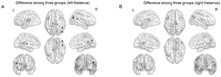
2                                                       LIU et al: FC IN PDN

(HbA1c) levels, homeostasis model assessment‑insulin               (total cholesterol and triglycerides) were measured by enzy‑
resistance and the estimated glomerular filtration rate were       matic assays (Olympus AU5400 autoanalyzer; Beckman
significantly associated with abnormal spontaneous activity        Coulter).
in the brain.
     The present study aimed to explore abnormalities of the       Nerve assessment. Three independently‑trained doctors
thalamo‑cortical FC in patients with PDN using rs‑fMRI.            assessed the nerve and mental conditions of the patients in
The study may provide novel information on the underlying          accordance with the pain symptoms and neurological signs:
mechanisms of PDN.                                                 i) The visual analogue scale (VAS) (12,14); ii) the Toronto
                                                                   Clinical Scoring System (TCSS) (12,14); and iii) the Leeds
Materials and methods                                              Assessment of Neuropathic Symptoms and Signs (12,15).

Participants. A cross‑sectional study was performed to             MRI acquisition. The whole‑blood oxygen level‑dependent
explore the FC of the thalamus in patients with PDN using          (BOLD) signals were collected using an Ingenia 3.0T MRI
rs‑fMRI at the Department of Endocrinology of Nanjing First        machine (Philips Medical Systems B.V.). The MRI scan‑
Hospital (Nanjing, China) between September 2016 and March         ning technique was performed as described previously (12).
2017. Patients were categorized into three groups: i) Patients     The parameters of T1‑weighted imaging were as follows:
with T2DM and PDN (Group PDN), ii) patients with T2DM              i) Repetition time (TR)/echo time (TE), 8.2/3.8 msec; ii) field
and non‑painful neuropathy (Group NDN) and iii) healthy            of view (FOV), 240x240 mm; iii) matrix, 240x222; iv) slice
subjects as a control (Group C). The protocol was approved         thickness, 1 mm; and v) scanning time, 5 min and 29 sec. For
by the Institutional Review Board of Nanjing First Hospital        subjects with no structural brain abnormalities, a resting‑state
(Nanjing, China). All procedures were in accordance with the       functional imaging scan was performed. Subjects were
Declaration of Helsinki from 1964 and its later amendments or      required to close their eyes during the scan, and stay awake and
comparable ethical standards. Written informed consent was         quiet without any further movements. The parameters were
obtained from all participants.                                    as follows: i) TR/TE, 2,000/30 msec; ii) FOV, 220x220 mm;
     The inclusion criteria for the patients were as follows:      iii) matrix, 72x70, slice thickness, 4 mm; and iv) scanning
i) Voluntary participation and written informed consent;           time, 12 min and 45 sec.
ii) age between 18 and 60 years with junior high school educa‑
tion or above; and iii) met the 1999 World Health Organization     MRI processing. The standard pre‑processing steps were
T2DM diagnostic criteria (fasting plasma glucose ≥7.0 mmol/l       performed using the Statistical Parametric Mapping (SPM)
or 2‑h postprandial glucose ≥11.1 mmol/l) (13).                    version 8 (http://www.fil.ion.ucl.ac.uk/spm/) and the Data
     The criteria for the diagnosis of PDN were as follows:        Processing Assistant for re‑fMRI on the MATLAB R2012b
i) Neuropathy occurred after diagnosis of diabetes; ii) patients   platform (MathWorks) (16).
had clinical symptoms, such as pain, numbness or abnormal              The initial 10 time‑points were removed to eliminate
sensation; and iii) patients had abnormalities in one of the       early detection interference. Subsequently, slice timing and
following five examinations: Ankle reflex, vibratory sensation,    head‑motion correction were performed. Head movements
pressure sensation, temperature sensation and acupuncture          were calibrated with 2‑mm translations and were angled at
pain (12).                                                         2 degrees to eliminate inconsistencies. Subsequently, the
     The inclusion criteria for the healthy controls were as       image space was normalized in accordance with the Montreal
follows: i) Voluntary participation and written informed           standard head anatomic template and was resampled to
consent; ii) age between 18 to 60 years with junior high school    a 3 mm x 3 mm x 3 mm size using a unified segmentation
education or above; iii) no history of diabetes and HbA1c          algorithm on the T1 image (12). Nuisance signals, including
levels of 4‑6%; and iv) normal results of anxiety and depres‑      white matter signals and cerebral spinal fluid, were regressed.
sion scales.                                                       Detrending and temporal band‑pass filtering (0.01.0.08 Hz)
     The exclusion criteria for all subjects were as follows:      were performed. A Gauss kernel function of 4 mm with full
i) Left‑hand writers; ii) neuropathy caused by other causes,       width and half height were used for spatial smoothing.
such as cervical spondylosis, cerebral infarction, Green Barre
syndrome, severe arteriovenous disease, drug neurotoxicity         FC analysis. The thalamus was divided into two subregions
and renal insufficiency; iii) patients with severe cerebral        (Thalamus_L and Thalamus_R) for selecting the regions of
vascular disease; iv) disorders such as depression, anxiety or     interest (ROIs). Masks of ROIs were obtained using the WFU
Alzheimer's disease; v) history of any serious medical, psychi‑    Pick Atlas 3.0.5 (http://fmri.wfubmc.edu/software/PickAtlas)
atric or neurologic disorders; vi) substance abuse; vii) head      from the Montreal Neurological Institute, which automatically
trauma or loss of consciousness; and viii) any contraindica‑       generated segmented atlas ROI templates.
tions to MRI.                                                          The mean time course for calculating thalamic ROI was
                                                                   determined by averaging the time course of the voxels within
Laboratory assessments. HbA1c was measured using a                 the thalamus (17). Subsequently, the thalamo‑cortical FC was
high‑performance liquid chromatography assay (D‑100                calculated. The z scores were obtained from the correlation
system; Bio‑Rad Laboratories, Inc.). C‑peptide was measured        coefficients by Fisher's transformation (18,19).
using a chemiluminescent immunometric assay, which
employs the Modular Analytics E170 (Roche Diagnostics              Statistical analysis. To examine inter‑group differences in
GmbH). Blood glucose, serum creatinine and lipid profiles          the clinical characteristics, one‑way analysis of variance
EXPERIMENTAL AND THERAPEUTIC MEDICINE 21: 509, 2021 3 Table I. Demographic and clinical characteristics of the participants. Item Group‑PDN (n=19) Group‑NDN (n=20) Group‑C (n=13) P‑value Sex (male/female) 12/7 13/7 7/6 0.800a Age (year) 53.8±8.1 54.1±6.4 53.9±5.3 0.994b Education (years) 9.6±3.4 10.5±3.7 10.4±2.9 0.724b Duration of disease (months) 109.4±65.5 100.4±66.9 ‑ 0.671c Fasting blood glucose (mmol/l) 9.2±2.9 7.4±3.5 ‑ 0.083c HbA1c (%) 8.5±1.9 8.7±1.7 ‑ 0.633c C‑peptide (ng/ml) 1.6±0.9 1.2±0.7 ‑ 0.100c Cholesterol (mmol/l) 4.9±1.2 5.1±1.2 ‑ 0.650c Triacylglycerol (mmol/l) 2.4±2.7 2.4±2.9 ‑ 0.970c Creatinine (µmol/l) 70.0±19.2 67.9±30.6 ‑ 0.990c VAS (score) 6.8±1.9 0.0±0.0 ‑ 2 mm during and NDN groups by treating age, sex and years of educa‑ MRI. The patient characteristics are presented in Table I. tion as covariates. Multiple comparative corrections were No significant difference in age, sex and years of educa‑ performed using Monte Carlo simulation in conjunction with tionwas present among the three groups. Furthermore, there the REST AlphaSim program. Voxels with cluster sizes ≥4 was no significant difference in the course of disease, fasting (108 mm 3) and P
4                                                                       LIU et al: FC IN PDN

Table II. ANOVA of differences in thalamic‑whole brain                             Table III. Analysis of the differences in thalamic‑whole
functional connectivity among three groups when taking the                         brain functional connectivity between the Group‑PDN and
bilateral thalamus as the region of interest.                                      Group‑NDN when taking the bilateral thalamus as the region
                                                                                   of interest.
A, FC with the left thalamus
                                                                                   A, FC with the left thalamus
                                 MNI coordinate
                                 ‑‑‑‑‑‑‑‑‑‑‑‑‑‑‑‑‑‑‑‑‑‑‑‑‑‑‑‑‑‑‑                                                                 MNI
Area                    Side       x            y           z      K    F‑value                                             coordinate
                                                                                                                         ‑‑‑‑‑‑‑‑‑‑‑‑‑‑‑‑‑‑‑‑‑‑‑‑‑‑‑
Vermis_10                 L        0         ‑45         ‑30       45   11.64      Area                         Side      x          y          z      K    t‑value
Parahippocampal           R       24           3         ‑21        7    9.44
Temporal_Inf              R       54         ‑39         ‑15        9   13.26      Angular gyrus                  R      48        ‑48        21       6     4.83
Fusiform                  R       24         ‑72          ‑6       15   12.52      Middle occipital gyrus         R      48        ‑75        27       5     4.28
Thalamus                  R        6          ‑9           3       26   10.8
Temporal_Mid              R       48         ‑48          18        6   11.34      B, FC with the right thalamus
Rolandic_Oper             L      ‑36         ‑36          18       10    9.76
Occipital_Mid             R       45         ‑75          27       10    9.49                                                    MNI
Cingulum_Mid              L        0         ‑30          36       12   10.19                                               coordinate
                                                                                                                         ‑‑‑‑‑‑‑‑‑‑‑‑‑‑‑‑‑‑‑‑‑‑‑‑‑‑‑
                                                                                   Area                         Side      x          y          z      K    t‑value
B, FC with the right thalamus
                                                                                   Angular gyrus                  R      48        ‑66          3      19    4.47
                                 MNI coordinate
                                 ‑‑‑‑‑‑‑‑‑‑‑‑‑‑‑‑‑‑‑‑‑‑‑‑‑‑‑‑‑‑‑                   x, y, z are the coordinates of primary peak locations in the MNI space.
Area                    Side       x            y           z      K    F‑value    The t statistical value of peak voxels indicated significant differences
                                                                                   in FC between the groups PDN and NDN (P
EXPERIMENTAL AND THERAPEUTIC MEDICINE 21: 509, 2021 5
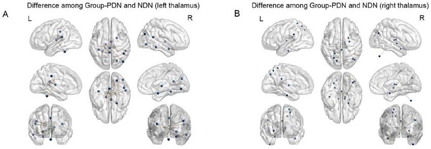
Figure 2. Differences of functional connectivity in the thalamus between Group‑PDN and Group‑NDN. The blue dots represent brain regions that had
significantly increased activity in Group PDN when compared with Group NDN. (A) Brain regions exhibiting different activity between the two groups when
considering the left thalamus as the ROI. (B) Brain regions exhibiting different activity between two groups when considering the right thalamus as the ROI.
ROI, region of interest; Group‑PDN, T2DM with painful neuropathy; Group‑NDN, T2DM with non‑painful neuropathy.
The thalamus is the central cortical‑subcortical connec‑ The present study also indicated that the thalamus had
tivity hub and serves as the gateway to the cerebral cortex. increased FC with the right middle occipital gyrus. The
The thalamus is considered to have a significant role in the thalamus is responsible for interpreting visual information and
process of pain sensation (26). The thalamus was indicated to forming conscious perception (34). Changes in the thickness
be associated with spontaneous and evoked pain in chronic of the middle occipital gyrus associated were determined
conditions (7). In 2011, it was reported that PDN was associ‑ in blind patients (35). Furthermore, local synchronicity and
ated with increased thalamic vascularity in T1DM (27). To abnormal occipital lobe function were reported in patients
date, there has been a scarcity of research focusing particularly with hemifacial spasm (36). Patients with classical trigeminal
on the thalamus and the role of PDN in T2DM using rs‑fMRI. neuralgia (CTN) had symptoms characterized by orbital pain,
Therefore, the present study may expand the current under‑ decreased corneal reflex and decreased vision (34). Individuals
standing of the underlying neurobiological mechanisms of with chronic post‑hysterectomy pain had decreased FC
these conditions. between the left sensorimotor insula and the right angular
The angular gyrus is one of the components within the and middle occipital gyri (MOG), as well as between the left
default mode network (DMN), which was reported to be chemosensory insula, the bilateral angular gyri and the MOG
involved in the process of episodic memory retrieval (28). following pain stimulation (37).
The angular gyrus is involved in several cognitive domains Furthermore, increased regional homogeneity (ReHo)
(i.e., language processing and attention) and serves as values in the right middle occipital gyrus were reported in
an important node of the DMN (29,30). The DMN was patients with CTN (34). Patients with drug‑induced head‑
indicated to be altered in chronic pain diseases such as aches exhibited an increased volume of the bilateral middle
fibromyalgia, which is considered to be the prototypical occipital gyrus (38). Furthermore, patients with pain‑related
central chronic pain syndrome (31). Another previous study conditions had altered neuronal activity or structural func‑
indicated that music was able to reduce pain and increase tions in the middle occipital gyrus (38,39). These results
the amplitude of rs‑fMRI BOLD signals in the left angular suggested that the middle occipital gyrus may participate in
gyrus in patients with fibromyalgia. The angular gyrus is the processes leading to the perception of pain in patients
involved in the top‑down regulation of the pain modulatory with diabetic polyneuropathy. The abnormal FC between
network by the DMN (32). Furthermore, increased activity the thalamus and other brain regions were indicated to be
in the left angular gyrus following verbally‑induced placebo associated with PDN.
analgesia was observed in patients with chronic pain (33). The present study has several limitations. First, the sample
These results suggested that the angular gyrus may have size was small. Furthermore, no associations between the
interactions with brain areas or pathways involved in pain altered brain regions and clinical characteristics were explored
modulation (33). in the present study. Further studies with a larger sample size
A previous multivariate analysis of resting FC demon‑ are required to understand the mechanisms of T2DM with
strated that the angular gyrus had similar connectivity to that PDN. Finally, it was not possible to exclude the potential influ‑
in the default mode network (DMN) (28). Another study deter‑ ences of medication, such as oral antidiabetic drugs, in the
mined that the angular gyrus were involved in numerous tasks present study.
and processes (29). In the present study, it was indicated that In conclusion, the present study revealed enhanced FC
in PDN group the bilateral thalamus had increased FC with between the bilateral thalamic‑angular gyrus and the left
the right angular gyrus, which was related to the pain modula‑ thalamic‑right MOG in patients with T2DM and PDN. The
tory network. Therefore, it was speculated that impaired FC increased thalamic‑whole brain FC may be involved in PDN.
between the thalamus and angular gyrus in PDN may be Furthermore, the abnormal thalamic‑angular gyrus FC may
related to the pain modulatory network within the DMN. be related to the DMN. These results may be helpful for6 LIU et al: FC IN PDN
understanding the central pathophysiological mechanisms of 6. Modha DS and Singh R: Network architecture of the long‑distance
pathways in the macaque brain. Proc Natl Acad Sci USA 107:
PDN in patients with T2DM. 13485‑13490, 2010.
7. Yen CT and Lu PL: Thalamus and pain. Acta Anaesthesiol
Acknowledgements Taiwan 51: 73‑80, 2013.
8. Selvarajah D, Wilkinson ID, Maxwell M, Davies J, Sankar A,
Boland E, Gandhi R, Tracey I and Tesfaye S: Magnetic resonance
Not applicable. neuroimaging study of brain structural differences in diabetic
peripheral neuropathy. Diabetes Care 37: 1681‑1688, 2014.
9. Biswal B, Yetkin FZ, Haughton VM and Hyde JS: Functional
Funding connectivity in the motor cortex of resting human brain using
echo‑planar MRI. Magn Reson Med 34: 537‑541, 1995.
The present study was supported by grants from the Nanjing 10. Zuo XN, Kelly C, Di Martino A, Mennes M, Margulies DS,
Bangaru S, Grzadzinski R, Evans AC, Zang YF, Castellanos FX
Science and Technology Development Project (grant and Milham MP: Growing together and growing apart: Regional
no. 201605027) and the Natural Science Foundation of Jiangsu and sex differences in the lifespan developmental trajectories of
Province (grant no. BK20170136). functional homotopy. J Neurosci 30: 15034‑15043, 2010.
11. Gorges M, Muller HP, Lule D, Ludolph AC, Pinkhardt EH and
Kassubek J: Functional connectivity within the default mode
Availability of data and materials network is associated with saccadic accuracy in Parkinson's
disease: A resting‑state FMRI and videooculographic study.
Brain Connect 3: 265‑272, 2013.
The datasets used and/or analyzed during the current study are 12. Zhang Q, Zhang P, Yan R, Xu X, Mao C, Liu X, Li F, Ma J, Ye L,
available from the corresponding author on reasonable request. Yao Z and Wu J: A single‑blinded trial using resting‑state func‑
tional magnetic resonance imaging of brain activity in patients
with type 2 diabetes and painful neuropathy. Diabetes Ther 10:
Authors' contributions 135‑147, 2019.
13. Gabir MM, Hanson RL, Dabelea D, Imperatore G, Roumain J,
XL, XX and CM acquired and interpreted the patient data, Bennett PH and Knowler WC: The 1997 American Diabetes
Association and 1999 World Health Organization criteria for
constructed the figures and wrote the manuscript. PZ, QZ, LJ, hyperglycemia in the diagnosis and prediction of diabetes.
YY, JM, LY and KL acquired and analyzed the patient data, Diabetes Care 23: 1108‑1112, 2000.
and revised the manuscript. JW and ZY designed the study 14. Essmat A and Hussein MS: Green tea extract for mild‑to‑moderate
diabetic peripheral neuropathy A randomized controlled trial.
and revised the manuscript. JW and ZY confirm the authen‑ Complement Ther Clin Pract 43: 101317, 2021.
ticity of all the raw data. All authors have read and approved 15. Garoushi S, Johnson MI and Tashani OA: Translation and cultural
the final manuscript. adaptation of the Leeds Assessment of Neuropathic Symptoms
and Signs (LANSS) pain scale into Arabic for use with patients
with diabetes in Libya. Libyan J Med 12: 1384288, 2017.
Ethics approval and consent to participate 16. Chao‑Gan Y and Yu‑Feng Z: DPARSF: A MATLAB toolbox
for ‘Pipeline’ data analysis of resting‑state fMRI. Front Syst
Neurosci 4: 13, 2010.
The protocol was approved by the Institutional Review Board 17. Salvador R, Suckling J, Schwarzbauer C and Bullmore E:
of Nanjing First Hospital (Nanjing, China). All procedures Undirected graphs of frequency‑dependent functional connec‑
were in accordance with the 1964 Helsinki Declaration and tivity in whole brain networks. Philos Trans R Soc Lond B Biol
Sci 360: 937‑946, 2005.
its later amendments or comparable ethical standards. Written 18. Liu Y, Yu C, Liang M, Li J, Tian L, Zhou Y, Qin W, Li K and
informed consent was obtained from all participants. Jiang T: Whole brain functional connectivity in the early blind.
Brain 130: 2085‑2096, 2007.
19. Hale JR, White TP, Mayhew SD, Wilson RS, Rollings DT,
Patient consent for publication Khalsa S, Arvanitis TN and Bagshaw AP: Altered thalamo‑
cortical and intra‑thalamic functional connectivity during light
Not applicable. sleep compared with wake. Neuroimage 125: 657‑667, 2016.
20. Song XW, Dong ZY, Long XY, Li SF, Zuo XN, Zhu CZ, He Y,
Yan CG and Zang YF: REST: A toolkit for resting‑state func‑
Competing interests tional magnetic resonance imaging data processing. PLoS One 6:
e25031, 2011.
21. Tzourio‑Mazoyer N, Landeau B, Papathanassiou D, Crivello F,
The authors declare that they have no competing interests. Etard O, Delcroix N, Mazoyer B and Joliot M: Automated
anatomical labeling of activations in SPM using a macroscopic
References anatomical parcellation of the MNI MRI single‑subject brain.
Neuroimage 15: 273‑289, 2002.
22. Zhu X, He Z, Luo C, Qiu X, He S, Peng A, Zhang L and Chen L:
1. Feldman EL, Nave KA, Jensen TS and Bennett DLH: New Altered spontaneous brain activity in MRI‑negative refractory
horizons in diabetic neuropathy: Mechanisms, bioenergetics, and temporal lobe epilepsy patients with major depressive disorder:
pain. Neuron 93: 1296‑1313, 2017. A resting‑state fMRI study. J Neurol Sci 386: 29‑35, 2018.
2. Gaskin DJ and Richard P: The economic costs of pain in the 23. Tose K, Yoshihara Y and Takahashi H: FMRI neurofeedback
United States. J Pain 13: 715‑724, 2012. and its application to psychiatric disorders. Brain Nerve 70:
3. Fayaz A, Croft P, Langford RM, Donaldson LJ and Jones GT: 1209‑1216, 2018.
Prevalence of chronic pain in the UK: A systematic review and 24. Tadayonnejad R, Deshpande R, Ajilore O, Moody T,
meta‑analysis of population studies. BMJ Open 6: e010364, 2016. Morfini F, Ly R, O'Neill J and Feusner JD: Pregenual ante‑
4. Dermanovic Dobrota V, Hrabac P, Skegro D, Smiljanic R, rior cingulate dysfunction associated with depression in
Dobrota S, Prkacin I, Brkljacic N, Peros K, Tomic M, OCD: An integrated multimodal f MRI/(1)H MRS study.
Lukinovic‑Skudar V and Basic Kes V: The impact of neuropathic Neuropsychopharmacology 43: 1146‑1155, 2018.
pain and other comorbidities on the quality of life in patients 25. Shen Z, Jiang L, Yang S, Ye J, Dai N, Liu X, Li N, Lu J, Liu F,
with diabetes. Health Qual Life Outcomes 12: 171, 2014. Lu Y, et al: Identify changes of brain regional homogeneity in
5. Tesfaye S, Selvarajah D, Gandhi R, Greig M, Shillo P, Fang F early and later adult onset patients with first‑episode depression
and Wilkinson ID: Diabetic peripheral neuropathy may not be as using resting‑state fMRI. PLoS One 12: e0184712, 2017.
its name suggests: Evidence from magnetic resonance imaging. 26. Head H and Holmes G: Sensory disturbances from cerebral
Pain 157 (Suppl 1): S72‑S80, 2016. lesions. Brain 34: 102‑254, 1911.EXPERIMENTAL AND THERAPEUTIC MEDICINE 21: 509, 2021 7
27. Selvarajah D, Wilkinson ID, Gandhi R, Griffiths PD and 35. Anurova I, Renier LA, De Volder AG, Carlson S and
Tesfaye S: Microvascular perfusion abnormalities of the Rauschecker JP: Relationship between cortical thickness
Thalamus in painful but not painless diabetic polyneuropathy: and functional activation in the early blind. Cereb Cortex 25:
A clue to the pathogenesis of pain in type 1 diabetes. Diabetes 2035‑2048, 2015.
Care 34: 718‑720, 2011. 36. Tu Y, Wei Y, Sun K, Zhao W and Yu B: Altered spontaneous
28. Bellana B, Liu Z, Anderson JAE, Moscovitch M and Grady CL: brain activity in patients with hemifacial spasm: A resting‑state
Laterality effects in functional connectivity of the angular gyrus functional MRI study. PLoS One 10: e0116849, 2015.
during rest and episodic retrieval. Neuropsychologia 80: 24‑34, 37. Ching YY, Wang C, Tay T, Loke YM, Tang PH, Sng BL and
2016. Zhou J: Altered sensory insular connectivity in chronic postsur‑
29. Seghier ML: The angular gyrus: Multiple functions and multiple gical pain patients. Front Hum Neurosci 12: 483, 2018.
subdivisions. Neuroscientist 19: 43‑61, 2013. 38. Chen X, Chen Z, Dong Z, Liu M and Yu S: Morphometric
30. Greicius MD, Supekar K, Menon V and Dougherty RF: changes over the whole brain in caffeine‑containing
Resting‑state functional connectivity reflects structural connec‑ combination‑analgesic‑overuse headache. Mol Pain 14:
tivity in the default mode network. Cereb Cortex 19: 72‑78, 2009. 1744806918778641, 2018.
31. Napadow V, LaCount L, Park K, As‑Sanie S, Clauw DJ and 39. Chen Y, Meng Z, Zhang Z, Zhu Y, Gao R, Cao X, Tan L, Wang Z,
Harris RE: Intrinsic brain connectivity in fibromyalgia is associated Zhang H, Li Y and Fan Q: The right thalamic glutamate level
with chronic pain intensity. Arthritis Rheum 62: 2545‑2555, 2010. correlates with functional connectivity with right dorsal ante‑
32. Garza‑Villarreal EA, Jiang Z, Vuust P, Alcauter S, Vase L, rior cingulate cortex/middle occipital gyrus in unmedicated
Pasaye EH, Cavazos‑Rodriguez R, Brattico E, Jensen TS and obsessive‑compulsive disorder: A combined fMRI and 1H‑MRS
Barrios FA: Music reduces pain and increases resting state fMRI study. Aust N Z J Psychiatry 53: 207‑218, 2019.
BOLD signal amplitude in the left angular gyrus in fibromyalgia
patients. Front Psychol 6: 1051, 2015. This work is licensed under a Creative Commons
33. Craggs JG, Price DD and Robinson ME: Enhancing the placebo Attribution-NonCommercial-NoDerivatives 4.0
response: Functional magnetic resonance imaging evidence of International (CC BY-NC-ND 4.0) License.
memory and semantic processing in placebo analgesia. J Pain 15:
435‑446, 2014.
34. Xiang CQ, Liu WF, Xu QH, Su T, Yong‑Qiang S, Min YL,
Yuan Q, Zhu PW, Liu KC, Jiang N, et al: Altered spontaneous
brain activity in classical trigeminal neuralgia as determined by
changes in regional homogeneity: A resting‑state functional MRI
study. Pain Pract 19: 397‑406, 2018.You can also read