The efficacy of Ovopet in the treatment of hip dysplasia in dogs
←
→
Page content transcription
If your browser does not render page correctly, please read the page content below
Vol. 10(8), pp. 198-207, August 2018
     DOI: 10.5897/JVMAH2018.0687
     Article Number: B13386657918
     ISSN: 2141-2529
     Copyright ©2018                                                Journal of Veterinary Medicine and Animal
     Author(s) retain the copyright of this article
     http://www.academicjournals.org/JVMAH                                                             Health

Full Length Research Paper

             The efficacy of Ovopet® in the treatment of hip
                            dysplasia in dogs
     Andrés Aguirre1*, Erena Gil-Quintana1, Marisa Fenaux1, Nuria Sanchez2 and Celina Torre2
 1
  Department of Production, Quality and R&D of Eggnovo S.L., Avenida los Tilos 5, 31132 Villatuerta (Navarra), Spain.
 2
  Department of Research of Affinity Petcare, Plaza Europa 54-56, 08902 L'Hospitalet de Llobregat, Barcelona, Spain.
                                               Received 16 April, 2018: Accepted 3 July, 2018

       Hip dysplasia is a widespread condition that can affect dogs of all ages. Hip dysplasia is caused by a
       subluxation in the hip joint. This leads to the development of osteoarthritis that causes inflammation
       and pain. At this sight, the efficacy of a supplement with Ovopet®, eggshell membrane, was evaluated
       together with its tolerability and safety. Forty client-owned medium sized arthritic dogs were treated
       daily for a period of 40 days with placebo or Ovopet®. Every ten days, the dogs were evaluated for
       functional limitation and joint mobility (hip functional scale), muscular atrophy and mobility range
       (extension-flexion rating). Dogs were also examined for blood analysis (inflammatory markers), and
       sonographies of the hip joint space were taken before and at the end of the study. Performances in
       daily life activities and vitality assessed by the owners were also recorded. Based on these
       observations, significant (p
Aguirre et al. 199
resultant OA (Gail et al., 2001). et al., 2016).
Hip dysplasia is associated with abnormal joint The goal of the present study is to assess the
structure and a laxity of the muscles, connective tissue, effectiveness of Ovopet® in the treatment of hip
and ligaments that would normally support the joint. As dysplasia and the consequent OA in dogs.
joint laxity develops, the articular surfaces of the two
bones lose contact with each other (Kyriazis and
Prassinos, 2016). This separation of the two bones within MATERIALS AND METHODS
the joint is called a subluxation, and this causes a drastic
Dog selection
change in the size and shape of the articular surfaces
(Butler and Gambino, 2017). OA of the hip is the result of A group of adults (63% female), privately owned arthritic dogs were
the degeneration of the joint due to a laxity caused by hip used in this study. Their age was between 1 to 13 years old, with an
dysplasia (Butler and Gambino, 2017). When a dog has average of 8±0.56 years. All the participants were medium-sized
hip dysplasia, the joint wears out abnormally and the dogs. Their body mass ranged from 15 to 40 kg, with an average of
protective cartilage on the surface of the joint deteriorates 34±1.05kg. The body condition of the 66% of the dogs was ideal
according to the body condition score published in the
and the resultant bone-to-bone contact creates pain manufacturer’s site
(Butler and Gambino, 2017). (http://www.affinitypetcare.com/veterinary/obesity/obesity_dog/flash
Moreover, the degenerative changes of OA appear /bcs.html). The German Shepherd was the most common breed
frequently causing inflammation and pain (Henrotin et al., (40% of dogs) followed by Labrador and Golden (15% of dogs
2014). Pain is also related with the inflammation of the each). All dogs had radiographic evidence of hip dysplasia. They
were recently diagnosed dogs in all cases, so they had not been
joint (synovitis) (Henrotin et al., 2014). This pain makes
previously treated with chondroprotective supplements or diets.
the dogs shift most of their body weight to the front end. Besides the afore mentioned stated inclusion criteria, the owner
As a result, dogs showing few clinical symptoms may had to describe at least one of the following signs: difficulty to stand
develop increased shoulder musculature while hind limb up, difficulty to jump, difficulty to climb stairs or clear lameness. The
musculature remains under-developed (Kyriazis and study protocol was performed in compliance with national
Prassinos, 2016). As damage to the joint progresses and guidelines for research onanimals. Throughout the study, dogs
remained with their owners. The owner’s consent was obtained at
secondary OA sets in, symptoms of stiffness and the beginning of the study.
lameness may be present (Kyriazis and Prassinos,
2016).
Some forms of degenerative joint disease can be Nutraceuticals
treated with surgery but drugs are the most common
therapy for OA treatment. Nevertheless, pharmacological Ovopet® was obtained from Eggnovo S.L. (Navarra, Spain) in a
sustainable and environmentally friendly manner without the use of
treatment is limited to clinical signs alleviation (Comblain chemicals. It consists of egg membranes separated from eggshells
et al., 2016). In this way, non-steroidal anti-inflammatory by a patented process. Compositional analysis of Ovopet® has
(NSAIDs) are commonly prescribed to address anti- identified a high content of protein (collagen types I-V-X, elastin,
inflammatory mechanisms. Unfortunately, the use of keratin) and moderate quantities of GAGs (chondroitin sulfate, HA)
NSAIDs may be associated with detrimental effects, and glucosamine. Snacks were prepared by a Spanish pet food
manufacturer.
especially gastrointestinal side effects (Comblain et al.,
2016). Some clinical studies have highlighted the
beneficial effects of dietary supplement for the treatment Study protocol
of OA in dogs (Comblain et al., 2016).
Glucosamine and chondroitin are two compounds that The randomized and double-blind with placebo study was carried
have been widely used for the treatment of OA in out in cooperation with Eudald Toralles and Sentmenat Veterinary
Clinics (Barcelona). The veterinary monitoring was performed at
animals. These aminosaccharides act as a preferred days 0, 10, 20, 30 and 40. Veterinary Clinics were in charge of
substrate for the biosynthesis of glycosaminoglycan(GAG) recruiting the dogs with hip dysplasia, getting the owners signed
chains, and subsequently for the production of aggrecan, consent and performing the standard clinical tests according to the
main proteoglycan used by chondrocytes to build the study protocol. If the inclusion criteria were met, the dogs could
cartilage extracellular matrix (ECM). They also exert anti- take part in the study. Firstly, the veterinary filled in the dogs’ data
inflammatory and anti-catabolic effects through the (breed, age, body condition, sex and diet), did a clinical
examination of the dog before starting the treatment and evaluated
inhibition of nuclear factor κB (NF-κB) binding activity the OA grade with the Kellgren-Lawrence scale (Kohn et al., 2016).
(Comblain et al., 2016). Based on the weight of the dog the veterinary established, the
Ovopet® is an innovative ingredient obtained from egg amount of fodder and the number of snacks that dogs had to intake
shell membranes at Eggnovo S.L. via a patented during the study. This amount was revised in every veterinary visit
process. Eggshell membrane discovery as a natural to adjust the dosage of fodder. The performed protocol in each
assessment day was as follows:
source of glycosaminoglycan’s, such as chondroitin
sulfate and hyaluronic acid among others, has led to the (1) To assess the muscle atrophy by measuring the perimeter of the
consideration of this product as a potential approach for rear legs
the treatment of OA as shown in a previous study (Blasco (2) To evaluate the extension-flexion range of the hip200 J. Vet. Med. Anim. Health
(3) To fill in a hip functional scale questionnaire jumps,pain during palpation and pain during movement.
(4) To fill in an evaluation questionnaire together with the owner
(5) To fill in the side effects questionnaire
(6) To draw blood for measuring inflammatory blood markers (at Evaluation questionnaire with the owner
day 0 and 40)
(7) To make a sonography of the hip (at day 0 and 40) The evaluation questionnaire that the veterinarian completed with
the owner is based partially on the Canine Brief Pain Inventory
Two groups of patients were established, one taking the (Brown et al., 2018) and is divided in three sections: description of
chondroprotective supplement with Ovopet® (N=30) and the other function, description of positive behaviour and visual scale.The
group taking the placebo supplement (the same recipe but without description of the function compiles different questions in relation to
Ovopet®) (N=10). The treatment lasted 40 days and the the general activity of the dog, the ability to standing up from lying
recommended daily dose of Ovopet® was 15 mg Kg-1 dog day-1. down, the ability to walk, the ability to run and the ability to climb
The only authorised pharmacological treatment, when the lameness stairs. The description of positive behaviour includes two questions:
was intense or the quality of life decreased markedly was Metacam. mood and feel like playing. The visual scale refers to the severity of
The owners and the veterinary noted down if the dog needed the pain that the owner thinks that his/her dog suffers. Each section
rescue drugs. has different items and each item has a numerical rating scale from
0 to 10. Zero means no pain (or that pain does not interfere) and
ten means extreme pain (or that pain interferes completely).
Besides individual measurements for each parameter we obtained
Kellgren-Lawrence classification of OA degree
an average value for all the parameters included in the description
of the function and another for the description of positive behaviour.
The OA degree (from 1 to 4) was classified using the Kellgren-
The improvement with respect to the beginning of the study was
Lawrence grading scale. The grades in the scale are described as
calculated for each treatment (mean day 40 – mean day 0) x100 /
follows:
mean day 0). We also calculated the difference in improvement
Grade 1: Doubtful narrowing of joint space and possible osteophytic between therapy and placebo (mean treatment – mean placebo).
lipping.
Grade 2: Definite osteophytes, definite narrowing of joint space.
Blood analysis
Grade 3: Moderate multiple osteophytes, definite narrowing of joint
space, some sclerosis and possible deformity of bone contour. Blood was obtained from the cephalic vein for biochemical analyses
Grade 4: Large osteophytes, marked narrowing of joint space, of inflammation markers. It was collected under aseptic conditions
severe sclerosis and definitedeformity of bone contour. in 5 ml tubes, and then centrifuged for 5 min at 2000 rpm to collect
serum. Serum samples were analysed for Tumour necrosis factor-α
(TNF-α) and nitric oxide (nitrite/nitrate) (NO). TNFα assay was
Assessment of muscular atrophy
performed using canine TNFα commercial ELISA set from R&D
The circumference of each thigh was measured at standard (Ref. CATA00, R&D systems, La Joya, CA), following the
anatomical references based on the Bioarth assessment scale manufacturer’s instructions. No assay was performed as previously
(Villaret al., 2016; Cuervo et al., 2014). The same investigator described (Miranda, Espeyand Wink, 2001; García-Robledo, Corzo
performed all measurements using a measure ribbon. and Papaspyrou, 2014).
Assessment of flexion-extension range Sonography
To evaluate the hip mobility range, the veterinarian measured the Sonographic evaluations were performed under routine sedation in
flexion and extension degrees of the hip every ten days based on all dogs using the Vivid I (GE Healthcare, Wauwatosa, WI, USA)
the Bioarth assessment scale (Villaret al., 2016; Cuervo et al., ultrasound system at day 0 and 40 after treatment. Linear
2014). The range of movement was measured bilaterally for the hip transducer 12L-RS of high frequency (5-13 MHz) was used for the
using a goniometer. These measurements were taken for the ultra sound examination. The animals were placed in dorsal
maximum possible extensionand flexion values. decubit, with posterior members in neutralposition (between 10°
flexion and 30° extension, 10° and 30° abduction and 0° and
10°external rotation), the knees forming an angle of 90° between
Hip functional scale the femur and the tibia/fibula. The images of the coxofemoral joints
(both hips) were obtained in three sagittal views parallel to the
The hip functional scale is an adaptation of the Bioarth assessment longitudinal axis of the lateral side of the hip, dorsal to the greater
scale (Villaret al., 2016) to quantify the degree of OA. This is the trochanter of the femur. The measurement of the distance between
questionnaire that the supplement manufacturer R&D department the femur head and the acetabulum was taken for each view and
trained personnel employs for researching. The questionnaire the major value was considered for statistical analysis.
contains two different sections: functional limitation and joint
mobility. The functional limitation assesses the lameness before
starting to walk, the lameness during walk, the resistance to walk, Statistical analysis
the resistance to run and play, the difficulty to climb stairs and the
difficulty in little jumps. The joint mobility assesses the pain at Data were analysed using the GraphPad (GraphPad Prism version
manual mobilisation of the hip, the pain during palpation and the 6.0 for Windows, GraphPad Software, Inc) program and the Epi Info
pain during movement. These parameters were assessed by the TM (Epi Info version 7 for Windows, CDC). Data were assessed for
veterinarian in the clinical visits every ten days. The scale for most normality with the D’Agostino and Pearson normality test, and for
of the questions goes from one (no functional limitation or no pain) homoscedasticity with the Bartlett’s test. Parametric analyses were
to four (complete functional limitation orextreme pain). However, performed with one-way ANOVA with repeated measures followed
some items use a three-point scale, such as, difficulty in little by Holm Sidak’s multiple comparison post-test. Non-parametricAguirre et al. 201
samples were compared with Friedman test for related samples. to walk, the resistance to run and play, the difficulty to
climb stairsand the difficulty in little jumps (Figure 2A, B,
C, D, E and F, respectively). In general, the pattern of
RESULTS each treatment was different. In the treated group of
dogs, for the lameness before starting to walk (p=0.005),
All participants completed the study. The percentage of the lameness during walk (p=0.0389), the resistance to
dogs taking part in the supplement study was similarly run and play (p=0.0038) and the difficulty in little jumps
distributed among grade 1, grade 2, grade 3 and grade 4 (p=0.01), there was a significant decrease while the
of OA. Most of the dogs fulfilled two of the following placebo group remained stable or even augmented,
inclusion criteria (48%): difficulty to stand up, difficulty to although not significantly (Figure 2A, B, D and F). The
jump, difficulty to climb stairs or clear lameness, where assessment of the resistance to walk (p=0.0015) and the
the difficulty to stand up was the most common sign. difficulty to climb stairs (p=0.0024) showed a progressive
Dogs were not administered rescue pain medication. The decrease after treatment which failed to reach statistical
possible side effects related to the treatment such as: significance in the ANOVA post hoc tests. The opposite
changes in appetite, vomits, diarrhoea and skin reactions occurred with the placebo group, which showed a
were assessed in each veterinary visit. No significant side tendency to increase progressively (resistance to walk) or
effects were seen during the treatment with Ovopet® and remained stable (difficulty to climb stairs) (Figure 2C and
the veterinary did not relate the observed symptoms with E).
Ovopet® as similar reactions were seen in the placebo The joint mobility assesses the pain at manual
group. Moreover, an initial test was carried out in 10 dogs mobilisation of the hip, the pain during palpation and the
giving them ten times the recommended daily dosage for pain during movement, parameters that the veterinarian
50 days to assess that Ovopet® was safe. measured in the clinical visits every ten days (Figure 3A,
B and C respectively). In the group treated with Ovopet®
there was a gradual decrease in pain since day 10 until
Assessment of muscular atrophy the end of the study. There were statistically significant
differences in the pain at manual mobilization of the hip
A gradual improvement in the muscular perimeter in the (p=0.0008) and in the pain during palpation (p=0.0193)
group treated with Ovopet® was observed along the while the pain during movement only showed a tendency
study reaching a significant improvement of 7.3% in the to decrease. Evaluation questionnaire with the owner
right (p202 J. Vet. Med. Anim. Health
Figure 1. Evolution of the muscular perimeter and the mobility range (flexion and extension) during
the treatment with Ovopet®. Muscular perimeter in the right (A) and in the left rear leg (B).Flexion of
the right (C) and the left rear leg (D).Extension of the right (E) and the leftrear leg (F). Black asterisks
indicate significant differences (p ≤ 0.05) when compared to basal values in the post-test. Values are
represented as mean ± SEM.
decline in pain in the treated group was 46,9% at the end Sonography
of the treatment compared to day 0. Moreover, the
difference in pain between the placebo and the Ovopet® The hip joint space from the legs was measured at day 0
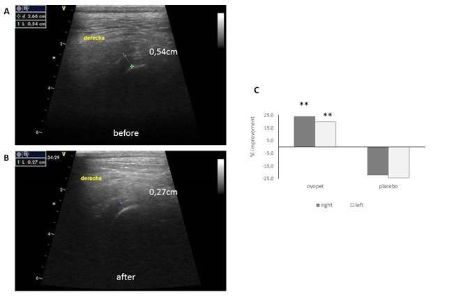
group at day 40 was 37,9% less pain in the Ovopet® and at day 40 using ultrasounds (Figure 7). The results
group. showed a significant decrease in the joint space of both
legs in the dogs treated with Ovopet® (Figure 7A and B)
while in the placebo group the joint space maintained or
increased. The improvement in synovitis at the end of the
Blood analysis study was 19.6 and 24% in the right and left legs of
Ovopet® treated dogs respectively (Figure 7C). The % of
Anti-inflammatory blood markers, such as NO and TNF-α, improvement in the Ovopet® group was statistically
were analysed before treatmentand at the end of the significant as compared with the placebo group in both
study. A change in NO levels from 19.4 to 35.6μmol/L legs (p=0.0019 and pAguirre et al. 203
Figure 2. Evolution of the functional limitation parameters in the hip functional scale during the treatment
with Ovopet®: lameness before starting to walk (A), lameness during walk (B), 0 resistance to walk (C),
resistance to run and to play (D), difficulty to climb up stairs (E) and difficulty in little jumps (F). Black
asterisks indicate significant differences (p ≤ 0.05) when compared to basal values in the post-test. Values
are represented as mean ± SEM.
introduction and approval of several new supplements One of the symptoms associated to OA is the muscle
and drugs (Scott et al., 2017). In the present study, we weakening or muscular atrophy (Arden and Nevitt, 2006).
have tested the efficacy of a novel dietary supplement In the present study, the muscular perimeter of rear legs
Ovopet®, obtained from eggshell membranes, for the was significantly improved in the Ovopet® group together
treatment of OA in dogs with hip dysplasia. OA is a with the range of motion (extension and flexion) of the
condition that causes pain, inflammation and stiffness in hip. The decrease in the extension angle of dogs
joints (Barrouin-Melo et al., 2016) that are associated suffering hip dysplasia has been proven to worsen each
with ligamentous laxity and muscle weakening (Arden year of a dog’s life leading even to the inability of the dog
and Nevitt, 2006). to run, jump or climb steps (Greene et al., 2014; Dycus et
Although there is a lack of consensus in the research al., 2017). Therapeutic exercises and diverse physical
community regarding the outcomes that should be therapies have been employed to improve the hip range
assessed in canine OA, validated and objective methods of motion and to decline OA symptoms such as muscle
are urged (Belshaw et al., 2016), for instance, the atrophy, pain, inflammation (Dycus et al., 2017). At the
goniometer to measure the hip range of motion (Jaegger sight of the results, Ovopet® appears as an alternative
et al., 2002), imaging (de Sousa et al., 2017) or the use treatment to improvethe mobility range of dogs suffering
of validated questionnaires (Belshaw et al., 2016). The OA and hip dysplasia.
efficacy assessment Innvacion-Eggnovo performed in the In this study, enhanced joint mobility and functionality
present study aims to measure both, objective and semi- were observed in treated dogs. The joint mobility
objective outcomes related to the known symptoms of OA parameter was based on a veterinary physical
and the physical limitations of the patients, to prove the examination to evaluate pain. To our knowledge,
efficacy of the natural and innovative eggshell-derived although the assessment of pain has not been validated
dietary supplement. to be completed by veterinary surgeons, there is a need204 J. Vet. Med. Anim. Health
Figure 3. Evolution of the joint mobility parameters in the hip functional scale during the
treatment with Ovopet®: pain at manual mobilisation of the hip (A), pain during palpation (B)
and pain during movement (C). Black asterisks indicate significant differences (p ≤ 0.05) when
compared to basal values in the post-test. Values are represented as mean ± SEM.
Figure 4. Evolution of the function description in the evaluation made by owners during the
treatment with Ovopet®: general activity (A), ability to rise to standing from lying down (B),
ability to walk (C), ability to run (D) and ability to climb up stairs (E). Black asterisks indicate
significant differences (p ≤ 0.05) when compared to basal values in the post-test.Values are
represented as mean ± SEM.Aguirre et al. 205
Figure 5. Evolution of the positive behaviour, mood (A) and feel like playing (B), and the pain that the owner thinks
that suffers the dog (C) during the treatment with Ovopet®. Black asterisks indicate significant differences (p ≤ 0.05)
when compared to basal values in the post test.Values are represented as mean ± SEM.
Figure 6. Serum levels of the inflammatory blood marker nitric oxide (NO) before
and 40 days after treatment with placebo or Ovopet®. The horizontal line represents
the mean ± SEM.206 J. Vet. Med. Anim. Health
Figure 7. Sonographic evaluation of the evolution of the hip joint space after the treatment with
Ovopet®. A: right hip of one treated dog before Ovopet® treatment. B: right hip of the same
treated dog after treatment with Ovopet®. C: % of improvement of the hip joint space at the end of
the study. (n = 30 for Ovopet®; n = 10 for placebo). Black asterisks indicate significant differences
(p ≤ 0.05) when compared to placebo.
to somehow measure pain by professionals as it is one of functional scale for functional limitation that was
the most common outcomes when measuring canine OA assessed by the veterinarian. They are also in
(Belshaw et al., 2016). A tendency to decrease pain was concordance with data of mobility range, which revealed
measured in Ovopet® group that was not observed in the an improvement of the flexion and extension angles, and
placebo group. The evaluation made by owners helped with muscular atrophy, which showed an increase of the
us to assess the function description and positive rear legs perimeter.
behaviour. In all the categories evaluated in the evolution In OA and in synovitis there is an increase in the
of the function description there was significant production of pro-inflammatory factors (Mobasheriet al.,
improvement only in the Ovopet® treated group while in 2017; Bhattaram and Chandrasekharan, 2017). The anti-
the dogs treated with placebo, all the parameters inflammatory properties attributed to eggshell membrane
remained stable or even worsened. This is in contrast to that are potentially responsible for reducing inflammatory
what previously was observed in the evaluation of a cytokines IL-1β and TFN-α are shown in rats (Ruff and
patient response to treatment, where there was a care De Vore, 2014). There is a need to prove these effects in
giver placebo effect (Scott et al., 2017). clinical studies with eggshell membrane products used as
Here, the placebo effect associated with the owners of nutraceutical compounds for dogs. Our finding of
the dogs regarding the function description was not decreased NO blood concentration in treated dogs,
observed in this study population which is in concordance although failed to besignificantly different, may indicate
with the statement of other researchers saying that when that a similar anti-inflammatory mechanism was
looking at the group average change in animal OA, there conferred by Ovopet®. Moreover, hip joint space was
is no a placebo effect (Gagnon et al., 2017). Similarly, to analysed by ultrasounds, revealing a significant
assess canine behavioural changes affected by pain, an narrowing of the hip joint space. The results obtained for
owner-reported instrument was employed. The detailed blood measurements of NO and the sonographic
behaviour-based assessment performed by the evaluation of hip joint space indicates that Ovopet® was
ownershave been extensively employed (Belshaw et al., capable of reducing the inflammation.
2014; Essner et al., 2017) and offer the advantage of an The goal of dietary supplements intended to treat OA is
extended assessment of dogs in their typical to provide relief to the major clinical signs of OA.
environmentand routine (Sharkey, 2013). These results Ovopet® arises as an effective supplement to decline
are in good concordance with the data obtained in the hip inflammatory pain, functional disability, lameness andAguirre et al. 207
inflammation associated to hip dysplasia and OA, and De Sousa EB, dos Santos GC, Duarte MEL, Moura Neto V, Aguiar DP
(2017).Metabolomics as a promising tool for early osteoarthritis
therefore, to improve the quality of life of dogs. The data
diagnosis. Brazilian Journal of Medical and Biological Research
obtained in the present study support the use of this 50(11):6485.
dietary supplement as an effective treatment for hip Dycus DL, Levine D, Marcellin-Little DJ (2017) Physical Rehabilitation
dysplasia and OA. Further research is needed to for the Management of Canine Hip Dysplasia. Veterinary Clinics:
Small Animal Practice 47:823-850.
ascertain and better understand the mechanisms
Gail K, Smith VMD, Philipp D (2001). Evaluation of risk factors for
underlying the mode of action of eggshell membrane in degenerative joint disease associated with hip dysplasia in German
osteoarthritic joints. For instance, the measurement of Shepherd Dogs, Golden Retrievers, Labrador Retrievers and
some biomarkers such as fragments of collagen type II Rottweilers. Journal of the American Veterinary Medical Association
219(12):1719-1724.
(de Sousa et al., 2017) could be useful to gain insights in García-Robledo E, Corzo A, Papaspyrou S (2014). A fast and
this complex mechanism. directspectrophotometric method for the sequential determination of
nitrate and nitrite at lowconcentrations in small volumes. Marine
Chemistry 162:30-36.
Greene LM, Marcellin-Little DJ, Lascelles BD (2013). Associations
CONFLICT OF INTERESTS
among exercise duration, lameness severity, and hip joint range of
motion in Labrador Retrievers with hip dysplasia. Journal of the
The authors have not declared any conflict of interests. American Veterinary Medical Association 242(11):1528-1533.
Henrotin Y, Lambert C, Richette P (2014). Importance of synovitis in
osteoarthritis: Evidence for the use of glycosaminoglycans against
synovial inflammation. Seminars in Arthritis and Rheumatism
ACKNOWLEDGEMENTS 43(5):579-587).
Jaegger G, Marcellin-Little DJ, Levine D (2002). Reliability of
The authors wishes to express their gratitudes to the goniometry in Labrador Retrievers. American journal of veterinary
research 63(7):979-986.
Eco-innovation Initiative of the European Union for co- Kohn MD, Sassoon AA, Fernando ND (2016). Classifications in Brief:
founding the research. Kellgren-Lawrence Classification of Osteoarthritis. Clinical
Orthopaedics andRelated Research 474:1886-1893.
Kyriazis A, Prassinos NN (2016). Canine hip dysplasia. Part I.
REFERENCES Aetiopathogenesis& diagnostic aproach. Hellenic Journal Companion
Animal Medicine 5(1):36-47.
Miranda KM, Espey MG, Wink DA (2001). Nitric Oxide: Biology
Arden N, Nevitt MC(2006).Osteoarthritis: Epidemiology. Best Practice
andChemistry. A Rapid, Simple Spectrophotometric Method for
and Research Clinical Rheumatology 24(1):15-26.
Simultaneous Detection of Nitrate and Nitrite 5(1):62-71.
Barrouin-Melo SM, Anturaniemi J, Sankari S (2016). Evaluating
Mobasheri A, Rayman MP, Gualillo O, Sellam J, van der Kraan P,
oxidative stress, serological-and haematological status of dogs
Fearon U (2017).The role of metabolims in the pathogenesis of
suffering from osteoarthritis, after supplementing their diet with fish or
osteoarthritis. Nature Reviews Rheumatology 13(5):302-311.
corn oil. Lipids in Health and Disease 15(1):139.
Ruff KJ, De Vore DP (2014). Reduction of pro-inflammatory cytokines in
Belshaw Z, Asher L, Dean RS (2016). Systematic Review of Outcome
rats following 7-day oral supplementation with a proprietary eggshell
Measures Reported in Clinical Canine Osteoarthritis Research.
membrane-derived product. Modern Research in Inflammation
Veterinary Surgery 45(4):480-487.
3(1):19-25.
Bhattaram P, Chandrasekharan U (2017). The joint synovium: a critical
Scott RM, Evans R, Conzemius MG (2017). Efficacy of an oral
determinant of articular cartilage fate in inflammatory joint diseases.
nutraceutical for the treatment of canine osteoarthritis. Veterinary and
In Seminars in Cell and Developmental Biology Academic Press.
Comparative Orthopaedics and Traumatology 30(05):318-323.
62:86-93.
Sharkey M (2013). The challenges of assessing osteoarthritis and
Blasco J, Aguirre A, Gil-Quintana E, Fenaux M (2016). The effect of
postoperative pain in dogs. The AAPS Journal 15(2):598-607.
daily administration of 300 mg of Ovomet® for treatment of arthritis in
Villar JM, Cuervo B, Rubio M (2016). Effect of intraarticular inoculation
elderly patients. International Journal of Clinical Rheumatology
of mesenchymal stem cells in dogs with hip osteoarthritis by means
11(5):077-081.
of objective force platform gait analysis: concordance with numeric
Brown DC, Boston RC, Coyne JC, Farrar JT (2008). Ability of the
subjective scoring scales. BMC veterinary Research 12(1):223.
Canine Brief Pain Inventory to detect response to treatment in dogs
with osteoarthritis. Journal of the American Veterinary Medical
Association 233(8):1278-83.
Butler JR, Gambino J (2017). Canine hip dysplasia: Diagnostic imaging.
Veterinary Clinics: Small Animal Practice 47(4):777-793.
Comblain F, Serisier S, Barthelemy N, Balligand M, Henrotin Y
(2016).Review of dietary supplements for the management of
osteoarthritis in dogs in studiesfrom 2004 to 2014. Journal of
veterinary pharmacology and therapeutics 39(1):1-15.
Cuervo B, Rubio M, Sopena (2014). Hip osteoarthritis in dogs: a
randomized study using mesenchymal stem cells from adipose tissue
and plasma rich in growth factors. International Journal of Molecular
Sciences 15:13437-13460.You can also read