Scholarly Commons @ Baptist Health South Florida
←
→
Page content transcription
If your browser does not render page correctly, please read the page content below
Baptist Health South Florida
Scholarly Commons @ Baptist Health South Florida

All Publications

6-5-2020

Association of Programmed Death 1 Protein Ligand (PD-L1)
Expression With Prognosis in Merkel Cell Carcinoma
Guilherme Rabinowits
Baptist Health Medical Group; Miami Cancer Institute, guilhermer@baptisthealth.net

Follow this and additional works at: https://scholarlycommons.baptisthealth.net/se-all-publications

Citation
Front Med (Lausanne) (2020) 7:198

This Article -- Open Access is brought to you for free and open access by Scholarly Commons @ Baptist Health
South Florida. It has been accepted for inclusion in All Publications by an authorized administrator of Scholarly
Commons @ Baptist Health South Florida. For more information, please contact Carrief@baptisthealth.net.
ORIGINAL RESEARCH
                                                                                                                                              published: 05 June 2020
                                                                                                                                       doi: 10.3389/fmed.2020.00198

                                              Association of Programmed Death 1
                                              Protein Ligand (PD-L1) Expression
                                              With Prognosis in Merkel Cell
                                              Carcinoma
                                              Glenn J. Hanna 1 , Alec J. Kacew 1 , Anusha R. Tanguturi 2 , Hans J. Grote 3 , Victoria Vergara 1 ,
                                              Beatrice Brunkhorst 4 , Guilherme Rabinowits 5 , Manisha Thakuria 6 , Nicole R. LeBoeuf 6 ,
                                              Christian Ihling 7 , James A. DeCaprio 8 and Jochen H. Lorch 1*
                                              1
                                               Head and Neck Oncology, Department of Medical Oncology, Dana-Farber Cancer Institute, Boston, MA, United States,
                                              2
                                               Boston University School of Medicine, Boston, MA, United States, 3 Merck KGaA, Darmstadt, Germany, 4 EMD Serono,
                                              Research and Development Institute, Billerica, MA, United States, 5 Hematology/Oncology, Miami Cancer Institute, Miami,
                                              FL, United States, 6 Dermatology, Cutaneous Oncology, Dana-Farber Cancer Institute and Brigham and Women’s Hospital,
                                              Boston, MA, United States, 7 Merck KGaA, Darmstadt, Germany, 8 Molecular and Cellular Oncology, Department of Medical
                                              Oncology, Dana-Farber Cancer Institute, Boston, MA, United States

                               Edited by:
                        Michel Goldman,       Background: Merkel cell carcinoma (MCC) is a rare neuroendocrine skin cancer. Prior to
Institute for Interdisciplinary Innovation
                                              the advent of immunotherapy, treatment options were limited. In our study, we evaluate
            in Healthcare (I3h), Belgium
                                              the impact of tumor cell PD-L1 expression and tumor immune microenvironment on
                         Reviewed by:
                                Xin Lai,      survival in MCC patients who were not treated with immune checkpoint inhibitors.
University Hospital Erlangen, Germany
                 Anca Maria Cimpean,
                                              Methods: Clinical data and tissue samples were collected from 78 patients with
  Victor Babes University of Medicine         confirmed MCC treated at Dana-Farber Cancer Institute. Specimens were analyzed
             and Pharmacy, Romania
                                              for the distribution of PD-L1 by immunohistochemistry staining (IHC) and standardized
                        Kotaro Nagase,
               Saga University, Japan         analysis. Results were correlated with survival data.
                  *Correspondence:            Results: In this study, membrane and cytoplasmic MCC tumor cell staining for PD-L1
                     Jochen H. Lorch
        jochen_lorch@dfci.harvard.edu
                                              was detected in 22.4% (15 of 67) of cases and PD-L1 staining of intratumoral
                                              microvessels and PD-L1 positive immune cells at the infiltrative margins of the tumor
                    Specialty section:        in 92.5% (62 of 67) of cases. In patients untreated with immune checkpoint inhibitors,
          This article was submitted to
                                              median overall survival was not different for patients based on PD-L1 expression (PD-L1+
                    Precision Medicine,
                a section of the journal      64 months vs. PD-L1- not reached; HR = 1.26, 95% CI: 0.46–3.45; p = 0.60).
                   Frontiers in Medicine
                                              Conclusion: PD-L1 expression is frequently detected in MCC tumor cells and tumor
         Received: 27 February 2018
            Accepted: 23 April 2020
                                              microenvironment. PD-L1 expression did not affect prognosis in this cohort that had not
           Published: 05 June 2020            received PD-1/L1 blockade.
                             Citation:        Keywords: PD-L1, merkel cell carcinoma, cancer, neuroendocrine carcinoma, prognostic biomarkers, merkel cell
  Hanna GJ, Kacew AJ, Tanguturi AR,           polyomavirus, MCPyV
    Grote HJ, Vergara V, Brunkhorst B,
             Rabinowits G, Thakuria M,
    LeBoeuf NR, Ihling C, DeCaprio JA
   and Lorch JH (2020) Association of
                                              INTRODUCTION
  Programmed Death 1 Protein Ligand
 (PD-L1) Expression With Prognosis in
                                              Merkel cell carcinoma (MCC) is a rare neuroendocrine skin neoplasm that resembles normal
                Merkel Cell Carcinoma.        Merkel cells that reside within the basal layer of the epidermis (1). MCC typically occurs in
                    Front. Med. 7:198.        sun-exposed areas of the head and neck and in patients with an altered immune system (2, 3).
       doi: 10.3389/fmed.2020.00198           Recently, the Merkel cell polyomavirus (MCPyV) was discovered to be associated with a majority

Frontiers in Medicine | www.frontiersin.org                                         1                                                June 2020 | Volume 7 | Article 198
Hanna et al. PD-L1 in Merkel Cell Carcinoma
of MCC cases (4). Viral genome integration into the tumor antibody), to characterize formaldehyde fixed paraffin embedded
genome precedes clonal expansion, supporting a potential (FFPE) tissue blocks from each patient. Individual patient
pathogenic role for this virus (5). The genomic alterations seen samples were arranged in triplicate as a tissue microarray (TMA)
in MCC overlap with small cell lung cancer, a more prevalent to facilitate group staining.
neuroendocrine carcinoma, with translocations and deletions in Specimens were analyzed for the presence and distribution of
chromosome 1 occurring in up to 40% of cases (6). PD-L1 immunoreactivity. Semi-quantitative scoring for PD-L1
Locally advanced MCC is typically treated with wide local utilized the H-score. The percentage of negative (0), weakly
excision with or without adjuvant radiotherapy, and cure rates stained (1), moderately stained (2), and strongly stained (3)
are high (7). However, recurrence rates for MCC approach tumor cells was estimated. Subsequently the H-score was
60% with most recurrences occurring within 2 years of primary calculated as follows: H-score = (weak)% × 1 + (moderate)%
treatment−52% are reported to occur in regional nodes and 34% × 2 + (strong)% × 3, as previously described (16). An H-score
represent distant recurrences (8). Even in the face of locoregional greater than zero was counted as positive PD-L1 staining.
recurrence, up to 60% of patients can be salvaged with re-excision Two independent surgical pathologists participated in verifying
and adjuvant radiotherapy (9). Factors associated with decreased the results (Ihling C and Grote HJ, Merck KGaA, Clinical
overall survival are age ≥75, male sex, tumor size ≥2 cm, positive Biomarkers, and Companion Diagnostics).
margins, and ≥1 positive node (10). Palliative chemotherapy is Given the relatively small sample size, we used general
usually not effective beyond first-line treatment. descriptive statistics to represent most data. Overall survival (OS)
The advent of checkpoint blockade therapy targeting was defined as time from study entry to death, otherwise this
programmed cell death protein-1 (PD-1) and its ligand (PD-L1) was censored at date last known alive. Kaplan-Meier statistics
have altered the treatment of many cancer types. Since MCC were applied using log-rank testing to evaluate outcome data.
occurs more frequently in patients who are immunosuppressed A Pearson coefficient was used to evaluate for association.
(e.g., solid organ transplant patients) and since tumor and All statistical testing used a significance cutoff ofHanna et al. PD-L1 in Merkel Cell Carcinoma
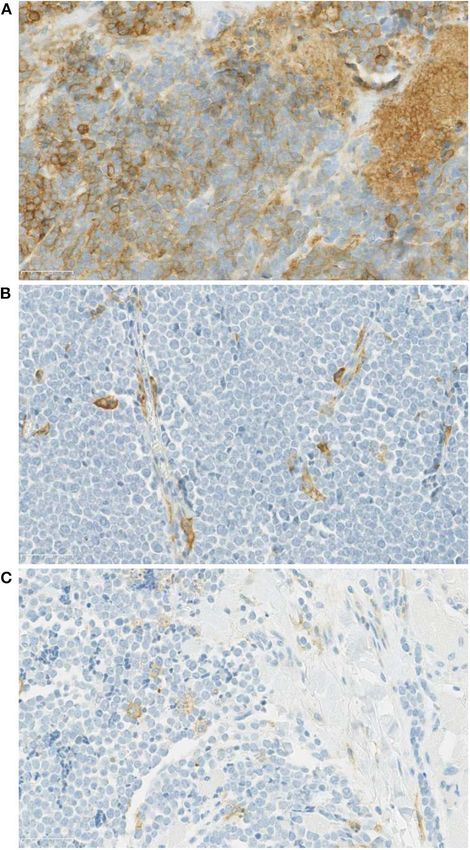
FIGURE 1 | (A) Merkel cell carcinoma case (40x) demonstrating typical morphology with tumor cells arranged in a diffuse fashion showing scant amphophilic or
eosinophilic cytoplasm and relatively uniform nuclei. The nuclei have finely dispersed chromatin without prominent nucleoli. (B) CK-20 staining pattern in patients with
Merkel cell carcinoma (cells are arranged in groups of three, with three horizontal cells representing triplicate samples from a single tumor). (C) Rabbit IgG isotype
immunohistochemistry (control). (D) PD-L1 staining in patients with Merkel cell carcinoma (cells are arranged in groups of three, with three horizontal cells representing
triplicate samples from a single tumor).
TABLE 2 | Immunohistochemical staining with anti-PD-L1 clone 73-10 in patients with Merkel cell carcinoma and intratumoral expression of PD-L1.
Subjects Tumor tissue (% cells) H-score Tumor tissue (% cells) membrane H-score Tumor infiltrating
cytoplasm cytoplasm membrane immune cells
(TIICs) (“+” or “-”)
Negative Weak Moderate Strong Sum (%) Negative Weak Moderate Strong Sum (%)
1 97 0 3 0 100 6 97 0 3 0 100 6 -
2 98 0 2 0 100 4 98 0 2 0 100 4 -
3 97 0 3 0 100 6 97 0 3 0 100 6 -
4 98 0 2 0 100 4 98 0 2 0 100 4 +
5 99 0 1 0 100 2 99 0 1 0 100 2 +
6* 98 0 2 0 100 4 98 0 2 0 100 4 +
7 99 0 1 0 100 2 99 0 1 0 100 2 +
8* 99 1 0 0 100 1 99 1 0 0 100 1 -
9∧ 99 0 1 0 100 2 99 0 1 0 100 2 +
10* 99 0 1 0 100 2 99 0 1 0 100 2 +
11 97 0 3 0 100 6 97 0 3 0 100 6 +
12 99 0 1 0 100 2 99 0 1 0 100 2 +
13 100 0 0 0 100 0 99 1 0 0 100 1 -
14 99 0 1 0 100 2 99 0 1 0 100 2 +
15 99 0 1 0 100 2 99 0 1 0 100 2 +
*Intratumoral microvessels staining for PD-L1.
∧ Nuclear staining of tumor cells for PD-L1.
Semi-quantitative scoring for PD-L1 utilized the H-score. The percentage of negative (0), weakly stained (1), moderately stained (2) and strongly stained (3) tumor cells was estimated.
Subsequently the H-score was calculated as follows: H-score = (weak)% × 1 + (moderate)% × 2 + (strong)% × 3. An H-score greater than zero was counted as positive PD-L1 staining.
Frontiers in Medicine | www.frontiersin.org 3 June 2020 | Volume 7 | Article 198Hanna et al. PD-L1 in Merkel Cell Carcinoma
TABLE 3 | Median survival depending on the PD-L1 expression and stage of
disease.
PD-L1+ PD-L1- Hazards 95% CI P-value
expression expression ratio
OS in PD-L1 64.0 months NR 1.26* 0.46–3.46 0.601
expression
months
Stages of the OS Between OS between 0.99* 0.41–2.39 0.986
MCC disease Stage 1 and 2 Stage 3 and 4
Expressed in 69.0 NR
months
NR, not reached; *HR changes with the change in the follow up time period; OS,
Overall survival.
staining was performed to exclude skin metastasis of small cell
lung cancer (Figure 1B). Rabbit isotypes were stained for PD-L1
to serve as controls (Figure 1C). A total of 67 out of 78 cases were
evaluable following immunohistochemical staining (cases were
excluded if tissue washed off or < 50 tumor cells were present).
PD-L1 staining in the tumor cells and in the surrounding
tumor infiltrating immune cells is shown in Figure 1D. Semi-
quantitative scoring for tumor cell PD-L1 using the H-score is
shown in Table 2. 15 of 67 (22.4%) patient tumor samples were
characterized as PD-L1 positive, with an H-score maximum of 6
for individual cases (range 1–6).
In 14 patients (20.9%), the tumor cells showed both
membrane-associated staining and cytoplasmic staining for PD-
L1 (Figure 2A). In several cases, prominent PD-L1 staining
of intratumoral microvessels was present (Figure 2B). Immune
infiltrates with PD-L1 positive cells of varying size and density
were present at the infiltrative margin of the tumor in 62
(92.5%) of 67 cases. By contrast, within the tumor specimens
we observed scattered PD-L1 positive immune cells but no
widespread or dense immune infiltrates (Figure 2C). Neither
membrane-associated nor cytoplasmic staining for PD-L1 by H-
score was correlated with the presence of infiltrating immune
cells (p-value = 0.17). Median overall survival was similar among
FIGURE 2 | (A) PD-L1 staining of a Merkel cell carcinoma (case 12-5684, 40x)
subgroups regardless of PD-L1 expression intensity (HR 1.26,
showing tumor cells with membrane associated as well as cytoplasmic PD-L1
staining. (B) A Merkel cell carcinoma sample with prominent PD-L1 staining of
95% CI: 0.46–3.45, p = 0.60) (Table 3, Figure 3) and regardless
intra-tumoral microvessels (40x). (C) Tumor cells lacking PD-L1 expression. By of disease site (Table 4).
contrast, within the tumor there are scattered PD-L1 positive immune cells
with weak membrane and cytoplasmic PD-L1 staining.
DISCUSSION
MCC represents an aggressive neuroendocrine carcinoma of
(26.9%) had stage II, 41 (52.6%) had stage III, and one (1.3%) the skin that is characterized by high rates of locoregional
had stage IV disease. The initial staging information for three recurrence and distant metastatic disease. While overall response
patients was unavailable. Median survival time was 40 (range: 5– rates of advanced disease to chemotherapy regimens are
113) months. Thirty-four patients (43.5%) had died at the point favorable, they are often of limited duration (17). The purpose
of analysis. of this study was to characterize tumoral PD-L1 expression
Tumor samples confirmed as MCC were composed of patterns observed in MCC tumors and to determine if
trabecular to insular or diffuse proliferations of tumor these expression patterns relate to outcomes in patients not
cells with scant amphophilic or eosinophilic cytoplasm and treated with immunotherapy. Notably, three landmark studies
relatively uniform nuclei sometimes exhibiting nuclear molding have recently demonstrated high response rates of MCC to
(Figure 1A). Additionally, CK-20 immunohistochemical checkpoint blockade therapy targeting PD-1 and PD-L1 in MCC.
Frontiers in Medicine | www.frontiersin.org 4 June 2020 | Volume 7 | Article 198Hanna et al. PD-L1 in Merkel Cell Carcinoma FIGURE 3 | (A) Overall survival (all evaluable patients) (B) Overall survival by PD-L1 expression (C) Overall survival by anatomic site. Understanding why MCC is so sensitive to PD-1 and PD-L1 In this study, membranous and/or cytoplasmic MCC tumor targeted therapy may bring insights to guide therapy in other cell staining for PD-L1 was detected in 22.4% of patients. We tumor types. found prominent PD-L1 staining associated with intratumoral Frontiers in Medicine | www.frontiersin.org 5 June 2020 | Volume 7 | Article 198
Hanna et al.                                                                                                                    PD-L1 in Merkel Cell Carcinoma

TABLE 4 | Median survival depending on site of disease.                                 breast cancer suggests that PD-L1 overexpression is associated
                                                                                        with lower OS (22), while data in non-small cell lung cancer
                            Median OS                95% CI        P-value (vs.
                            in months                             rest of cohort)
                                                                                        suggests PD-L1 is not a robust prognostic marker (23). PD-L1
                                                                                        expression is associated with longer disease-free survival in head
Upper extremity                  NR                   44-NR            0.43             and neck cutaneous squamous cell carcinomas (24). Indeed, our
Lower extremity                  131                  27-NR            0.29             conclusion that PD-L1 is not an independent prognostic marker
Head & neck                       69                  38-NR            0.93             in MCC is at odds with an earlier, smaller study in MCC, which
Other                             5                   5-NR             1.00             suggested that PD-L1 expression was associated with higher
                                                                                        OS (25).
CI, confidence interval; NR, not reached; OS, overall survival.
                                                                                            Recent work suggests that PD-L1 may serve as prognostic
                                                                                        marker when considered in multivariate analysis with other
microvessels, which raises the possibility that these structures                        immune-related markers. In non-small cell lung cancer, patients
may play a role in the immunologic defense of the tumor. In                             with both high PD-L1 expression and high CD8+ TIL density
addition, we noted PD-L1 positive immune cells at the infiltrative                      experienced particularly long OS, while patients with high PD-L1
margins of the tumor in 92.5% of cases. PD-L1 expression in the                         expression and low CD8+ TIL density experienced particularly
membrane and cytoplasm of tumor cells did not affect prognosis                          low OS (26). Further study in MCC may show that analysis
in this cohort that had not received immunotherapy agents.                              involving multiple immune components at once may hold
Given the relatively large sample size in an untreated population                       prognostic value for patients.
of MCC, this data may remain an important point of reference as                             There are several lines of evidence that suggest that MCC
a control group for future prospective studies.                                         is particularly dependent on immune evasion. For example,
   PD-L1 expression has been previously described on immune-                            the identification of MCPyV has led to several epidemiologic
infiltrating cells rather than on the surface of tumor cells and                        studies that demonstrate that the virus is widely prevalent and
was frequently overexpressed in peripheral tumor-infiltrating                           viral capsid proteins are readily recognized by the immune
lymphocytes (TILs). Similar observations have been noted in                             system (27). It has further been demonstrated that MCC tumor
melanoma, in which PD-L1 positive tumor cells are often                                 cells express the Large T (LT) and Small T (ST) antigens;
localized near TILs (18).                                                               a MCPyV protein constitutively expressed by virally infected
   Afanasiev and colleagues determined the presence of PD-                              tumor cells (28). Antibodies to ST serve a useful biomarker
L1 within MCC tumors and characterized CD8 mRNA                                         for following disease status in patients with virus-positive
expression to detect CD8+ lymphocytic infiltration (19). Biopsy                         MCC (29).
specimens from 69% of patients (9 of 13) were at least                                      A phase 2 study with 26 MCC patients with with stage IIIb
weakly positive for PD-L1 by immunohistochemistry. They                                 and IV disease received pembrolizumab as first-line treatment
demonstrated that high levels of PD-L1 tumoral staining                                 observed an overall response rate (ORR) of 56%. In this study,
correlated with CD8 lymphocytic infiltration (p < 0.05)—                                neither PD-L1 expression on tumor cells nor expression on
suggesting a high likelihood of inhibitory ligand matching                              infiltrating immune cells correlated significantly with clinical
in the tumor microenvironment. Dowlatshahi and colleagues                               response. Virus-positive tumors were three times more likely
similarly demonstrated that 50% of MCC T-cells expressed PD-                            to express PD-L1 compared with virus-negative tumors (71
1 within the tumoral microenvironment (20). However, they                               vs. 25%, p = 0.049). Intratumoral CD8 T-cell infiltration did
characterized CD69 and CD25 expression patterns on the surface                          not correlate significantly with clinical response or with viral
of tumor-specific T-cells, which are markers of activation. They                        status (13).
found severely decreased levels of these markers, suggesting                                The PD-L1 inhibitor avelumab was tested in a phase 2
suppression of T-cell activation within the MCC tumor                                   study which included 88 patients with stage IV chemotherapy-
microenvironment—likely reflecting T-cell exhaustion (21).                              refractory, histologically confirmed metastatic MCC. The initial
   The presence of MCPyV-specific T-cells correlates with MCC                           report from this study led to FDA approval in MCC in 2016. In a
disease burden—such that MCPyV-specific T-cell increased                                1-year update with median follow-up at 16.4 months and analysis
with growing tumor burden and coexpression of immune                                    ongoing, an ORR of 33.0% was observed. In 29 patients who
checkpoint receptors, namely PD-1, was high within the MCC                              had a response, 10 patients had complete response. The median
tumor microenvironment (19). PD-1 was expressed in 71% of                               duration of response had not been reached (range 2.8–23.3+
MCC tumor-infiltrating lymphocytes and 96% of circulating                               months) (30). Amongst 58 patients who were positive for PD-L1
MCyV-specific T-cells. The inhibitory receptor Tim-3 was                                in the original publication, objective response was achieved in 20
also coexpressed more commonly in these cell types. CTLA-4                              (34.5%). In the recent update, post-hoc subgroup analyses were
expression was generally low. However, studies assessing PD-1                           reported. The objective response rate among those who were
and PD-L1 expression by IHC were complicated by differences in                          PD-L1 positive remained stable at 34.5%. Six-month duration of
staining properties between antibodies and a lack of standardized                       response (DOR) was estimated at 100% and 6-month PFS rate at
criteria for analysis.                                                                  43%, thus suggesting responses had occurred later in treatment.
   Across tumors, including those that have been studied in                             In our analysis, which included only patients prior to the advent
much larger patient populations compared with MCC, the                                  of immunotherapy treatment, there was no difference in overall
proposed prognostic value of PD-L1 varies widely. Data in                               survival regardless of PD-L1 positivity (p = 0.60).

Frontiers in Medicine | www.frontiersin.org                                         6                                         June 2020 | Volume 7 | Article 198
Hanna et al. PD-L1 in Merkel Cell Carcinoma
DATA AVAILABILITY STATEMENT AUTHOR CONTRIBUTIONS
The raw data supporting the conclusions of this GH conceived of the presented idea. Members of the Merck
article will be made available by the authors, without KGaA/EMD Serono team (HG, BB, and CI) performed
undue reservation. laboratory testing for PD-L1. GH analyzed the data with the
support of AT and VV. GH wrote the manuscript with support
from AK. All authors discussed the results and contributed to the
ETHICS STATEMENT final manuscript. JL supervised the project with support from GR,
NL, MT, and JD.
This study was carried out in accordance with the
recommendations of DOH principles, Dana-Farber Institutional FUNDING
Review Board. The protocol was approved by the Dana-
Farber Institutional Review Board. All subjects gave written This work was supported in part by the US Public Health Service
informed consent in accordance with the Declaration R35CA232128, R01CA63113, R01CA173023, and P01CA203655
of Helsinki. to JD.
REFERENCES Annual Meeting 2017. Washington, DC; Philadelphia, PA: AACR; Cancer Res.
(2017). 77(13 Suppl):CT074. doi: 10.1158/1538–7445.AM2017-CT074
1. Foschini MP, Eusebi V. Divergent differentiation in endocrine and 15. Kaufman HL, Russell J, Hamid O, Bhatia S, Terheyden P, D’Angelo SP, et al.
nonendocrine tumors of the skin. Semin Diagn Pathol. (2000) 17:162. Avelumab in patients with chemotherapy-refractory metastatic Merkel cell
2. Suntharalingam M, Rudoltz MS, Mendenhall WM, Parsons JT, Stringer SP, carcinoma: a multicentre, single-group, open-label, phase 2 trial. Lancet Oncol.
Million RR. Radiotherapy for Merkel cell carcinoma of the skin of the head (2016) 17:1374–85. doi: 10.1016/S1470–2045(16)30364–3
and neck. Head Neck. (1995) 17:96–101. doi: 10.1002/hed.2880170204 16. Thike AA, Chng MJ, Fook-Chong S, Tan PH. Immunohistochemical
3. Janice Ma E, Jerry D. Merkel cell carcinoma in immunosuppressed patients. expression of hormone receptors in invasive breast carcinoma: correlation
Cancers. (2014) 6:1328–50. doi: 10.3390/cancers6031328 of results of H-score with pathological parameters. Pathology. (2001) 33:21–
4. Leroux-Kozal V, Lévêque N, Brodard V, Lesage C, Dudez O, Makeieff M, et 5. doi: 10.1080/00313020123290
al. Merkel cell carcinoma: histopathologic and prognostic features according 17. Crown J, Lipzstein R, Cohen S, Goldsmith M, Wisch N, Paciucci PA, et
to the immunohistochemical expression of Merkel cell polyomavirus al. Chemotherapy of metastatic Merkel cell cancer. Cancer Investig. (1991)
large T antigen correlated with viral load. Hum Pathol. (2015) 46:443– 9:129–32. doi: 10.3109/07357909109044222
53. doi: 10.1016/j.humpath.2014.12.001 18. Taube JM, Anders RA, Young GD, Xu H, Sharma R, McMiller TL, et al.
5. Feng H, Shuda M, Chang Y, Moore PS. Clonal integration of a Colocalization of inflammatory response with B7-h1 expression in human
polyomavirus in human Merkel cell carcinoma. Science. (2008) 319:1096– melanocytic lesions supports an adaptive resistance mechanism of immune
100. doi: 10.1126/science.1152586 escape. Sci Transl Med. (2012) 4:127ra37. doi: 10.1126/scitranslmed.3003689
6. Vortmeyer AO, Merino MJ, Böni R, Liotta LA, Cavazzana A, Zhuang Z. 19. Afanasiev OK, Yelistratova L, Miller N, Nagase K, Paulson K,
Genetic changes associated with primary Merkel cell carcinoma. Am J Clin Iyer JG, et al. Merkel polyomavirus-specific T cells fluctuate with
Pathol. (1998) 109:565–70. doi: 10.1093/ajcp/109.5.565 merkel cell carcinoma burden and express therapeutically targetable
7. Tai PT, Yu E, Tonita J, Gilchrist J. Merkel cell carcinoma of the skin. J Cutan PD-1 and Tim-3 exhaustion markers. Clin Cancer Res. (2013)
Med Surg. (2000) 4:186–95. doi: 10.1177/120347540000400403 19:5351–60. doi: 10.1158/1078–0432.CCR-13–0035
8. Gillenwater AM, Hessel AC, Morrison WH, Burgess M, Silva EG, Roberts D, 20. Dowlatshahi M, Huang V, Gehad AE, Jiang Y, Calarese A, Teague JE, et al.
et al. Merkel cell carcinoma of the head and neck: effect of surgical excision Tumor-specific T cells in human Merkel cell carcinomas: a possible role for
and radiation on recurrence and survival. Arch Otolaryngol Head Neck Surg. Tregs and T-cell exhaustion in reducing T-cell responses. J Invest Dermatol.
(2001) 127:149–54. doi: 10.1001/archotol.127.2.149 (2013) 133:1879–89. doi: 10.1038/jid.2013.75
9. Müller A, Keus R, Neumann N, Lammering G, Schnabel T. Management of 21. Jin HT, Ahmed R, Okazaki T. Role of PD-1 in regulating T-cell immunity.
Merkel cell carcinoma: case series of 36 patients. Oncol Rep. (2003) 10:577–85. Curr Top Microbiol Immunol. (2011) 350:17–37 doi: 10.1007/82_2010_116
10. Chen MM, Roman SA, Sosa JA, Judson BL. The role of adjuvant therapy 22. Zhang M, Sun H, Zhao S, Wang Y, Pu H, Wang Y, et al. Expression of
in the management of head and neck merkel cell carcinoma: an analysis PD-L1 and prognosis in breast cancer: a meta-analysis. Oncotarget. (2017)
of 4815 patients. JAMA Otolaryngol Head Neck Surg. (2015) 141:137– 8:31347–54. doi: 10.18632/oncotarget.15532
41. doi: 10.1001/jamaoto.2014.3052 23. Lin G, Fan X, Zhu W, Huang C, Zhuang W, Xu H, et al. Prognostic
11. Cirillo F, Buononato M, Lima G, Cafaro I, Alquati P. Clinical experience significance of PD-L1 expression and tumor infiltrating lymphocyte in
on eight cases of Merkel cell carcinoma. Tumorionolgy. (2003) 89:146– surgically resectable non-small cell lung cancer. Oncotarget. (2017) 8:83986–
51. doi: 10.1177/030089160308900208 94. doi: 10.18632/oncotarget.20233
12. Paulson KG, Carter JJ, Johnson LG, Cahill KW, Iyer JG, Schrama D, et 24. Roper E, Lum T, Palme CE, Ashford B, Ch’ng S, Ranson M, et al.
al. Antibodies to merkel cell polyomavirus T antigen oncoproteins reflect PD-L1 expression predicts longer disease free survival in high risk head
tumor burden in merkel cell carcinoma patients. Cancer Res. (2010) 70:8388– and neck cutaneous squamous cell carcinoma. Pathology. (2017) 49:499–
97. doi: 10.1158/0008–5472.CAN-10–2128 505. doi: 10.1016/j.pathol.2017.04.004
13. Nghiem PT, Bhatia S, Lipson EJ, Kudchadkar RR, Miller NJ, Annamalai L, et 25. Lipson EJ, Vincent JG, Loyo M, Kagohara LT, Luber BS, Wang H, et al. PD-L1
al. PD-1 blockade with pembrolizumab in advanced merkel-cell carcinoma. N expression in the Merkel cell carcinoma microenvironment: association with
Engl J Med. (2016) 374:2542–52. doi: 10.1056/NEJMoa1603702 inflammation, Merkel cell polyomavirus and overall survival. Cancer Immunol
14. Topalian SL, Bhatia S, Hollebecque A, Awada A, De Boer JP, Kudchadkar Res. (2013) 1:54–63. doi: 10.1158/2326–6066.CIR-13–0034
RR, et al. Non-comparative, open-label, multiple cohort, phase 1/2 study 26. Yang H, Shi J, Lin D, Li X, Zhao C, Wang Q, et al. Prognostic value of
to evaluate nivolumab (NIVO) in patients with virus-associated tumors PD-L1 expression in combination with CD8+ TILs density in patients with
(CheckMate 358): Efficacy and safety in Merkel cell carcinoma (MCC) surgically resected non-small cell lung cancer. Cancer Med. (2018) 7:32–
[abstract]. In: Proceedings of the American Association for Cancer Research 45. doi: 10.1002/cam4.1243
Frontiers in Medicine | www.frontiersin.org 7 June 2020 | Volume 7 | Article 198Hanna et al. PD-L1 in Merkel Cell Carcinoma
27. Tolstov YL, Pastrana DV, Feng H, Becker JC, Jenkins FJ, Moschos Conflict of Interest: HG, CI, and BB are employees of Merck KGaA, a co-
S, et al. Human Merkel cell polyomavirus infection II. MCV is a developer of the PD-L1 inhibitor avelumab. JD received research funding from
common human infection that can be detected by conformational capsid Constellation Pharmaceuticals. JD has served as a consultant to Merck & Co. and
epitope immunoassays. Int J Cancer. (2009) 125:1250–6. doi: 10.1002/ij EMD Serono.
c.24509
28. Houben R, Shuda M, Weinkam R, Schrama D, Feng H, Chang Y, et al. Merkel The remaining authors declare that the research was conducted in the absence of
cell polyomavirus-infected Merkel cell carcinoma cells require expression of any commercial or financial relationships that could be construed as a potential
viral T antigens. J Virol. (2010) 84:7064–72. doi: 10.1128/JVI.02400–09 conflict of interest.
29. Paulson KG, Lewis CW, Redman MW, Simonson WT, Lisberg A, Ritter D,
et al. Viral oncoprotein antibodies as a marker for recurrence of Merkel Copyright © 2020 Hanna, Kacew, Tanguturi, Grote, Vergara, Brunkhorst,
cell carcinoma: a prospective validation study. Cancer. (2017) 123:1464– Rabinowits, Thakuria, LeBoeuf, Ihling, DeCaprio and Lorch. This is an open-access
74. doi: 10.1002/cncr.30475 article distributed under the terms of the Creative Commons Attribution License (CC
30. Kaufman HL, Russell JS, Hamid O, Bhatia S, Terheyden P, D’Angelo BY). The use, distribution or reproduction in other forums is permitted, provided
SP, et al. Updated efficacy of avelumab in patients with previously the original author(s) and the copyright owner(s) are credited and that the original
treated metastatic Merkel cell carcinoma after ≥1 year of follow-up: publication in this journal is cited, in accordance with accepted academic practice.
JAVELIN Merkel 200, a phase 2 clinical trial. J Immunother Cancer. (2018) No use, distribution or reproduction is permitted which does not comply with these
6:7. doi: 10.1186/s40425–017-0310-x terms.
Frontiers in Medicine | www.frontiersin.org 8 June 2020 | Volume 7 | Article 198You can also read