Detection of brain tumor using K-nearest neighbor (KNN) based classification model and self-organizing map (SOM) algorithm - ijitee
←
→
Page content transcription
If your browser does not render page correctly, please read the page content below
International Journal of Innovative Technology and Exploring Engineering (IJITEE)
ISSN: 2278-3075, Volume-8 Issue-8 June, 2019
Detection of brain tumor using K-nearest
neighbor (KNN) based classification model and
self-organizing map (SOM) algorithm
S. G. Raja, K. Nirmala
Abstract: Knowledge discovery is also known as Data mining in
databases, in recent years that technique plays a major role in One of the steps in KDD is data mining which contains
research area. Data mining in healthcare domain has noteworthy analysis of information and computation of previously
usage in real world. The mining method can enable the healthcare
field for the enhancement of institutionalization of its gathered information. Cluster analysis is an essential
administrations and become quicker with best in class approach for database mining. It is either used as a unique tool
technologies. Innovation utilization isn't restricted to basic to understand the distribution of an information index, e.g. to
leadership in undertakings, yet spread to different social statuses focus as well as support, data analysis and data preparation, or
in all fields. In this paper a novel approach for the detection of as a pre-handling adventure for various manipulations in the
brain tumor is proposed. The novel approach uses the
classification technique of K-nearest neighbor (KNN) and for
recognized groups.[2]
ignoring the error of the dataset image SOM (self-organizing Data mining is a major approach to recognize the real, novel,
map) algorithm has been used. Discrete wavelet transform (DWT) potential significances in addition to sensible design in
is used for transforming input image data set, in which RGB color information. Data mining, the separation of the concealed
of input data image has been converted into gray scale. Then it perceptive data from tremendous database, is an
has been classified using KNN after that the error avoiding
extraordinary new advancement with potential to analyze
algorithm has been carried out. This will help to differentiate
tumor cells and the normal cells. The presence of tumor in brain crucial data in the data distribution center. The term data
image is detected using parametric analysis by simulation. mining is referred to discovery of relevant and valued data
from database. The mining methods can be applied on
Keyword: data mining, medical data mining, K-nearest neighbor cognitive science to study human brain. Greater proportions
(KNN), SOM (self-organizing map) algorithm, DWT (discrete of information are accumulated and stored in databases
wavelets transform) growing the necessity for profitability and convincing
investigation techniques to make use of the data enclosed.
I. INTRODUCTION
The most necessary action for information investigation is
Recent decade has encountered a revolution in data group investigation. It helps the customer to know about the
accessibility and transfer of it through computer network. In construction of data in database. Therefore, in the recent
the same spirit, many organizations of different industries years the progress of enhanced group investigation has got a
have commenced to gather data relevant to their respective great deal of attention. [2]
field. The database technologists are in search of efficient In our human body brain plays a multipart organ. In human
ways for storing, recovering and manipulating data. In order brain more than 100 billion nerves are located and
to learn and acquire knowledge from the data the machine overlapped. The identification of tumor in the brain is a
learning developing method is essential. In strategic planning difficult task because of its complex structure. An
for research and development that the data is plays a vital role irrepressible development of cells in the brain is known as
in knowledge Discovery database (KDD) and data mining tumor. Malignant tumor and benign tumor are two kinds of
(DM).Occasionally, the data can be plays an important key brain tumor, these are smaller in size. Depends upon their
planning for innovative work which is stated as Data Mining growth pattern tumor are recognized. Benign tumor develops
(DM) and Knowledge Discovery in Databases (KDD). gradually within well-defined boundaries. This type of tumor
Data mining is an important area of research relevant to can be completely detached by surgical procedure, such that
problem of discovering vague knowledge from real-time it’s not spread into any parts of brain, the spinal cord, or any
databases [1]. In enhancement of database technology, the other portion of the human body. The next type of tumor is
input as well as output information of respective field has to known as malignant tumor, is rapidly-growing and affects the
be stored instantaneously. various cells of the healthy brain. In spinal cord this type of
tumor is spread. It causes very harmful effects and may
Revised Manuscript Received on June 05, 2019 remain unprocessed. [3]
S. G. Raja, Research Scholar, Department of Computer Science , Vels
University, Chennai, India
Brain tumors can be either benign (non-cancerous) or
Dr. K. Nirmala, Research Supervisor, Department of Computer malignant (cancerous).
Science , Quaid-e-millath college for women, Chennai, India Gliomas and malignant
meningiomas, there are two
Published By:
Blue Eyes Intelligence Engineering
Retrieval Number F3816048619/19©BEIESP 787 & Sciences Publication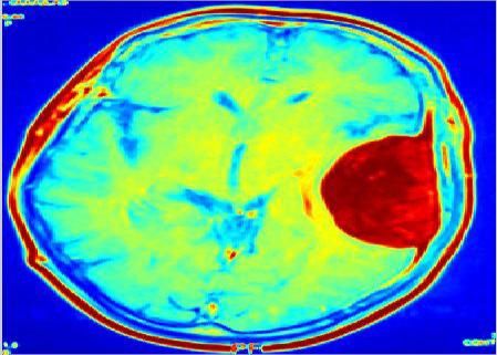
Detection of brain tumor using K-nearest neighbor (KNN) based classification model and self-organizing map (SOM)
                                                    algorithm

primary types of brain tumor or brain cancer. In malignant         quality of services. [6] Ming-Ni Wu1 Chia-Chen Lin2
tumor, Gliomas is a familiar one. It includes different kinds of   Chin-Chen Chang, In K- means clustering technique it
cells from which they existed: oligodendrogliomas                  utilized in color-based segmentation approach offers in order
,astrocytomas and ependymomas. Meningiomas started from            to recognized the tumor substance in magnetic resonance
the meninges, which surround the external part of the spinal       (MR).The vital thought of the proposed algorithm is to
cord and brain, which are tissues. The mainstream of
                                                                   change the grey dimension MR image into a shading space
meningiomas is benign and can be cured by surgery. It has
                                                                   image what's more it likewise achieves division for MR image
numeral of brain tumors, with medulloblastomas, which
develop from the primitive stem cells of the cerebellum and        by utilizing histogram-clustering and K-implies clustering.
are most often seen in children. Primary and secondary             [7]
tumors are the two kinds of malignant tumors. Auxiliary brain      Saumya Chauhan[1],Aayushi More[2], RitumbhraUikey[3],
tumors as a rule begin in a few sections of the body and next      Pooja Malviya[4], Asmita Moghe, 2017,suggests a median
metastasize to brain. The reasons for brain tumors incorporate     filter for pre-processing MRI brain images. Edge detection
head injuries, genetic syndromes, immune suppression,              and the color based segmentation are being performed for
delayed introduction to ionizing radiation, electromagnetic        segregation of lesion from image. The extraction schemes,
fields, cell phones, or synthetic substances like formaldehyde     such as gray level co-event lattice and histogram of situated
and vinyl chloride. Manifestations of brain tumors
                                                                   gradients are utilized to extricate the different highlights of
incorporate determined headache, nausea and heaving, visual
                                                                   the pictures. All the removed highlights are put away in a
perception, hearing as well as discourse issues, strolling as
well as parity troubles, identity changes, memory lapses,          debatable database. To this database, Instance based
issues with cognizance and focus, and seizures. Meningioma         K-Nearest utilizing Log and Gaussian weight Kernels
is an assortment of tumor that creates from the meninges. The      (IBKLG) classifier has been connected utilizing WEKA 3.9
dura mater, arachnoid and Pia mater are the layers of              instrument, to classify the tumor into benign or malignant. [8].
meninges. Meningiomas are considered as benign tumors,             Samee Azad, Shaikh Anowarul Fattah, Naqib Sad Pathan
with the 10% being atypical or malignant. Benign                   2016 proposes automatic method based on voxel statistics, for
meningioma spreads slowly. A meningioma grows to a larger          division of region of interest(ROI. The region of interest is a
size before causing any symptoms. Other meningiomas                region encompassing the cerebrum tumor and its
develop very fast, and will have sudden growth spurts. There
                                                                   neighborhood. In the proposed technique, first conceivable
are no proper methods for manipulating the growth for a
                                                                   applicant determination is performed using power qualities of
meningioma. Glioma is a tumor that arises from glial cells
within the brain or spine. [4].                                    tumor region in the T1 and FLAIR pictures of MRI
                                                                   documents. In the following stage, a cubic shaped 3D mean
               II. LITERATURE REVIEW                               separating task is connected all in all volumetric data to get
                                                                   sifted volume are some irregular power conduct is relied upon
RuomingJin, Ge Yang, and Gagan Agrawal 2005, preformed
                                                                   to be dispensed with. At last, from the subsequent 3D FLAIR
an inside and out evaluation of arrangement of techniques,
                                                                   data, region of interest is removed dependent on combined
These techniques are investigated utilizing data mining
                                                                   dispersion capacity of power. It is discovered that the isolated
algorithms, for example, fp-tree-based association mining,
                                                                   region of interest offers real decrease of the general MRI
and apriori, k-means clustering, k-nearest neighbor classifier,
                                                                   volume without losing tumor data. The proposed ROI
and decision tree construction. The investigation completed
                                                                   extraction plot is attempted on 20 real high survey tumor
in this paper accomplishes the accompanying: 1) Of the
                                                                   cases gained from a comprehensively used database and an
different techniques for parallelization of data mining
                                                                   exceptionally tasteful execution is gotten as far as
algorithm, three techniques to be specific, full replication,
                                                                   segmentation precision, generally speaking volume decrease
optimized full locking, and cache sensitive locking can do
                                                                   and computational time.[9] M. Murugesan, Dr. (Mrs.)
very much contrasted with different strategies relying on
                                                                   R.Sukanesh2009 A programmed structure for expert
machine and dataset parameters 2) The overhead of the
                                                                   recognition of brain tumors in electroencephalograms (EEG)
interface is inside 10 percent in practically all cases 3) Good
                                                                   signals utilizing artificial neural networks (ANNs). The ANN
parallel effectiveness is accomplished for every one of the
                                                                   utilized in the projected structure is feed forward back spread
four data mining algorithms. 4) For phenomenal execution,
                                                                   neural system. Usually, the EEG signals will undoubtedly
the blend of different strategy swings to be basic if there
                                                                   contain various historical facts about subject and devices
should arise an occurrence of decision tree construction
                                                                   impedances from the start with imperative data in regards to
algorithm.[5] B.V.Kiranmayee, Dr. T.V.Rajinikanth,
                                                                   variations from the norm and brain action (reactions to
S.Nagini, 2016 presented an approach consisting of training
                                                                   specific stimuli). At first, adaptable separating is associated
and testing stage to detect the brain tumor. The functionality
                                                                   with confine the ancient rarities available in the EEG signal.
of proposed algorithm have been validated by constructing a
                                                                   In order to use spectral
prototype application. The results of developed prototype
                                                                   estimation the common
show that it can be combined to choice emotionally
                                                                   features in the EEG signal
supportive networks in medicinal services area for enhancing

                                                                         Published By:
                                                                         Blue Eyes Intelligence Engineering
   Retrieval Number F3816048619/19©BEIESP                      788       & Sciences Publication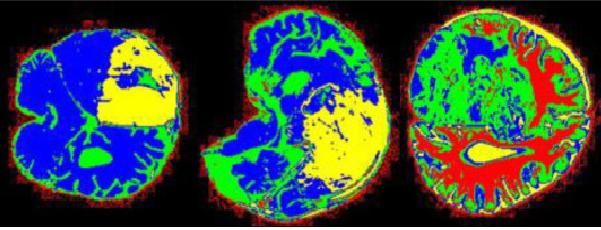
International Journal of Innovative Technology and Exploring Engineering (IJITEE)
                                                                          ISSN: 2278-3075, Volume-8 Issue-8 June, 2019

are segregated. Definitely, the fast fourier transform (FFT)                    III. RESEARCH METHODOLOGY
analysis is carried out to extract the signal highlights covered   In this paper we suggest the novel move toward for the
in a varied group of noise. In this manner the perfect EEG         detection of brain tumor. Here we use the classification
information obtained is utilized as preparing contribution to      technique of K-nearest neighbor (KNN) and for ignoring the
the feed forward back engendering neural network. At the           error of the dataset image SOM (self-organizing map)
point when this prepared information is sustained with a test      algorithm has been used. The image of input data set is
EEG signal, it adequately distinguishes the nearness of brain      transformed from RGB color into gray scale by DWT.
tumor in the EEG signal. [10]                                      Then it is classified using KNN after that the error avoiding
SushmaV. Telrandhe, Divya Chikate, Pooja Banode,2015,              algorithm has been carried out. This will helps to differentiate
explains an approach dependent on back propagations neural         tumor cells and the normal cells. Simulation results show the
system procedure for the order of MRI picture. The approach        parametric analysis of the brain image shows if there is a
is designed utilizing image enrichment, segmentation,              presence of tumor. The architecture for proposed system is as
registration, and characteristic identification and segregation    follows:
techniques.
The morphological operations and thresholding process are
considered during segmentation method. The training and
experiment of MRI image is analyzed by back propagation
neural network technique to recognize the presence of tumor.
[11]
Neha Rani, Sharda Vashisth 2016 presents the work, in which
to process the MRI images morphological and thresholding
techniques are used. Feed-forward back propagation neural
network is utilized to organize the presentation of tumor
section of the image. By using the proposed method, high
accuracy and less iteration for detection of tumor are achieved
which in turn lessens the consumption time. [12]                              Fig.1 Block Diagram for proposed system.
Viktor Losing, Barbara Hammer and HeikoWersing2016
suggest the SELF ADJUSTING MEMORY (SAM) display                    A. Image conversion from RGB to gray scale feature
for the K NEAREST NEIGHBOR (KNN) algorithm since                   using DWT
KNN builds up a confirmed classifier inside the                    The gray level features are of two types i.e. edge mapping and
non-stationary information stream setting. SAM - KNN can           energy. An edge mapping is a process in which fundamental
manage unrelated idea point, for example, utilizing                implementation in image process, gives the contour of the
organically motivated memory models and their coordination.        object in an image. The result of edge mapping can trace the
It very well may be effectively connected by and by since an       boundary of the object as well as the curve surface. Edge
enhancement of the Meta parameters isn't fundamental. The          mapping is used for image segmentation. An energy level find
fundamental thought is to build committed models for the           by discrete wavelet change. The wavelet is amazing numerical
present and previous ideas and apply them as indicated by the      instrument for highlight extraction of MRI image. The
requests of the given circumstance.                                wavelet transform is used to give the information about the
The different benchmarks, including fake streams with known        signal both in time domain and frequency. DWT (discrete
drift includes just as genuine world datasets are generally        wavelet transforms) is applied on image to get energy level.
                                                                   H= undefined if max=min
assessed. Along these lines, we correctly include new
benchmarks for exact assessment on different kinds of drift.       H= 60

The very focused results got from all examinations stress the      H= 60
robustness of SAM-kNN just as its capacity to oversee
heterogeneous drift idea. [13] WANG Huai-bin, YANG                 H= 60
Hong-liang1,2XU Zhi-jian1,2 YUAN Zheng 2010, focus on
                                                                   H= 60
improving intrusion detection rate and on SOM to give the
accurate clustering results. Hence it Presents a new algorithm,
in which SOM is used to gain generally clusters and focus of       S=

clusters, at that point, utilizing K-Means to refine the
clustering in the SOM stage. The proposed algorithm is tested      V=MAX
using KDD CUP-99 dataset. The test results illustrates that
the proposed algorithm can achieve clustering accuracy and         The following steps are
good stability of efficiency. [14]                                 followed for conversing,

                                                                           Published By:
                                                                           Blue Eyes Intelligence Engineering
    Retrieval Number F3816048619/19©BEIESP                     789         & Sciences Publication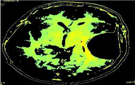
Detection of brain tumor using K-nearest neighbor (KNN) based classification model and self-organizing map (SOM)
algorithm
RGB color of image to gray scale feature using DWT IV. PERFORMANCE ANALYSIS
1. Initially RGB image is changed into YCbCr, follow In this section we discuss the results obtained using
by segregation of the Y, Cb and Cr mechanism. simulation. The performance analysis explains that the images
2. Discrete Wavelet transformation is functional in the Y of RGB color are transformed to gray scale using DWT and
constituent, which results in 7 uneven substitute-images. the parametric analysis of K-NN classification model and the
3. The components Cr and Cb are scaled at 25% of error rejection using SOM.
preliminary dimension, to extract Cb + as well as Cb- and
similarly Cr+ as well as Cr- images. Optimistic/negative
values of pixels are derivative from Cb and Cr components
and others are equaled with 0.
4. The quarter from right base of left best sub-image is
replaced with Cb-, the correct best sub-image with Cr+, the
sub-image from left-base with Cb+ and sub-image from
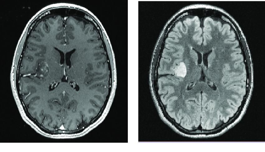
right-base with Cr. Fig.2 Brain Image dataset
5. DWT change is connected to the subsequent picture, to get
the favored gray scale picture.
B. K-nearest neighbor Classification model
KNN is a simple classification method which works well for
real time application. The training procedure is very easy and
it sample includes class labels and set of tuples interrelated
with that. This algorithm works for random number of
Fig.3 affected part of brain
module. Distance function is used by KNN classification
model for mapping the samples with classes.
In order to calculate distance among the assumed test
illustration X with that existing samples y1,y2…yk, the
Classification process of KNN is used. The nearest neighbor
around the test instance are identified and depends on the
selection of neighbors, the majority neighborhood lecture is
allotted to the test samples. The distance function is applied
between the samples using Euclidean method or Manhattan
method or Minkowski method. These methods are employed
when the values are continuous. Depends on number of Fig.4 RGB colored Brain image
neighbor that the sample X probability is assigning. The
probability of assigning a sample X to that of a class C is
based on the number of neighbors considered, denoted as K.
Probability of X to a class C =
C. Self-organizing map for error rejection:
SOM algorithm undergoes the following stages to reject
errors: Fig.5 Gray scale image
1. actualization– Introduce irregular qualities for the
underlying weight vectors wj.
2. Selection– Segregate an example preparing input vector x
from the information space.
3. Identical– Compare the weight vector closest to that of the
input vector and find the winning neuron I(x).that is the least
value of dji.xi.wji,
4. Modernizing–Apply the weight refresh
equation
whereTj, I(x)(t) is a Gaussian neighborhood and h(t) is the Fig.6 KNN classified image
education rate.
5. Continuance– Repeat Step2 until the feature map stops
varying.
Published By:
Blue Eyes Intelligence Engineering
Retrieval Number F3816048619/19©BEIESP 790 & Sciences Publication
International Journal of Innovative Technology and Exploring Engineering (IJITEE)
                                                                          ISSN: 2278-3075, Volume-8 Issue-8 June, 2019

                                                                 out to differentiate tumor cells and the normal cells. From the
                                                                 experimental results it is clear that the MRI brain image has
                                                                 been classified using K-NN model and error rejected using
                                                                 SOM technique to detect the tumor in the brain image.

                                                                 REFERENCES
                                                                 1.    Dr. Rajni Jain, “Introduction to Data Mining Techniques”.
   Fig.7 SOM error rejections in detected brain image            2.    Shivangi Bhardwaj, May 2017, “Data Mining Clustering Techniques –
                                                                       A Review”, IJCSMC, Vol. 6, Issue. 5, pg.183 – 186.
                                                                 3.    Shubhangi S. Veer (Handore)1, Pradeep M. Patil, | Dec-2015 ,“BRAIN
                                                                       TUMOR              CLASSIFICATION          USING         ARTIFICIAL
                                                                       NEURALNETWORK ON MRI IMAGES”, IJRET: International
                                                                       Journal of Research in Engineering and Technology, Volume: 04 Issue:
                                                                       12.
                                                                 4.    C.Ramalakshmi† and A.JayaChandran,May 2014, “Automatic Brain
                                                                       Tumor Detection in MR Images Using Neural Network Based
                                                                       Classification”, IJCSNS International Journal of Computer Science and
                                                                       Network Security, VOL.14 No.5.
                                                                 5.    RuomingJin, Ge Yang, and Gagan Agrawal, Jan 2005, “Shared Memory
                                                                       Parallelization of Data Mining Algorithms: Techniques, Programming
                                                                       Interface, and Performance”, IEEE TRANSACTIONS ON
                                                                       KNOWLEDGE AND DATA ENGINEERING, VOL. 17, NO. 1.
                                                                 6.    B.V.Kiranmayee, Dr. T.V.Rajinikanth, S.Nagini,2016, “A Novel Data
                                                                       Mining        Approach       for     Brain      Tumor      Detection”,
                                                                       978-1-5090-5256-1/16/$31.00_c IEEE.
                                                                 7.    Ming-Ni Wu1 Chia-Chen Lin2 Chin-Chen Chang, “Brain Tumor
       Fig. 8 SOM map for brain tumor detection                        Detection Using Color-Based K-Means Clustering Segmentation”
                                                                 8.    8.Saumya Chauhan[1], Aayushi More[2], RitumbhraUikey[3], Pooja
Fig.2 shows the brain image dataset, Fig.3 shows the affected          Malviya[4], AsmitaMoghe,2017 “Brain Tumor Detection and
                                                                       Classification in MRI Images using Image and Data Mining”, ISBN
part of brain without noise, Fig.4 shows the RGB colored               978-1-5090-4760-4/17/$31.00©2017 IEEE.
brain image, Fig. 5 shows the gray scale image, Fig.6 shows      9.    9.Samee Azad, Shaikh Anowarul Fattah, Naqib Sad Pathan, 2016 “An
the K-NN classified image, Fig.7 SOM error rejections in               Efficient Scheme for Detecting Region of Interest Encompassing the
                                                                       Brain Tumor from 3D MRI Data Based on Voxel Statistics”, 2016
tumor detected brain image, and Fig.8 shows the SOM map                IEEE International WIE Conference on Electrical and Computer
for brain tumor detection.                                             Engineering (WIECON-ECE).
                                                                 10.   M. Murugesan, Dr. (Mrs.) R.Sukanesh, 2009, “Automated Detection
                                                                       of Brain Tumor in EEG Signals Using Artificial Neural Networks”,
Table-1 parametric analysis of K-NN and error rejection                2009 International Conference on Advances in Computing, Control,
                                                                       and Telecommunication Technologies.
                        using SOM                                11.   11.Sushma V. Telrandhe, 2Divya Chikate,May-2015 “AUTOMATED
                                                                       BRAIN TUMOR DETECTION USING BACK PROPAGATION
                                                                       NEURAL NETWORK”, International Journal of Soft Computing and
                                          Rejection                    Artificial Intelligence, ISSN: 2321-404X, Volume-3, Issue-1.
                             K-NN         of error               12.   12.Neha Rani, Sharda Vashisth, July 2016“Brain Tumor Detection and
                         classification    using                       Classification      with    Feed    Forward      Back-Prop    Neural
         Parameters          model         SOM                         Network”,International Journal of Computer Applications (0975 –
                                                                       8887) Volume 146 – No.12.
          Accuracy            88.6         0.056                 13.   13.Viktor Losing, Barbara Hammerand HeikoWersing, 2016,“KNN
            (%)                                                        Classifier with Self Adjusting Memory for Heterogeneous Concept
          Sensitivity        92.3            0.067                     Drift”, 2016 IEEE 16th International Conference on Data Mining.
                                                                 14.   WANG Huai-bin, YANG Hong-liang1,2 XU Zhi-jian1,2 YUAN
             (%)                                                       Zheng, 2010, “A clustering algorithm use SOM and K-Means in
          Specificity        78.5            0.089                     Intrusion Detection”, 2010 International Conference on E-Business
                                                                       and E-Government.
             (%)
The table-1 represents the analysis of K-NN classifier and
error rejecting algorithm using SOM of parameters such as
accuracy, sensitivity and specificity.

                    V. CONCLUSION
In this paper suggests the novel method for the finding of
brain tumor. In this manuscript, classification technique of
K-nearest neighbor (KNN) and SOM (self-organizing map)
algorithm for ignoring the error of the dataset image has been
used. Using the proposed technique, the input image dataset,
converts RGB color into gray scale using discrete wavelet
transform (DWT). In next stage, it has been classified using
KNN and later the error avoiding algorithm has been carried

                                                                        Published By:
                                                                        Blue Eyes Intelligence Engineering
    Retrieval Number F3816048619/19©BEIESP                   791        & Sciences Publication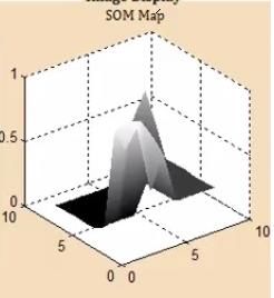
You can also read


