Freiburg Neuropathology Case Conference
←
→
Page content transcription
If your browser does not render page correctly, please read the page content below
Clin Neuroradiol (2020) 30:189–195
https://doi.org/10.1007/s00062-020-00885-3

    CLINICAL CASE

Freiburg Neuropathology Case Conference

Intraventricular Mass Lesion in a Child

C. A. Taschner1 · D. Erny2 · O. Schnell3 · H. Urbach1 · I. E. Duman1 · M. Prinz2

Published online: 26 February 2020
© The Author(s) 2020

Keywords Choroid plexus tumor · Subependymal giant cell astrocytoma · Supratentorial ependymoma · Central
neurocytoma · Pilocytic astrocytoma

Case Report                                                        field loss occurred; otherwise no new neurological deficit
                                                                   was encountered. The second operation for tumor removal
An 11-year-old girl presented with increasing headaches            was performed 8 weeks later. After opening of the dura
that had started approximately 4 weeks earlier. These              a soft predominantly greyish tumor with a sharp border
headaches of high intensity occurred on a daily basis and          to the surrounding ependymal lining, especially in the di-
were predominantly located in the left frontal region. The         rection of the cella media of the left lateral ventricle was
patient reported an episode of severe pain at night, with          developed. In a rostral direction a sharp border to the thala-
a state of disorientation and limited motor control that had       mus was visible. A larger tumor node was visualized in the
first occurred 2 weeks prior to admission. An electroen-           direction of the temporal horn of the left ventricle which
cephalography (EEG) was performed at that time and did             appeared to be covered by a thin layer of parenchyma. After
not reveal any pathological findings. Neither nausea nor           fenestration of the parenchyma under neuronavigation, the
vomiting occurred at any time.                                     greyish tumor of soft consistency could easily be separated
    On admission the clinically and neurological examina-          from the surrounding tissue and was removed. Finally, the
tions were strictly normal. Brain magnetic resonance imag-         tumor portion directed to the midline, adjacent to the inter-
ing (MRI) showed an intraventricular mass lesion (Figs. 1, 2       nal cerebral veins was prepared. Here, the tumor borders
and 3). The indications for tumor resection were established       seemed less well defined. The consistency of the tissue ap-
at the weekly interdisciplinary tumor board meeting.               peared hardened and was of more diffuse yellowish-greyish
    The operation was performed applying intraoperative            appearance. Altered tissue was removed via a cavitronic ul-
neuronavigation, intraoperative ultrasound and intraoper-          trasonic surgical aspirator (CUSA), under neuronavigation
ative neuromonitoring. Despite an initially unremarkable           and electrophysiological monitoring. Due to increasingly
course of surgery, the first operation for tumor resection         diffuse tumor infiltration and no indication of clear tumor
had to be aborted shortly after dural opening due to a newly       residues, resection was stopped and wound closure accom-
occurring epidural hematoma. Postoperatively a new visual          plished. The postoperative course was uneventful and the
                                                                   patient was discharged home in good clinical condition.

                                                                   Imaging
 C. A. Taschner
      christian.taschner@uniklinik-freiburg.de
                                                                   Axial (Fig. 1a–c) and coronal (Fig. 1d) T2-weighted images
1
      Department of Neuroradiology, Medical Center – University    showed an intraventricular tumor extending from the tem-
      of Freiburg, Breisacher Straße 64, 79106 Freiburg, Germany   poral horn through to the cella media of the left lateral ven-
2
      Department of Neuropathology, Medical Center – University    tricle (Fig. 1a–d, arrow). The tumor presents heterogeneous
      of Freiburg, Freiburg, Germany                               signal intensities with isointense portions next to smaller
3
      Department of Neurosurgery, Medical Center – University of   cystic components. The tumor led to subsequent dilata-
      Freiburg, Freiburg, Germany                                  tion of the left lateral ventricle (Fig. 1a, d, arrowhead). On

                                                                                                                       K
190 C. A. Taschner et al. Fig. 1 Axial (a–c) and coro- nal (d) T2-weighted images show an intraventricular tumor extending from the temporal horn through to the cella media of the left lateral ventricle (a–d, arrow). The tumor presents het- erogeneous signal intensities with isointense portions next to smaller cystic components. The tumor leads to subsequent dilatation of the left lateral ven- tricle (a, d, arrow head) coronal T1-weighted non-contrast images (Fig. 2a) the le- Differential Diagnosis sion had a relatively homogeneous appearance and seemed isointense compared to the grey matter (Fig. 2a, arrow). On Choroid Plexus Tumor coronal (Fig. 2b) and axial (Fig. 2c–e) T1-weighted images after i.v. administration of Gadolinium the lesion revealed Choroid plexus tumors (CPT) are rare, highly vascularized marked and homogeneous contrast enhancement (arrow). neoplasms of the central nervous system (CNS) represent- The dilatation of the left lateral ventricle was clearly visu- ing
Freiburg Neuropathology Case Conference                                                                                                     191

Fig. 2 On coronal T1-weighted non-contrast images (a) the lesion has a relatively homogeneous appearance and seems isointense compared to
the grey matter (a arrow). On coronal (b) and axial (c–e) T1-weighted images obtained after i.v. administration of gadolinium the lesion reveals
marked and homogeneous contrast enhancement (arrow). The dilatation of the left lateral ventricle is again clearly visualized (a–c arrowhead)

cinomas (CPC, WHO grade III) [4]. These tumors occur in                   tomography (CT) shows an intraventricular lobular mass
the ventricular system with the lateral (50%) and fourth                  with a periventricular interstitial edema due to ventricular
(40%) ventricles being the most common locations, af-                     obstruction or overproduction of cerebrospinal fluid (CSF).
fording these tumors the ability to cause hydrocephalus                   On magnetic resonance imaging (MRI), choroid plexus tu-
and/or metastasize through the ventricles. Nausea, vomit-                 mors show a well-delineated T1-isointense to hypointense
ing, headache, and obtundation are the most common pre-                   lobular mass with a marked and homogeneous enhance-
senting features most likely related to a rise in intracranial            ment. In addition, occasional cysts, calcifications and small
pressure. The typical imaging pattern consists of a usually               foci of necrosis can be present [5]. Imaging cannot reliably
large lobulated (cauliflower-like) mass within the atrium                 distinguish CPP from aCPP or CPC. Extensive invasion
of the lateral ventricle with a strong enhancement and in-                of the brain parenchyma, heterogeneity, early CSF spread
ternal linear and branching vascular flow voids. Computed                 suggests CPC.

                                                                                                                                    K
192 C. A. Taschner et al.
frequently YAP1 fusions (affecting childrenFreiburg Neuropathology Case Conference 193
[9]. A strong association with neurofibromatosis type 1 Histology
(NF 1) is known. Pilocytic astrocytomas are most com-
monly seen in the cerebellum (60%), followed by the optic Histology and Immunohistochemistry
pathway (25–30%) being the next most common location,
particularly in patients with NF1. Other less common lo- In the hematoxylin-eosin (H&E) stained sections of the
cations are the brainstem, cerebral hemispheres particularly formaldehyde-fixed and paraffin-embedded initial intraop-
in adults, cerebral ventricles, velum interpositum and spinal erative biopsy material, a relatively isomorphic neoplasm
cord (spinal pilocytic astrocytomas). Clinical presentation with slightly increased cellularity without signs of necrosis
varies with location. Headache, nausea and vomiting are was observed and classified as pilocytic astrocytoma. The
the most common symptoms due to the raised intracranial following biopsy material yielded more tumor tissue which
pressure, mainly related to hydrocephalus. Visual symptoms exhibited similar histomorphological features: the astrocytic
are related with the optic pathway lesions, while ataxia and tumor shows predominantly a solid growth pattern, region-
cerebellar signs are seen in cerebellar lesions. The typi- ally a mucoid matrix and appears to be relatively sharp de-
cal imaging pattern consists of a usually large, cystic mass marcated from few smaller fragments of the adjacent CNS
with a strong enhancing mural nodule. Overall morphol- tissue. The tumor cells display mostly small and round-
ogy is often determined by cystic component. Computed oval shaped chromatin dense nuclei (Fig. 4). Eosinophilic
tomography shows a cystic and solid mass with relatively Rosenthal fibers and protein droplets can be found region-
little peripheral edema. The solid part appears hypodense ally (Fig. 4). Occasional dystrophic calcifications are seen
to isodense in non-enhanced CT compared to gray mat- locally in the present tissue as well as a few and mostly
ter and sometimes shows calcification. after contrast agent small hemorrhages. Few hemosiderin deposits indicating
administration, they show variable enhancement patterns older bleedings can be detected focally. Mitotic figures are
with variable involvement of cystic components and mu- not detectable. Accordingly, the proliferation index is only
ral nodules. The MRI shows in 50% of cases a character- modestly increased and in total less than 5% of the tumor
istic non-enhancing cyst, strongly enhancing mural nod- cells are marked in the Ki67/MIB1 immunohistochemical
ule appearance with T1-weighted isointense/hypointense reaction (Fig. 5). Blood vessels feature partially thickened
and T2-weighted hyperintense solid portions. Solid portions hyalinized walls. Microvascular proliferation is not present
show intense but heterogeneous enhancement after gadolin- in several regions. Marked desmoplastic components are
ium injection. Cyst contents appear in T2-weighted images absent in the present tissue.
isointense/hyperintense, in fluid-attenuated inversion recov- Staining for GFAP (glial fibrillary acidic protein) is pre-
ery (FLAIR) images hyperintense to CSF and shows occa- dominantly positive in the tumor tissue (Fig. 6). Tumor
sionally enhancement [10]. cells show OLIG2 expression, indicative among others for
gliomas, including pilocytic astrocytoma and high-grade
astrocytoma or oligodendroglioma (Fig. 7). The immuno-
histochemical reaction against neurofilament confirms ab-
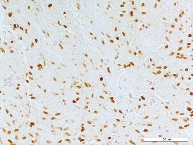
sence of neuronal structures within the tumor tissue and
Fig. 4 Hematoxylin-eosin stained section showing an astrocytic tumor Fig. 5 MIB1 staining. The proliferation index is slightly increased tag-
with Rosenthal fibers (arrowheads). Size bar: 100 µm ging less than 5% of tumor cells. Size bar: 100 µm
K194 C. A. Taschner et al.
Fig. 6 Tumor cells and a GFAP-rich tumor matrix are visualized by Fig. 8 The immunohistochemical reaction against neurofilament re-
immunohistochemical reaction against GFAP. Size bar: 100 µm mains negative within the tumor tissue (asterisk) and shows a positive
reaction in sharply demarcated adjacent gliotic CNS tissue. Size bar:
100 µm
tomas [12, 13]. The diagnosis was independently confirmed
by the brain tumor reference center in Berlin (Germany).
Differential diagnoses for the current tumor include pri-
marily tumors usually found in the lateral ventricles during
childhood, e.g. ependymal tumors (such as ependymoma,
WHO grade II), tumors derived from choroid plexus (such
as choroid plexus papilloma, WHO grade I) or central neu-
rocytoma, WHO grade II. The SEGAs typically appear in
the wall of lateral ventricles and are associated with the
tuberous sclerosis syndrome and harbor large ganglionic
astrocytic cells [13, 14]. As another differential diagnosis
diffuse gliomas can be considered since they may be located
predominantly in the ventricle system [13].
Fig. 7 Immunohistochemical staining against OLIG2 reveals positiv- Concerning these differential diagnoses, perivascular
ity in various tumor cells. Size bar: 100 µm
pseudorosettes a common feature of ependymoma are not
present in the specimen. Furthermore, OLIG2 expression
highlights a relatively sharp border to adjacent gliotic CNS is present in the tumor cells and described to be absent
tissue (Fig. 8). Staining using a mutation-specific (R132H) in ependymal tumors [15]. The morphological features of
antibody against isocitrate dehydrogenase 1 (IDH1), indica- choroid plexus tumors are quite distinct with a papillary
tive for IDH-mutated astrocytomas and oligodendrogliomas pattern composed of a single layer of epithelial tumor
[11], revealed no specific reaction in the present tumor (not cells. In contrast, the present tumor displays mainly a solid
shown). The nuclear expression of the transcriptional regu- growth pattern with monomorphic glial tumor cells. Ad-
lator ATRX is retained in the tumor cells (not shown). ditionally, expression of neuronal markers is absent in
In summary, the histopathological finding of a glial, iso- the present specimen and oppose the diagnosis of tumors
morphic, relatively well-demarcated tumor with slightly el- with an at least partial neuronal differentiation like cen-
evated cellularity exhibiting characteristic Rosenthal fibers tral neurocytoma and SEGA. The differential diagnoses of
together with low proliferative activity as well as absence of diffuse gliomas including the highly malignant pediatric
microvascular proliferation and necrosis leads consequently tumor diffuse midline glioma are ruled out by a lack of
to the diagnosis of pilocytic astrocytoma, WHO grade I. histomorphological hallmarks including increased mitotic
Signs of anaplastic transformation are not found. The di- rate, diffuse infiltration. Furthermore, H3 K27M mutation
agnosis is further supported by detection of the BRAF- and mutations in the IDH1 and IDH2 genes are absent. Pi-
KIAA1549 fusion that constitutively activates the MAPK lomyxoid astrocytomas are a more rare variant of pilocytic
pathway and occurs in approx. 70% of pilocytic astrocy- astrocytoma [16]. In contrast to the present tumor they are
KFreiburg Neuropathology Case Conference 195
histologically characterized by an angiocentric composition References
of the tumor cells and prominent myxoid alterations.
1. Bettegowda C, Adogwa O, Mehta V, Chaichana KL, Weingart J,
Carson BS, Jallo GI, Ahn ES. Treatment of choroid plexus tumors:
a 20-year single institutional experience. J Neurosurg Pediatr.
Diagnosis 2012;10:398–405.
2. Asai A, Hoffman HJ, Hendrick EB, Humphreys RP, Becker LE.
Intraventricular Pilocytic Astrocytoma (WHO I) Primary intracranial neoplasms in the first year of life. Childs Nerv
Syst. 1989;5:230–3.
3. Jooma R, Hayward RD, Grant DN. Intracranial neoplasms during
Intraventricular tumors are relatively symptomless until the first year of life: analysis of one hundred consecutive cases.
they enlarge and obstruct the pathways of CSF, produc- Neurosurgery. 1985;14:31–41.
ing obstructing hydrocephalus and leading to an increased 4. Louis DN, Ohgaki H, Wiestler OD, Cavenee WK, Burger PC, Jou-
vet A, Scheithauer BW, Kleihues P. The 2007 WHO classifica-
intracranial pressure [17]. Pilocytic astrocytomas are the tion of tumours of the central nervous system. Acta Neuropathol.
most common glioma present in children and young adults 2007;114:97–109.
and composed of neoplastic glial cells [18]. They are cir- 5. Naeini RM, Yoo JH, Hunter JV. Spectrum of choroid plexus lesions
cumscribed, benign, slowly growing and mainly located in children. AJR Am J Roentgenol. 2009;192:32–40.
6. Koeller KK1, Sandberg GD; Armed Forces Institute of Pathol-
in infratentorial regions and cerebral midline structures. ogy. From the archives of the AFIP. Cerebral intraventricu-
Tumors localized in the lateral ventricles of the cerebral lar neoplasms: radiologic-pathologic correlation. Radiographics.
hemispheres account for less than 1% of all intracranial 2002;22:1473–505.
tumors but are relatively more frequent in the pediatric 7. Kalantari BN, Salamon N. Neuroimaging of tuberous sclerosis:
spectrum of pathologic findings and frontiers in imaging. AJR Am
population [17]. Pilocytic astrocytomas rarely present as J Roentgenol. 2008;190:W304–9.
intraventricular tumors and should be included in the 8. Smith A, Smirniotopoulos J, Horkanyne-Szakaly I. From the radi-
differential diagnosis in a pediatric population. The intra- ologic pathology archives: intraventricular neoplasms: radiologic-
ventricular location of a glial tumor leads to the assumption pathologic correlation. Radiographics. 2013;33:21–43.
9. AlRayahi J, Zapotocky M, Ramaswamy V, Hanagandi P, Bran-
that the present tumor initially originated from the brain son H, Mubarak W, Raybaud C, Laughlin S. Pediatric brain tu-
parenchyma close to the ventricle system and subsequently mor genetics: what radiologists need to know. Radiographics.
grew into the ventricles [13]. 2018;38:2102–22.
10. Koeller KK, Rushing EJ. From the archives of the AFIP: pilocytic
Funding Open Access funding provided by Projekt DEAL. astrocytoma: radiologic-pathologic correlation. Radiographics.
2004;24:1693–708.
11. Capper D, Weissert S, Balss J, Habel A, Meyer J, Jäger D, Acker-
Compliance with ethical guidelines mann U, Tessmer C, Korshunov A, Zentgraf H, Hartmann C, von
Deimling A. Characterization of R132H mutation-specific IDH1
Conflict of interest C. A. Taschner, D. Erny, O. Schnell, H. Urbach, antibody binding in brain tumors. Brain Pathol. 2010;20:245–54.
I. E. Duman and M. Prinz declare that they have no competing interests. 12. Bar EE, Lin A, Tihan T, Burger PC, Eberhart CG. Frequent gains
at chromosome 7q34 involving BRAF in pilocytic astrocytoma.
Ethical standards All investigations described in this manuscript were J Neuropathol Exp Neurol. 2008;67:878–87.
carried out with the approval of the responsible ethics committee and 13. Louis DN, Ohgaki H, Wiestler OD, Cavenee WK. World Health Or-
in accordance with national law and the Helsinki Declaration of 1975 ganization histological classification of tumours of the central ner-
(in its current revised form). Informed consent was obtained from the vous system. France: International Agency for Research on Cancer;
patient in this case if identifiable from images or other information 2016.
within the manuscript. In the case of the underage patient in this report, 14. Roth J, Roach ES, Bartels U, Jóźwiak S, Koenig MK, Weiner HL,
informed consent was obtained from the legal representatives. Franz DN, Wang HZ. Subependymal giant cell astrocytoma: diag-
Open Access This article is licensed under a Creative Commons At- nosis, screening, and treatment. Recommendations from the inter-
tribution 4.0 International License, which permits use, sharing, adapta- national tuberous sclerosis complex consensus conference. Pediatr
tion, distribution and reproduction in any medium or format, as long as Neurol. 2012;2013:439–44.
you give appropriate credit to the original author(s) and the source, pro- 15. Otero JJ, Rowitch D, Vandenberg S. OLIG2 is differentially ex-
vide a link to the Creative Commons licence, and indicate if changes pressed in pediatric astrocytic and in ependymal neoplasms. J Neu-
were made. The images or other third party material in this article are rooncol. 2011;104:423–38.
included in the article’s Creative Commons licence, unless indicated 16. Tihan T, Fisher PG, Kepner JL, Godfraind C, McComb RD, Goldth-
otherwise in a credit line to the material. If material is not included waite PT, Burger PC. Pediatric astrocytomas with monomorphous
in the article’s Creative Commons licence and your intended use is not pilomyxoid features and a less favorable outcome. J Neuropathol
permitted by statutory regulation or exceeds the permitted use, you will Exp Neurol. 1999;58:1061–8.
need to obtain permission directly from the copyright holder. To view 17. Sattar S, Akhunzada NZ, Javed G, Uddin Z, Khan YA. Pilocytic as-
a copy of this licence, visit http://creativecommons.org/licenses/by/4. trocytoma: a rare presentation as intraventricular tumor. Surg Neu-
0/. rol Int. 2017;8:116.
18. Ostrom QT, Gittleman H, Liao P, Rouse C, Chen Y, Dowling J,
Wolinsky Y, Kruchko C, Barnholtz-Sloan J. CBTRUS statistical re-
port: primary brain and central nervous system tumors diagnosed in
the United States in 2007–2011. Neuro Oncol. 2014;16:iv1–63.
KYou can also read