Desmoplastic small round cell tumor of the kidney: a case report - Diagnostic ...
←
→
Page content transcription
If your browser does not render page correctly, please read the page content below
Ertoy Baydar et al. Diagnostic Pathology   (2020) 15:95
https://doi.org/10.1186/s13000-020-01015-w

 CASE REPORT                                                                                                                                        Open Access

Desmoplastic small round cell tumor of the
kidney: a case report
Dilek Ertoy Baydar1*† , Ayse Armutlu1†, Oguz Aydin2, Ayhan Dagdemir3 and Yarkin Kamil Yakupoglu4

  Abstract
  Background: Desmoplastic small round cell tumor (DSRCT) is a rare, aggressive neoplasm seen in children and
  young adults, usually manifested by involvement of abdominal serosa. Here, we present an unusual case of primary
  DSRCT of kidney.
  Case presentation: The patient was an 8-year-old girl with a large renal mass which was confused with primitive
  neuroectodermal tumor (PNET) in the needle biopsy. The tumor had a variegated histology revealing frequent
  pseudo-rosette formations, pseudopapillary architecture, rhabdoid, clear or pleomorphic cells in addition to typical
  small round cell morphology and desmoplasia. It showed immunohistochemical features of DSRCT, and EWSR1 re-
  arrangement.
  Conclusions: Proffering this diagnosis is particularly difficult for tumors of viscera because of the incognizance of
  the entity in these locations. Moreover, DSRCT is a great mimicker and may get easily confused with more common
  kidney malignancies of childhood such as Wilms tumor, PNET/EWS, rhabdoid tumor, clear cell sarcoma, and other
  small round cell tumors as well as renal cell carcinomas. The distinction is critical as the accurate therapeutic
  approach will require correct diagnosis.
  Keywords: Desmoplastic small round cell tumor, Kidney, WT1, EWSR1, Case report

Background                                                                             are extremely rare with only a few cases reported in
Desmoplastic small round cell tumor (DSRCT) is a rare,                                 lungs, ovary, soft tissues, bones, intracranial and sinona-
distinct entity that was first described by Gerald and                                 sal locations [2, 3].
Rosai in 1989 [1]. Predilection for adolescent males,                                    DSRCT primary of the kidney was first described by
predominant intraabdominal location involving serosal                                  Su, et al. [4] in 2004 and since then only a total of
surfaces, nesting pattern of growth, focal rhabdoid                                    12 cases have been reported in the literature (Table 1)
morphology, prominent desmoplastic reaction, immuno-                                   [5–12]. Herein, we present the thirteenth case of
histochemical reactivity for epithelial, neural and muscle                             renal DSRCT that had variant histological features
markers, and highly aggressive clinical behavior are its                               mimicking various types of other neoplasia. The
main features. DSRCT shows a specific reciprocal                                       pathologic diagnosis of this entity can be markedly
chromosomal translocation, t(11;22)(p13;q12) (EWSR1-                                   challenging when it develops in visceral organs such
WT1 fusion) which generates a chimerical protein with                                  as kidney and especially if diverse and confounding
transcriptional regulatory activity. Extraserosal DSRCTs                               microscopic features are present.

* Correspondence: dertoy@kuh.ku.edu.tr; dilekertoy@yahoo.com                           Case presentation
†
 Dilek Ertoy Baydar and Ayse Armutlu contributed equally to this work.                 Clinical history
1
 Department of Pathology, Koc University School of Medicine, Topkapi,
34010 Istanbul, Turkey                                                                 An 8-year-old girl complained of abdominal pain and an
Full list of author information is available at the end of the article                 ultrasonography found a large mass in her left kidney.
                                        © The Author(s). 2020 Open Access This article is licensed under a Creative Commons Attribution 4.0 International License,
                                        which permits use, sharing, adaptation, distribution and reproduction in any medium or format, as long as you give
                                        appropriate credit to the original author(s) and the source, provide a link to the Creative Commons licence, and indicate if
                                        changes were made. The images or other third party material in this article are included in the article's Creative Commons
                                        licence, unless indicated otherwise in a credit line to the material. If material is not included in the article's Creative Commons
                                        licence and your intended use is not permitted by statutory regulation or exceeds the permitted use, you will need to obtain
                                        permission directly from the copyright holder. To view a copy of this licence, visit http://creativecommons.org/licenses/by/4.0/.
                                        The Creative Commons Public Domain Dedication waiver (http://creativecommons.org/publicdomain/zero/1.0/) applies to the
                                        data made available in this article, unless otherwise stated in a credit line to the data.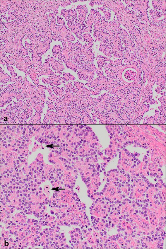
Table 1 Clinicopathologic features of the cases described in the literature (a)
Age Clinical R / L Gross Findings Microscopic Findings Immunohistochemistry Molecular Therapy Follow-up Publication
/ presentation side pathology
Sex
Case 41 / Incidental Left Renal mass at hilar region, Nests of small round blue cells CK (AE1/AE3) (+), Desmin (+), RT-PCR Surgery Alive (NED) at Su, et al.,
1 F 6x5x5 cm, invaded into within desmoplastic stroma; Vimentin (+), NSE (+), EMA (−), showing 18th mo. [4]
perirenal soft tissue occasional rosette-like forma- CK7 and 20 (−), CD99 (−), S100 EWSR1-WT1
tions; necrosis and numerous (−), Chromogranin (−), fusion
mitotic figures Synaptophysin (−)
Case 7 / F Gross Left Confined within renal Small round undifferentiated Desmin (+), WT-1 (+), CD99 (+), RT-PCR Surgery + CT Alive (NED) at Eaton,
2 hematuria capsule, 3.7 × 3.7 × 3.2 cm cells, necrosis and epithelioid SMA (+), EMA (+), Myogenin (−) showing 12th mo. et al., [5]
following a fall component EWSR1-WT1
fusion
Ertoy Baydar et al. Diagnostic Pathology
Case 6 / F Renal mass Left 3.7 cm mass confined to Nests, sheets or cords of small CK (+), Desmin (+), Vimentin (+), Dual color FISH Neoadjuvant Alive (NED) Egloff,
3b (no details kidney undifferentiated cells; WT1 (+), FLI-1 (+), CD56 (+); EMA showing CT + Surgery 24th mo. et al., [6]
about clinical numerous mitotic figures, no (−), CD99 (−), Myogenin (−), S100 EWSR1-WT1 and Wang,
presentations) desmoplasia (−), Chromogranin (−), Synapto- translocation et al., [7]
physin (−) and RT-PCR
showing
(2020) 15:95
Case 6/F Left 13.4 cm mass showing CK (+), EMA (+), Desmin (+), Surgery + CT Pulmonary Wang,
EWSR1-WT1
4 renal sinus invasion Vimentin (+), CD99 (+), WT1 (+), fusion metastasis at et al., [7]
FLI-1 (+), CD56 (+), Myogenin (−), 32nd mo. CT,
S100 (−), Chromogranin (−), stem cell
Synaptophysin (−) transplantation.
NED a year later
Case 6/F Left 9 cm mass with perirenal CK (+), EMA (+), Desmin (+), Alive (NED) at
5 soft tissue and renal sinus Vimentin (+), CD99 (+), WT1 (+), 22nd mo
invasion FLI-1 (+), CD56 (+), Myogenin (−),
S100 (−), Chromogranin (−),
Synaptophysin (−)
Case 8/ Left 9.2 cm mass with renal EMA (+), Desmin (+), Vimentin (+), Intraabdomimal
6 M sinus invasion CD99 (+), WT1 (+), FLI-1 (+), CD56 recurrence and
(+), CK (−), Myogenin (−), S100 liver metastasis
(−), Chromogranin (−), Synapto- at 20th mo.
physin (−) CT.
AWD
Case 14 / Gross Left 17.5x12x11 cm mass Small ovoid-spindle blue cells EMA (+), Desmin (+), Vimentin (+), RT-PCR Surgery + Liver and lung Collardeau-
7 F hematuria, invading renal sinus and in solid sheets and large nests; WT1 (+), CD56 (+), Chromogranin showing CT + local RT metastases at Frachon,
fever and self- perinephric fat, and focally rare rosette-like structures; nu- (focal +), Synaptophysin (rare +), EWSR1-WT1 8th mo. et al., [8]
disvovered ab- extending to Gerota’s merous mitotic figures; tumor CK (AE1/AE3) (−), CD99 (−), S100 fusion
dominal mass fascia; metastatic lymph thrombi in perinephric blood (−)
node in hilar region vessels
Case 10 / Gross Right 14 × 11 cm mass Small blue round cells with CK (+), Desmin (+), CD99 (−), WT1 RT-PCR Surgery + Liver, lung, da Silva,
8 M hematuria, desmoplasia; occasional (+), FLI-1 (+); S100 (−), Chromo- showing CT + local RT bone, lymph et al., [9]
abdominal rosette-like formations. granin (−), Synaptophysin (−) EWSR1-WT1 node
pain, palpable fusion metastases.
mass AWD at 12th
mo
Page 2 of 9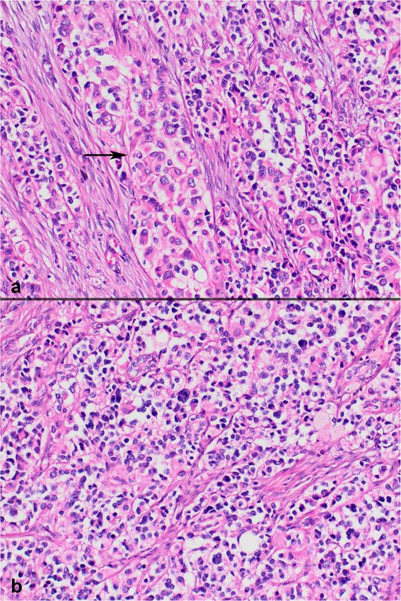
Table 1 Clinicopathologic features of the cases described in the literature (a) (Continued)
        Age     Clinical            R / L Gross Findings                     Microscopic Findings                 Immunohistochemistry                     Molecular        Therapy       Follow-up           Publication
        /       presentation        side                                                                                                                   pathology
        Sex
Case    20 /    Renal mass and Right 8 cm mass with areas of                 Elongated to round cells with        CK (+), Desmin (+), Vimentin (+),        FISH showing     Surgery       Pulmonary           Rao, et al.,
9       M       pulmonary            hemorrhage and necrosis,                scant cytoplasm in sheets and        CD56 (+); WT1 (cytoplasmic +),           EWSR11                         metastases at       [10]
                nodules              invading renal vein grossly             occasionally a vague nodular         CD99 (−), MyoD1 (−), NSE (−),            rearrangement                  presentation,
                (no details                                                  pattern, frequent mitotic            RCC Ag (−), EMA (−), Myogenin            and RT-PCR                     local recurrence
                about clinical                                               activity, lacked prominent           (−), S100 (−)                            showing                        after surgery.
                presentations)                                               desmoplasia                                                                   EWSR1-WT1                      Exitus at 2nd
                                                                                                                                                           fusion                         year
Case    7/      Gross               Left    Polypoid mass confined to Spindled and polygonal tumor CD99 (+), Vimentin (+), Desmin                          FISH showing     Surgery +     Alive (NED)         Eklund
10      M       hematuria,                  the renal collecting system, cells, rare rosettes, low mitotic (focal+), Actin (focal+), WT1                   EWSR1            CT + RT       (duration           et al., [11]
                                                                                                                                                                                                                             Ertoy Baydar et al. Diagnostic Pathology

                microscopic                 extending into proximal      rate                              (focal+), PAX2 (+); PAX8 (−)                    rearrangement,                 unknown)
                hematuria and               and mid ureter, no                                                                                             karyotyping
                intermittent                involvement of renal                                                                                           showing t(11;
                back pain 3                 parenchyma                                                                                                     22) (p13;q12).
                years priorly
Case    6/      Facial swelling     Right 5.7 × 5.5 × 4.7 cm mass       Sheets of poorly differentiated           Bcl-2 (+), CD99 (+), desmin (+),         RT-PCR           No            Metastatic          Walton,
                                                                                                                                                                                                                             (2020) 15:95

11      M       and pain,                 with large areas of central round cells, no desmoplastic                vimentin (+), CD56 (+), and FLI-1        showing          information   disease             et al., [12]
                headache,                 necrosis, invading hilar soft stroma                                    (+), WT1 (−), Synaptophysin (−),         EWSR1-WT1                      (multiple, bone
                decreased oral            tissues                                                                 SMA (−), Myogenin (−), Myo-D1            fusion                         and lungs) at
                intake                                                                                            (−), CD31 (−), CD34 (−), Napsin                                         presentation.
                                                                                                                  (−)                                                                     No further
                                                                                                                                                                                          follow-up
                                                                                                                                                                                          information
Case    8 / F Abdominal pain Left           11x9x7 cm mass with renal Nests, cords, sheets of small               CK (+), EMA (+), Desmin (+),             FISH showing   Neoadjuvant     Multiple            Current
12                                          pelvis, perirenal fat tissue round cells within                       Vimentin (+), CD56 (+), WT1 (+),         EWSR1 gene re- CT + surgery    metastases          case
                                            and adrenal gland invasion desmoplastic stroma; frequent              CD99 (−), Bcl-2 (−), MUC4 (−),           arrangement    + adjuvant      (liver, lungs and
                                                                         rosette-like structures, psedo-          Myogenin (−), Myo-D1 (−), SMA                           CT              lymph nodes).
                                                                         papillary appearance and fre-            (−), S100 (−), Chromogranin (−),                                        Exitus at 30th
                                                                         quent rhabdoid cells.                    Synaptophysin (−)                                                       mo.
NED No evidence of disease, AWD Alive with disease, CT Chemotherapy, RT Radiotherapy
a
 Table does not include a case reported by Janssens E, et al. (2009) [13] as the article could not be reached by any means
b
  Case 3 was first reported by Egloff, et al. (2005) [6] and also included among 4 patients in the case series published a year later by Wang, et al. (2007) [7]
                                                                                                                                                                                                                             Page 3 of 9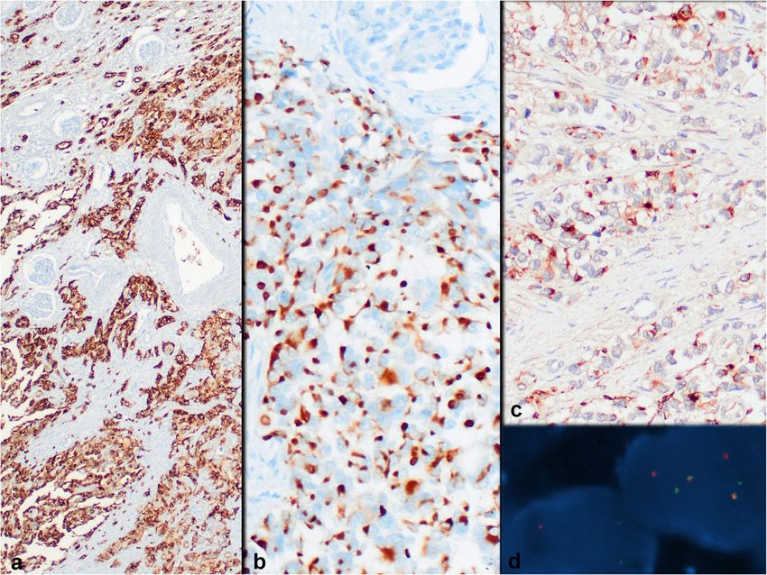
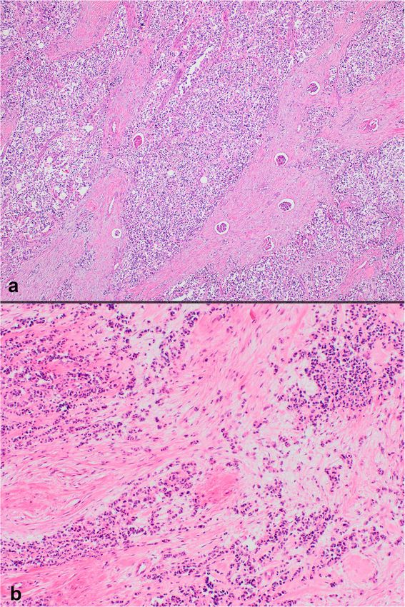
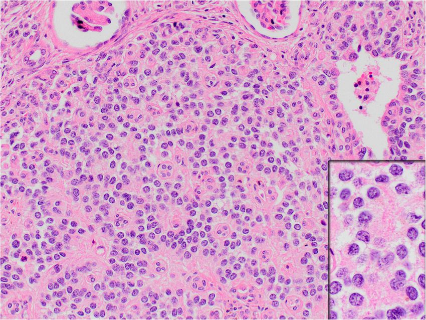
Ertoy Baydar et al. Diagnostic Pathology      (2020) 15:95                                                                  Page 4 of 9

Abdomimal MRI showed that it was a heterogenous lob-                        Pathology
ulated solid lesion measuring 80x92x118 mm in size                          On histopathologic examination, neoplastic cells formed
with cystic and necrotic areas. Needle biopsy from the                      nests, cords and sheets within desmoplastic stroma
tumor was diagnosed in an outside center as a small                         (Fig. 1). Tumor also revealed intermittent areas of primi-
round blue cell tumor consistent with PNET/EWS.                             tive tubule or rosette-like structures (Fig. 2). Further-
The patient had multiple lung, liver, adrenal and                           more, occasional foci appeared to have (psedo) papillary
lymph node metastases at initial presentation. After 6                      architecture with foamy histiocytes which was possibly
cycles of neoadjuvant chemotherapy, left radical neph-                      due to drop-outs and loss of cohesion between cells
rectomy was performed. Macroscopic examination                              (Fig. 3). Most neoplastic cells were small with narrow
showed 11x9x7 cm grey-white solid mass that occu-                           cytoplasm and round monotonous hyperchromatic nu-
pied most of the organ parenchyma, invading also                            clei. However, there were areas that contained unusually
renal pelvis, perirenal soft tissue and adrenal gland                       large amounts of eosinophilic, clear or vacuolated
extensively. Paraffin blocks of both needle biopsy and                      cytoplasm. Some cells revealed rhabdoid features, or
nephrectomy material were sent to our institution for                       pleomorphic, even multilobated nuclei (Fig. 4). Immuno-
consultation.                                                               histochemically, neoplasm diffusely expressed EMA,

 Fig. 1 Sheets, nests and cords of neoplastic cells in a desmoplastic stroma (a H&E × 40; b H&E × 100)
Ertoy Baydar et al. Diagnostic Pathology      (2020) 15:95                                                                 Page 5 of 9

 Fig. 2 Rosette or tubule-like formations (H&E × 200; inset: H&E × 400)

pan-cytokeratin, CD56, vimentin and desmin (para-                         epithelial (EMA and keratin), neural (NSE) and mesen-
nuclear dot-like) whereas it stained negative for                         chymal (vimentin and desmin) markers. The cytogenet-
synaptophysin, chromogranin, S100, CD99, bcl-2,                           ics of a case of DSRCT, featuring a diploid DNA content
myo-D1, GATA3 and PAX8 (Fig. 5a-b). Nuclear INI1                          and t(11;22)(p13;q12) was first reported in 1992 by
was intact. Antibodies directed to N-terminus of                          Sawyer JR, et al. [14] In1994, Ladanyi and Gerald defined
WT1 protein stained cytoplasm of the tumor cells                          the consistent fusion between EWSR1 and WT1 genes in
non-specifically without nuclear immunoreactivity                         DSRCTs [15]. In a publication in 1995, they proved that
(Fig. 5c). FISH analysis with a break-apart probe                         DSRCT represents the third tumor type associated with
proved EWSR1 gene re-arrangement in the neoplastic                        EWSR1 translocation, and it is the only malignancy
cells (Fig. 5d). Our final diagnosis was desmoplastic                     holding EWSR1 - WT1 rearrangement [16].
small round cell tumor of the kidney.                                       Urogenital DSRCT may involve bladder, ureters, pros-
                                                                          tate and paratesticular structures [17]. Primary DSRCT
Follow-up                                                                 of the kidney was first noted by Su, et al. [4] in 2004 and
The child was given multiple cycles and different combi-                  12 cases have been reported in the literature thereafter
nations of adjuvant chemotherapy. She had a local re-                     [5–12] albeit one of them has an inaccessible publica-
lapse in the 2nd year that underwent salvage resection.                   tion. Although male predilection is emphasized in ab-
Chemotherapy was continued thereafter, but she died in                    dominal DSRCTs, most of renal cases including ours
the 30th month from her first operation due to disease                    have been female (F/M = 7/5). Majority were detected
progression and wide-spread metastases.                                   incidentally or presented with gross hematuria and/or
  Informed consent was obtained from the parents of                       abdominal pain. Most tumors had infiltrative appearance
the deceased child for the publication of the patient in-                 with a size ranging from 3.7 to 17.5 cm in diameter. Two
formation and microscopic images.                                         cases were confined to kidney [5, 7] and one case was
                                                                          limited to the renal collecting system without parenchy-
Discussion and conclusions                                                mal involvement [12]. Interestingly, 9 out of 12 renal
DSRCT is a rare, aggressive sarcoma placed in the family                  DSRCTs preferred left kidney.
of small round cell tumors, typically seen in children                      DSRCTs may manifest unusual histologies such as tu-
and young adults with a male predilection. The disease                    bule or rosette-like structures, papillary formations,
most commonly originates in the abdominal or pelvic                       abundant rhabdoid cells, predominantly spindle cell
serosa. Primary DSRCT in extraserosal sites is extremely                  morphology, ample clear cytoplasm and absence of sig-
unusual. Classical histology of DSCRT is the nesting pat-                 nificant desmoplasia. The recognition of the morpho-
tern of small round to oval cells separated by prominent                  logic diversity is important to avoid a misinterpretation
desmoplastic stroma, focal rhabdoid features, and immu-                   during pathologic diagnosis especially when tumors are
nohistochemical profile with peculiar coexpression of                     located in unusual sites. DSRCT of kidney appears to be
Ertoy Baydar et al. Diagnostic Pathology (2020) 15:95 Page 6 of 9 Fig. 3 Pseudo-papillary pattern with foamy histiocytes (arrows) (a H&E × 100; b H&E × 200) a childhood neoplasm as almost all cases were pediatric, with antibodies against carboxy terminus of WT1 pro- 6 to 8 years being the most frequent, with only two adult tein. Yet, immunohistochemistry is still useful in the dis- patients. Given this and also overlapping histopathologic crimination: While WT is characterized by dual nuclear features, main differential diagnosis of DSRCT is Wilms immunoreactivity for both amino- and carboxy-terminus tumor (WT) in the kidney, the most common renal ma- WT1 antibodies, neoplastic nuclei in DSRCT do not lignancy of children. Although the peak incidence of stain with antibody recognizing amino-terminal of WT1 Wilms tumor is between 2 and 3 years, it can occasion- although non-specific cytoplasmic positivity can be seen ally occur at later ages. Blastemal-predominant WT can as in our case. This can be explained by the fusion of be challenging to distinguish histologically from DSRCT the EWSR1 gene to the last three exons (carboxy- and pseudo-rosette or tubule-like arrangements in terminus) of WT1 in DSRCT. The re-arrangement pro- DSRCT may mimic epithelioid component of WT. duces a protein containing the zinc finger region of DSRCT localized to the kidney can lack desmoplastic re- WT1 which needs C-terminal antibodies for recognition. action or WT can demonstrate desmoplasia. Both tu- It has been shown that blastemal WT may express para- mors show nuclear positivity immunohistochemically nuclear desmin in 50% of cases [18], however this is
Ertoy Baydar et al. Diagnostic Pathology     (2020) 15:95                                                                              Page 7 of 9

 Fig. 4 Rhabdoid cells (pointed by an arrow) (a) and cells with clear cytoplasm or pleomorphic nuclei (b) (a H&E × 200; b H&E × 200)

usually not widespread. Extensive (> 75%) paranuclear                      diagnosis in our patient, given to the needle biopsy taken
dotlike desmin positivity in addition to negative PAX8                     from the mass, which was mainly suggested by the pres-
and bland nuclear features will suggest DSRCT [18].                        ence of frequent pseudorosettes in the tumor. These two
  Other pediatric renal tumors, clear cell sarcoma and                     different tumor types have similar age distribution, simi-
rhabdoid tumor, may also need to excluded from DSRC                        lar cytology and both harbor EWSR1 rearrangements.
T in the kidney although these two preferentially occur                    Keratin expression may be seen nearly in 25% of Ewing
in infancy or at very early childhood. DSRCT lacks capil-                  sarcomas, but desmin positivity is exceedingly rare and
lary network of clear cell sarcoma and large nucleolus or                  keratin plus desmin coexpressing Ewing sarcoma has
prominent cytoplasmic inclusions of rhabdoid tumor. It                     not been asserted. DSRCTs show more variable expres-
has intact nuclear INI1 protein expression contrary to                     sion of CD99, rather than the diffuse membranous posi-
rhabdoid tumor. Clear cell sarcomas are negative for epi-                  tivity typical of Ewing sarcoma. The characteristic
thelial and muscle markers, and they have recently been                    translocation of Ewing sarcoma involves EWSR1 and the
shown to overexpress nuclear BCOR protein [19, 20].                        ETS family of transcription factors, not WT1, and it
  DSRCT may mimic PNET/EWS, especially when it                             lacks nuclear WT1 expression. The break-apart FISH
has a solid growth pattern. PNET/EWS was the first                         assay for EWSR1 will not be helpful in the differential
Ertoy Baydar et al. Diagnostic Pathology      (2020) 15:95                                                                                 Page 8 of 9

 Fig. 5 Strong EMA (a), perinuclear dot-like desmin (b) and non-specific cytoplasmic (non-nuclear) WT1 expression (c) by neoplastic cells, (d) is
 FISH analysis showing ESWR1 rearrangement (a Immunohistochemistry, anti-EMA Ab × 40; b Immunohistochemistry, anti-desmin Ab × 200; c
 Immunohistochemistry, anti-WT1 (N-terminal) Ab × 200; d Dual Color Break Apart specific locus FISH probe targeting EWSR1 gene at 22q12.2
 chromosomal region; green and red signals mark the 5′ and 3′ ends of the gene respectively)

diagnosis between DSRCT and PNET/EWS as one fu-                              and the majority of alveolar rhabdomyosarcoma have
sion partner in both tumors is this same gene. Given the                     FOXO1 fusions.
previous reports of a few curious cases carrying hybrid                         In our case, there were areas of cellular discohesion
features of both DSRCT and PNET/EWS but with                                 with groups of foamy histiocytes, leading to focal pseu-
EWSR1-FLI1 or EWSR1-ERG fusion [21, 22], the gold                            dopapillary architecture and bringing papillary type renal
standard for the definitive diagnosis of DSRCT would be                      cell carcinoma (RCC) into consideration. Additionally,
demonstration of the EWSR1-WT1 fusion by RT-PCR                              we have observed some small nests consisted of neoplas-
when feasible. It was not possible in our case due to low                    tic cells with clear cytoplasm, reminiscent of clear cell
quality of extracted RNA from paraffin block.                                RCC. Strong cytokeratin and EMA expression might
  Lymphoma/leukemia,         metastatic   neuroblastoma,                     favor an epithelial neoplasm, however negative immuno-
poorly differentiated synovial sarcoma and rhabdomyo-                        reactivity for PAX8 turned us away from the renal cell
sarcoma are the other tumors that need to be considered                      origin in the first round.
in the differential diagnosis of renal DSRCT. Lymph-                            DSRCT is known to have a poor prognosis. Our pa-
oma/leukemia often demonstrate a diffuse growth pat-                         tient who presented with multiple distant metastases
tern and do not exhibit the cohesion and nuclear                             at the initial diagnosis died at the 30th month despite
features of DSRCT, and can be excluded by a panel of                         radical operation and intensive chemotherapy. How-
lymphoid markers or TdT. Neuroblastoma occurs in                             ever, the detection of the disease at early stage and
very young children, over 90% being diagnosed below 5                        complete resectability may provide significant prog-
years of age. Clinical and laboratory evaluation will usu-                   nostic benefit as previously reported: 6 out of 11
ally reveal an adrenal mass and elevated catecholamine                       renal DSRCTs were stated alive without disease, keep-
metabolites in urine. Neuroblastoma lacks the specific                       ing in mind that the follow-up durations are too
chromosomal translocation and all show HISL-19 ex-                           short to drive a reliable conclusion. The best thera-
pression. Synovial sarcoma characteristically harbors                        peutic modality has yet to be explored for renal
SYT-SSX gene fusion (t(X;18)(p11;q11)). Rhabdomyosar-                        DSRCT. A combination of total resection and chemo-
coma generally does not have a desmoplastic stroma and                       therapy seems to be the most preferred strategy at
unlike DSRCT, it will express myogenin and MyoD1,                            the moment.
Ertoy Baydar et al. Diagnostic Pathology            (2020) 15:95                                                                                         Page 9 of 9

   As a conclusion, DSRCT is a rare disease, but should                                   a case report and review of the literature. Appl Immunohistochem Mol
be considered in the differential diagnosis of small round                                Morphol. 2009;17:557–62.
                                                                                    10.   Rao P, Tamboli P, Fillman EP, Meis JM. Primary intra-renal desmoplastic
cell tumors of the kidney in pediatric patients. This is                                  small round cell tumor: expanding the histologic spectrum, with special
important as each one of those tumors has different                                       emphasis on the differential diagnostic considerations. Pathol Res Pract.
clinical behavior, prognosis, and treatment implications.                                 2014;210:1130–3.
                                                                                    11.   Eklund MJ, Cundiff C, Shehata BM, Alazraki AL. Desmoplastic small round
Immunohistochemical and molecular studies have par-                                       cell tumor of the kidney with unusual imaging features. Clin Imaging. 2015;
ticular guidance for the right analytic approach, and                                     39:904–7.
documentation of EWSR1-WT1 fusion is the “gold                                      12.   Walton WJ, Flores RR. Desmoplastic small round cell tumor of the
                                                                                          kidney: AIRP best cases in radiologic-pathologic correlation.
standard” for the diagnosis of DSRCT as it appears ex-                                    Radiographics. 2016;36:1533–8.
ceedingly characteristic for this disease.                                          13.   Janssens E, Desprechins B, Ernst C, De Smet K, De Mey J. Desmoplastic small
                                                                                          round cell tumor of the kidney. JBR-BTR. 2009;92:60.
Abbreviations                                                                       14.   Sawyer JR, Tryka AF, Lewis JM. A novel reciprocal chromosome translocation
DSRCT: Desmoplastic small round cell tumor; PNET: Primitive                               t (11;22)(p13;q12) in an intraabdominal desmoplastic small round-cell tumor.
neuroectodermal tumor; EWS: Ewing sarcoma; WT: Wilms tumor; RT-                           Am J Surg Pathol. 1992;16:411–6.
PCR: Reverse transcription polymerase chain reaction                                15.   Ladanyi M, Gerald W. Fusion of the EWS and WT1 genes in the
                                                                                          desmoplastic small round cell tumor. Cancer Res. 1994;54:2837–40.
Acknowledgements                                                                    16.   Gerald WL, Rosai J, Ladanyi M. Characterization of the genomic breakpoint
Not applicable.                                                                           and chimeric transcripts in the EWS-WT1 gene fusion of desmoplastic small
                                                                                          round cell tumor. Proc Natl Acad Sci U S A. 1995;92:1028–32.
Authors’ contributions                                                              17.   Furman J, Murphy WM, Wajsman Z, Berry AD 3rd. Urogenital involvement
The report was designed, written, and reviewed by DEB and AA. All authors                 by desmoplastic small round-cell tumor. J Urol. 1997;158:1506–9.
contributed to the data collection, data analysis, and interpretation. The          18.   Arnold MA, Schoenfield L, Limketkai BN, Arnold CA. Diagnostic pitfalls of
manuscript was approved by all authors.                                                   differentiating desmoplastic small round cell tumor (DSRCT) from Wilms
                                                                                          tumor (WT): overlapping morphologic and immunohistochemical features.
Funding                                                                                   Am J Surg Pathol. 2014;38:1220–6.
None.                                                                               19.   Ueno-Yokohata H, Okita H, Nakasato K, et al. Consistent in-frame internal
                                                                                          tandem duplications of BCOR characterize clear cell sarcoma of the kidney.
Availability of data and materials                                                        Nat Genet. 2015;47:861–3.
Stained and unstained slides of the case can be provided if required.               20.   Khan MZ, Akhtar N, Hassan U, Mushtaq S. Diagnostic utility of BCOR
                                                                                          antibody in clear cell sarcomas of kidney. Int J Surg Pathol. 2020;28:477–81.
                                                                                    21.   Katz RL, Quezado M, Senderowicz AM, Villalba L, Laskin WB, Tsokos M. An
Consent for publication
                                                                                          intra-abdominal small round cell neoplasm with features of primitive
Informed consent was obtained from the parents of the deceased child.
                                                                                          neuroectodermal and desmoplastic round cell tumor and a EWS/FLI-1
                                                                                          fusion transcript. Hum Pathol. 1997;28:502–9.
Competing interests                                                                 22.   Ordi J, de Alava E, Torné A, et al. Intraabdominal desmoplastic small
The authors declare that they have no competing interests.                                round cell tumor with EWS/ERG fusion transcript. Am J Surg Pathol.
                                                                                          1998;22:1026–32.
Author details
1
 Department of Pathology, Koc University School of Medicine, Topkapi,
34010 Istanbul, Turkey. 2Department of Pathology, Ondokuz Mayis University          Publisher’s Note
School of Medicine, Samsun, Turkey. 3Department of Pediatric Oncology,              Springer Nature remains neutral with regard to jurisdictional claims in
Ondokuz Mayis University School of Medicine, Samsun, Turkey. 4Department            published maps and institutional affiliations.
of Urology, Ondokuz Mayis University School of Medicine, Samsun, Turkey.

Received: 21 April 2020 Accepted: 14 July 2020

References
1. Gerald WL, Rosai J. Case 2. Desmoplastic small cell tumor with divergent
    differentiation. Pediatr Pathol. 1989;9:177–83.
2. Syed S, Haque AK, Hawkins HK, Sorensen PH, Cowan DF. Desmoplastic small
    round cell tumor of the lung. Arch Pathol Lab Med. 2002;126:1226–8.
3. Altal OF, Aleshawi AJ, Tashtush NA, Alhowary A. A 23-year-old Joradanian
    woman with a desmoplastic small round cell tumor involving the ovary. Am
    J Case Rep. 2019;20:1675–8.
4. Su MC, Jeng YM, Chu YC. Desmoplastic small round cell tumor of the
    kidney. Am J Surg Pathol. 2004;28:1379–83.
5. Eaton SH, Cendron MA. Primary desmoplastic small round cell tumor of the
    kidney in a 7-year-old girl. J Pediatr Urol. 2006;2:52–4.
6. Egloff AM, Lee EY, Dillon JE, Callahan MJ. Desmoplastic small round cell
    tumor of the kidney in a pediatric patient: sonographic and multiphase CT
    findings. AJR Am J Roentgenol. 2005;185:1347–9.
7. Wang LL, Perlman EJ, Vujanic GM, et al. Desmoplastic small round cell
    tumor of the kidney in childhood. Am J Surg Pathol. 2007;3:576–84.
8. Collardeau-Frachon S, Ranchère-Vince D, Delattre O, et al. Primary
    desmoplastic small round cell tumor of the kidney: a case report in a 14-
    year-old girl with molecular confirmation. Pediatr Dev Pathol. 2007;10:320–4.
9. da Silva RC, Medeiros Filho P, Chioato L, Silva TR, Ribeiro SM, Bacchi CE.
    Desmoplastic small round cell tumor of the kidney mimicking Wilms tumor:
You can also read