Meningioma in mature cystic teratoma of the ovary: clinical and computed tomography findings - Cancer ...
←
→
Page content transcription
If your browser does not render page correctly, please read the page content below
Liu et al. Cancer Imaging     (2020) 20:15
https://doi.org/10.1186/s40644-020-0291-8

 RESEARCH ARTICLE                                                                                                                                 Open Access

Meningioma in mature cystic teratoma of
the ovary: clinical and computed
tomography findings
Yi-yang Liu1, Pan Liang1, Jiang Ji2, Kui-sheng Chen1, Li-ming Li1, Jian-bo Gao1* and Liu-liang Yong1

  Abstract
  Background: Mature cystic teratoma (MCT) with meningioma of the ovary is a very rare benign tumor. There is
  only 3 reports of this disease until June 2019. The aim of the present study was to describe a ovarian mature cystic
  teratoma containing meningioma and nests of neuroblasts in a 15-year-old girl.
  Methods: The method used in the present study consists of description of the clinical history, image lab features,
  and pathological result.
  Results: The patient complained of a 2-month history of irregular vaginal bleeding. Abdominal computed
  tomography (CT) showed a large oval cystic-solid mass with septations and fat density shadow, in abdomen pelvic
  cavity. The cystic part was the main component in the mass. The tumoral solid parts and its internal division could
  be seen intensified from slight to moderate on contrast-enhanced CT images compared with those on precontrast
  images, and the solid parts showed heterogeneous enhancement. Neighbouring intestinal tract and the uterus
  displaced by compression. The pathological examination confirmed the diagnosis.
  Conclusions: The clinical feature of ovarian mature cystic teratoma with meningioma includes a lack of specificity.
  Only meticulous recording of the gross features, histopathological examination including immunohistochemistry
  and supportive clinical and radiological findings to arrive at a correct diagnosis in case of unconventional tumours.
  If necessary, preoperative puncture can be performed.
  Keywords: Mature cystic teratoma, Meningioma, Ovary, Computed tomography

Background                                                                             appearance of the similar tumor [3, 4]. Moreover, to the
Mature cystic teratomas are the germ cell tumor deriv-                                 best of our knowledge, meningioma co-existing with the
ing from primordial germ cells and composes of tissues                                 nests of neuroblasts in the same mature cystic teratoma
arising from endoderm, mesoderm, and ectoderm in                                       has not been reported. Due to the low incidence, under-
terms of histology [1]. Malignant elements or the sec-                                 standing of the pathogenesis of other tumors or/and le-
ondary benign tumors in MCTs can also be seen [1, 2].                                  sion arising within MCTs remains rudimentary, with
However, the incidence of their co-exist in MCTs is ex-                                each new case potentially bringing additional insight [5].
tremely rare. An extensive literature search has revealed                              Furthermore, it is significant to distinguish between be-
only three cases of mature cystic teratoma with meningi-                               nign and malignant components that arise in teratomas
oma of the ovary reported in medical literature, two of                                because of the therapeutic and prognostic importance of
which are in English. And the majority of them have fo-                                their identification. In the present study, we discussed
cused on the pathology and clinical manifestation; previ-                              the computed tomography (CT) findings, pathologic
ous reports have not described the radiological                                        characteristics and the clinical features of a rare case of
                                                                                       MCTs with reviewing the relative references.
* Correspondence: fccgaojb@zzu.edu.cn
1
 Department of Radiology, The First Affiliated Hospital of Zhengzhou
University, Zhengzhou 450052, Henan Province, China
Full list of author information is available at the end of the article

                                         © The Author(s). 2020 Open Access This article is distributed under the terms of the Creative Commons Attribution 4.0
                                         International License (http://creativecommons.org/licenses/by/4.0/), which permits unrestricted use, distribution, and
                                         reproduction in any medium, provided you give appropriate credit to the original author(s) and the source, provide a link to
                                         the Creative Commons license, and indicate if changes were made. The Creative Commons Public Domain Dedication waiver
                                         (http://creativecommons.org/publicdomain/zero/1.0/) applies to the data made available in this article, unless otherwise stated.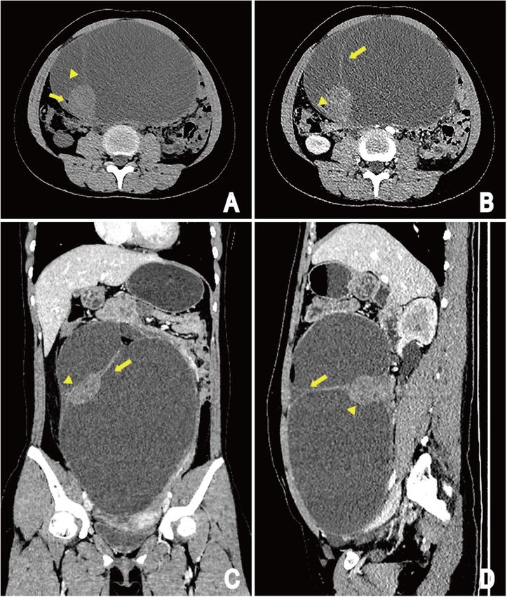
Liu et al. Cancer Imaging   (2020) 20:15                                                                          Page 2 of 7

Methods                                                         Pathological features and follow-up
A case report of meningioma and nests of neuroblasts            Laparoscopic left salpingo-oophorectomy and Separation
arising from mature cystic teratoma of the ovary in an          of pelvic adhesions were performed. Surgical exploration
adolescent female was presented. We searched PubMed,            revealed a large cystic mass located at the left ovary. Fat,
Medline, Google Scholar, Chinese Biomedicine Data-              hair, scalp-like tissue could be seen when the tumors
base, and the China Journal Full Text Database without          were dissected, with dark red and bloody fluid inside, as
language restriction. The search terms included (men-           well as a solid nodule.
ingioma [MeSH]) AND Ovarian teratoma [MeSH]). The                 On gross description, the Pelvic cavity showed a multi-
demographic, clinical features and imaging findings of          loculated cystic mass measuring 20 × 13 × 27 cm in lar-
both the newly described and previously reported cases          gest dimension, with gray brown solid areas measuring
are summarized and discussed.                                   4 × 4.2 × 3.9 cm. The uterus, bilateral tubes, and right
                                                                ovary were unremarkable. There was hair-like sample
Results                                                         with a diameter of about 1.1 cm. The thickness of the
Our case description                                            inner wall was 0.4–0.5 cm, and the inner wall was
Clinical characteristics                                        smooth.
A 15-year-old girl presented with a 2-month history of            Microscopic examination of the cystic areas shown se-
irregular vaginal bleeding. She complained of abdominal         baceous glands, hair follicle, and squamous epithelium
distension and progressive aggravation, occasional ab-          (Fig. 3). Sections from the solid areas revealed spindle
dominal pain. On examination, a large mass was found            cells in lobular growth pattern, syncytium-like appear-
in abdomen pelvic cavity.                                       ance due to poorly defined cell borders, scattered clear
  Physical examination revealed the abdominal cavity            nuclear holes, and occasional intranuclear pseudoinclu-
had giant mass with the upper boundary between the              sions, suggesting a meningothelial meningioma (WHO
belly button and the xiphoid process. The margin of the         grade I) (Fig. 4). These cells were immunoreactive for
mass was clear and the activity was poor. Laboratory            somatostatin receptor (SSTR2), Progesterone receptor
blood tests were as follows: platelet count 366 × 109/L,        (PR), epithelial cadherin (E-cadherin) but negative for
hematocrit count 0.348 L/L, and fibrin degradation prod-        glial fibrillary acidic protein (GFAP). The labeling index
ucts count 18.34 mg/L. No abnormalities were revealed           of Antigen KI67 (Ki-67) was less than 10%. The men-
except for cancer antigen 125 (121.20 U/mL) and post-           ingioma component was found entirely within the con-
menopausal roman index in (39.50%) the elevated tumor           fines of the tumor mass.
markers.                                                          In addition, a few epithelioid cell nests were found on
                                                                the wall of the cyst, and the cell heterosexuality was ob-
Imaging findings                                                viously accompanied by necrosis. The tumour cells was
Ultrasonography (US) displayed a huge pelvic cavity             small with a round or polygonal, darkly staining nucleus
solid-cystic mass measuring 250 × 195 × 86 mm with              with signs of division (Fig. 5a). Immunohistochemical
separation, and an echoic nodule in the wall. The solid         staining supported Source of neuroblasts with positive
component measuring 52 × 32 mm. The mass was                    staining for S-100 protein and CD56 (Fig. 5b and c).
mainly composed of cystic components, which were                  Final pathological diagnosis of this tumor was a ma-
poor in sound transmission, and dense light spot echo           ture ovarian cystic teratoma containing meningioma and
could be detected. Color Doppler flow imaging (CDFI)            nests of neuroblasts.
showed Punctate blood flow signal in the tumoral nod-             After 4 months of follow-up by ultrasonography, no
ule, wall and internal division (Fig. 1a, b). Finally, ultra-   complication or recurrence was observed.
sound diagnosis was cystic and solid mass in the
abdomen pelvic cavity.                                          Literature review
   CT scan of the abdomen revealed a large oval cystic-         We initially identified 3 relevant items in PubMed, Med-
solid mass (19.6 × 12.5 × 26.3 cm) with septations and fat      line, Google Scholar, Chinese Biomedicine Database, and
density shadow in abdomen pelvic cavity. The cystic             the China Academic Journals Full-Text Database. Publica-
parts was the main component in the mass and the mar-           tion dates ranged from 1968 to June 2019. After reviewing
gin of the solid parts (3.8 × 4.1 × 3.6 cm) was clear           each publication, we selected 2 original studies in English.
(Fig. 2a). The tumoral solid parts and its internal div-        The characteristics of these patients with ovarian teratoma
ision could be seen intensified from slight to moderate         containing meningioma was shown in Table 1.
on contrast-enhanced CT images, with the solid parts
showing heterogeneous enhancement. Neighbouring in-             Discussion
testinal tract and the uterus displaced by compression          MCTs are common ovarian neoplasms characterized by
(Fig. 2b, c and d).                                             the presence of elements of the 3 germ layers. They are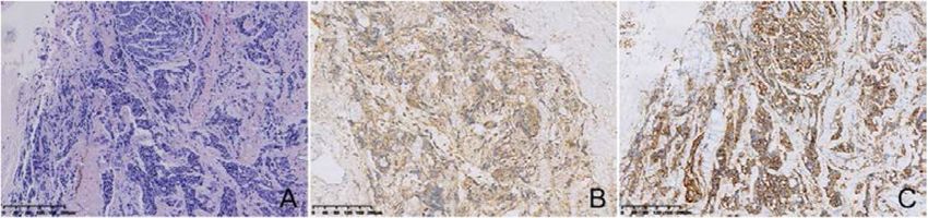
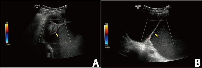
Liu et al. Cancer Imaging     (2020) 20:15                                                                                               Page 3 of 7

 Fig. 1 US shows a huge pelvic cavity solid-cystic mass with separation (arrow) (b), and an echoic nodule (arrow) in the wall (a). The mass is
 mainly composed of cystic components with poor sound transmission, and dense light spot echo could be detected. (CDFI) shows Punctate
 blood flow signal in the tumoral nodule, wall and internal division

diverse neoplasms with a wide range of epidemiology,                        In a review of published work until May 2019, we found
histological characteristics, and biological behavior. A                    only two English case reports about meningioma in a
variety of tissue types derived from one or more germ                       mature cystic ovarian teratoma [3, 4].
cell layers can be observed microscopically, but the most                      The mean age of the patients with MCT was 37.5
common elements consist of cartilage, tissue of the cen-                    years. The patient’s age in the present study was only 15
tral nervous system, and epithelium including respira-                      years compared to 52.5 years in the mean age of the two
tory, gastrointestinal, and cutaneous systems [6–8].                        previously published patients [11]. The clinical manifest-
Tumor or benign and malignant components can also                           ation was not typical, where by it can be hidden for a
be observed. To date, the secondary benign tumors in-                       long time [3]. Pain and enlargement of abdomen was the
cluding mucinous cystadenomas, compound nevus, blue                         most common symptoms. Irregular vaginal bleeding may
nevus, prolactinoma, ACTH-producing pituitary aden-                         be caused by neuroendocrine dysfunction regulating
oma, epithelioid hemangioma, sebaceous adenoma and                          reproduction. Serum tumor markers played an import-
benign skin adnexal tumor arising in mature cystic tera-                    ant role in the differential diagnosis of malignant trans-
tomas of the ovary have been described [3].                                 formation arising from MCT and MCT. Kikkawa et al.
  Teratomas containing meningiomas have been found                          [11] reported that there were significant differences
only in the ovary and testes [2–4, 9]. In addition, tera-                   levels of squamous cell carcinoma antigen (SCC),
toma containing neuroblastoma or immature neuroepi-                         CA125, CEA, and CA19–9 between MCT and squamous
thelial elements has also been reported in ovary. Among                     cell carcinoma arising from MCT. Although this case
teratoma, secondary benign and malignant neoplasms or                       was a mature cystic teratoma, CA125 was significantly
elements may arise from the tissues constituting terato-                    elevated, about 121.20 U/ml. From a clinical point of
mas. The histogenesis of such case of meningioma can                        view, CA125 was also significantly elevated in benign
be considered as a benign or malignant transformation                       ovarian tumors, but usually less than 200 U/ml.
of the components of mature cystic teratoma. The                               Due to the rarity of this tumor, imaging has not made an
arachnoid cells of some organs can be considered as the                     accurate diagnosis or even detailed description. Ultrasonog-
source of meningiomas, such as the dura, nasal and                          raphy is the most common, economical, and simplest
paranasal sinuses, skin, lungs, mediastinum, and periph-                    method for clinical diagnosis of ovarian cystic teratomas.
eral nerves [3]. Therefore, arachnoid cells of some tis-                    Specifically, the US findings of mature cystic teratomas are
sues in teratomas have been the origin of this tumor. In                    various. For instance, a cystic lesion with a densely echogenic
addition, our case reconfirmed that immature neuroepi-                      tubercle projecting into the cyst lumen, or with the seba-
thelial elements have been present in the some mature                       ceous material and hair in the cyst cavity, it presents as a dif-
teratoma [10].                                                              fusely or partially echogenic mass with the echogenic area
  Teratoma with meningioma of the reproductive organs                       normally showing sound attenuation, or a cyst cavity with
was originally described by Takeshima et al. [3] in 2004.                   multiple thin echogenic bands. However, definite diagnosis
Teratoma containing immature neuroepithelial elements                       may be limited by only assessing the internal structure and
in the reproductive organs was firstly described by                         echogenicity of ovarian mass. Therefore, this diagnostic op-
McCullough et al. in 1963. Nevertheless, malignant                          tion is not appropriate in the present study [12, 13]. In
transformation of teratoma components only can be                           addition, CT seems to be the best modality to assist in the
seen in 3–6% of metastatic germ cell tumors. Secondary                      diagnosis of ovarian cystic teratoma [14], it has been com-
benign neoplasm in teratomas is also extremely rare [2].                    monly used to study the possible communication between
Liu et al. Cancer Imaging     (2020) 20:15                                                                                             Page 4 of 7

 Fig. 2 CT images of patient: Unenhanced CT image shows a large oval cystic-solid mass in abdomen pelvic cavity, originating from the left ovary,
 the margin of the mass is clear and the cystic part is the main component in the mass; The round-like solid component shows inhomogeneous
 density shadow, CT value is about 39 HU, with smooth edge yet. The cystic part has internal division change (arrowhead) and fatty lesions (arrow)
 (a). The tumoral solid components (arrowhead) and its internal division (arrow) could be seen intensified from slight to moderate on contrast-
 enhanced CT images compared with those on precontrast images, and the solid component shows heterogeneous enhancement. Adjacent
 organs such as the intestine, the uterus displaced by compression (b, c and d). (b) Arterial phase of contrast enhancement image; (c and d)
 portal phase of contrast enhancement image

the mass and the ovary, adjacent organs, and to further de-                cavity of MCT, just like the soft tissue component in this
scription of the mass.                                                     case, which is called the Rokitansky protuberance [12]. It
  In the present case, the tumor manifested as a multilo-                  is important to recognize malignant transformation of
culated cystic mass with mild to moderate degree en-                       Rokitansky nodule that arise in teratomas because of re-
hancement of solid part and internal division in the cyst,                 lated to the prognosis of patients, especially when Roki-
and fat components can be seen, without calcification.                     tansky nodule are tumors. Patients in whom the
The histopathological examination shows that the tumor                     malignant component is localized to the organ of origin
consists of soft tissue composed of spindle cells and a                    do well, but patients in whom the non-germ cell compo-
cystic space, with the thin-walled cystic lesion has con-                  nent metastasizes do poorly [15].
nective septa covered by fibrocyte. Moreover, a few epi-                     For all these reasons, we are committed to differentiat-
thelioid cell nests are founded in the wall of the cyst.                   ing secondary benign component from malignant compo-
Generally, there is a raised protuberance in the cyst                      nent arising in ovarian cystic teratoma on CT findings.
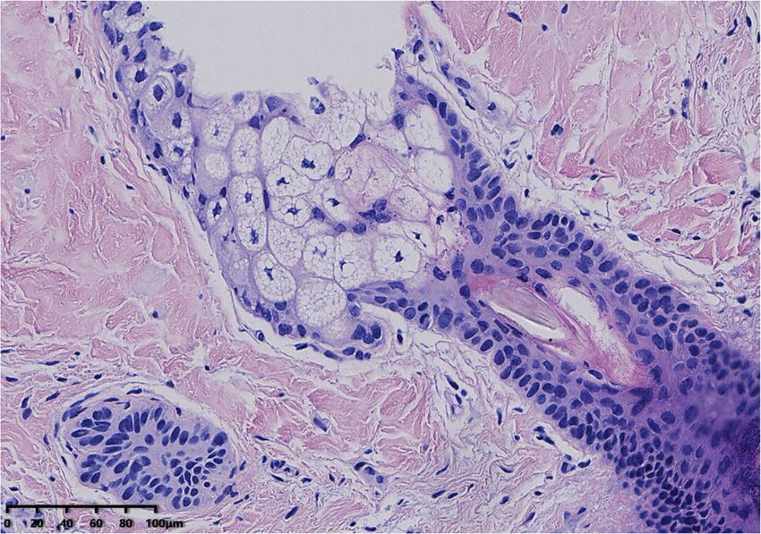
Liu et al. Cancer Imaging      (2020) 20:15                                                                                               Page 5 of 7

 Fig. 3 Microscopic examination of the cystic areas. Image shows sebaceous glands, hair follicle, and squamous epithelium. (HE, × 200)

  The imaging manifestations of secondary malignant                          wall of the cyst, as well as extracapsular tumor growth
tumors in cystic teratomas have been discussed previ-                        with extension into adjacent structures or disseminated
ously. Imaging findings of the tumor usually presented                       metastasis [13]. Buy et al. [14] reported two cases of ma-
as: larger than 9.9 cm of the largest diameter, the pres-                    lignant transformation of teratomas about CT findings,
ence of enhancing soft tissue components, an obtuse                          the suspected cause of the malignancy was a solid mass
angle between the soft tissue components and the inner                       larger than 5 cm in diameter with irregular borders

 Fig. 4 Histological appearance (hematoxylin and eosin stain) of Sections from the solid areas. Histological appearance of the solid areas reveals
 spindle cells in lobular growth pattern, syncytium-like appearance due to poorly defined cell borders, scattered clear nuclear holes, and
 occasional intranuclear pseudoinclusions, suggesting a meningothelial meningioma. (H&E, × 100)
Liu et al. Cancer Imaging        (2020) 20:15                                                                                                   Page 6 of 7

 Fig. 5 Microscopic and immunohistochemical features of the local cyst wall. The tumour cells was small with a round or polygonal, darkly
 staining nucleus with signs of division (H&E, × 100) (a). Immunohistochemical staining supported Source of neuroblasts with positive staining for
 S-100 protein (b) and CD56 (c)

forming an obtuse angle with the inner wall of the cyst                         hysterectomy, omentectomy and pelvic-paraaortic
in one case and with uptake of contrast medium in an-                           lymph node dissection. After the operation is com-
other case. In our case, although the maximum diameter                          pleted, radiotherapy or chemotherapy can be per-
of the soft tissue composition was more than 5 cm as                            formed according to the actual condition of the
well as the border of the enhancing soft tissue compo-                          patient [18] .However, when there is no malignant
nents made an obtuse angle laterally with the cyst wall,                        transformation in MCT, laparoscopic surgery is usu-
the soft tissue appearance is smooth and regular. Finally,                      ally performed [11]. Therefore, knowledge of any pos-
according to the WHO classification of tumors of the                            sible malignant transformation of a mature teratoma
nervous systems [16], the meningioma constituents is                            and its relationship to adjacent organs could be valu-
considered to be a benign meningioma (WHO grade I).                             able for surgical planning [19]. Inaddition, for young
Therefore, Rokitansky protuberance is an important CT                           women of childbearing age or fertility requirements
finding.                                                                        with pure ovarian MCT, surgical procedures are often
  Ovarian teratoma containing meningioma is a rarely                            used to ovarian cysts decollement and preserve normal
seen tumor. Pure ovarian cysts can be identified by                             ovarian tissue on the affected side. Takeshima et al.
ultrasound alone. Pure ovarian cysts usually have a                             [3] reported a 60-year-old woman with microcystic
circular or elliptical clear liquid sound transmission                          meningioma arising in a mature cystic teratoma of the
area with clear boundaries and thin, smooth cyst wall                           right ovary, who performed salpingo-oophorectomy.
with a diameter of 3–8 cm. An acoustic enhancement                              Mandal et al. [4] reported a 45-year-old female with
effect was observed on the back wall and posterior of                           psammomatous meningioma arising in a mature cystic
the cyst. CDFI showed no blood flow in the cyst wall                            teratoma of the left ovary, who carried out total ab-
[17]. Nevertheless, the definitive diagnosis of ovarian                         dominal hysterectomy with bilateral saplingo-
mature cystic teratoma containing meningioma and                                ophorectomy. While recurrence of meningioma in ma-
nests of neuroblasts depended on a combination of                               ture cystic teratoma of the ovary had not been re-
clinical, radiological, and histopathological evidences.                        ported previously, the benign elements of cystic
Despite mature cystic teratoma containing a low grade                           teratoma can be transformed into malignant elements
meningeoma being a benign disease, there is a possi-                            and malignant epithelium that develop within a pre-
bility of recurrence and malignant transformation. In                           existing teratoma can continue to proliferate and
the terms of secondary malignant transformation aris-                           clone [4]. Therefore, strict follow-up after surgery is
ing from mature cystic teratoma. The surgical resec-                            necessary.
tion appears to be the most effective treatment,
especially when the tumor increases in size and applies                         Conclusion
pressure on the surrounding tissues. Specific methods                           Ovarian mature cystic teratoma containing both meningi-
including bilateral salpingo-oophorectomy, total                                oma and neuroblasts components is extremely rare, but
Table 1 Clinical and imaging findings analysis of mature cystic teratoma with meningioma in 3 patients
Case                      Age,     Size, cm              solid areas                                   Trentment       Prognosis     Other component
                          y
                                                         Border(I/R)    Size, cm          Enhance
1 Takeshima et al. [3]    60       10 × 10 × 8           NE             3x3x2             NE           RSO             NR            NO
2 Shramana et al. [4]     45       12.4x11x9.5           NE             4 × 3.2 × 3       NE           HBSO            NR            NO
3 present case            15       19.6 × 12.5 × 26.3    Regular        3.8 × 4.1 × 3.6   (+)          LSO             NR            Nests of neuroblasts
RSO Right salpingo-oophorectomy, HBSO hysterectomy with bilateral saplingo-oophorectomy, LSO Left salpingo-oophorectomy, NE no evaluation, NR
no recurrence
Liu et al. Cancer Imaging (2020) 20:15 Page 7 of 7
preoperative diagnosis is very important because it involves 5. Borak SG, Ross JR, Bell WC. Granular cell tumor within an ovarian mature
the choice of treatment options and prognosis of patients. cystic Teratoma: report of a unique case and review of the literature. Int J
Gynecol Pathol. 2017;36:453–8.
It is recommended to determine the pathology through 6. Roy S, Mukhopadhayay S, Gupta M, Chandramohan A. Mature cystic
preoperative puncture when fat and enhanced soft tissue Teratoma with co-existent mucinous Cystadenocarcinoma in the same
components are found in cystic tumors during CT examin- ovary-a diagnostic dilemma. J Clin Diagn Res. 2016;10:ED11–ED3.
7. Feng X, Xu L. Rare case of squamous cell carcinoma arising in a recurrent
ation. Despite it being a benign disease, there is a possibility ovarian mature cystic teratoma of a young woman: a case report and
of recurrence and malignant transformation. Radical surgi- review of the literature. Medicine (Baltimore). 2018;97:e10802.
cal resection is needed to treat this tumor and discreetly 8. Gurda GT, VandenBussche CJ, Yonescu R, Gonzalez-Roibon N, Ellis CL,
Batista DA, et al. Sacrococcygeal teratomas: clinico-pathological
follow-up with CT or ultrasound is essential. characteristics and isochromosome 12p status. Mod Pathol. 2014;27:562–8.
9. Allen EA, Burger PC, Epstein JI. Microcystic meningioma arising in a mixed
Abbreviations germ cell tumor of the testis: a case report. Am J Surg Pathol. 1999;23:
ACTH: Adreno-cortico-tropic-hormone; CA125: Carbohydrate antigen 125; 1131–5.
CA19–9: Carbohydrate antigen 19–9; CD56: Carbohydrate antigen 56; 10. Reid HA, van der Walt JD, Fox H. Neuroblastoma arising in a mature cystic
CDFI: Color Doppler flow imaging; CEA: Carcinoembryonic antigen; teratoma of the ovary. J Clin Pathol. 1983;36:68–73.
CT: Computed tomography; E-cadherin: Epithelial cadherin; GFAP: Glial 11. Kikkawa F, Nawa A, Tamakoshi K, Ishikawa H, Kuzuya K, Suganuma N, et al.
fibrillary acidic protein; GFAP: Glial fibrillary acidic protein; H&E: Hematoxylin Diagnosis of squamous cell carcinoma arising from mature cystic teratoma
and eosin; Ki67: Antigen KI67; MCT: Mature cystic teratoma; PR: Progesterone of the ovary. Cancer. 1998;82:2249–55.
receptor; S100: Soluble protein-100; SCC: Squamous cell carcinoma antigen; 12. Sahin H, Abdullazade S, Sanci M. Mature cystic teratoma of the ovary: a
SSTR2: Somatostatin receptor; US: Ultrasound; WHO: World Health cutting edge overview on imaging features. Insights Imaging. 2017;8:
Organization 227–41.
13. Park SB, Kim JK, Kim KR, Cho KS. Preoperative diagnosis of mature cystic
Acknowledgements teratoma with malignant transformation: analysis of imaging findings and
None. clinical and laboratory data. Arch Gynecol Obstet. 2007;275:25–31.
14. Buy JN, Ghossain MA, Moss AA, Bazot M, Doucet M, Hugol D, et al. Cystic
Authors’ contributions teratoma of the ovary: CT detection. Radiology. 1989;171:697–701.
YYL: manuscript preparation, literature research, and data analysis. PL: 15. Ahmed T, Bosl GJ, Hajdu SI. Teratoma with malignant transformation in
literature research and data analysis. JJ: Imaging data collection and analysis. germ cell tumors in men. Cancer. 1985;56:860–3.
KSC: Guidance of pathological knowledge. LML: data acquisition. LLY: 16. Riemenschneider MJ, Perry A, Reifenberger G. Histological classification and
manuscript editing. JBG: study conception and design, manuscript review molecular genetics of meningiomas. Lancet Neurol. 2006;5:1045–54.
and guarantor of integrity of the entire study. All authors have read and 17. Zhang Y, Yuan Z, Sun K, Li P. Ultrasonic and pathological characteristics of
approved the final manuscript. ovarian mucinous cystic tumors with malignant mural nodules: two cases
report. Medicine (Baltimore). 2017;96:e8636.
Funding 18. Hackethal A, Brueggmann D, Bohlmann MK, Franke FE, Tinneberg HR,
National Natural and Science Fund of China (NO. 81671682). Munstedt K. Squamous-cell carcinoma in mature cystic teratoma of the
ovary: systematic review and analysis of published data. Lancet Oncol. 2008;
Availability of data and materials 9:1173–80.
Not applicable. 19. Wang LJ, Chu SH, Ng KF, Wong YC. Adenocarcinomas arising from primary
retroperitoneal mature teratomas: CT and MR imaging. Eur Radiol. 2002;12:
Ethics approval and consent to participate 1546–9.
The study protocol was approved by the institutional review board and due
to the retrospective nature of the study, the informed consent requirement
was abandoned. Publisher’s Note
Springer Nature remains neutral with regard to jurisdictional claims in
Consent for publication published maps and institutional affiliations.
Not applicable.
Competing interests
The authors declare that they have no competing interests.
Author details
1
Department of Radiology, The First Affiliated Hospital of Zhengzhou
University, Zhengzhou 450052, Henan Province, China. 2Department of
Radiology, General Hospital, Ningxia Medical University, Yinchuan 750004,
China.
Received: 29 September 2019 Accepted: 15 January 2020
References
1. Maharjan S. Mature cystic teratoma of ovary with squamous cell carcinoma
arising from it. Clin Case Rep. 2019;7:668–71.
2. Castro Pereira FM, de Oliveira MG, Almeida Ldo C, Pires BC, de Bessa Junior
J. A rare component of psammomatous meningioma in a testicular
teratoma. Case Rep Pathol. 2013;2013:645415.
3. Takeshima Y, Kaneko M, Furonaka O, Jeet AV, Inai K. Meningioma in mature
cystic teratoma of the ovary. Pathol Int. 2004;54:543–8.
4. Mandal S, Dhingra K, Gupta P, Khurana N. Rare growth of a psammomatous
meningioma in a mature ovarian teratoma: a case report. Pathol Res Pract.
2010;206:322–4.You can also read