Gynecological malignancy mimicking a thyroid lymph node metastasis
←
→
Page content transcription
If your browser does not render page correctly, please read the page content below
S Pederzoli and others                Neck lymph node metastasis:              ID: 20-0055; January 2021
                                                                                not always thyroid                       DOI: 10.1530/EDM-20-0055

Gynecological malignancy mimicking a
thyroid lymph node metastasis

Simone Pederzoli1,2, Giorgia Spaggiari1, Giuditta Bernardelli3, Francesco Mattioli4,
Cinzia Baldessari5, Antonino Maiorana3, Vincenzo Rochira1,2 and Daniele Santi 1,2
1Unit  of Endocrinology, Department of Medical Specialties, Azienda Ospedaliero-Universitaria of Modena, Modena,
                                                                                                                         Correspondence
Italy, 2Unit of Endocrinology, Department of Biomedical, Metabolic and Neural Sciences, 3Department of Pathology,
                                                                                                                         should be addressed
University of Modena and Reggio Emilia, Modena, Italy, 4Department of Otorhinolaryngology-Head and Neck
                                                                                                                         to D Santi
Surgery, and 5Department of Oncology and Haematology, Azienda Ospedaliero-Universitaria of Modena,
                                                                                                                         Email
Modena, Italy
                                                                                                                         daniele.santi@unimore.it

Summary
We present the case of a 69-year-old woman who attended the Endocrinology Unit of Modena for a suspicious lymph
node in the left cervical compartment discovered during the follow-up of a recurrent gynecological malignancy. At neck
ultrasonography, a thyroid goiter was detected, and the further cytological examination was inconclusive for thyroid
nodule and compatible with a localization of an adenocarcinoma with papillary architecture for the lymph node. The
histological examination after a left neck dissection confirmed the presence of an intracapsular metastasis of a papillary
carcinoma immunohistochemically focally positive for thyroid transcription factor 1 and paired box 8 and negative for
thyroglobulin. Subsequently, in the suspicion of a thyroid primitiveness, a total thyroidectomy was performed, revealing
an intraparenchymal follicular variant of papillary thyroid carcinoma of 2 mm in the right lobe. During the follow-up,
the appearance of a suspected cervical metastatic lesion led to another neck dissection, histologically compatible with
a papillary carcinoma localization, immunohistochemically focally positive for thyroid transcription factor 1 and paired
box 8, and negative for thyroglobulin. The histological revision of surgical specimens suggests the cervical recurrence of
the prior gynecological cancer, rather than a thyroid carcinoma metastasis. The case described shows how carefully the
cytological, histological and immunoistochemical results must be evaluated in oncological management, considering the
whole patient’s history.

  Learning points:
  •• Neck lymph node metastases occasionally originate from anatomically distant primary sites, such as breast, lung,
     gastro-intestinal tract, genito-urinary tract and CNS.
  •• Histological and immunohistochemical evaluations play an important role to identify the primary malignant site,
     although in some cases they could mislead the clinicians.
  •• A multidisciplinary approach and the evaluation of the whole medical history of the patient are mandatory to
     guide the diagnostic-therapeutic path and to avoid unnecessary treatments.

Background
Cervical lymph nodes are a typical site of metastases for                       tract, thyroid, salivary gland and skin (2). However, neck
malignant tumors originating from the head–neck region                          lymphatic metastases occasionally originate from remote
(1). Commonly, malignancies most frequently showing                             primary sites, such as breast, lung, gastro-intestinal tract,
cervical lymphatic spread arise from upper aero-digestive                       genito-urinary tract and CNS (3). In the management

                               This work is licensed under a Creative Commons    © 2021 The authors                           https://edm.bioscientifica.com/
                               Attribution-NonCommercial-NoDerivatives 4.0                                                     Published by Bioscientifica Ltd
                               International License.
                                                                                                             Downloaded from Bioscientifica.com at 02/03/2021 11:50:53AM
                                                                                                                                                           via free access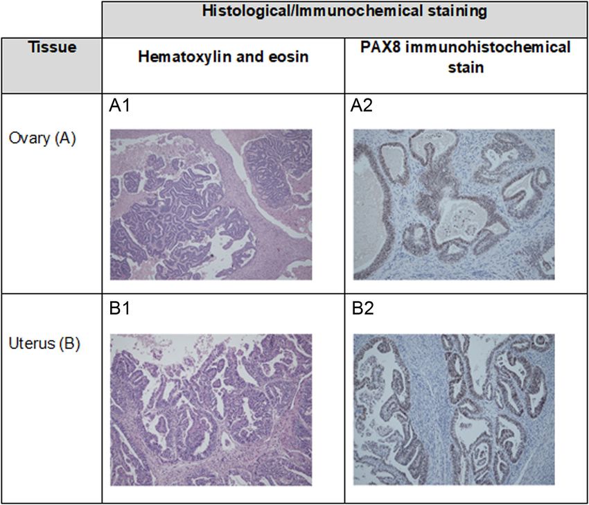
S Pederzoli and others       Neck lymph node metastasis:                 ID: 20-0055; January 2021
                                                              not always thyroid                          DOI: 10.1530/EDM-20-0055

of cervical lymph node metastasis, the first mandatory        2009. Histologically, the cystadenocarcinoma was
step is the identification of the tumor’s primary site,       mainly endometroid-like with squamous metaplasia
influencing the survival, the clinical outcome and the        areas and papillary features (Fig. 1, panels A and B). The
consequent follow-up. However, since the metastases           carcinoma affected both ovaries, the myometrium and
could be anatomically far from the primitive site (3), it     six on 23 lymph nodes removed. After the surgery, the
is crucial to improve the diagnostic work-up considering      patient underwent paclitaxel-carboplatin chemotherapy
several potential tools. Indeed, cytology, histology and      until June 2010, then repeated from February to June
immunohistochemistry could play a considerable role to        2013. Meanwhile, two further lymph node dissections
identify the malignant primary site, both by defining the     involving retroperitoneal, lumbar and pelvic regions
neoplasm architecture and using tissue-specific markers       were performed in September 2011 and in January 2012
(4). Secondary disease locations usually share the growth     for the persistence of multiple node metastases. Finally,
structural architecture with primary tumors, and they         the disease persistence was treated with radiotherapy
could express, in several cases, immunohistochemical          from September to December 2014. Since then, no other
markers typical of the native tissue by which the             signs of persistence/recurrence of neoplastic disease were
neoplasm originates (5). However, especially in case of       reported.
poorly differentiated carcinomas or particular histological       Neither familial history nor prior episodes of thyroid
types (i.e. adenocarcinomas), even the comprehensive          diseases emerged during the anamnestic interview. The
evaluation of cyto-histological and immunohistochemical       patient’s chronic therapy consisted of acetylsalicylic acid
features could be inconclusive (2).                           and statin in primary prevention for the presence of non-
     Several immunohistochemical markers are currently        hemodynamically significant carotid stenosis and eye
considered representative of a specific tissue or organ.      drops for bilateral glaucoma.
However, many of these markers could be expressed in
variable percentages also in different types of tumors,
                                                              Investigation
originating from different sites (6). This overlap
represents the main challenge in the clinical application     At the first visit, the neck physical examination was
of immunohistochemistry within the diagnostic work-up         unremarkable, in absence of palpable nodules in the
of distant metastases.                                        thyroid region. While normal values of thyroid-stimulating
     Here, we present a clinical case in which the
histological and immunohistochemical features of
a lymph node metastasis oriented the diagnostic
pathway to the wrong primary tumor. In particular, a
thyroid primitiveness was suspected and supported by
immunohistochemical evaluations, although the patient’s
clinical history suggested a more likely persistence of the
prior gynecological malignancies than a new thyroid
implication.

Case presentation
A 69-year-old Caucasian woman attended the
Endocrinology Unit of Modena in August 2017 for a
multinodular thyroid goiter newly diagnosed. This
incidental finding occurred for the detection of a
fluorodeoxyglucose PET (FDG-PET)-positive lymph
node in the left cervical compartment, subsequently
investigated with a neck ultrasound. Indeed, she was in       Figure 1
oncological follow-up for an ovarian and uterine mixed        Histological and immunohistochemical findings during the oncological
                                                              follow-up of the patient. Pathology preparations of ovary (panel A) and
cystadenocarcinoma treated with a total laparoscopic
                                                              uterus (panel B). Panels 1 refer to hematoxylin and eosin stain, panels 2
hysterectomy and a bilateral oophorectomy with pelvic         refer to immunohistochemical positivity for PAX8. Light microscope
and lumbar-aortic lymphadenectomy in December                 images; original magnification: 100×. (PAX8 = paired-box gene 8).

https://edm.bioscientifica.com/2
                                                                                              Downloaded from Bioscientifica.com at 02/03/2021 11:50:53AM
                                                                                                                                            via free access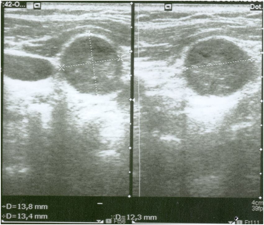
S Pederzoli and others Neck lymph node metastasis: ID: 20-0055; January 2021
not always thyroid DOI: 10.1530/EDM-20-0055
hormone (TSH) were detected (0.6 µIU/mL, reference
Microcalcifications
range 0.27–4.20 µIU/mL), the sonographic evaluation
revealed a thyroid of normal size, with an inhomogeneous
echo-structure due to the presence of multiple nodules.
Present
Absent
Absent
Absent
Absent
In particular, four nodules were detected in the right
thyroid lobe and one in the left one. Ultrasound features
of the nodules were reported in Table 1. Three nodules
(B, C, D) were classified as intermediate-risk thyroid
lesions, while the nodule A and E belonged to the high-
risk category according to Gharib et al. (7). Moreover, at
Vascularization
Intranodular
Intranodular
the third left level of Robbins corresponding to the PET
Peripheral
Peripheral
Peripheral
uptake, an inhomogeneous hypoechoic roundish lesion
compatible with a suspicious lymph node was detected
(Fig. 2). Ultrasound evaluation was performed by Esaote®
My Lab25 Gold (Malmesbury, Wiltshire, UK). According
Irregular
Irregular
Irregular
Irregular
Irregular
Margins
to the sonographic characteristics, an ultrasound-guided
fine-needle aspiration (FNA) biopsy of nodules A and
E and of the left cervical lesion was performed. The
cytological examination of the nodule A highlighted
an indeterminate lesion (Tir3a according to the Italian
Iso-hypoechoic
Iso-hypoechoic
Iso-hypoechoic
Iso-hypoechoic
hypoechoic
Ultrasonographic characteristics of the thyroid nodules detected after the neck ultrasound.
Echogenicity
thyroid cytology classification system, 2013) (8),
suspected for the follicular nature, while the sample of the
Marked
nodule E resulted inadequate (Tir1). The histology of the
cervical lymph node was compatible with a localization
of adenocarcinoma with papillary features, not otherwise
Longitudinal
specified (Fig. 3, panel C1). An immunohistochemical
15.5
7.9
9.3
11.9
12.1
positivity for the paired box 8 (PAX8) (Fig. 3, panel C2)
and for the thyroid transcription factor1 (TTF1) (Fig. 3,
panel C3) suggested a possible thyroid primitiveness of
Diameters (mm)
the left cervical lesion. However, the thyroglobulin (Tg)
Transverse
concentration in washout fluid from FNA biopsy (FNA-Tg)
16.0
8.0
6.9
10.7
9.4
was undetectable and the Tg immunohistochemistry was
negative on the cytological sample. Since the clinical
picture remained unclear, the FNA examination was
Anteroposterior
repeated, confirming the cancerous nature of the cervical
lymph node, in absence of evident cytological atypia of
12.5
5.9
7.1
9.1
8.6
the thyroid nodules evaluated.
Treatment
Middle third
Middle third
Middle third
Upper third
Lower third
Following the cytological result, in October 2017 the
Location
patient underwent a left neck dissection (levels II–IV of
Robbins) at the Unit of Otorhinolaryngology of Modena.
The histological examination revealed intracapsular
metastasis of papillary carcinoma in two nodes, resulting
Right
Right
Right
Right
Lobe
immunohistochemically diffusely positive for PAX8 (Fig.
Left
3, panel D2), focally positive for TTF1 (Fig. 3, panel D3),
negative for Wilms tumor 1 (WT1), estrogen and Tg. The
Table 1
Nodule
molecular characterization of the metastases revealed a
mutation in codon 12 of KRAS (c.35 guanine>alanine),
D
B
A
C
E
https://edm.bioscientifica.com/3
Downloaded from Bioscientifica.com at 02/03/2021 11:50:53AM
via free accessS Pederzoli and others Neck lymph node metastasis: ID: 20-0055; January 2021
not always thyroid DOI: 10.1530/EDM-20-0055
an umpteenth disease recurrence, a PET-computerized
tomography (CT) was performed, confirming a
pathological glucose uptake in correspondence of the
detected lesion (standardized uptake value 20.7).
After the surgical removal of the mass, the
histological examination revealed a localization of
papillary carcinoma without any evidence of nodal
residues, immunohistochemically diffusely positive for
PAX8, focally positive for TTF1 and p53, and negative
for estrogen receptor and cancer antigen 125 (Fig. 3,
panels E). Considering (i) the overall oncologic history,
(ii) the very low probability of metastatic disease due
to the very small primary thyroid cancer, and (iii) the
immunophenotype of metastases, which was also similar
to the prior gynecological tumor profile, a pathological
revision of the case was carried out, suggesting a more
likely ovarian/uterine cystadenocarcinoma persistence of
Figure 2
disease, ‘exonerating’ the thyroid primitiveness.
Ultrasound image of the lymph node on the third left level of Robbins,
corresponding to the positron emission tomography (PET) uptake. The Considering the aggressive disease behavior, the
lesion appears rounded, markedly hypoechoic and inhomogeneous with seeming unique metastatic site, as well as the good general
increased vascularization. Under the image, the anteroposterior,
conditions of the patient, an adjuvant radiotherapy on
transverse and longitudinal diameter were reported.
the left cervical region was further proposed and then
while no alterations were detected on BRAF and NRAS performed in January 2019.
genes. Since PAX8 and TTF1 could also be expressed At the last follow-up evaluation in September 2019,
in different kind of tumors (Table 2), such as thyroid, there were no signs of relapse for both malignancies,
ovarian and uterine ones, there was no certainty about that is, the ovarian/uterine adenocarcinoma and the
the primitive origin of the neoplasia. Nevertheless, in thyroid microcarcinoma. In particular, the biochemical
the suspicion of a thyroid-native malignancy, a total assessment documented Tg serum levels close to zero (0.2
thyroidectomy associated with bilateral central neck ng/mL), in absence of anti-Tg antibodies, and a negative
dissection was performed. After the surgery, the patient ultrasound neck scan. Moreover, a normal phospho-calcic
started the levothyroxine replacement therapy at the profile was detected suggesting a recovery of parathyroid
posology of 75 µg daily and a calcium-vitamin integration functionality, allowing the calcium-vitamin therapy
for the occurrence of a post-surgical hypoparathyroidism. discontinuation.
The thyroid histological examination revealed an
intraparenchymal follicular variant of papillary thyroid
Discussion
carcinoma of 2 mm in the right lobe (pT1as N0a M0 –
stage 1 according to the 8th edition of the American Joint The case described represents an emblematic example
Committee on Cancer staging system) (9). Considering of the limits of histological and immunohistochemical
the low stage of the thyroid disease, a further ablative evaluations in oncological pathology. In our patient, the
treatment with iodine-131 was deemed unnecessary, and TTF1 and PAX8 positivity at the immunohistochemical
a biochemical and sonographic follow-up was proposed. analysis of the neck metastatic lymph node focused
the clinical attention to the thyroid gland. Indeed, the
combined TTF1 and PAX8 positivity could be identified
Outcome and follow-up
in thyroid carcinomas (10). TTF1 belongs to the NKx2
After 7 months, the patient referred the appearance of a family of homeodomain transcription factors and
further palpable cervical swelling at the left Vb level of mediates the transcription of the Tg (11). Although an
Robbins. A neck ultrasound scan showed a vascularized immunohistochemical positivity for TTF1 and Tg is
nodule of 28 mm of longitudinal diameter with a detectable in the majority of thyroid tumors with similar
mixed component, both fluid and solid, in the context sensitivity, TTF1 is more expressed in poorly differentiated
of the sternocleidomastoid muscle. In the suspect of carcinomas and related metastasis compared to Tg (11). In
https://edm.bioscientifica.com/4
Downloaded from Bioscientifica.com at 02/03/2021 11:50:53AM
via free accessS Pederzoli and others Neck lymph node metastasis: ID: 20-0055; January 2021
not always thyroid DOI: 10.1530/EDM-20-0055
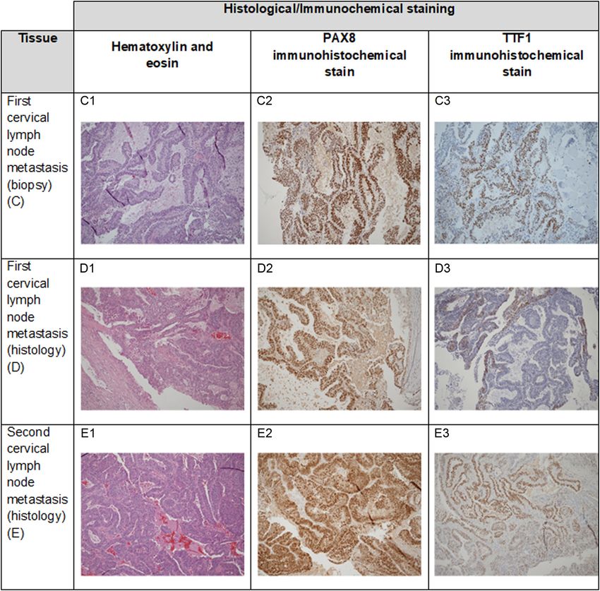
Figure 3
Histological and immunohistochemical findings during the oncological follow-up of the patient. Pathology preparations of first cervical lymph node
metastasis (biopsy: panel C, histology: panel D) and second cervical lymph node metastasis (panel E) were reported. Panels 1 refer to hematoxylin and
eosin stain, panels 2 refer to immunohistochemical positivity for PAX8, and panels 3 refer to immunohistochemical positivity for TTF1. Light microscope
images; original magnification: 100×. (PAX8 = paired-box gene 8; TTF1 = thyroid transcription factor 1).
our case, we detected a TTF1+/Tg− in the first metastasis, female and male reproductive system (13). Thus, the PAX8
thus the possible existence of a poorly differentiated positivity detected in our case in the first lymph node
thyroid cancer could not be a priori excluded. On the metastasis could not rule out a primitive thyroid cancer.
other hand, since PAX8 is a fundamental transcription However, both TTF1 and PAX8 immunohistochemical
factor for organogenesis of the thyroid gland, kidney, positivity is not pathognomonic of thyroid primitiveness,
and Mullerian-derived organs (12), its expression is not since their expression could be present in variable
surprisingly detectable in a relevant percentage of tumor percentage in several tumors (Table 2) (14). This
originating from thyroid, renal-urinary tract, breast and challenge could be further complicated by the histologic
https://edm.bioscientifica.com/5
Downloaded from Bioscientifica.com at 02/03/2021 11:50:53AM
via free accessS Pederzoli and others Neck lymph node metastasis: ID: 20-0055; January 2021
not always thyroid DOI: 10.1530/EDM-20-0055
Table 2 Expression of thyroid transcription factor 1 (TTF1) pathological revision has classified this microcarcinoma
and paired-box gene 8 (PAX8) in different types of human as an incidental finding. This condition is not infrequent
cancer. for thyroid carcinomas, particularly for microcarcinomas,
since the fortuitous post-surgery histological identification
Expression Cancer type
of these malignancies is described in large series of patients
TTF1
thyroidectomized for benign thyroid diseases (18). Indeed,
100% Glioma
thyroid tumors present generally a low aggressiveness and
Thyroid cancer
Carcinoid a slow growth, requiring often several years to spread to
Colorectal cancer locoregional lymph nodes and/or to other organs. As a
Head and neck cancer confirmation, the clinician attitude at treating this kind of
Testis cancer
tumors is actually debated in the scientific community and
Breast cancer
90% Prostate cancer an increasingly conservative approach is proposed (18).
Cervical cancer Cervical lymph nodes metastases typically originate
80% Lung cancer from head and neck malignancies, due to anatomical
Stomach cancer
proximity and to the structure of the lymphatic drainage
Skin cancer
78% Urothelial cancer pathways from these sites (2). However, anatomically-
Melanoma distant primary sites are described (2). Although the
70% Ovarian cancer physio-pathological mechanism of atypical lymphatic
Pancreatic cancer
spreads remains unclear, the correct identification of the
60% Liver cancer
50% Lymphoma primitive malignancy exerts a significant impact on the
Endometrial caner patient’s management (2). Considering gynecological
45% Renal canner malignancies, even if the main lymphatic dissemination
PAX8
79–90 % Thyroid cancer
route involves the retroperitoneal pelvic and para-aortic
88–100 % Renal cancer lymph nodes, cases of further extension to the cervical
56–74 % Carcinoid nodes are reported (19), especially for cervical and ovarian
79–100% Ovarian cancer (primary) cancers, rather than endometrial ones (2). For anatomical
70–96 % Ovarian cancer (metastatic)
reasons, left-sided neck metastases are more frequent (20),
but cervical lymph node involvement from gynecological
appearance. Indeed, a micro-papillary histologic structure malignancies remains rare and only few cases are reported
could be common in thyroid cancers and also in ovarian/ in the literature (21).
endometrial carcinomas (15). In our case, the discovery of In the case described, a controversial cyto-histological
an adenocarcinoma in the cervical lymph node dissected profile together with an atypical cervical localization of a
with the peculiar immunohistochemistry (i.e. TTF1+/ gynecological malignancy misled the clinicians, causing a
Tg-/PAX8+) could be explained by either a thyroid or probably inappropriate thyroid surgical removal. Although
ovarian/endometrial primitiveness. Considering that the the incidental finding of a thyroid microcarcinoma
papillary-featured adenocarcinoma represents the most partially reduces the inadequacy of the procedure, the
common histological type of thyroid malignancies (16) patient has been exposed to thyroid surgery complications,
and the concomitant presence of thyroid nodules with such as hypoparathyroidism and recurrent laryngeal nerve
undetermined cytology, a possible thyroid primitiveness injury (22), and to the need for chronic levothyroxine
was suspected. In contrast, FNA-Tg was negative on the replacement therapy. Thus, in presence of an histologically
cervical lymph node metastasis. However, a false negativity documented thyroid microcarcinoma and cervical
in FNA-Tg could be detected in thyroid-malignant lymph lymph nodes metastases other primary cancer should be
nodes in several cases (17). considered according to patient’s medical history, because
Considering all previous histological and the metastases of these non-thyroidal adenocarcinomas
immunohistochemical features of the cervical lymph may mask a metastasis due to thyroid cancer.
node, a thyroid dissection was performed, revealing a In conclusion, cyto-histological analyses and
follicular variant of intraparenchymal micro-papillary immunohistochemical evaluations represent fundamental
thyroid carcinoma at the histological examination. tools in clinical oncology management. However, an
However, the further medical history and the subsequent adequate approach cannot disregard the evaluation of the
https://edm.bioscientifica.com/6
Downloaded from Bioscientifica.com at 02/03/2021 11:50:53AM
via free accessS Pederzoli and others Neck lymph node metastasis: ID: 20-0055; January 2021
not always thyroid DOI: 10.1530/EDM-20-0055
previous medical history, together with the findings derived 6 Kalampokas E, Payne F, Nomikos A & Gurumurthy M. An update
on the use of immunohistochemistry and molecular pathology in
from physical, biochemical and radiological examinations.
the diagnosis of pre-invasive and malignant lesions in gynecological
In a controversial clinical presentation, a multidisciplinary oncology. Gynecologic Oncology 2018 150 378–386. (https://doi.
approach is more than ever mandatory to guide the org/10.1016/j.ygyno.2018.05.023)
7 Gharib H, Papini E, Garber JR, Duick DS, Harrell RM, Hegedus L,
diagnostic-therapeutic path, avoiding unnecessary
Paschke R, Valcavi R & Vitti P. American Association of Clinical
treatments and the possible related complications. Endocrinologists, American College of Endocrinology, and
Associazione Medici Endocrinologi Medical Guidelines for Clinical
Practice for the diagnosis and management of thyroid nodules
– 2016 update. Endocrine Practice 2016 22 1–60. (https://doi.
Declaration of interest org/10.4158/EP161208.GL)
The authors declare that there is no conflict of interest that could be 8 Nardi F, Basolo F, Crescenzi A, Fadda G, Frasoldati A, Orlandi F,
perceived as prejudicing the impartiality of the research reported. Palombini L, Papini E, Zini M, Pontecorvi A, et al. Italian consensus
for the classification and reporting of thyroid cytology. Journal
of Endocrinological Investigation 2014 37 593–599. (https://doi.
org/10.1007/s40618-014-0062-0)
Funding 9 Zanoni DK, Patel SG & Shah JP. Changes in the 8th edition of the
This work did not receive any specific grant from any funding agency in the American Joint Committee on Cancer (AJCC) staging of head and
public, commercial or not-for-profit sector. neck cancer: rationale and Implications. Current Oncology Reports
2019 21 52. (https://doi.org/10.1007/s11912-019-0799-x)
10 Nikiforov YE. Molecular diagnostics of thyroid tumors. Archives of
Pathology and Laboratory Medicine 2011 135 569–577. (https://doi.
Patient consent org/10.1043/2010-0664-RAIR.1)
Written informed consent for publication of her clinical details was 11 Bejarano PA, Nikiforov YE, Swenson ES & Biddinger PW. Thyroid
obtained from the patient. transcription factor-1, thyroglobulin, cytokeratin 7, and cytokeratin
20 in thyroid neoplasms. Applied Immunohistochemistry and Molecular
Morphology 2000 8 189–194. (https://doi.org/10.1097/00129039-
200009000-00004)
12 Tacha D, Zhou D & Cheng L. Expression of PAX8 in normal and
Author contribution statement
neoplastic tissues: a comprehensive immunohistochemical study.
S P, G S and D S wrote the manuscript and were involved in the endocrine
Applied Immunohistochemistry and Molecular Morphology 2011 19
care of the patient. V R revised the manuscript. F M performed the
293–299. (https://doi.org/10.1097/PAI.0b013e3182025f66)
neck surgery on the patient. G B and A M provided histological and
13 Xiang L & Kong B. PAX8 is a novel marker for differentiating
immunohistochemical images and performed the anatomo-pathological
between various types of tumor, particularly ovarian epithelial
analyses during the patient management. C B followed the patient within
carcinomas. Oncology Letters 2013 5 735–738. (https://doi.
the oncological team.
org/10.3892/ol.2013.1121)
14 Zhang Y, Yang J, Zhang M, Meng Z, Song W, Yang L, Li L, Wang D
& Shi T. Thyroid follicular carcinoma-like renal tumor: a case
report and literature review. Medicine 2018 97 e10815. (https://doi.
References org/10.1097/MD.0000000000010815)
1 Pilborough AE, Lambert DW & Khurram SA. Extranodal extension in 15 Kaku T, Ogawa S, Kawano Y, Ohishi Y, Kobayashi H, Hirakawa T
oral cancer: a role for the nodal microenvironment? Journal of Oral & Nakano H. Histological classification of ovarian cancer. Medical
Pathology and Medicine 2019 48 863–870. (https://doi.org/10.1111/ Electron Microscopy 2003 36 9–17. (https://doi.org/10.1007/
jop.12870) s007950300002)
2 Lopez F, Rodrigo JP, Silver CE, Haigentz M, Jr, Bishop JA, Strojan P, 16 Baloch ZW & LiVolsi VA. Cytologic and architectural mimics of
Hartl DM, Bradley PJ, Mendenhall WM, Suarez C, et al. Cervical papillary thyroid carcinoma. Diagnostic challenges in fine-needle
lymph node metastases from remote primary tumor sites. Head aspiration and surgical pathology specimens. American Journal of
and Neck 2016 38 (Supplement 1) E2374–E2385. (https://doi. Clinical Pathology 2006 125 (Supplement) S135–S144. (https://doi.
org/10.1002/hed.24344) org/10.1309/YY72M308WPEKL1YY)
3 Sanchez-Iglesias JL, Capote S, Cubo-Abert M, Carbonell-Socias M, 17 Moon JH, Kim YI, Lim JA, Choi HS, Cho SW, Kim KW, Park HJ,
Cabrera S, Illan-Hernandez L, Perez-Benavente MA, Monreal-Clua S Paeng JC, Park YJ, Yi KH, et al. Thyroglobulin in washout fluid
& Gil-Moreno A. A giant superinfected uterine angioleiomyoma from lymph node fine-needle aspiration biopsy in papillary thyroid
with distant septic metastases: an extremely rare presentation of a cancer: large-scale validation of the cutoff value to determine
benign process and a systematic review of the literature. Archives malignancy and evaluation of discrepant results. Journal of Clinical
of Gynecology and Obstetrics 2019 300 841–847. (https://doi. Endocrinology and Metabolism 2013 98 1061–1068. (https://doi.
org/10.1007/s00404-019-05267-w) org/10.1210/jc.2012-3291)
4 Miguel AFP, Mello FW, Melo G & Rivero ERC. Association between 18 Reinke R, Mathiesen JS, Larsen SR, Hahn CH, Pedersen HB,
immunohistochemical expression of matrix metalloproteinases Bentzen J, Schytte S, Godballe C, Londero SC & A study from
and metastasis in oral squamous cell carcinoma: systematic review The Danish Thyroid Cancer Group – DATHYRCA (part of the
and meta-analysis. Head and Neck 2020 42 569–584. (https://doi. DAHANCA organization). Incidental and non-incidental papillary
org/10.1002/hed.26009) thyroid microcarcinoma in Denmark 1996–2015: a national study
5 Torlakovic EE. Fit-for-purpose immunohistochemical biomarkers. on incidence, outcome and thoughts on active surveillance.
Endocrine Pathology 2018 29 199–205. (https://doi.org/10.1007/ Cancer Epidemiology 2019 60 46–50. (https://doi.org/10.1016/j.
s12022-018-9529-4) canep.2019.03.011)
https://edm.bioscientifica.com/7
Downloaded from Bioscientifica.com at 02/03/2021 11:50:53AM
via free accessS Pederzoli and others Neck lymph node metastasis: ID: 20-0055; January 2021
not always thyroid DOI: 10.1530/EDM-20-0055
19 Chen CW, Torng PL, Chen CL & Chen CA. Clinical features and lymph node metastasis from endometrial carcinoma. World Journal of
outcomes of neck lymphatic metastasis in ovarian epithelial Surgical Oncology 2012 10 143. (https://doi.org/10.1186/1477-7819-
carcinoma. World Journal of Surgical Oncology 2013 11 255. (https:// 10-143)
doi.org/10.1186/1477-7819-11-255) 22 Bove A, Panaccio P, Palone G, Esposito L, Marino L & Bongarzoni G.
20 Diddle AW. Carcinoma of the cervix uteri with metastases to the Impact of the new guidelines of the American Thyroid Association
neck. Cancer 1972 29 453–455. (https://doi.org/10.1002/1097- on the treatment of the differentiated thyroid tumor in an Italian
0142(197202)29:23.0.co;2-7) Center with medium-high volume thyroid surgery. BMC Surgery
21 Kojima M, Yokoyama J, Ito S, Ohba S, Fujimaki M & Ikeda K. Impact 2019 18 (Supplement 1) 127. (https://doi.org/10.1186/s12893-018-
of middle and lower jugular neck dissection on supraclavicular 0462-8)
Received in final form 31 August 2020
Accepted 16 December 2020
https://edm.bioscientifica.com/8
Downloaded from Bioscientifica.com at 02/03/2021 11:50:53AM
via free accessYou can also read