Adverse effects of iron deficiency anemia on pregnancy outcome and offspring development and intervention of three iron supplements - Nature
←
→
Page content transcription
If your browser does not render page correctly, please read the page content below
www.nature.com/scientificreports
OPEN Adverse effects of iron deficiency
anemia on pregnancy outcome
and offspring development
and intervention of three iron
supplements
Qi Zhang1,4, Xiao‑Min Lu1,4, Min Zhang1,4, Chen‑Ying Yang1, Si‑Yuan Lv1, Shi‑Fen Li3,
Cai‑Yun Zhong1,2* & Shan‑Shan Geng1,2*
Iron deficiency anemia (IDA) is a common micronutrient deficiency among pregnant women with
severe consequences including impaired immuno-inflammatory system, premature birth, fetal death
etc. The present study aimed to investigate the effects of three iron supplements on IDA female
rats and their offspring. The IDA female rat model was established with low iron diet and the rats
were then mated. After pregnancy, rats were fed diets containing different iron supplements (iron
polysaccharide complex, iron protein succinylate and ferrous sulfate) until their offspring were 42 days
old. Pregnancy outcomes, haematological, iron metabolism, physical and neurological development
indexes were determined. The results showed that all three iron supplements improved the levels of
hematological parameters of both mother and offspring rats. After iron supplementation, serum iron,
transferrin saturation and serum ferritin levels were increased compared with the IDA group. The level
of ferritin light chain in the liver and spleen of both mother and offspring rats in iron supplemented
groups was significantly higher than that of the IDA group. The average number of born alive per litter
in the iron treatment groups was significantly higher than that in the IDA group. Iron supplements
also improved the physical growth and neurobehavioral development of offspring rats. It was also
found that iron supplementation improved the expression of ferritin light chain and the synaptic
growth associated proteins in the brain and hippocampus. No significant difference was found in the
efficacy of three iron supplements. These results suggest that pregnant and postpartum IDA affects
pregnancy outcomes, offspring physical development and causes neural impairment. Sufficient iron
supplementation can significantly improve IDA and its adverse effects on both mother and offspring.
Iron is an essential micronutrient in human body and its deficiency leads to anemia along with a myriad of serious
consequences1. Lack of adequate iron in diet or malabsorption will cause iron deficiency anemia (IDA), which
affects millions of people throughout the world, especially among pregnant women. Because of the increased
iron requirements during pregnancy, pregnant women are recognized as the group most vulnerable to IDA.
Estimated by the World Health Organization (WHO), the prevalence of anemia in pregnant women is 38%1.
IDA during pregnancy can severely impair maternal and fetal outcomes. In the mother, IDA is associated
with reduced physical performance, increased fatigue level, reduced cognitive performance, increased risk of
infection and hospitalization, and inhibited lactation2. Also, pregnant women with anemia are at a greater risk of
perinatal mortality and m orbidity3,4. Adverse consequences for the fetus include spontaneous abortion, premature
delivery, intrauterine fetal death, low birth weight, small for gestational-age babies, hypertension, neurologic
impairment, etc.5.
1
Department of Nutrition and Food Safety, Center for Global Health, School of Public Health, Nanjing Medical
University, 101 Longmian Avenue, Jiangning District, Nanjing 211166, China. 2Center for Global Health, School
of Public Health, Nanjing Medical University, Nanjing 211166, China. 3Safety Assessment and Research Center
for Drug, Pesticide and Veterinary Drug of Jiangsu Province, Nanjing Medical University, Nanjing 211166,
China. 4These authors contributed equally: Qi Zhang, Xiao-Min Lu and Min Zhang. *email: cyzhong@njmu.edu.cn;
gss9814@njmu.edu.cn
Scientific Reports | (2021) 11:1347 | https://doi.org/10.1038/s41598-020-79971-y 1
Vol.:(0123456789)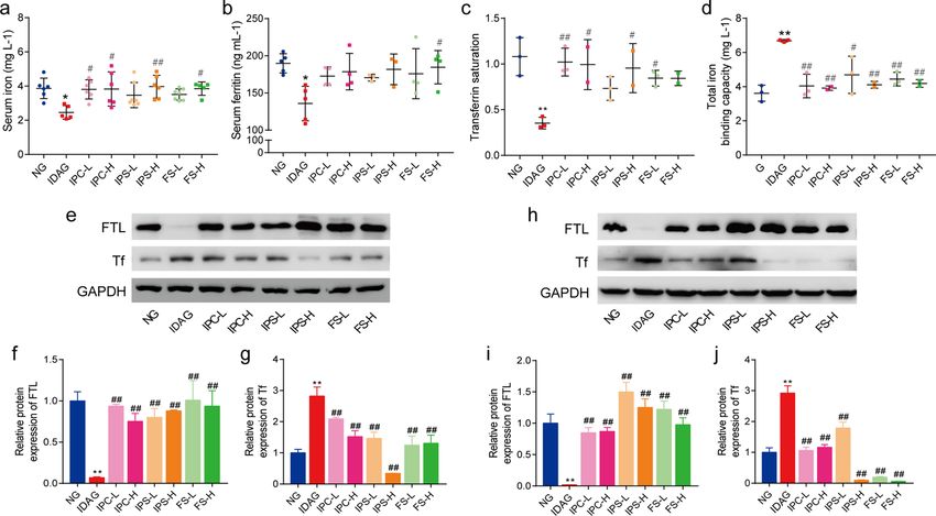
www.nature.com/scientificreports/
Figure 1. Establishment of iron deficiency anemia model in female rats. (a) Scheme of IDA model
establishment and iron supplement treatment. The whole blood of maternal rats was collected and
haematological indexes were tested. (b) HGB. (c) HCT. (d) MCV. (e) CHCM. f CH. (g) RDW. The serum of
maternal rats was collected and tested. (h) SI. (i) SF. (j) TS. (k) TIBC. HGB haemoglobin, HCT Hematocrit,
MCV mean corpuscular volume, CHCM cell haemoglobin concentration mean, CH haemoglobin content of red
blood cell, RDW red cell volume distribution width, SI serum iron, SF serum ferritin, TS transferrin saturation,
TIBC total iron binding capacity. Data are presented as mean ± SD (n = 12). **p < 0.01, compared with NG.
Unpaired t-test was used for comparison between two groups.
Oral iron supplementation is an effective treatment for IDA during pregnancy6. The most frequently used
oral iron preparations are ferrous sulfate (FS), ferrous fumarate, ferrous glycine sulfate, and ferrous gluconate. As
early as 1998, the efficacy and tolerability of iron protein succinylate (IPS) in the treatment of iron deficiency in
children were reported. Then, the use of succinic acid in the treatment of adults, pregnant women and premature
IDA was reported7–9. In 2019, Córdova A et al. showed that supplementation with IPS improved haematological
indexes in professional a thletes10. However, the effects of IPS on pregnancy outcome and offspring physical and
neural development have not been reported. Iron polysaccharide complex (IPC) is composed of low molecular
weight polysaccharide and iron, in which the iron content is 46%. IPC does not contain free iron ions, so there
is no corrosion and irritation to gastrointestinal mucosa caused by iron ions. Studies have shown that IPC can
effectively treat IDA and improve haematological parameters11–13. In China, some studies reported the effects
of IPC on IDA in pregnant women and its effects on pregnancy outcomes, but few studies have examined the
growth and development of newborns after birth.
In this study, we initially established a rat model with IDA by using a combination of low iron diet with blood-
letting and deionized water. IDA female rats were then allowed to proceed mating with males and conceiving.
Next, in addition to determining the effects of IPS, IPC, and FS on pregnancy outcomes, we also examined the
haematological and immuno-inflammatory indexes of the mother rats and offspring rats, as well as the physical
and neural development of the offspring rats with those iron supplements.
Results
Establishment of female rat model with iron deficiency anemia. IDA model establishment and
iron supplement treatment scheme is shown in Fig. 1a. After 8 weeks of treatment with low iron diet plus deion-
ized water and weekly bloodletting, the IDA model group (IDAG) rats had significantly lower levels of hae-
moglobin (HGB) (P < 0.001), hematocrit (HCT) (P < 0.001), mean corpuscular volume (MCV) (P < 0.001), cell
haemoglobin concentration mean (CHCM) (P < 0.001), haemoglobin content of red blood cell (CH) (P < 0.001)
and significantly higher red cell volume distribution width (RDW) (P < 0.001) than normal control group (NG)
(Fig. 1b–g). Further analysis revealed that the levels of serum iron (SI), serum ferritin (SF) and transferrin satu-
Scientific Reports | (2021) 11:1347 | https://doi.org/10.1038/s41598-020-79971-y 2
Vol:.(1234567890)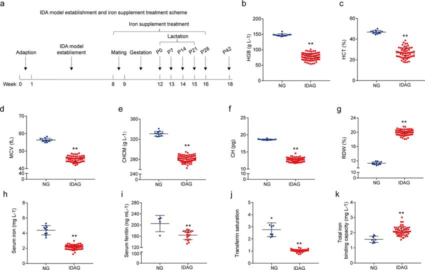
www.nature.com/scientificreports/
NG IDAG IPC-L IPC-H IPS-L IPS-H FS-L FS-H
Parturition rate (%) 79.26 44.83 80 66.7 73.33 80.00 86.67 60.00
Average No. of total # # ##
7.89 ± 1.94 3.77 ± 2.00** 5.83 ± 2.25 6.40 ± 2.01 6.09 ± 2.17 6.75 ± 3.05 6.92 ± 1.60 8.00 ± 2.45##
born per litter
Average No. of total
7.78 ± 1.99 2.92 ± 1.44** 4.79 ± 2.83 4.50 ± 2.84 6.00 ± 2.10# 6.42 ± 2.94## 6.85 ± 1.63## 7.33 ± 3.61##
born alive per litter
Table 1. Pregnancy outcome of maternal rats. * p < 0.05, ** p < 0.01, compared with NG, # p < 0.05, ## p < 0.01,
compared with IDAG. One-way ANOVA followed by Tukey multiple comparison test was used for comparison
among 8 different groups.
Figure 2. Haematological indexes of maternal rats after iron supplement treatment. The whole blood of
maternal rats was collected and the haematological indexes were tested. (a) HGB. (b) HCT. (c) MCV. (d)
CHCM. (e) CH. (f) RDW. (g) NEU. (h) LYM. (i) NLR. (j) TNF-α. HGB haemoglobin, HCT Hematocrit, MCV
mean corpuscular volume, CHCM cell haemoglobin concentration mean, CH haemoglobin content of red
blood cell, RDW red cell volume distribution width, NEU neutrophil, LYM lymphocyte, NLR neutrophil-to-
lymphocyte ratio, TNF-α tumor necrosis factor α. Data are presented as mean ± SD (n = 5). *p < 0.05, **p < 0.01,
compared with NG, #p < 0.05, ##p < 0.01, compared with IDAG. One-way ANOVA followed by Tukey multiple
comparison test was used for comparison among 8 different groups.
ration (TS) in IDA group were significantly lower than those in normal control group, and total iron binding
capacity (TIBC) was significantly higher in IDA group (Fig. 1h–k).
Effects of iron supplementation on maternal rats. Pregnancy outcomes. To determine whether ma-
ternal iron supplement could improve the pregnancy outcomes, the parturition rate, average number of total
born per litter, and average number of total born alive per litter were recorded in anemia rats supplemented with
or without iron during the entire gestation period. As shown in Table 1, the parturition rate of the IDA group
was 44.83%, which was lower than that of NG (79.26%). In IDA group, the average number of total born per litter
and average number of total born alive per litter were significantly lower than that in NG (Table 1). The average
number of total born per litter in IPC high-dose group (IPC-H), IPS high-dose group (IPS-H), FS low-dose
group (FS-L), FS high-dose group (FS-H), and the average number of total born alive per litter in IPS-L, IPS-H,
FS-L, and FS-H groups were significantly higher than that in IDA group (Table 1). These results suggested that
maternal iron supplementation significantly improved pregnancy outcomes.
Haematological and immune‑inflammatory indexes of maternal rats. Figure 2 shows the haematological indexes
measured in blood collected from different groups. The levels of HGB, HCT, MCV, CHCM and CH in the IDA
group were significantly lower, and RDW was significantly higher than those in normal control group. After
intervention with three iron supplements, HGB, HCT, MCV, CHCM, CH levels were significantly increased and
RDW was significantly decreased (Fig. 2a–f).
As shown in Fig. 2g–j, the IDA group had significantly lower lymphocyte (LYM) level, and significantly higher
neutrophil (NEU), neutrophil-to-lymphocyte ratio (NLR) and TNF-α levels than NG group. After intervention,
the levels of NEU, NLR and TNF-α in the six iron supplemented groups were significantly lower than those in
the IDA group (p < 0.05), while the level of LYM was significantly higher (p < 0.05). These results suggested that
iron supplements improved immune-inflammatory status altered by IDA.
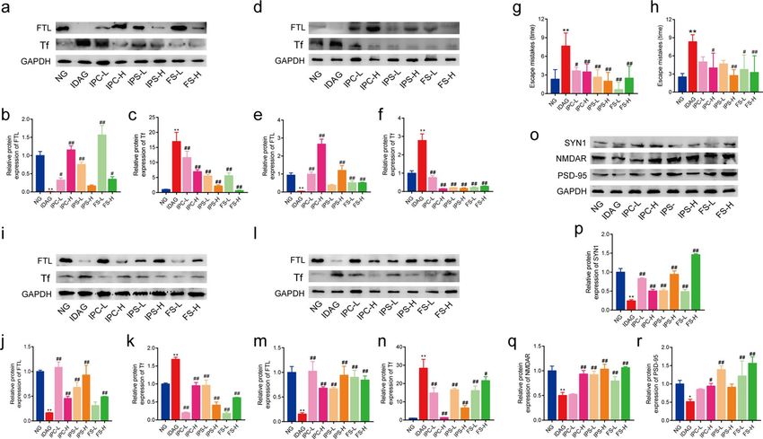
SI, TIBC, TS and SF levels. SI, TIBC, TS and SF levels are shown in Fig. 3a–d. At 28 days postpartum, the levels
of SF, SI and TS in the IDA group were still significantly lower than those in NG (p < 0.05). In contrast, TIBC level
in the IDA group was significantly higher (p < 0.05). After iron supplementation, the levels of SI, TS and SF in
Scientific Reports | (2021) 11:1347 | https://doi.org/10.1038/s41598-020-79971-y 3
Vol.:(0123456789)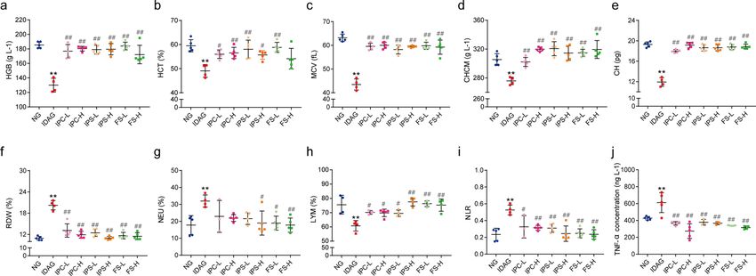
www.nature.com/scientificreports/

                                            Figure 3.  Iron related indexes of maternal rats after iron supplement treatment. The serum of maternal rats
                                            was collected and tested. (a) SI. (b) SF. (c) TS. (d) TIBC. Data are presented as mean ± SD (n = 6). Western blot
                                            analysis for FTL and Tf in liver (e–g) and spleen (h–j). The quantification of western blotting was provided in
                                            supplementary material. Data of Western blot analysis (mean ± SD) are expressed as the ratio of the relative
                                            contents between the value from IDA group and NG group and six iron treatment groups (n = 3). The relative
                                            contents of target proteins were quantified using the ratio between the optical density (OD) of target protein and
                                            the amount of the housekeeping protein GAPDH. SI serum iron, SF serum ferritin, TS transferrin saturation,
                                            TIBC total iron binding capacity, FTL ferritin light chain, Tf transferrin. *p < 0.05, **p < 0.01, compared with NG,
                                            #
                                             p < 0.05, ##p < 0.01, compared with IDAG. One-way ANOVA followed by Tukey multiple comparison test was
                                            used for comparison among 8 different groups.

                                            IPC, IPS, and FS groups were increased compared with the IDA group, while TIBC level in these iron treatment
                                            groups was significantly decreased.

                                            Liver and spleen ferritin light chain (FTL) and transferrin (Tf) levels. Figure 3e–j shows iron accumulation and
                                            iron uptake in the liver and spleen of different groups. The level of FTL in the liver and spleen of rats in the IDA
                                            group was significantly lower than the NG group and the iron supplemented groups, while the level of Tf was
                                            significantly higher. These results suggested that iron accumulation in liver and spleen were reduced and iron
                                            transport capacity was increased in IDA group, and iron stores in the liver and spleen was increased after iron
                                            supplementation.

                                            Effects of iron supplementation on offspring rats. Haematological indexes of offspring. At 28 days
                                            after birth, the haematological parameters of the offspring were similar to those of the mother rats. The HGB,
                                            HCT, MCV, CHCM, and CH levels of the rats in IDA group were significantly lower than those in NG group,
                                            while RDW level was significantly higher. After iron supplementation, the levels of HGB, HCT, MCV, CHCM,
                                            and CH in the offspring rats were significantly higher than those in the IDA group, and RDW level was signifi-
                                            cantly lower (Fig. 4a–f).

                                            Immune‑inflammatory indexes of offspring. Similarly, increased NEU and NLR levels and decreased LYM and
                                            WBC levels in the offspring rats of the IDA group were observed. The levels of these indexes in iron supple-
                                            mented groups were improved, which indicated that iron intervention improved the immune-inflammatory
                                            status in the offsprings of IDA rats (Fig. 4g–j).

                                            FTL and Tf levels in liver and spleen of offspring. As shown in Fig. 5a–c, the protein expression of FTL in the
                                            liver and spleen of the offspring rats in IDA group was significantly lower than NG group, which was reversed
                                            by iron supplement. The expression of Tf in the liver and spleen of IDA offspring rats was significantly higher
                                            than that of NG and iron supplemented groups (Fig. 5d–f). These results suggested that the offsprings of IDA
                                            rats were also in a state of iron deficiency, which indicated the effects of IDA during pregnancy and lactation.

          Scientific Reports |   (2021) 11:1347 |                https://doi.org/10.1038/s41598-020-79971-y                                                   4

Vol:.(1234567890)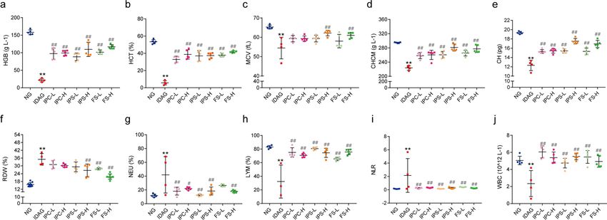
www.nature.com/scientificreports/

                                  Figure 4.  Haematological indexes of offspring rats after iron supplement treatment. The whole blood of
                                  offspring rats was collected and the haematological indexes were tested. (a) HGB. (b) HCT. (c) MCV. (d)
                                  CHCM. (e) CH. (f) RDW. (g) NEU. (h) LYM. (i) NLR. (j) WBC. HGB haemoglobin, HCT Hematocrit, MCV
                                  mean corpuscular volume, CHCM cell haemoglobin concentration mean, CH haemoglobin content of red
                                  blood cell, RDW red cell volume distribution width, NEU neutrophil, LYM lymphocyte, NLR neutrophil-to-
                                  lymphocyte ratio, WBC white blood cells count. Data are presented as mean ± SD (n = 5). *p < 0.05, **p < 0.01,
                                  compared with NG, #p < 0.05, ##p < 0.01, compared with IDAG. One-way ANOVA followed by Tukey multiple
                                  comparison test was used for comparison among 8 different groups.

                                  Figure 5.  Iron related indexes and neural development of offspring rats after iron supplement treatment.
                                  Western blot analysis for FTL and Tf in liver (a–c) and spleen (d–f). Morris water maze test for day 1 escape
                                  latency (g) and day 2 escape latency (h). Western blot analysis for FTL and Tf in brain (i–k) and hippocampus
                                  (l–n), and for SYN1, NMDAR, PSD-95 in hippocampus (o–r). The quantification of western blotting was
                                  provided in supplementary material. FTL ferritin light chain, Tf transferrin, SYN1 synapsin 1, NMDAR
                                  N-methyl-D-aspartate receptor, PSD-95 postsynaptic density protein 95. Data of Western blot analysis
                                  (mean ± SD) are expressed as the ratio of the relative contents between the value from IDA group and NG
                                  group and six iron treatment groups (n = 3). The relative contents of target proteins were quantified using the
                                  ratio between the optical density (OD) of target protein and the amount of the housekeeping protein GAPDH.
                                  **p < 0.01, compared with NG, #p < 0.05, ##p < 0.01, compared with IDAG. One-way ANOVA followed by Tukey
                                  multiple comparison test was used for comparison among 8 different groups.

Scientific Reports |   (2021) 11:1347 |               https://doi.org/10.1038/s41598-020-79971-y                                                    5

                                                                                                                                             Vol.:(0123456789)
www.nature.com/scientificreports/
Average weight of pups born alive at Average weight of pups born alive at Average weight of pups born alive at Average weight of pups born alive at
Group PND7 (g) PND14 (g) PND21 (g) PND28 (g)
NG 15.61 ± 1.704 30.74 ± 3.896 47.52 ± 7.016 64.98 ± 9.381
IDAG 9.823 ± 2.012** 15.55 ± 3.947** 19.94 ± 5.614** 23.63 ± 3.217**
IPC-L 14.95 ± 2.524## 28.18 ± 5.102## 43.64 ± 6.821## 65.04 ± 8.777##
IPC-H 16.85 ± 4.691## 34.21 ± 6.12## 51.35 ± 8.356## 71.28 ± 13.25##
## ## ##
IPS-L 15.85 ± 3.656 31.83 ± 5.042 48.67 ± 6.861 65.96 ± 10.55##
IPS-H 16.02 ± 2.135## 32.06 ± 3.813## 45.86 ± 7.129## 60.92 ± 11.95##
FS-L 16.47 ± 2.016## 33.09 ± 3.079## 49.98 ± 6.874## 70.41 ± 8.834##
FS-H 16.38 ± 2.072## 34.2 ± 5.055## 50.79 ± 6.595## 62.34 ± 11.47##
Table 2. The body weight of offspring rats. * p < 0.05, ** p < 0.01, compared with NG, # p < 0.05, ## p < 0.01,
compared with IDAG. One-way ANOVA followed by Tukey multiple comparison test was used for comparison
among 8 different groups.
Group Out of the hair (d) Tooth eruption (d) Eyes open (d) Rate o born alive per litter at PND28 (%)
NG 3.313 ± 0.6021 7.176 ± 0.809 14.62 ± 0.5064 0.9851 ± 0.04342
IDAG 4.545 ± 1.128** 8.545 ± 1.508** 14.44 ± 0.7265 0.2303 ± 0.3959**
IPC-L 3.083 ± 0.5149## 6.917 ± 0.5149## 14.33 ± 0.7785 0.9861 ± 0.04811##
## ##
IPC-H 3.1 ± 0.7379 6.6 ± 0.6992 14.22 ± 1.394 0.9732 ± 0.05663##
# ##
IPS-L 3.6 ± 0.5164 6.727 ± 0.9045 14.44 ± 0.8819 0.9886 ± 0.03769##
## ##
IPS-H 3.091 ± 0.5394 6.909 ± 0.8312 14.36 ± 0.809 0.9467 ± 0.1069##
## ##
FS-L 3.308 ± 0.7511 7.077 ± 0.8623 14.25 ± 0.6216 0.9890 ± 0.03962##
FS-H 3 ± 0.5345## 7.375 ± 0.5175# 14 ± 0.8944 1.000 ± 0.0##
Table 3. The development and PND 28 survival of offspring rats. * p < 0.05, ** p < 0.01, compared with NG,
# p < 0.05, ## p < 0.01, compared with IDAG. One-way ANOVA followed by Tukey multiple comparison test
was used for comparison among 8 different groups.
Group Surface righting reflex (d) Cliff avoidance reflex (d) Negative geotropism (d) Air righting reflex (d)
NG 3.526 ± 0.7723 4.684 ± 0.8201 4.632 ± 0.8951 10.75 ± 1.342
IDAG 5.273 ± 1.009** 6.909 ± 1.375## 6.818 ± 1.168## 12.67 ± 1.323**
# ##
IPC-L 4.083 ± 0.6686 5.333 ± 0.7785 5.25 ± 0.7538## 10.67 ± 1.303#
## ## ##
IPC-H 3.6 ± 0.6992 4.5 ± 0.7071 4.5 ± 0.7071 10.2 ± 1.476##
IPS-L 3.545 ± 0.8202## 4.364 ± 0.809## 4.364 ± 0.809## 10.64 ± 1.027#
IPS-H 3.455 ± 0.5222## 4.273 ± 0.6467## 4.364 ± 0.6742## 9.818 ± 1.834##
FS-L 3.538 ± 1.506## 4.692 ± 0.6304## 4.615 ± 0.5064## 10.6 ± 1.506#
FS-H 3.125 ± 0.3536## 4.5 ± 0.5345## 4.5 ± 0.5345## 10.63 ± 1.598#
Table 4. The nerve growth and development of offspring rats. * p < 0.05, ** p < 0.01, compared with NG,
# p < 0.05, ## p < 0.01, compared with IDAG. One-way ANOVA followed by Tukey multiple comparison test
was used for comparison among 8 different groups.
Physical growth and development of offspring. At postnatal day (PND) 7, 14, 21 and 28, the offspring’s body
weight of the IDA group was significantly lower than that of NG group and iron supplemented groups. The
offspring of the IDA group gained only about 14 g of body weight in 21 days, while iron supplemented groups
gained more than 50 g in 21 days (Table 2). The time of out of hair and tooth eruption in the IDA group was later
than that in NG group and iron supplemented groups. At PND 28, the rate of born alive per litter in the IDA
group was 23%, which was about 98% in NG and iron supplemented groups. These results indicated that the
anemic dams not only delivered fewer but also lower survival rate (Table 3).
Neural growth and development of offspring. The onset time of surface righting reflex, cliff avoidance, negative
geotropism and air righting reflex was observed and recorded to evaluate the neural growth and development
of the offspring. As shown in Table 4, the onset time of surface righting reflex, cliff avoidance, negative geotro-
pism and air righting reflex in the IDA group was later than that in NG group. However, the onset time of these
Scientific Reports | (2021) 11:1347 | https://doi.org/10.1038/s41598-020-79971-y 6
Vol:.(1234567890)www.nature.com/scientificreports/
indexes in the iron supplemented groups was significantly earlier than that in the IDA group, and there was no
significant difference with NG group.
Morris water maze test. On day 1 of the probe trial, the offspring rats of the IDA group showed increased
escape mistakes compared to NG group, indicating impaired spatial learning ability. Compared with the IDA
group, iron intervention groups showed a significant decrease in escape mistakes (Fig. 5g). On day 2 of the probe
trial, the escape mistakes of the rats in the IDA group were still more than that in NG group and iron interven-
tion groups (Fig. 5h). These results indicated that IDA affected spatial learning ability in offspring and this
adverse effect was improved by iron supplementation.
Expression of iron related proteins and synaptic growth associated proteins in brain and hippocampus. We exam-
ined the expression of iron related proteins, such as FTL and Tf, in the brain and hippocampus of offspring rats.
The level of FTL was significantly lower in IDA group than that of other groups, and the level of Tf was oppositely
altered (Fig. 5i–n). Then we detected the synaptic growth associated proteins in hippocampus. As expected, the
levels of SYN1, NMDAR and PSD-95 were significantly decreased in IDA rats (Fig. 5o–r), while the expres-
sion of these proteins was increased in iron intervention groups. Taken together, the IDA offspring rats showed
abnormal and retarded neurodevelopment, which was improved by iron supplement.
Discussion
IDA during pregnancy is a common health problem, even in high-income countries. Iron is an essential micro-
nutrient which is important not only for carrying oxygen but also for the catalytic activity of various enzymes.
Therefore, iron deficiency anemia during pregnancy has both short term and long term effects on the pregnant
woman, the puerperal woman, the fetus and the i nfant14–18. In the present study, based on the establishment of
IDA female rat model, the adverse effects of IDA on mother and offspring as well as the improvement effects of
iron supplementation were observed.
In this study, the IDA model was established in a period of 8 weeks with low iron diet (9 ppm), deionized
drinking water and bleeding once a week. HGB is the most commonly used indicator of iron deficiency anemia.
In our experiments, HGB < 90 g/L was used as the successful criterion of the IDA model. Our study showed that
this method had about a rate of 73% IDA (HGB < 90 g/L) at eight weeks. In addition, some other indicators such
as HCT, MCV, CHCM, CH, and RDW also showed significant changes. Serum iron, serum ferritin, TS, and TIBC
are also important indicators reflecting the iron metabolism in vivo. There were also significant alterations in
these indicators in our IDA animal model.
The liver and spleen are the principal iron storage organs and critical for the regulation of systemic iron
homeostasis. Hepatic iron deposition is properly controlled by the liver iron uptake and export system. Hepatic
iron uptake depends on Tf, transferrin receptor 1/2, et al.18,19. The excess iron is stored as ferritin which consists
of 24 subunits of heavy (FTH) or light (FTL) isoforms in a spherical shell that plays a central role in the intracel-
alance20–22. In this study, Tf protein expression level was up-regulated and FTL was down-regulated in
lular iron b
the liver and spleen of IDA rats. These results indicated that iron stores in the liver and spleen decreased during
pregnancy and lactation up to 14 weeks.
Iron deficiency affects performance during pregnancy and delivery, lactation performance, and immuno-
inflammatory status2. Severe anemia can also increase perinatal maternal mortality. In this study, although no
maternal death was observed in the IDA group, the abnormality of immuno-inflammatory parameters was
observed. Serum neutrophils, lymphocytes and TNF-α levels are common markers of immuno-inflammatory
response. NLR is a simple parameter that makes it easy to assess the inflammatory state of a subject. It has been
shown to be a useful predictor and marker of inflammation, infection, postoperative complications, e tc23–25. In
this study, the levels of neutrophils, NLR and TNF-α in the IDA group were significantly higher than those in
control group and iron supplemented groups, along with significant lower level of LYM. These data suggested
that IDA impaired the immune-inflammatory status. As a key nutrient for the developing fetus, neonate, infant,
and child, the demand of iron is high during the early stages of life because it is critical for the production of new
red blood cells and muscle cells as well as brain development. A recent study of anemia and iron deficiency in
pregnancy in Southern India showed that IDA in pregnancy was associated with higher risk of low birth weight,
preterm birth, gestational age at birth and infant WAZ s cores26. Our study also showed that the number of live
births per litter in IDA group was significantly lower than that in the normal control group, and the body weight
and 28-day weight gain were significantly lower than those in other groups with sufficient iron. The time of tooth
eruption, eye opening and hair growth of the rats in the IDA group was also later than that in the normal con-
trol group and iron supplemented groups. The 28-day survival rate in IDA group was only 23%, in comparison
to about 98% in NG and iron supplemented groups. These results suggested that maternal iron deficiency and
anemia during pregnancy affected the growth and development of offspring.
In early embryonic life, iron is already necessary for normal brain development due to the proliferation,
migration, and differentiation of neural progenitor cells. Animal models of prenatal iron deficiency show abnor-
malities in brain structure, neurotransmitter system and myelin formation, resulting in acute brain dysfunction
during the period of deficiency and persistence of various postnatal neurobehavioural abnormalities27. Studies of
fully developed infants have shown that iron deficiency experienced during development can have chronic and
irreversible damage to cognitive, memory and motor skills, indicating widespread effects of iron deficiency early
in life on neurodevelopment. Our results illustrated that the IDA group had a lower level of neurodevelopment
than iron supplemented groups. Synaptic growth and development proteins, including SYN1, NMDAR and PSD-
95, were severely decreased in IDA rats. These alterations in turn retarded the neurobehavioral development,
Scientific Reports | (2021) 11:1347 | https://doi.org/10.1038/s41598-020-79971-y 7
Vol.:(0123456789)www.nature.com/scientificreports/
such as surface righting reflex, cliff avoidance, negative geotropism and air righting reflex, and spatial learning
and memory ability.
Iron protein succinylate, iron polysaccharide complex and ferrous sulfate are all iron derivatives for the oral
treatment of IDA. However, there was no study to determine the effects of these three iron supplements on preg-
nancy outcomes and offspring growth. Cancelo-Hidalgo et al. reported the incidence of overall adverse reactions
was 7.3% for iron protein succinylate and 32.3% for ferrous sulfate, indicating that iron protein succinylate had a
lower incidence of adverse effects than ferrous s ulfate28. Since iron polysaccharide complex does not contain free
iron ions, it causes little corrosion and irritation to gastrointestinal mucosa. In an intervention trial for anemia
of prematurity, no notable adverse events were observed in either iron protein succinylate or iron polysaccharide
complex group7. In the present study, we did not observe significant differences in the improvement effects of
these three iron supplements. In the cases of severe iron deficiency anemia, iron supplementation can signifi-
cantly improve maternal and offspring outcomes. Therefore, iron supplementation or not is more important for
maternal and offspring than the type of iron supplement.
In conclusion, the effects of iron deficiency anemia on pregnant females were severe, leading to premature
birth, miscarriage, significant reduction in the number of litters per litter and birth weight, as well as abnormali-
ties in maternal immune-inflammatory status. It also had significant effects on the growth and development of
offspring, both physically and neurologically. Therefore, when pregnancy anemia is diagnosed, iron supplements
should be given to prevent and correct the adverse effects. Iron polysaccharide complex, iron protein succinylate
and ferrous sulfate could significantly improve pregnancy outcomes and iron nutrition status in maternal IDA
rats, and are beneficial for the growth and development of offspring. Therefore, they can be used as effective iron
supplements for pregnant women.
Materials and methods
Materials. Iron protein succinylate (IPS) (5.46% iron content) was purchased from Raw Material Medi-
cine Reagent Co., LTD (Nanjing, Jiangsu, China). Iron polysaccharide complex (IPC) (46% iron content) was
purchased from Shanghai Pharmaceutical Group, Qingdao Growful Pharmaceutical Co., Ltd. (Qingdao, Shan-
dong, China). Ferrous sulfate (FS) was purchased from Sigma Aldrich (St. Louis, MO, USA). Primary antibodies
against transferrin (Tf), ferritin light chain (FTL) were obtained from Wuhan Sanying Biology Technology Co.,
Ltd (Wuhan, Hubei, China). Primary antibodies against Synapsin 1 (SYN1), N-methyl-D-aspartate receptor
(NMDAR) and postsynaptic density protein 95 (PSD-95) were obtained from Affinity (OH, USA). The pri-
mary antibody for GAPDH was from Biogot Technology Co., Ltd. (Nanjing, Jiangsu, China). TNF-α ELISA Kit
was purchased from SenBeiJia Biological Technology Co., Ltd (Nanjing, Jiangsu, China). Serum iron (SI) test
kit, total iron binding capacity (TIBC) test kit, and serum ferritin (SF) test kit were purchased from Nanjing
Jiancheng Bioengineering Inst (Nanjing, Jiangsu, China).
Animals. All animals used in the study were purchased from Shanghai SLAC Laboratory Animal Co.,Ltd
[Licence No: SCXK (Hu)2017-0005]. Five-week-old, female and male Wistar rats were housed in pathogen free
environment in the animal house facility of Nanjing Medical University at regulated temperature 22 to 26 °C and
under 12 h/12 h light/dark cycles. Free access to laboratory diet and drinking water was provided to the animals.
Protocol adopted in the study was approved by the Institutional Animal Care and Use Committee (IACUC) of
Nanjing Medical University. The animal study protocol approval number was IACUC-1812017. All experiments
were performed in accordance with the approved guidelines and regulations by IACUC of Nanjing Medical
University.
Iron deficiency anemia model and treatment. Female rats were randomly divided into normal control
group (NG) and iron deficiency anemia model group. Normal control animals were fed with normal laboratory
diet containing 50 ppm iron during the experimental period. The animals in the IDA group were fed with low
iron diet containing 9 ppm iron (Jiangsu Xie Tong Pharmaceutical Bio-engineering Co., Ltd., China) and deion-
ized water ad libitum and followed by bleeding once a week for 8 weeks.
After 8 weeks, orbital blood was collected and haemoglobin (HGB) content was measured using an automatic
biochemistry analyzer (ADVIA 2120i, Siemens, Germany). Rats with haemoglobin content below 90 g/L were
considered to be IDA and were used in subsequent experiments. Except NG rats, the anemia animals were ran-
domly divided into seven group: IDA model group (IDAG), low dose of iron polysaccharide complex group at
iron dose of 30 mg·kg−1 (IPC-L), high dose of iron polysaccharide complex at iron dose of 50 mg·kg−1 (IPC-H),
low dose of iron protein succinylate group at iron dose of 30 mg·kg−1 (IPS-L), high dose of iron protein succinylate
at iron dose of 50 mg·kg−1 (IPS-H), low dose of ferrous sulfate at iron dose of 30 mg·kg−1 (FS-L), high dose of
ferrous sulfate at iron dose of 50 mg·kg−1 (FS-H).
After grouping, female rats from each group and male rats were mated by 3:1. Mating was confirmed by
detection of a vaginal plug at 8:00 in the morning. After mating, female rats were feed separately. NG rats were
fed with normal diet. IDAG were fed with low-iron diet. Six treatment groups were fed with iron-containing
diet (30 mg·kg−1 and 50 mg·kg−1) respectively. All groups of mother rats were fed until the 28th day after the
birth of their offspring.
After birth, the number of live offspring per litter was recorded. The weight of the offspring was recorded
weekly for 28 days after birth. The growth and development indexes, such as tooth eruption, eye opening and
villus growth, were observed and recorded. The nerve reflex and motor coordination function indexes, such as
the time of surface righting reflex, cliff avoidance, negative geotropism, and aerial righting reflex were tested
and recorded. On the 28th day, the number of the surviving offspring was recorded and the survival rate was
Scientific Reports | (2021) 11:1347 | https://doi.org/10.1038/s41598-020-79971-y 8
Vol:.(1234567890)www.nature.com/scientificreports/
calculated. Most of the offspring in each group were sacrificed and blood and tissues were collected. Six rats in
each group were fed to 42 days after birth and their learning ability was measured by Morris water maze test.
Sample collection. On the 28th day, blood of the mother rats and the offspring rats was collected. The
blood samples were collected in non-heparinized/heparinized microcapillary tubes from the rats’ retro-orbital
plexus. A small amount of blood from each rat was immediately added to the automated hematology analyzer
for determination. The remaining blood samples were centrifuged to obtain serum and were frozen at − 20 °C for
further analysis. Next, all mother and offspring rats were sacrificed and the liver, spleen, brain and hippocampus
were removed, rinsed with phosphate-buffered saline, weighed, and stored at − 80 °C for further analysis.
Morris water maze test. The Morris water maze was a black rectangular flume, in which several dia-
phragms formed the maze, and one corner of the maze had a group of steps as the end point. Before the experi-
ment, the maze was filled with water, with water depth of 10 cm and water temperature of 25 °C. On the first day
of training, rats were placed on the step for 10 s to make them understand the existence of this safe area, rats were
then placed at the starting point to let them swim freely, and the mistakes of climbing the safe step within 2 min
were recorded. On the next day, the training of the first day was repeated again, and the mistakes of reaching the
end point were recorded and the learning ability of the offspring rats was evaluated.
Haematological analysis. Haemoglobin (HGB), hematocrit (HCT), mean corpuscular volume (MCV),
red cell volume distribution width (RDW), cell haemoglobin concentration mean (CHCM), haemoglobin con-
tent of red blood cell (CH), lymphocyte (LYM), neutrophil (NEU) and white blood cells count (WBC) levels
were measured using an automated hematology analyzer (ADVIA 2120i, Siemens, Germany).
SI, TIBC and SF levels. Serum iron (SI) concentration and total iron binding capacity (TIBC) were meas-
ured using SI test kit and TIBC test kit (Nanjing Jiancheng Bioengineering Inst., Nanjing, Jiangsu, China)
respectively according to the manufacturer’s instructions. Transferrin saturation (TS) was calculated from SI to
TIBC ratio as follows: TS (%) = [SI (mg/L)/TIBC (mg/L)] × 100%. Serum ferritin (SF) level was measured using
double-antibody sandwich enzyme-linked immunosorbent assay (ELISA) kit according to the manufacturer’s
instructions (SenBeiJia Biological Technology Co., Ltd, Nanjing, Jiangsu, China).
Inflammatory cytokine. The level of tumor necrosis factor α (TNF‐α) in rat serum was measured by
ELISA kits according the manufacturer’s instructions (SenBeiJia Biological Technology Co., Ltd, Nanjing,
Jiangsu, China).
Western blotting. Total proteins were extracted from the liver, spleen, brain and hippocampus tissues
using RIPA buffer (Beyotime, Shanghai, China). Lysates from each sample were run on gels, electrotransferred
onto polyvinylidene difluoride (Millipore, Billerica, MA, USA), and immunoblotted with primary antibodies.
Horseradish peroxidase (HRP)-conjugated goat anti-rabbit IgG (ZSGB‐BIO, China) was used as secondary anti-
body. Immunoreactive proteins were visualized by enhanced chemiluminescence (Cell Signaling Technology,
USA). Quantification of the immunoreactive bands was performed by using Image-Pro Plus 5.0.1.9 software
(Media Cybernetics inc., Rockville, MD, USA).
Statistical analysis. Data are expressed as means ± standard deviation (SD). Unpaired t-test and one-way
ANOVA analyses were performed to compare the difference between two or multiple groups using GraphPad
Prism 7.0 software. A p value of < 0.05 was considered statistically significant.
Consent for publication. Consent for publication was obtained from all authors.
Data availability
All data generated or analyzed during this study are included in this published article.
Received: 17 April 2020; Accepted: 15 December 2020
References
1. Stevens, G. A. et al. Global, regional, and national trends in haemoglobin concentration and prevalence of total and severe anaemia
in children and pregnant and non-pregnant women for 1995–2011: A systematic analysis of population-representative data. Lancet.
Global health 1, e16-25. https://doi.org/10.1016/S2214-109X(13)70001-9 (2013).
2. Viteri, F. E. The consequences of iron deficiency and anaemia in pregnancy on maternal health, the foetus and the infant. SCN
News 2, 14–18 (1994).
3. Lee, H. S., Kim, M. S., Kim, M. H., Kim, Y. J. & Kim, W. Y. Iron status and its association with pregnancy outcome in Korean
pregnant women. Eur. J. Clin. Nutr. 60, 1130–1135. https://doi.org/10.1038/sj.ejcn.1602429 (2006).
4. Shao, J. et al. Maternal serum ferritin concentration is positively associated with newborn iron stores in women with low ferritin
status in late pregnancy. J. Nutr. 142, 2004–2009. https://doi.org/10.3945/jn.112.162362 (2012).
5. Srour, M. A., Aqel, S. S., Srour, K. M., Younis, K. R. & Samarah, F. Prevalence of anemia and iron deficiency among Palestinian
pregnant women and its association with pregnancy outcome. Anemia 2018, 9135625. https://doi.org/10.1155/2018/9135625
(2018).
6. Camaschella, C. Iron-deficiency anemia. N. Engl. J. Med. 373, 485–486. https://doi.org/10.1056/NEJMc1507104 (2015).
Scientific Reports | (2021) 11:1347 | https://doi.org/10.1038/s41598-020-79971-y 9
Vol.:(0123456789)www.nature.com/scientificreports/
7. Xing, Y. & Tong, X. M. Clinical study of iron protein succinylate oral solution for preventing and treating anemia of prematurity.
Zhongguo dang dai er ke za zhi Chi. J. Contemp. Pediatr. 15, 1059–1063 (2013).
8. Sifakis, S. et al. The efficacy and tolerability of iron protein succinylate in the treatment of iron-deficiency anemia in pregnancy.
Clin. Exp. Obstet. Gynecol. 32, 117–122 (2005).
9. Pujol, F. R. et al. Iron protein-succinylate in the treatment of adult iron-deficiency anemia. Anales de medicina interna 19, 651–652
(2002).
10. Cordova, A. et al. Effect of iron supplementation on the modulation of iron metabolism, muscle damage biomarkers and cortisol
in professional cyclists. Nutrients https://doi.org/10.3390/nu11030500 (2019).
11. Powers, J. M. et al. Effect of low-dose ferrous sulfate vs iron polysaccharide complex on hemoglobin concentration in young
children with nutritional iron-deficiency anemia: A randomized clinical trial. JAMA 317, 2297–2304. https://doi.org/10.1001/
jama.2017.6846 (2017).
12. Zhang, Y. et al. Characterization of a novel polysaccharide-iron(III) complex and its anti-anemia and nonspecific immune regulat-
ing activities. Mini. Rev. Med. Chem. 17, 1677–1683. https://doi.org/10.2174/1389557517666170424130327 (2017).
13. Cui, J. et al. A novel low molecular weight Enteromorpha polysaccharide-iron (III) complex and its effect on rats with iron defi-
ciency anemia (IDA). Int. J. Biol. Macromol. 108, 412–418. https://doi.org/10.1016/j.ijbiomac.2017.12.033 (2018).
14. Col Madendag, I. et al. The effect of iron deficiency anemia early in the third trimester on small for gestational age and birth
weight: A retrospective cohort study on iron deficiency anemia and fetal weight. Biomed. Res. Int. 2019, 7613868. https://doi.
org/10.1155/2019/7613868 (2019).
15. ElAlfy, M. S., El-Farrash, R. A., Taha, H. M., Ismail, E. A. & Mokhtar, N. A. Auditory brainstem response in full-term neonates born
to mothers with iron deficiency anemia: Relation to disease severity. J. Matern.–Fetal Neonat. Med. https://doi.org/10.1080/14767
058.2018.1533940 (2018).
16. Munoz, P. & Humeres, A. Iron deficiency on neuronal function. Biometals 25, 825–835. https: //doi.org/10.1007/s10534 -012-9550-x
(2012).
17. Rufer, E. S. et al. Adequacy of maternal iron status protects against behavioral, neuroanatomical, and growth deficits in fetal alcohol
spectrum disorders. PLoS ONE 7, e47499. https://doi.org/10.1371/journal.pone.0047499 (2012).
18. Worthen, C. A. & Enns, C. A. The role of hepatic transferrin receptor 2 in the regulation of iron homeostasis in the body. Front.
Pharmacol. 5, 34. https://doi.org/10.3389/fphar.2014.00034 (2014).
19. Chua, A. C. et al. Iron uptake from plasma transferrin by a transferrin receptor 2 mutant mouse model of haemochromatosis. J.
Hepatol. 52, 425–431. https://doi.org/10.1016/j.jhep.2009.12.010 (2010).
20. Li, W. et al. Systemic and cerebral iron homeostasis in ferritin knock-out mice. PLoS ONE 10, e0117435. https://doi.org/10.1371/
journal.pone.0117435 (2015).
21. Ahmad, S. et al. Ferritin L and Ferritin H are differentially located within hepatic and extra hepatic organs under physiological
and acute phase conditions. Int. J. Clin. Exp. Pathol. 6, 622–629 (2013).
22. Malik, I. A., Wilting, J., Ramadori, G. & Naz, N. Reabsorption of iron into acutely damaged rat liver: A role for ferritins. World J.
Gastroenterol. 23, 7347–7358. https://doi.org/10.3748/wjg.v23.i41.7347 (2017).
23. Forget, P. et al. What is the normal value of the neutrophil-to-lymphocyte ratio?. BMC Res. Notes 10, 12. https://doi.org/10.1186/
s13104-016-2335-5 (2017).
24. Celik, T. Neutrophil-to-lymphocyte ratio in thyroid ophthalmopathy. Bratisl. Lek. Listy 118, 495–498. https://doi.org/10.4149/
BLL_2017_095 (2017).
25. Santos, T. D., Jammal, M. P., Silveira, T. P., Murta, E. F. C. & Nomelini, R. S. Stromal IL2 is related to the neutrophil/lymphocyte
ratio in epithelial ovarian cancer. Pathologica 111, 62–66. https://doi.org/10.32074/1591-951X-62-18 (2019).
26. Finkelstein, J. L. et al. Anaemia and iron deficiency in pregnancy and adverse perinatal outcomes in Southern India. Eur. J. Clin.
Nutr. 74, 112–125. https://doi.org/10.1038/s41430-019-0464-3 (2020).
27. Alwan, N. A. & Hamamy, H. Maternal iron status in pregnancy and long-term health outcomes in the offspring. J. Pediatr. Genet.
4, 111–123. https://doi.org/10.1055/s-0035-1556742 (2015).
28. Cancelo-Hidalgo, M. J. et al. Tolerability of different oral iron supplements: A systematic review. Curr. Med. Res. Opin. 29, 291–303.
https://doi.org/10.1185/03007995.2012.761599 (2013).
Acknowledgements
The authors would like to thank Xia Zhengrong and the staff at Safety Assessment and Research Center for Drug,
Pesticide and Veterinary Drug of Jiangsu Province, Nanjing Medical University, particularly Zhang Jingshu.
Author contributions
Z.C.Y. and G.S.S. contributed to the study conception and design. The experiments were performed by Z.Q.,
L.X.M., Z.M. and L.S.F. Material preparation, data collection and analyses were performed by Z.Q., L.X.M., Z.M.,
Y.C.Y. and L.S.Y. The draft of the manuscript was written by G.S.S. and Z.Q. and revised by Z.C.Y. All authors
commented on previous versions of the manuscript. All authors read and approved the final manuscript.
Funding
This study was supported by National Natural Science Foundation of China (81872607, 81773431). The funders
had no role in study design, data collection and analysis, decision to publish, or preparation of the manuscript.
Competing interests
The authors declare no competing interests.
Additional information
Supplementary Information The online version contains supplementary material available at https://doi.
org/10.1038/s41598-020-79971-y.
Correspondence and requests for materials should be addressed to C.-Y.Z. or S.-S.G.
Reprints and permissions information is available at www.nature.com/reprints.
Publisher’s note Springer Nature remains neutral with regard to jurisdictional claims in published maps and
institutional affiliations.
Scientific Reports | (2021) 11:1347 | https://doi.org/10.1038/s41598-020-79971-y 10
Vol:.(1234567890)www.nature.com/scientificreports/
Open Access This article is licensed under a Creative Commons Attribution 4.0 International
License, which permits use, sharing, adaptation, distribution and reproduction in any medium or
format, as long as you give appropriate credit to the original author(s) and the source, provide a link to the
Creative Commons licence, and indicate if changes were made. The images or other third party material in this
article are included in the article’s Creative Commons licence, unless indicated otherwise in a credit line to the
material. If material is not included in the article’s Creative Commons licence and your intended use is not
permitted by statutory regulation or exceeds the permitted use, you will need to obtain permission directly from
the copyright holder. To view a copy of this licence, visit http://creativecommons.org/licenses/by/4.0/.
© The Author(s) 2021
Scientific Reports | (2021) 11:1347 | https://doi.org/10.1038/s41598-020-79971-y 11
Vol.:(0123456789)You can also read