A comparative study of asleep and awake deep brain stimulation robot-assisted surgery for Parkinson's disease - Nature
←
→
Page content transcription
If your browser does not render page correctly, please read the page content below
www.nature.com/npjparkd

                  ARTICLE                 OPEN

                  A comparative study of asleep and awake deep brain
                  stimulation robot-assisted surgery for Parkinson’s disease
                  Hai Jin1,4, Shun Gong1,4, Yingqun Tao 1 ✉, Hua Huo2 ✉, Xiao Sun1,4, Dandan Song3, Ming Xu3, Zhaozhu Xu3, Yang Liu1, Shimiao Wang1,
                  Lijia Yuan1, Tingting Wang1, Weilong Song1 and He Pan1

                     To compare the differences between asleep and awake robot-assisted deep brain stimulation (DBS) surgery for Parkinson’s Disease
                     (PD), we conducted this retrospective cohort study included 153 PD patients undergoing bilateral robot-assisted DBS from June
                     2017 to August 2019, of which 58 cases were performed under general anesthesia (GA) and 95 cases under local anesthesia (LA).
                     Procedure duration, stimulation parameters, electrode implantation accuracy, intracranial air, intraoperative electrophysiological
                     signal length, complications, and Unified PD Rating Scale (UPDRS) measurements were recorded and compared. The clinical
                     evaluation was conducted by two raters who were blinded to the choice of anesthesia. Procedure duration was significantly shorter
                     in the GA group, while on stimulation off medication motor scores (UPDRS-III) were significantly improved in both the GA and LA
                     group. ANCOVA covariated for the baseline UPDRS-III and levodopa challenge exhibited no significant differences. In terms of
                     amplitude, frequency, and pulse width, the stimulation parameters used for DBS power-on were similar. There were no significant
                     differences in electrode implantation accuracy, intraoperative electrophysiological signal length, or intracerebral hemorrhage (no
1234567890():,;

                     occurrences in either group). The pneumocephalus volume was significantly smaller in the GA group. Six patients exhibited
                     transient throat discomfort associated with tracheal intubation in the GA group. The occurrence of surgical incision infection was
                     similar in both groups. Compared with the awake group, the asleep group exhibited a shorter procedure duration with a similar
                     electrode implantation accuracy and short-term motor improvement. Robot-assisted asleep DBS surgery is a promising surgical
                     method for PD.
                     npj Parkinson’s Disease (2020)6:27 ; https://doi.org/10.1038/s41531-020-00130-1

                  INTRODUCTION                                                                           group) and 14.8 ± 7.3 (LA group). The preoperative levodopa daily
                  Deep brain stimulation (DBS) surgery is an effective treatment for                     doses were 652.4 ± 400.3 mg/d (GA group) and 577.6 ± 340.8 mg/d
                  Parkinson’s disease (PD)1,2. To date, awake surgery has been                           (LA group). The preoperative levodopa responses (UPDRS-III
                  typically performed with intraoperative test stimulations3.                            improvement) were 50.0 ± 17.8 % (GA group) and 48.8 ± 16.2%
                  Recently, asleep surgery has been performed under general                              (LA group). There were no significant differences in the baseline
                  anesthesia (GA) with intraoperative magnetic resonance imaging                         values between the two groups apart from sex and disease
                  (iMRI)4 or computed tomography (iCT)5 to confirm the position of                        duration (Table 1).
                  the electrode tip. However, limited research and clinical experi-
                  ence has been reported regarding robot-assisted asleep surgery.
                  Herein, this study reports robot-assisted DBS surgical procedures
                                                                                                         Procedure duration, pneumocephalus volume, levodopa
                  and clinical results in detail.
                                                                                                         equivalent of daily dose reduction, and Tao’s DBS surgery scale
                                                                                                         All 306 electrodes of the 153 cases were implanted successfully
                  RESULTS                                                                                without adjustment of the electrode position during the ROSA
                  General data                                                                           robot-assisted surgery. The duration of the procedure was
                                                                                                         significantly shorter in the GA group (1.09 ± 0.46 h) than in the
                  In total, 153 patients were retrospectively included in this study,
                                                                                                         LA group (1.54 ± 0.57 h, p < 0.0001). The total MER time was
                  with 58 cases in the GA group and 95 cases in the LA group. The
                  mean ages were 63.5 ± 6.6 years (GA group) and 63.2 ± 9.7 years                        significantly shorter in the GA group (12.03 ± 1.77 min) than in the
                  (LA group). Males represented 37.9% (22/58) of the GA group and                        LA group (12.89 ± 2.73 min, p = 0.0338). There was no significant
                  58.9% (56/95) of the LA group. The disease durations were 10.5 ±                       difference in the levodopa equivalent of daily dose reduction
                  5.0 years (GA group) and 8.7 ± 4.6 years (LA group). The Hoehn &                       between the two groups (162.8 ± 431.6 mg (GA) vs. 208.0 ±
                  Yahr (H & Y) stages were 3.01 ± 0.27 (GA group) and 2.99 ± 0.23 (LA                    403.5 mg (LA), p = 0.5141). The pneumocephalus volume was
                  group). The preoperative UPDRS scores (OFF) were 63.8 ± 13.6 (GA                       significantly smaller in the GA group (4.35 ± 5.61 cm3) than in the
                  group) and 62.2 ± 16.3 (LA group). The preoperative UPDRS-III                          LA group (12.25 ± 13.76 cm3, p < 0.0001). The Tao’s DBS surgery
                  scores (OFF) were 28.9 ± 6.9 (GA group) and 28.6 ± 9.0 (LA group),                     scale6 was significantly higher in the GA group (85.2 ± 9.3) than in
                  and the preoperative UPDRS-III scores (ON) were 14.3 ± 6.6 (GA                         the LA group (76.5 ± 8.0, p < 0.0001, Fig. 1).

                  1
                   Department of Neurosurgery, The General Hospital of Northern Theater Command, 83 Wenhua Road, 110016 Shenyang, China. 2Department of Clinical Trail Management Agency,
                  The General Hospital of Northern Theater Command, 83 Wenhua Road, 110016 Shenyang, China. 3Department of Anesthesia, The General Hospital of Northern Theater Command, 83
                  Wenhua Road, 110016 Shenyang, China. 4These authors contributed equally: Hai Jin, Shun Gong, Xiao Sun. ✉email: yingquntao@163.com; huohua_northern@163.com

                  Published in partnership with the Parkinson’s Foundation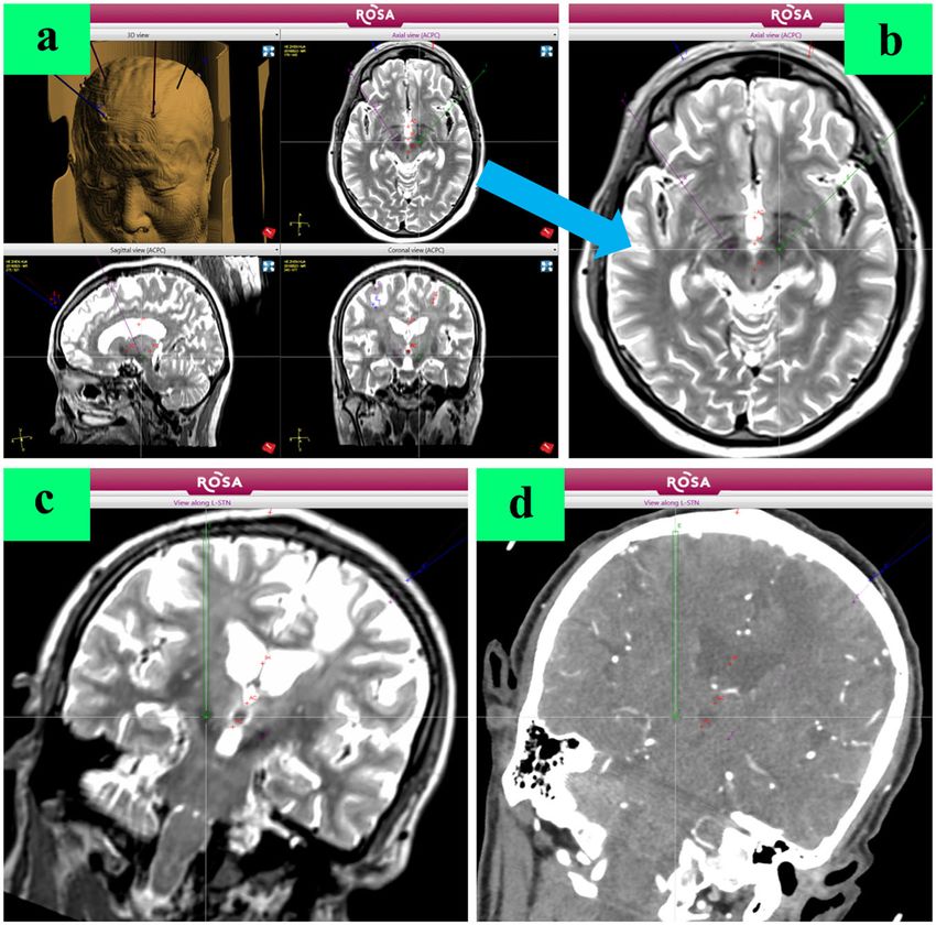
H. Jin et al.
                  2
                      Electrode implantation accuracy                                                   did not differ between the groups (for STN, 5.90 ± 1.45 mm (GA)
                      The electrode implantation accuracy did not differ between the                    vs. 5.47 ± 0.82 mm (LA), p = 0.0630; for Gpi, 8.68 ± 2.52 mm (GA)
                      GA group (0.71 ± 0.25 mm) and LA group (0.76 ± 0.23 mm, p =                       vs. 9.48 ± 2.39 mm (LA), p = 0.2525).
                      0.3031). The intraoperative electrophysiological signal length also
                                                                                                        UPDRS and UPDRS-III
                       Table 1.   Demographic and clinical characteristics of the PD patients.          The on stimulation off medication UPDRS scores significantly
                                                                                                        improved in both groups (GA: 26.84 ± 12.23, p < 0.0001; LA:
                      Characteristic                      GA group        LA group         P value      25.87 ± 12.59, p < 0.0001). ANCOVA covariated for the baseline
                                                          (n = 58)        (n = 95)                      UPDRS and levodopa challenge revealed no significant differences
                      Age-year                            63.5 ± 6.6      63.2 ± 9.7       0.8710
                                                                                                        (ANCOVA F = 0.009, Bonferroni p = 0.923). The percentage
                                                                                                        improvement was similar in both groups (42.8 ± 19.0% (GA) and
                      Male sex-no. (%)                    22 (37.9)       56 (58.9)        0.0116
                                                                                                        41.6 ± 15.9% (LA), ANCOVA F = 0.163, Bonferroni p = 0.687). The
                      Disease duration - Y                10.5 ± 5.0      8.7 ± 4.6        0.0304       on stimulation off medication motor scores (UPDRS-III) signifi-
                      H & Y stage                         3.01 ± 0.27     2.99 ± 0.23      0.6429       cantly improved in both group GA (18.31 ± 6.83, p < 0.0001) and
                      UPDRS scores (Med OFF)              63.8 ± 13.6     62.2 ± 16.3      0.5352       LA (16.24 ± 7.14, p < 0.0001). ANCOVA covariated for the baseline
                      UPDRS-III scores (Med OFF)          28.9 ± 6.9      28.6 ± 9.0‘      0.8383       UPDRS-III and levodopa challenge revealed no significant differ-
                      UPDRS-III scores (Med ON)           14.3 ± 6.6      14.8 ± 7.3       0.6795       ences (ANCOVA F = 1.233, Bonferroni p = 0.269). The percentage
                      Levodopa daily dose (mg/d)          652.4 ± 400.3   577.6 ± 340.8 0.2197          improvement was similar in both groups (GA: 63.4 ± 19.2%; LA:
                                                                                                        57.0 ± 17.3%; ANCOVA F = 0.718, Bonferroni p = 0.398).
                      Levodopa response (UPDRS-III        50.0 ± 17.8     48.8 ± 16.2      0.6649
                      improvement) (%)
                      MoCA scores                         26.7 ± 1.6      27.2 ± 1.3       0.0501       Stimulation parameters
                      PDQL-39 scores                      86.6 ± 10.4     90.2 ± 11.6      0.0547       In terms of amplitude, frequency, and pulse width, the stimulation
1234567890():,;

                      DBS target-STN vs Gpi               38 vs. 20       61 vs. 34        0.8697       parameters used for DBS power-on were similar (p > 0.05). The
                       PD Parkinson’s disease, DBS deep brain stimulation, STN subthalamic              amplitude stimulation parameter used for neuromodulation was
                       nucleus, Gpi globus pallidus internus, Y year, UPDRS Unified Parkinson’s          significantly lower in the GA-STN group (2.33 ± 0.62 V) than in the
                       disease Rating Scales, MoCA Montreal Cognitive Assessment Scale, PDQL-39         LA-STN group (2.77 ± 0.70 V, p = 0.0018), yet not significantly
                       PD quality of life, H & Y stage Hoehn & Yahr stage.                              different between the GA-GPi (3.94 ± 0.45 V) and LA-GPi groups
                                                                                                        (3.73 ± 0.70 V, p = 0.2504, Table 2).

                      Fig. 1 Preoperative planning. a DBS surgical planning in ROSA Robot system. b target shown in MRI T2 image. c Trajectory planning relying
                      on MRI image. d Trajectory planning relying on contrast-enhanced CT image to avoid damage to cerebral vessels.

                      npj Parkinson’s Disease (2020) 27                                                               Published in partnership with the Parkinson’s Foundation
H. Jin et al.
3
Table 2. Comparison of the stimulation parameters between two groups.
DBS targets Characteristic GA LA P value
STN (GA = 38, LA = 61) Mean amplitude (V) 1.47 ± 0.42 1.47 ± 0.51 0.9832
Mean frequency (Hz) 145.0 ± 21.5 136.5 ± 22.8 0.0698
Mean pulse width (μs) 62.1 ± 5.2 61.3 ± 8.5 0.6054
Amplitude stimulation for neuromodulation (V) 2.33 ± 0.62 2.77 ± 0.70 0.0018
Gpi (GA = 20, LA = 34) Mean amplitude (V) 1.96 ± 0.46 1.72 ± 0.43 0.0574
Mean frequency (Hz) 134.0 ± 24.2 137.1 ± 24.8 0.6602
Mean pulse width (μs) 65.5 ± 9.6 68.5 ± 9.2 0.2542
Amplitude stimulation for neuromodulation (V) 3.94 ± 0.45 3.73 ± 0.70 0.2504
DBS deep brain stimulation, STN subthalamic nucleus, Gpi globus pallidus internus, GA general anesthesia, LA local anesthesia.
Postoperative side effects and complication rates accuracy obtained by merged iMRI or iCT data cannot reflect the
The postoperative side effects during neuromodulation included real accuracy, which can only be confirmed in postoperative
muscle contraction (n = 7), dysarthria (n = 1), oculomotor dysfunc- images (after 1–2 weeks) after the brain shift resolves.
tion (n = 12), dysesthesia (n = 1), dizziness (n = 23), palpitation Electrode implantation accuracy is a key point related to the
(n = 10), and dyskinesia (n = 27) in the GA group and muscle prognosis of DBS surgery. The procedures of awake and asleep
contraction (n = 11), dysarthria (n = 2), oculomotor dysfunction DBS surgeries rely upon different methods for the verification of
(n = 27), dysesthesia (n = 11), dizziness (n = 48), palpitation the intended target acquisition. There are many factors that affect
(n = 3), and dyskinesia (n = 34) in the LA group. accuracy, such as head position17, surgical procedures, pneumo-
In the LA group, one participant (a 65-year-old male) was cephalus, and brain shift. In our study, the electrode implantation
allergic to lidocaine. After local administration of lidocaine, the accuracy was 0.71 ± 0.25 mm in the GA group and 0.76 ± 0.23 mm
patient immediately exhibited dyspnea, loss of consciousness, and in the LA group. Our results indicate that asleep DBS robot-
decreased blood pressure. The subsequent rescue was successful. assisted surgery can provide adequate accuracy. In our experience,
Because of the precise positioning of the robot and fine surgical the specific surgical procedures ensured accuracy by using a
operations, no intracranial hematomas occurred in any of the modified registration, intraoperative registration, and simulated
cases. Incision infection was similar in both groups (two cases in target verification6,18. Furthermore, intraoperative MER was used
the GA and two cases in the LA group). In the GA group, one case to confirm the final position of the electrode.
(a 71-year-old female) exhibited rejection such as swelling and Despite the support of the above surgical techniques, we used a
discomfort at the site of skin incision and six patients exhibited single-channel microelectrode to record the electrophysiological
transient throat discomfort associated with tracheal intubation. signals and thus to confirm the target location. Under GA
monitored by BIS, many factors can interfere with the MER signal.
Sometimes, there was even no MER signal at all. Previously, we
DISCUSSION reduced the depth of GA. When the BIS value score is >70, BIS
In the last 30 years, awake surgery has been widely performed7, monitoring is turned off to avoid interference and to thus obtain a
most often under LA with intraoperative test stimulation3. During typical MER signal. Once no signal can be recorded, the accuracy
awake surgery and without intraoperative imaging, neurosur- of electrode implantation is verified by the merged result of 2 h of
geons determine electrode placement by relying on electrophy- postoperative CT and preoperative planning images. Of note, in
siological typical signals and the patients’ symptom relief without our 153 cases no adjustment for electrode position was required
any side effects. In recent years, there have been some reports on during surgery.
the use of intraoperative imaging of DBS under GA. In our current A meta-analysis found that there were no significant differences
study, we report the application of a robot for DBS of PD patients between the clinical outcomes of the MER and non-MER groups17.
under GA, with which we achieved good results. In a retrospective study, the mean (±SD) duration of the procedure
The main difference between asleep and awake DBS surgery is for all 323 cases under iCT was 2.51 ± 1.04 h19. Mirzadeh et al.19
the method of intraoperatively verifying the position of electrode found that the MER was an independent contributor to increased
implantation. Some neurosurgical centers have performed asleep procedure durations (+44 min) and that improved accuracy was
DBS surgery under GA with iMRI4,8,9 or iCT merged together with associated with shorter procedure durations. In our study, the
preoperative MRI to verify the accuracy of electrode implanta- procedure duration was significantly shorter in the GA group. On
tion10,11. It has been demonstrated that the clinical outcomes and the basis of the above results, a possible ideal asleep DBS surgery
complication rates of asleep surgery are comparable to those in may be performed without MER and intraoperative imaging to
historical studies using MER to guide or confirm lead placement reduce excessive surgical procedures. Such an ideal asleep DBS
under LA12–15. An advantage of iCT or iMRI guidance is the ability surgery is most likely to be achieved under the following
to account for brain shift following dural opening and cerebrosp- conditions: correction registration, intraoperative registration,
inal fluid loss16. The ideal intraoperative imaging modality for use and simulated target verification of the robot. Such surgery
during asleep DBS surgery remains to be confirmed, and further should be performed by skilled neurosurgeons and their teams.
data are needed to provide accurate comparisons between the Nevertheless, for this type of surgery to be approved, more
outcomes of iCT and iMRI. The disadvantages of iCT and iMRI prospective clinical studies are necessary.
include longer surgeries and longer times under anesthesia as well Most studies that investigated outcomes after DBS asleep
as higher risks related to imaging procedures and inevitable surgery and that found comparable results to awake surgery had
errors. The most important disadvantage of iCT and iMRI is that no control group but rather compared their outcomes with
intraoperative images under pneumocephalus or loss of cere- previous studies8,13,20,21. In a retrospective study, Tsai et al.22
brospinal fluid can merely reflect the real electrode position. The found that, in terms of UPDRS score improvement, levodopa
Published in partnership with the Parkinson’s Foundation npj Parkinson’s Disease (2020) 27H. Jin et al.
4
equivalent of daily dose reduction and stimulation parameters did accuracy and short-term motor improvement. In general, DBS
not show significant differences between groups after 5 years. surgery should still be performed with the technique that the
Blasberg et al.13 found a significant difference in the percentage neurosurgeon and team members are most familiar with, because
reduction of UPDRS-III motor scores due to stimulation after this provides the patients with the best possible outcome. Robot-
3 months but not after 1 year. Using the baseline values of UPDRS assisted asleep DBS surgery is a promising surgical method for PD
and levodopa challenge as covariates, we found that the short- in the future. However, high-quality epidemiological data are
term UPDRS-III improvement rate in the GA group was similar to lacking. Thus, a prospective randomized controlled trial with a
that of the LA group. larger patient population and longer follow-up is needed to
The short-term clinical outcomes in this study were consistent confirm the findings and conclusions of this study.
with our previous findings regarding the DBS surgical scoring
method (Tao’s DBS surgery scale)6. The scale of asleep DBS surgery
was higher than that of awake DBS surgery in this study. Although METHODS
patients undergo extensive pre- and postoperative evaluation, the Patient selection
field lacks a robust scoring system for quantifying DBS surgery. To All PD patients who underwent bilateral subthalamic nucleus (STN) or
determine whether a practical scale could assess DBS surgery and globus pallidus internus (Gpi) DBS surgery from June 2017 to August 2019
predict its clinical significance, we designed the Tao’s DBS surgery at the General Hospital of Northern Theater Command were included.
scale. The scale draws upon multi-factor statistical analysis of These patients met the diagnostic criteria of the United Kingdom PD Brain
factors that affect the efficacy of DBS surgery in patients with PD Bank, in which at least two of the cardinal symptoms were present. Before
and was designed to evaluate the quality of DBS surgery, as well surgery, each patient underwent a levodopa test to ensure a positive
levodopa response (Unified PD Rating Scale (UPDRS) part III >30%
as to help improve its efficacy. It consists of the following parts:
improvement in scores). The same DBS team, with one senior neurosur-
electrode implantation duration, postoperative pneumocephalus geon (Professor YQ Tao) and one senior anesthesiologist (Doctor DD Song),
volume, and electrode fusion error. At present, it is derived from performed all DBS procedures for these patients at our hospital. The
single-center data and thus requires further research and surgical procedures have been described in our previous reports6,18.
verification. Because of the lack of DBS surgery guidelines regarding GA or local
The current study demonstrated no significant differences in anesthesia (LA), this choice was made by the patient after we informed the
postoperative “freezing” and “speech” between groups. The patients and their families about the potential benefits and risks of both
compared clinical outcome statements still require further GA and LA. The clinical evaluation was conducted by two raters (Doctors
observation and long-term follow-up of motor/non-motor and Yang Liu and Shimiao Wang) who were blinded to the choice of
neuropsychological symptoms and side effects (e.g., cognitive, anesthesia.
mood, and behavioral effects). Taken together, although most of
these studies showed comparable results for both procedures, Preoperative planning
they were limited because of the absence of control groups or at All patients underwent preoperative MRI (Siemens MAGNETOM Verio 3T
best unmatched groups with different baseline characteristics, Tim) and head contrast-enhanced CT using the parameters7 before DBS
small sample sizes for direct and matched comparisons, or short surgery. Five metal markers (2023-VG, The ALCIS Company, Besancon,
postoperative observation periods13. France) were fixed on the patients’ skulls before contrast-enhanced CT
The literature on the impact of complications associated with scanning. CT data were imported into the ROSA® (Robot of Surgery
Assistant, Medtech S.A.S, France) software to create an image fused with
the use of GA during asleep DBS surgery is limited. Recent the preoperative MRI according to three different blood vessels on the
studies15,17,23,24 have demonstrated that the incidence of intracer- plane of the intended target. MRI and CT images were imported into the
ebral hemorrhages, infections, and epilepsy were similar between ROSA system, and a surgical trajectory was designed according to the
asleep and awake DBS surgery, which is generally consistent with location of the nuclei and the optimal cortical puncture point, i.e., where
our results. In this study, the volume of intracranial air was the cortical gyrus was closest to the dura mater, simultaneously avoiding
significantly lower in the GA group than that in the LA group, the sulci and blood vessels (Fig. 2). The preoperative planning was led by
which is consistent with previous reports17,25. Awake DBS resulted Professor YQ Tao, and Dr. Hai Jin and Xiao Sun were responsible for
in significantly larger cortical brain shifts25. Additionally, awake inspection and verification.
DBS surgery has the disadvantage of potential local anesthetic
drug allergies26. The robot-assisted neurosurgical procedure
A literature review20 published recently revealed that there are Preoperative data processing: as metal artifacts of bone markers are
no significant differences in cost between awake and asleep DBS present in CT images, we used a modified registration method of robot-
surgery. In contrast, in a single academic medical center cost assisted DBS surgery, which can reduce the registration error and electrode
analysis, asleep DBS surgery was associated with lower costs in vector error, as published by our center18. The main surgical procedure: all
comparison with the awake procedures27. The cost was influenced patients underwent surgery in the supine position with the head elevated
by the use of iCT, iMRI, or a robot, as well as anesthesia-related at 10–20°. A stereotactic head frame (Leksell Modell G, Elekta Instruments,
expenses and postoperative incidents or complications. Inc., GA) was fixed to the patients’ skulls and was mainly used to fix the
patients’ heads and then tightly fixed to the connecting rod of the robot.
Certain limitations were present in our study design. First, this After the laser localization of the robotic manipulator to determine the
study was a retrospective cohort study, which limits its external position of the burr hole, the operator cut the scalp and drilled the burr
validity. Nevertheless, the patients in the two groups were hole. Preoperative registration (the first registration) of markers was
consecutively recruited and returned to all postoperative follow- performed (Fig. 3a). Next, intraoperative registration (the second registra-
ups. There was no randomization for the group assignment tion) was performed to avoid head shift errors (Fig. 3b); this guaranteed a
(asleep vs. awake), which implies a selection bias. Second, we minimum registration error compared with the preoperative registration.
investigated robot-assisted asleep DBS surgery for PD in a single Then the microelectrode on the robotic manipulator verified an implanta-
center, even though this technique is still not widely used. We tion error of 0.4 mm (the edge of the bone marker notch), it was slightly corrected by
Third, the follow-up time for UPDRS score evaluation (including
adjusting the screw direction on the microelectrode thruster base to
subscale scores) was 6 months on average. Long-term follow-ups ensure the minimization of any errors before opening the dura mater. The
are still needed for both groups. The lack of good-quality dura mater was opened to a diameter of 2–3 mm so that one parallel steel
randomized clinical trials warrants further research in this field. cannula could be inserted. Then the microelectrode recording (MER) was
Compared with the awake group, the asleep group exhibited a performed intraoperatively using the alpha-omega microelectrode record-
shorter procedure duration and a similar electrode implantation ing system to confirm that the target was correct. The final placement
npj Parkinson’s Disease (2020) 27 Published in partnership with the Parkinson’s FoundationH. Jin et al.
5
Fig. 2 Registration and verification. (There was consent to use the photographs). a Preoperative registration (the first registration) of markers
fixed on the skull. b After skin incision and skull hole drilling, intraoperative registration (the second registration) of markers.
c, d Intraoperative verification of simulated target before electrode implantation.
The duration was recorded for the following procedures: (1) skin
incision-stage 1 (frontal scalp incisions); (2) dural incision-first side; (3) dural
incision-second side; (4) skin closure; (5) skin incision-stage 2 (Impulse
Generator, IPG); and (6) skin closure. The duration of the procedure was
defined as the interval between skin incision and closure (steps 1–6).
Anesthesia
Awake surgery patients received LA with lidocaine. Asleep surgery patients
received GA with endotracheal intubation. Anesthesia was induced by
administration of fentanyl (1.5 µg/kg), propofol (1.5 mg/kg), and rocur-
onium (1 mg/kg). Desflurane inhalation was maintained during surgery
and used to keep minimal alveolar concentration during scalp incision and
skull hole creation. The depth of GA was adjusted by reducing minimal
alveolar concentration of inhalational anesthetics to 0.6 during MER with a
bispectral index (BIS) monitor (scores >70). During MER, the BIS monitor
was turned off for ~10 min on each side.
Fig. 3 Asleep compared with awake DBS surgery. a Procedure
duration was significantly shorter in the GA group (GA 1.09 ± 0.46 h Postoperative check
vs. LA 1.54 ± 0.57 h, p < 0.0001). b Tao’s DBS surgery scale were Postoperative CT (Discovery CT750, GE Healthcare), with spiral scanning,
significantly higher in the GA group (GA 85.2 ± 9.3 vs. LA 76.5 ± 8.0, 100 KV, 350 mA, and 2.0 mm slice thickness, was performed both 2 h and
p < 0.0001). 1 week after surgery to assess the electrode position, pneumocephalus
volume, and complications such as intracranial hemorrhage or electrode
depth of the electrode was determined according to the intraoperative offset. The electrode fusion error compared with preoperative planning
electrophysiological signal length or the patients’ symptom relief during was defined as the Euclidean difference between the intended and actual
surgery. When the steel cannula was pushed out after successful trajectories of electrodes on the axial plane of the intended target. The
implantation of the electrode, the burr hole was immediately closed by deviation in X- and Y- coordinate vectors of the DBS lead on the intended
bone wax and the lead was fixed. Next, bilateral pulse generators were target Z-plane were measured on each side of the fused images both 2 h
implanted into subcutaneous pockets of the infraclavicular region. We and 1 week after surgery, and the total deviation (D) of the electrodes
recorded the duration of surgery beginning with scalp incision and ending p ffiffiffiffiffiffiffiffiffiffiffiffiffiffiffiffi the intended and actual trajectories was calculated as D =
between
with skin suture completion. Electrode implantation duration (~10 min on X2 þ Y2 . The Tao’s DBS surgery scale, including electrode implantation
each side) was recorded beginning with the opening of the dura mater duration, postoperative pneumocephalus volume, and electrode fusion
and ending with burr hole closure on each side. error, was used to assess the DBS surgery.
Published in partnership with the Parkinson’s Foundation npj Parkinson’s Disease (2020) 27H. Jin et al.
6
Clinical evaluation 7. Yin, Z. et al. Is awake physiological confirmation necessary for DBS treatment of
The UPDRS, the Montreal Cognitive Assessment Scale, and the PD quality Parkinson’s disease today? A comparison of intraoperative imaging, physiology,
of life (PDQL-39) scale were all preoperatively performed on patients. The and physiology imaging-guided DBS in the past decade. Brain Stimul. 12,
time of follow-up for postoperative neuromodulation was usually every 893–900 (2019).
3 months. Most patients only required proper adjustment of the original 8. Mirzadeh, Z. et al. Parkinson’s disease outcomes after intraoperative CT-guided
parameters, while some patients required adjustments to the stimulating “asleep” deep brain stimulation in the globus pallidus internus. J. Neurosurg. 124,
contacts. For certain patients (with dizziness, blurred vision, or unsatisfac- 902–907 (2016).
tory improvement of gait disturbance), special stimulation modes (such as 9. Chircop, C. et al. MRI-verified “asleep” deep brain stimulation in Malta through
cross electric pulse, variable frequency stimulation, and low frequency cross border collaboration: clinical outcome of the first five years. Br. J. Neurosurg.
stimulation) were used. Furthermore, different program groups were set 32, 365–371 (2018).
up for patients to use in different situations. At the 6-month follow-up, we 10. Cui, Z. et al. Intraoperative MRI for optimizing electrode placement for deep brain
assessed the short-term clinical efficacy of PD patients with UPDRS and stimulation of the subthalamic nucleus in Parkinson disease. J. Neurosurg. 124,
UPDRS-III. 62–69 (2016).
11. Bot, M. et al. Accuracy of intraoperative computed tomography during deep
brain stimulation procedures: comparison with postoperative magnetic reso-
Statistical analysis nance imaging. Stereotact. Funct. Neurosurg. 95, 183–188 (2017).
Statistical analysis was performed using GraphPad Prism version 8.0 for 12. Chen, T. et al. Clinical outcomes following awake and asleep deep brain stimu-
Mac OS X (GraphPad Software, San Diego, CA, USA). Continuous data were lation for Parkinson disease. J. Neurosurg. 16, 1–12 (2018).
expressed as the mean ± SD, and binary data were provided as 13. Blasberg, F. et al. Comparison of awake vs. asleep surgery for subthalamic deep
percentages. The χ2 test or Fisher’s exact test were used for the binary brain stimulation in Parkinson’s disease. Neuromodulation 21, 541–547 (2018).
data, and the paired t-tests or Wilcoxon signed-rank test were used for pre- 14. Brodsky, M. A. et al. Clinical outcomes of asleep vs awake deep brain stimulation
and postoperative continuous data. The unpaired independent sample for Parkinson disease. Neurology 89, 1944–1950 (2017).
t-test or Mann–Whitney U test were used for independent continuous data 15. Kochanski, R. B. & Sani, S. Awake versus asleep deep brain stimulation surgery:
(depending on whether the variable met the parametric assumptions). technical considerations and critical review of the literature. Brain Sci. 8, E17 (2018).
Analysis of covariance (ANCOVA) was performed between groups for 16. Caio, M. M. et al. Deep brain stimulation outcomes in patients implanted under
UPDRS and UPDRS-III improvement comparison, and baseline UPDRS and general anesthesia with frame-based stereotaxy and intraoperative MRI. J. Neu-
the levodopa challenge were selected as covariates. The Bonferroni rosurg. 129, 1572–1578 (2018).
correction was performed for multiple comparisons. P < 0.05 (two-tailed) 17. Liu, Z., He, S. & Li, L. General anesthesia versus local anesthesia for deep brain
was considered statistically significant. stimulation in Parkinson’s disease: a meta-analysis. Stereotact. Funct. Neurosurg.
97, 381–390 (2019).
18. Feng, X. et al. Improved accuracy using a modified registration method of ROSA
Statement regarding ethics committee approval and patient in deep brain stimulation surgery. Neurosurg. Focus 45, E18 (2018).
consent 19. Mirzadeh, Z. et al. Procedural variables influencing stereotactic accuracy and
This study was approved by the local ethics committee of the General efficiency in deep brain stimulation surgery. Oper. Neurosurg. 17, 70–78 (2019).
Hospital of Northern Theater Command and was conducted in accordance 20. Jun, W. et al. Comparison of awake and asleep deep brain stimulation for Par-
with the Declaration of Helsinki. All participants provided written informed kinson’s disease: a detailed analysis through literature review. Neuromodulation
consent to take part in this study, and the participants in Fig. 3 provided 23, 444–450 (2020).
consent for the photo to be published. 21. Harries, A. M. et al. Deep brain stimulation of the subthalamic nucleus for
advanced Parkinson disease using general anesthesia: long-term results. J. Neu-
rosurg. 116, 107–113 (2012).
Reporting summary
22. Sheng-Tzung, T. et al. Five-year clinical outcomes of local versus general anes-
Further information on research design is available in the Nature Research thesia deep brain stimulation for Parkinson’s disease. Parkinson’s Dis. 2019,
Reporting Summary linked to this article. 5676345 (2019).
23. Ho, A. et al. Awake versus asleep deep brain stimulation for Parkinson’s disease: a
critical comparison and meta-analysis. J. Neurol. Neurosurg. Psychiatry 89,
DATA AVAILABILITY 687–691 (2018).
The datasets generated and analyzed during the current study are available from the 24. Chen, T. et al. Complication rates, lengths of stay, and readmission rates in
corresponding author upon reasonable request. “awake” and “asleep” deep brain stimulation. J. Neurosurg. 127, 360–369 (2017).
25. Ko, A. L. et al. Asleep deep brain stimulation reduces incidence of intracranial air
during electrode implantation. Stereotact. Funct. Neurosurg. 96, 83–90 (2018).
Received: 29 February 2020; Accepted: 2 September 2020;
26. Chen, T., Mirzadeh, Z. & Ponce, F. A. “Asleep” Deep Brain Stimulation surgery:
acritical review of the literature. World Neurosurg. 105, 191–198 (2017).
27. Jacob, R. L. et al. Cost analysis of awake versus asleep deep brain stimulation: a
single academic health center experience. J. Neurosurg. 124, 1517–1523 (2016).
REFERENCES
1. Michelle, P. et al. Two-year clinical outcomes associated with robotic-assisted
subthalamic lead implantation in patients with Parkinson’s disease. J. Robot Surg.
ACKNOWLEDGEMENTS
14, 559–565 (2020). This study was supported by grants from the National Natural Science Foundation of
2. Sato, K. et al. Balance and gait improvements of postoperative rehabilitation in China (81870890 and 81701227), and the Doctoral Research Initiation Foundation of
patients with Parkinson’s Disease treated with Subthalamic Nucleus Deep Brain Liaoning province (2020-BS-031).
Stimulation (STN-DBS). Parkinsons Dis. 2019, 7104071 (2019).
3. Sammartino, F., Rege, R. & Krishna, V. Reliability of intraoperative testing during
Deep Brain Stimulation surgery. Neuromodulation 23, 525–529 (2020).
4. Southwell, D. G. et al. Comparison of Deep Brain Stimulation lead targeting
AUTHOR CONTRIBUTIONS
accuracy and procedure duration between 1.5-and 3-Tesla interventional mag- H.J., S.G., and X.S. contribute equally to this article. H.J.: research project conception and
netic resonance imaging systems: an initial 12-Month experience. Stereotact. writing of the first draft of the manuscript. S.G.: design, execution, and review of
Funct. Neurosurg. 94, 102–107 (2016). statistical analysis; and review of the manuscript. X.S.: data collecting and review of the
5. Kremer, N. I. et al. Accuracy of intraoperative computed tomography in Deep manuscript. Y.T.: research project conception, organisation, and execution; and review of
Brain Stimulation-a prospective non-inferiority study. Neuromodulation 22, the manuscript. H.H.: design and supervision. D.S., M.X., and Z.X.: anesthesia, review and
472–477 (2019). critique, and final approval. Y.L. and S.W.: patients’ follow-up and clinical evaluation. L.Y.
6. Shun, G. et al. Assessment of Deep Brain Stimulation implantation surgery: a and T.W.: research project-execution and data acquisition. W.S. and H.P.: review and
practical scale. World Neurosurg. 134, e1121–e1129 (2020). critique, and final approval.
npj Parkinson’s Disease (2020) 27 Published in partnership with the Parkinson’s FoundationH. Jin et al.
7
COMPETING INTERESTS Open Access This article is licensed under a Creative Commons
The authors declare no competing interests. Attribution 4.0 International License, which permits use, sharing,
adaptation, distribution and reproduction in any medium or format, as long as you give
appropriate credit to the original author(s) and the source, provide a link to the Creative
ADDITIONAL INFORMATION Commons license, and indicate if changes were made. The images or other third party
material in this article are included in the article’s Creative Commons license, unless
Supplementary information is available for this paper at https://doi.org/10.1038/
indicated otherwise in a credit line to the material. If material is not included in the
s41531-020-00130-1.
article’s Creative Commons license and your intended use is not permitted by statutory
regulation or exceeds the permitted use, you will need to obtain permission directly
Correspondence and requests for materials should be addressed to Y.T. or H.H.
from the copyright holder. To view a copy of this license, visit http://creativecommons.
org/licenses/by/4.0/.
Reprints and permission information is available at http://www.nature.com/
reprints
© The Author(s) 2020
Publisher’s note Springer Nature remains neutral with regard to jurisdictional claims
in published maps and institutional affiliations.
Published in partnership with the Parkinson’s Foundation npj Parkinson’s Disease (2020) 27You can also read